DIGESTIVE SYSTEM II ACCESSORY DIGESTIVE ORGANS
|
|
|
- Allyson Mills
- 5 years ago
- Views:
Transcription

1 DIGESTIVE SYSTEM II ACCESSORY DIGESTIVE ORGANS Dr. Larry Johnson Texas A& M University
2 Objectives Distinguish between the parotid and submandibular salivary glands. Understand and identify the structural organization of the pancreas. Characterize the structural organization of the liver and relate it to a classical lobule, portal lobule and hepatic acinus. Identify the gall bladder and describe its function.

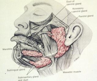
3 Major salivary glands Copyright McGraw-Hill Companies

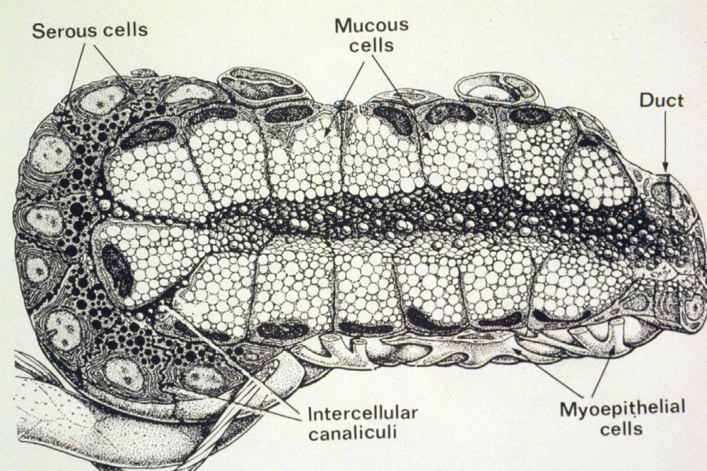
4 Slide 71: Parotid gland Serous acini Ducts

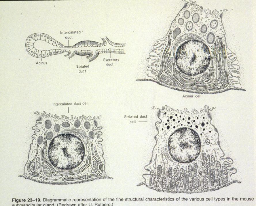
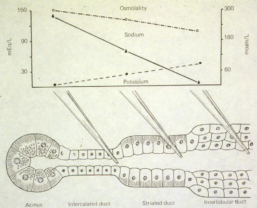
5 Slide 72: Submandibular gland Intralobular ducts Striated duct Mucous acini Serous acini

6 Slide 72 : Submandibular gland Stratified cuboidal/ columnar epithelium of Serous acini larger interlobular duct Mucous acini Serous demilune

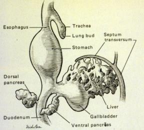
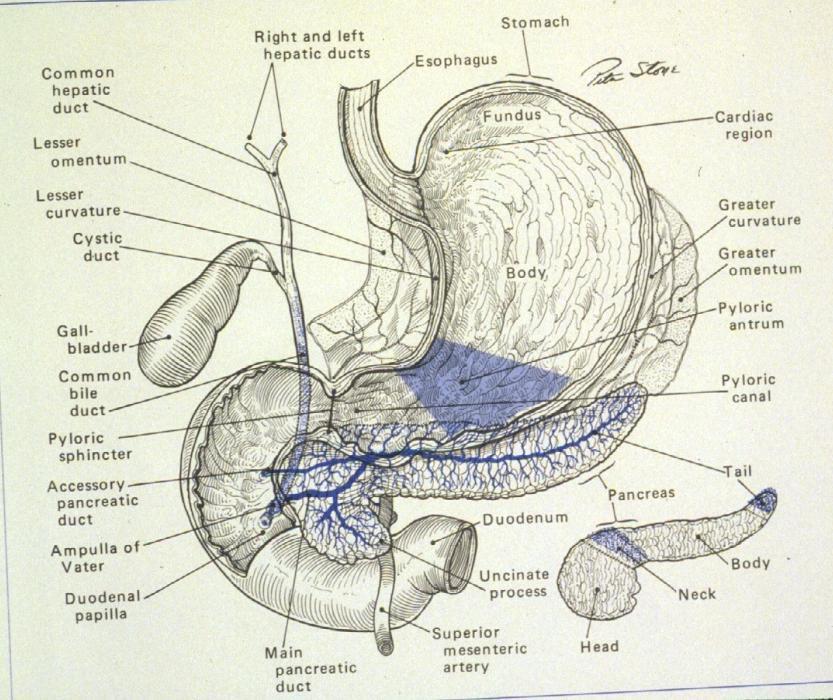
7 Pancreas and duodenum Copyright McGraw-Hill Companies

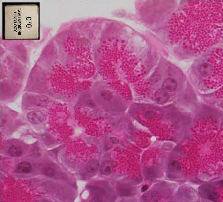
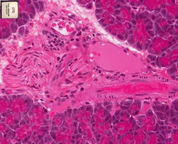
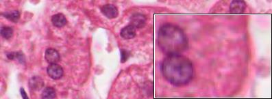
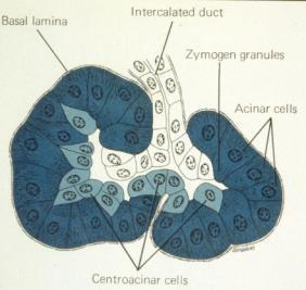
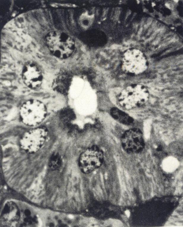
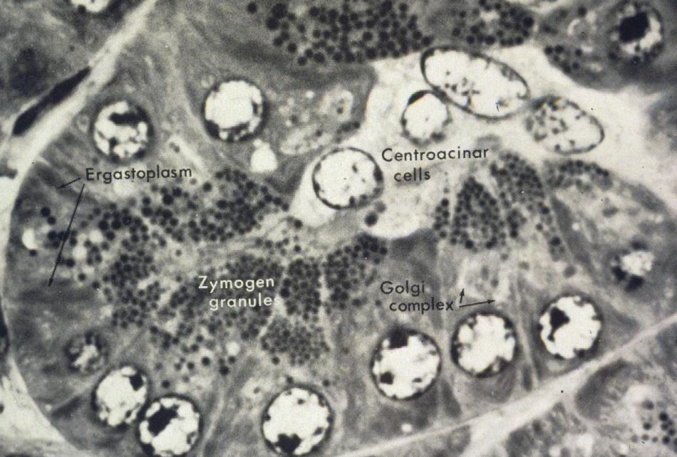
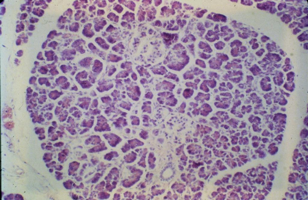
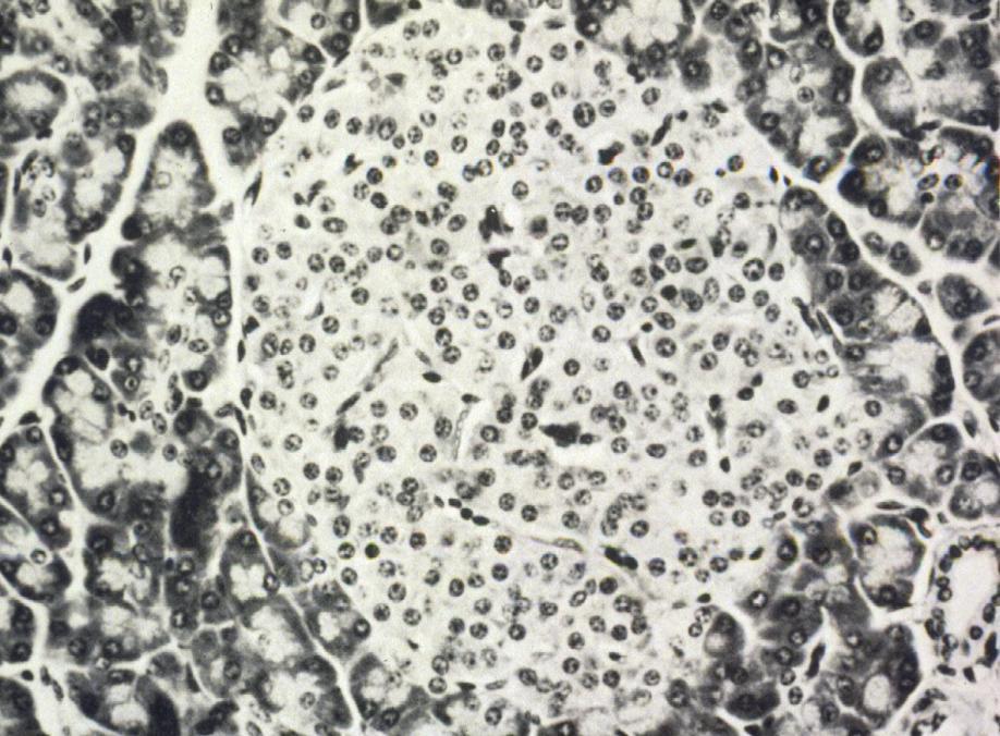
8 Slide 70: Pancreas (plastic-embedded) Exocrine acini Endocrine Zymogen granules Identify these features
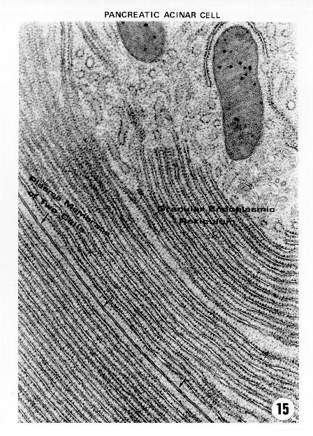
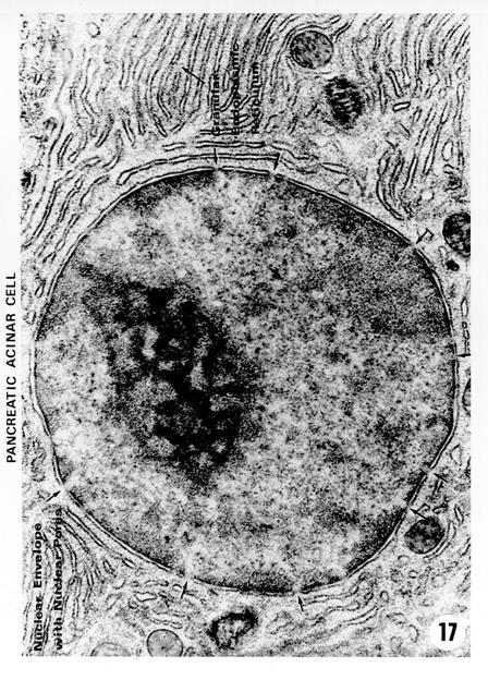
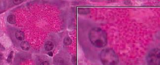
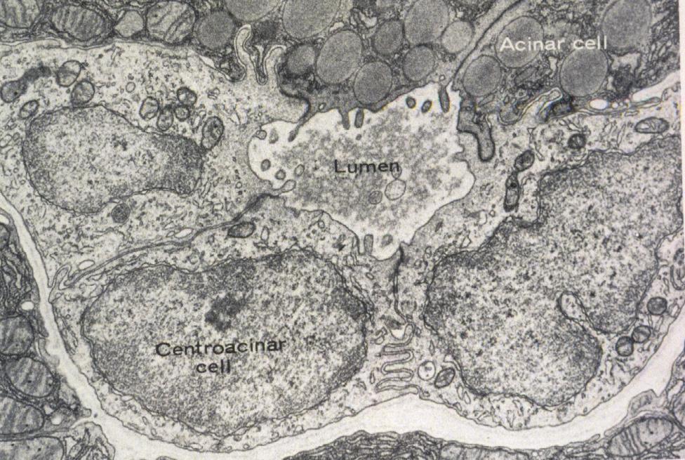
9 EM 15: Pancreatic acinar cells 70
10 34218

11 Biliary tract and gallbladder Copyright McGraw-Hill Companies


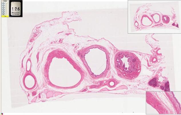
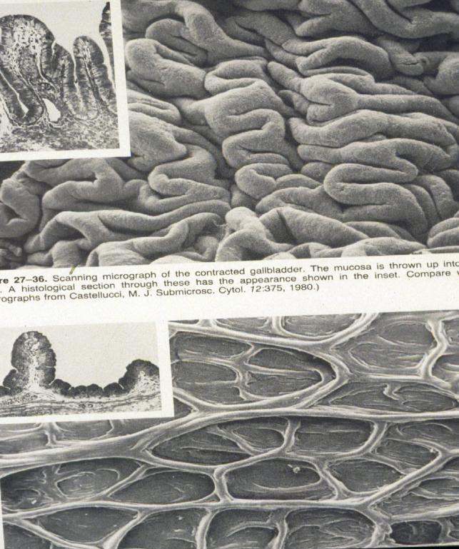
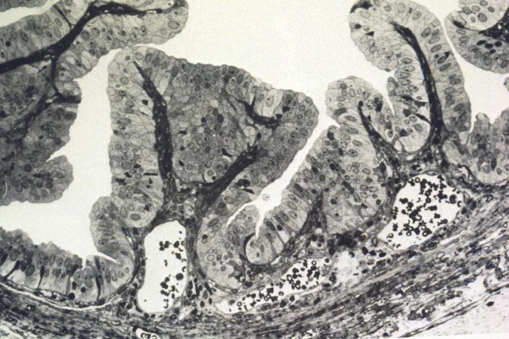
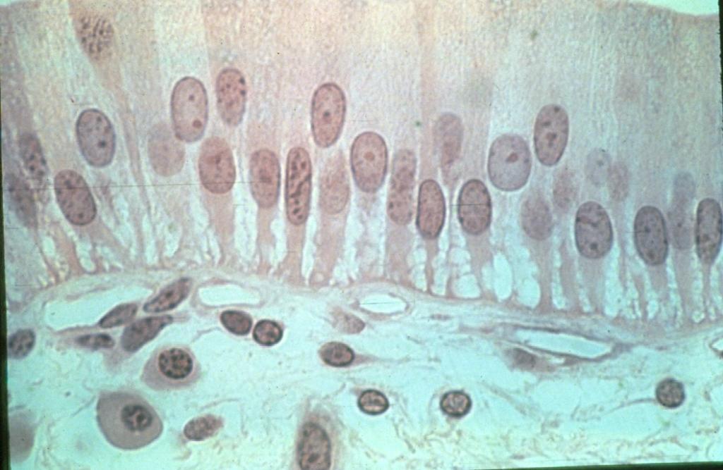
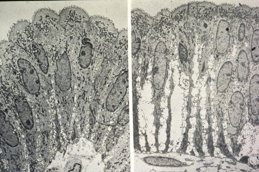
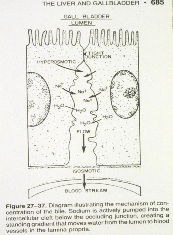
12 Slide 69: Gall bladder Brush border of simple columnar epithelium Lamina propria Irregular smooth muscle of muscularis externa Adventitia/Serosa Mucous glands

13 Liver Copyright McGraw-Hill Companies

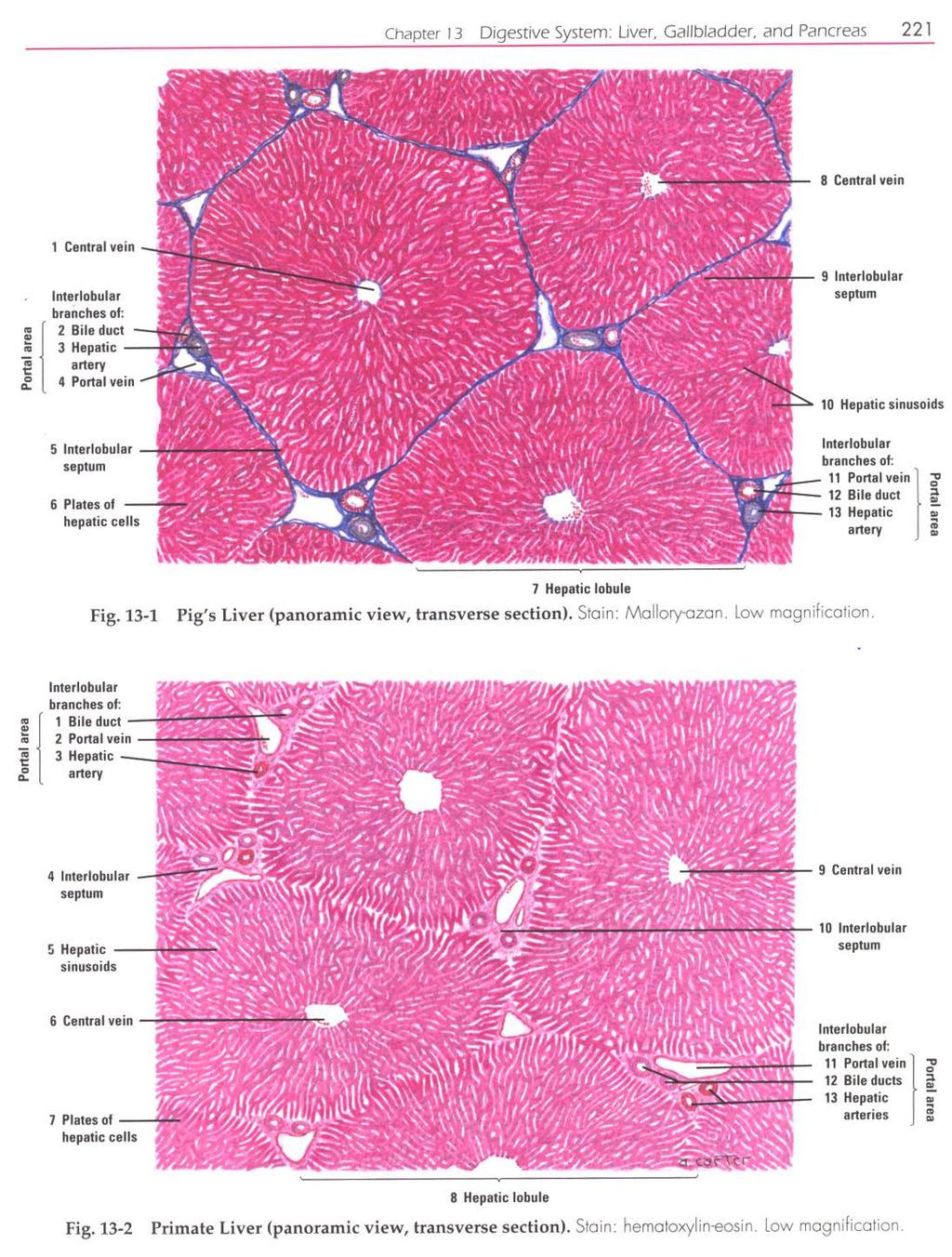
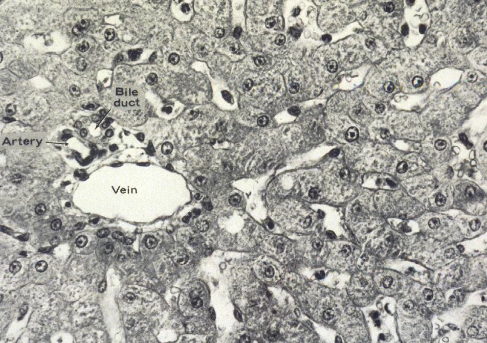
14 Slide 67: Pig liver Portal triad Hepatic artery Bile duct Portal vein Classic lobule Central vein


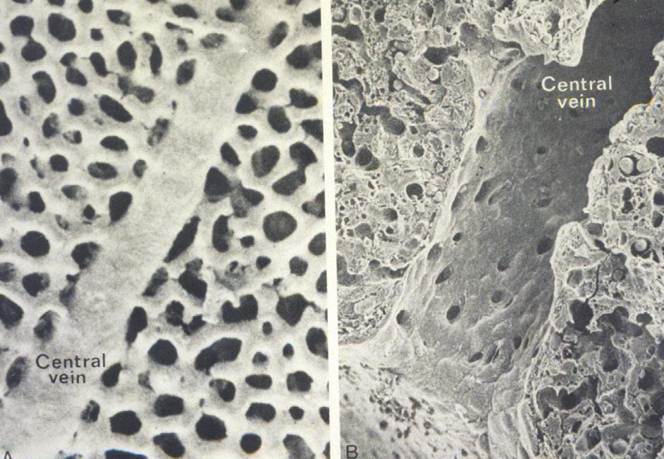
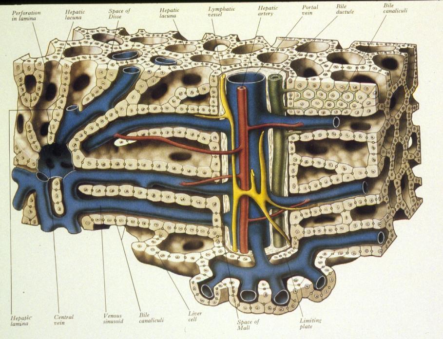
15 Slide 67 : Pig liver Space of Disse beneath endothelial cell Hepatic sinusoids Hepatic macrophage Hepatocytes Hepatic capsule (Kupffer cell) (Glisson s)

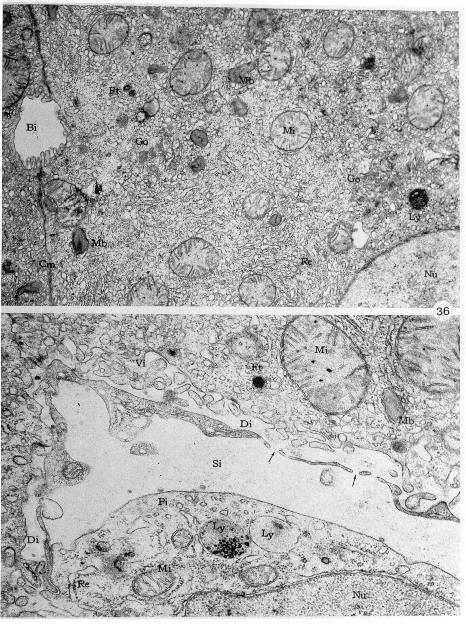
16 EM 11 and 36
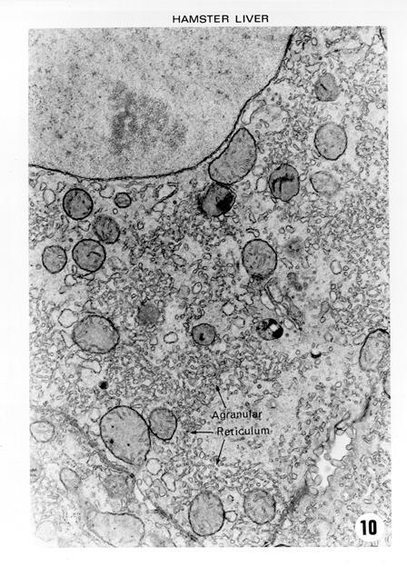
17 68 EM 10


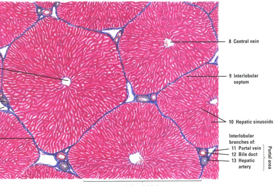
18 Slide 68: Human Liver Hepatic lobule Central vein Hepatic capsule Hepatic sinusoids Hepatic cords Portal vein Hepatic artery Bile duct
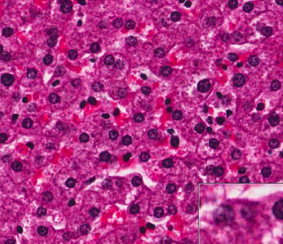
19 Clinical Correlation The most common cause of liver cirrhosis is chronic alcoholism. Which zone of the liver acinus would be most severely affected by alcohol? What would a biopsy of liver tissue from a patient with alcoholic cirrhosis look like? Read Exploring Alcohol s Effects on Liver Function by JACQUELYN J. MAHER, M.D. Copyright McGraw-Hill Companies
20 Clinical Correlation The most common cause of liver cirrhosis is chronic alcoholism. Which zone of the liver acinus would be most severely affected by alcohol? Alcohol metabolism requires increased oxygen utilization, thereby reducing the availability of oxygen (hypoxia) for cells in zone III of the liver. The relative lack of oxygen (hypoxia) in zone III encourages fibrosis in cirrhotic livers. What would a biopsy of liver tissue from a patient with alcoholic cirrhosis look like? Read Exploring Alcohol s Effects on Liver Function by JACQUELYN J. MAHER, M.D. Copyright McGraw-Hill Companies
 for cells in zone III of the liver.")

21 Clinical Correlation The most common cause of liver cirrhosis is chronic alcoholism. Which zone of the liver acinus would be most severely affected by alcohol? Alcohol metabolism requires increased oxygen utilization, thereby reducing the availability of oxygen (hypoxia) for cells in zone III of the liver. The relative lack of oxygen (hypoxia) in zone III encourages fibrosis in cirrhotic livers. What would a biopsy of liver tissue from a patient with alcoholic cirrhosis look like? The liver tissue from a patient with alcoholic cirrhosis would demonstrate extensive fibrosis that distorts the liver structure. Read Exploring Alcohol s Effects on Liver Function by JACQUELYN J. MAHER, M.D. Copyright McGraw-Hill Companies
22 The End!
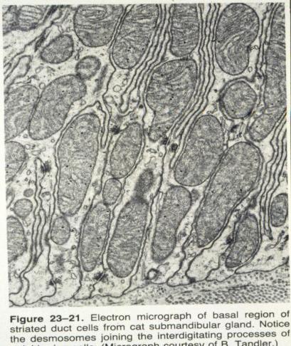
23 Answers to questions in lab manual 1. Identify an important enzyme produced by the parotid gland. Serous cells of parotid glands secrete abundant alpha-amylase, that initiates hydrolysis of carbohydrates, and prolinerich proteins with antimicrobial and other protective properties. 2. Identify an enzyme secreted by the cells forming the demilune and its function. Lysozyme, functions in bacterial wall hydrolysis. 3. What is the functional significance of these infoldings? The infoldings of the basal cell membrane seen in striated ducts contain numerous elongated mitochondria. These structures are characteristic feature of cells that transport fluids and electrolytes across cell membranes. These mitochondria within the folds supply energy for rapid ion uptake from saliva. 4. What is the significance of this (lactoferrin secretion by some submandibular glands? Lactoferrin binds iron, thereby preventing bacterial growth (which requires iron). 5. What is the function of the rough endoplasmic reticulum? Modifies, transports, and stores proteins produced by attached ribosomes; these proteins are secreted, become components of the plasma membrane, or serve as enzymes of lysosomes. 6. What are zymogen granules? Secretory granules with dense contents of inactive precursors of digestive enzymes
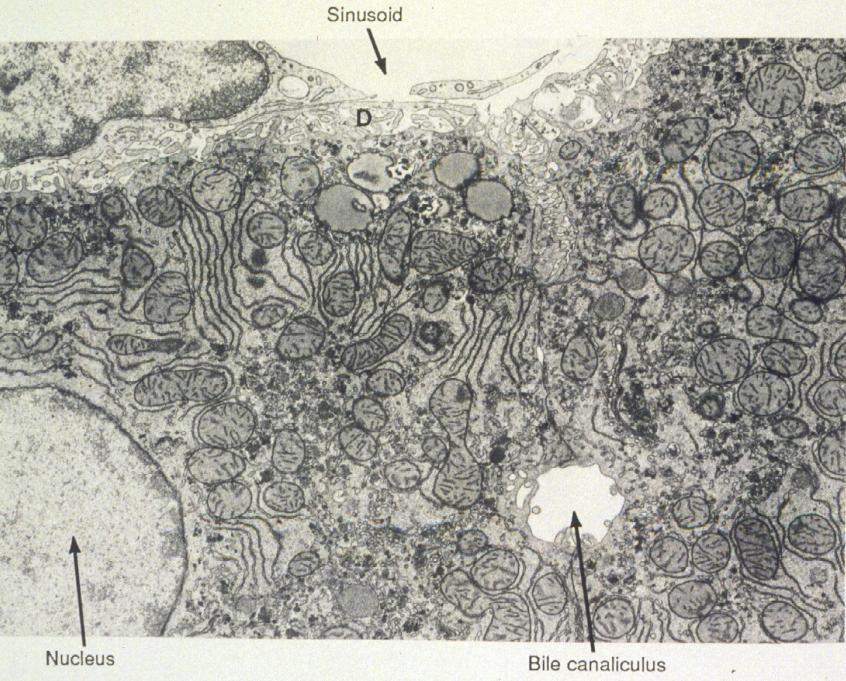
24 Answers to questions in lab manual 7. What hormone causes contraction of the smooth muscle layer and where is that hormone produced? Cholecystokinin (CCK) causes contraction of the smooth muscle layer. CCK is produced by enteroendocrine cells of the small intestine and is stimulated by the presence of ingested fats. 8. What is the difference between a serosa and an adventia? Serosa: Thin layer of loose connective tissue with a simple squamous mesothelium. The serosa is continuous with mesenteries, which are continuous with the peritoneum, which lines the abdominal cavity. Surrounds digestive organs that are suspended within the abdominal cavity. Adventitia: Thick connective tissue layer that merges with surrounding tissues and lacks mesothelium. Surrounds digestive organs that are not suspended within the abdominal cavity, but rather are bound directly to adjacent structures. 9. Identify the function of the gall bladder. The function of the gallbladder is to store bile. 10. Is the hepatic capsule (Glisson s) present? Yes. 11. What is found in the Space of Disse? Irregular microvilli projecting from the hepatocytes fill the Space of Disse. This direct contact between hepatocytes and plasma facilitates most key hepatocyte functions that involve uptake and release of nutrients, proteins, and potential toxins.
25 Answers to questions in lab manual 12. Are lymphatics part of the portal triad? Where are they found in the liver? Not part of the portal triad per say (portal vein, hepatic artery, bile duct), but most peripheral portal areas do contain lymphatics. 13. Which zone of the liver acinus would be most severely affected by alcohol? Alcohol metabolism requires increased oxygen utilization, thereby reducing the availability of oxygen (hypoxia) for cells in zone III of the liver. The relative lack of oxygen (hypoxia) in zone III encourages fibrosis in cirrhotic livers. 14. What would a biopsy of liver tissue from a patient with alcoholic cirrhosis look like? The liver tissue from a patient with alcoholic cirrhosis would demonstrate extensive fibrosis that distorts the liver structure.
26 LIVER, GALLBLADDER, PANCREAS, AND SALIVARY GLANDS
27


28 OBJECTIVES Learn of the GENERAL AND UNIQUE STRUCTURAL FEATURES OF GLANDS ASSOCIATED WITH DIGESTIVE TRACT ORIGIN OF THESE GLANDS AND HOW STRUCTURAL FEATURES OF THESE GLANDS CONTRIBUTE TO THEIR FUNCTION IN DIGESTION AND ABSORPTION OF FOOD STUFFS
29 ORIGIN AND DISTRIBUTION OF EPITHELIUM ECTODERM - EPIDERMIS OF SKIN AND EPITHELIUM OF CORNEA TOGETHER COVERS THE ENTIRE SURFACE OF THE BODY; SEBACEOUS AND MAMMARY GLANDS ECTODERM
30 ORIGIN AND DISTRIBUTION OF EPITHELIUM ECTODERM - EPIDERMIS OF SKIN AND EPITHELIUM OF CORNEA TOGETHER COVERS THE ENTIRE SURFACE OF THE BODY; SEBACEOUS AND MAMMARY GLANDS ECTODERM ENDODERM - ALIMENTARY TRACT, LIVER, PANCREAS, GASTRIC GLANDS, INTESTINAL GLANDS ENDOCRINE GLANDS - LOSE CONNECTION WITH SURFACE ENDODERM
31 ORIGIN AND DISTRIBUTION OF EPITHELIUM ECTODERM - EPIDERMIS OF SKIN AND EPITHELIUM OF CORNEA TOGETHER COVERS THE ENTIRE SURFACE OF THE BODY; SEBACEOUS AND MAMMARY GLANDS ECTODERM ENDODERM - ALIMENTARY TRACT, LIVER, PANCREAS, GASTRIC GLANDS, INTESTINAL GLANDS ENDOCRINE GLANDS - LOSE CONNECTION WITH SURFACE MESODERM MESODERM ENDOTHELIUM - LINING OF BLOOD VESSELS MESOTHELIUM - LINING SEROUS CAVITIES ENDODERM

32 CONNECTIVE TISSUE CLASSIFICATION from the mesoderm mesoderm

33 in of glands
34
35
36

37 454 Liver - vein
38
39 liver
40 Liver

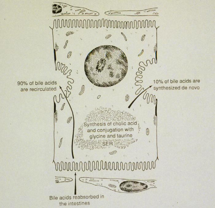
41 LIVER FUNCTION - LARGEST GLAND EXOCRINE - BILE ACIDS, BILIRUBIN ENDOCRINE - ALBUMIN, FIBRINOGEN, ETC.

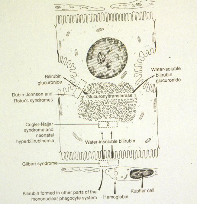
42 LIVER FUNCTIONS BLOOD FILTRATION 1.2 X 10 7 KUPFFER CELLS/G BLOOD STORAGE - LIVER SIZE AND SINUSOIDS EXPAND MAINTAIN NORMAL BLOOD GLUCOSE CONCENTRATIONS METABOLISM AND TRANSPORT OF LIPIDS SECRETE PLASMA PROTEINS - BLOOD CLOTTING NUTRITIONAL METABOLISM AND BILE SECRETION DRUG METABOLISM - SER DRUG TOLERANCE EXCRETION OF BILIRUBIN - JAUNDICE SECRETE BILE - EMULSIFYING FATS
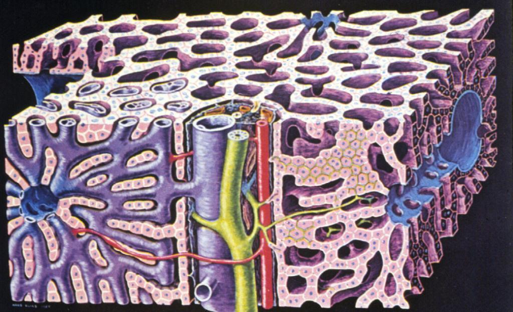
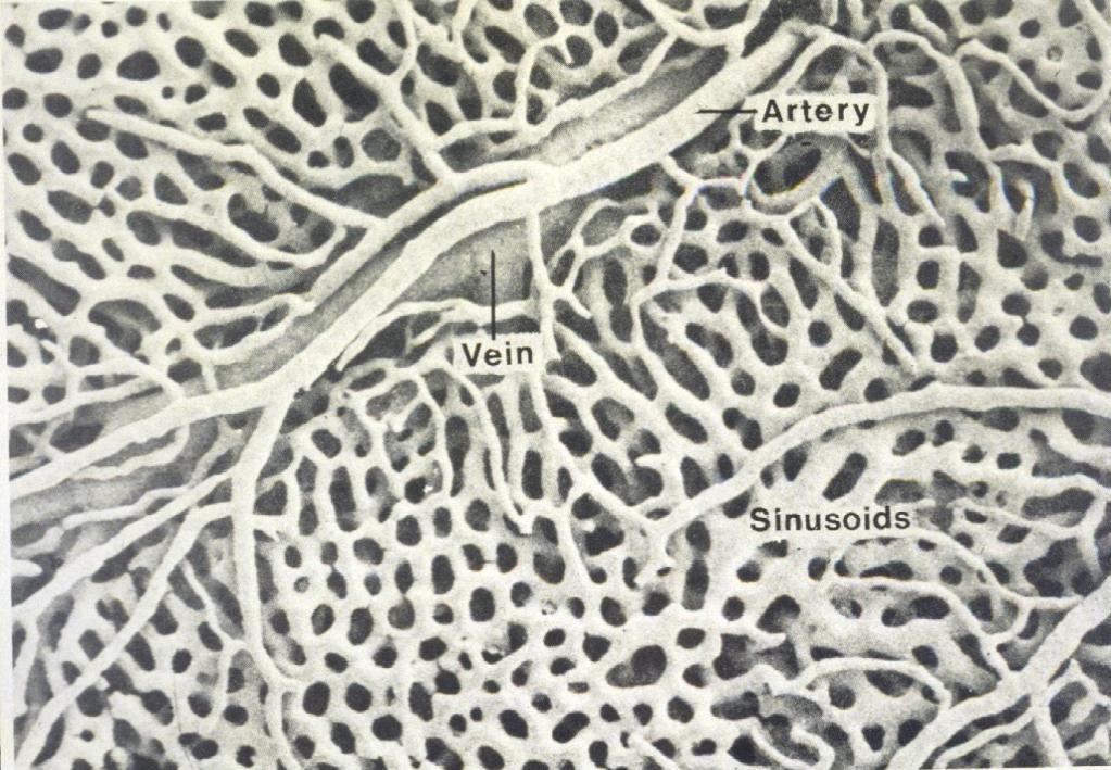
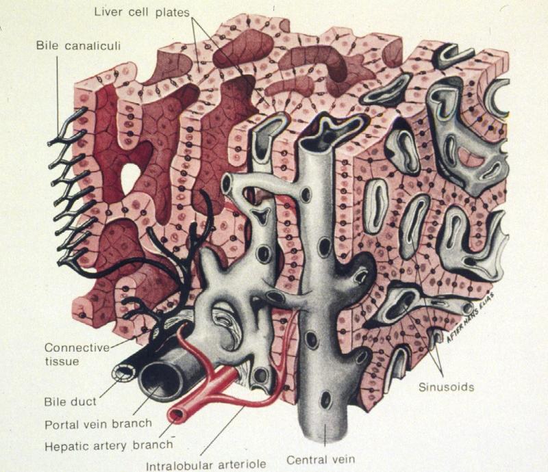
43 LIVER LOBULE PORTAL TRIAD BLOOD SUPPLY
44 LIVER LOBULE PORTAL TRIAD BLOOD SUPPLY CENTRAL VEIN HEPATIC SINUSOIDES

45 LIVER LOBULE PORTAL TRIAD BLOOD SUPPLY CENTRAL VEIN HEPATIC SINUSOIDES ZONATION OF THE LIVER

46 Central vein in center Classical lobule in the pig who has separating connective tissue
47
48

49
50 liver
51 liver
52
53
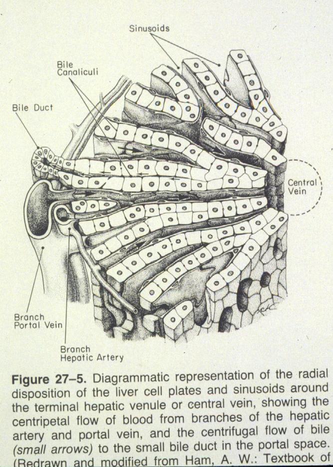
54 Acinus with portal vein and artery in center ZONATION OF THE LIVER 1. Classical lobule
55 Acinus with portal vein and artery in center ZONATION OF THE LIVER 1. Classical lobule Portal lobule with triad in center
56 Acinus with portal vein and artery in center ZONATION OF THE LIVER 1. Classical lobule Portal lobule with triad in center Acinus layers between two central veins


57 ZONATION OF THE LIVER Classical lobule

58 Portal lobule with triad in center

59 Acinus with portal vein and artery in center
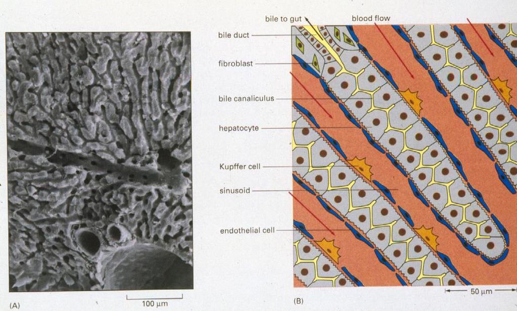
60 CELLS OF THE LIVER LOBULE A. HEPATOCYTE B. KUPFFER AND FAT-STORING CELLS C. ENDOTHELIAL CELL
61 CELLS OF THE LIVER LOBULE A. HEPATOCYTE B. KUPFFER AND FAT-STORING CELLS C. ENDOTHELIAL CELL HEPATOCYTE
62 CELLS OF THE LIVER LOBULE A. HEPATOCYTE B. KUPFFER AND FAT-STORING CELLS C. ENDOTHELIAL CELL HEPATOCYTE KUPFFER CELLS

63 CELLS OF THE LIVER LOBULE A. HEPATOCYTE B. KUPFFER AND FAT-STORING CELLS C. ENDOTHELIAL CELL HEPATOCYTE KUPFFER CELLS ENDOTHELIAL CELL


64 CELLS OF THE LIVER LOBULE A. HEPATOCYTE B. KUPFFER CELLS C. ENDOTHELIAL CELL
65
66 Liver
67 Liver
68 HEPATOCYTE
69 HEPATOCYTE
70

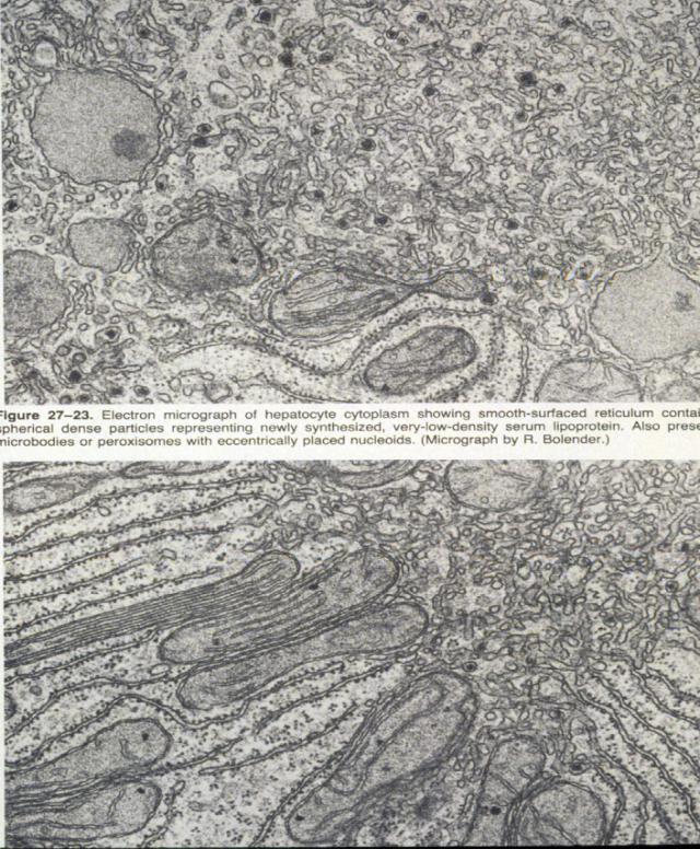
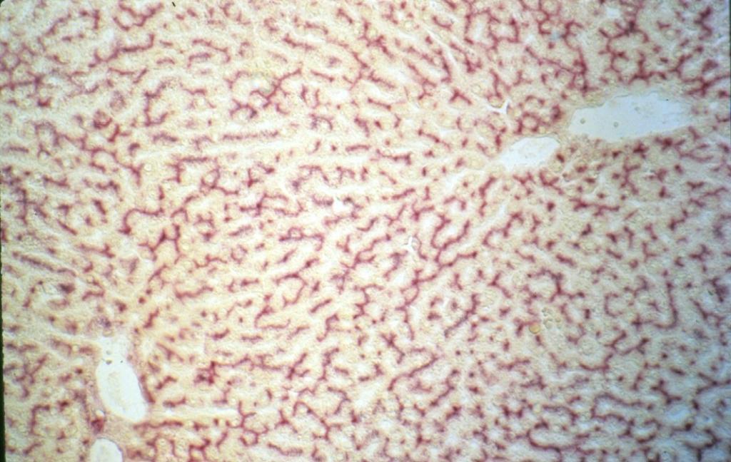
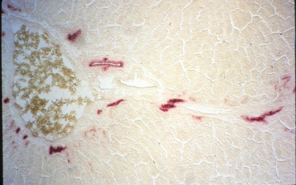
71 Histological reaction for peroxidase HEPATOCYTE


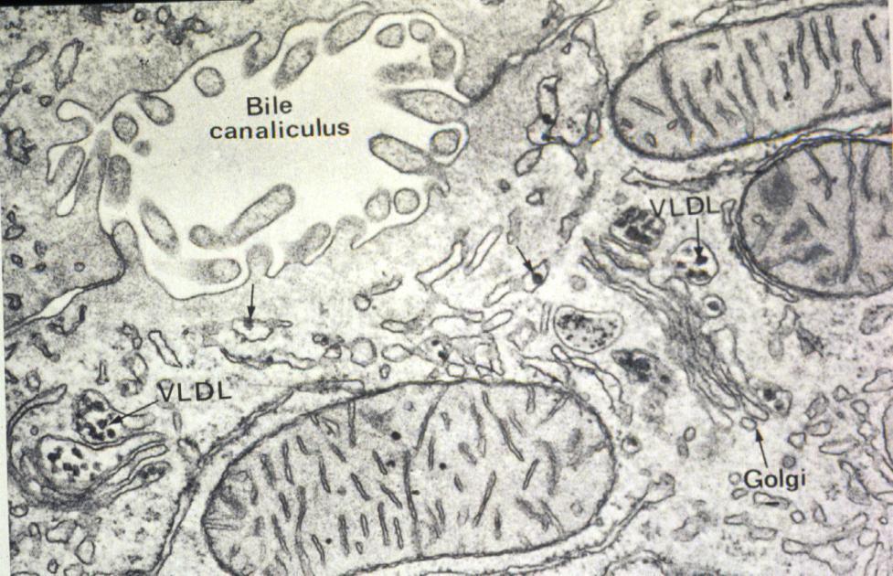
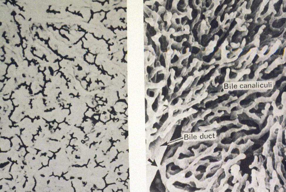
72 HEPATOCYTE SPACE OF DISSE BILE CANALICULI

73 SPACE OF DISSE
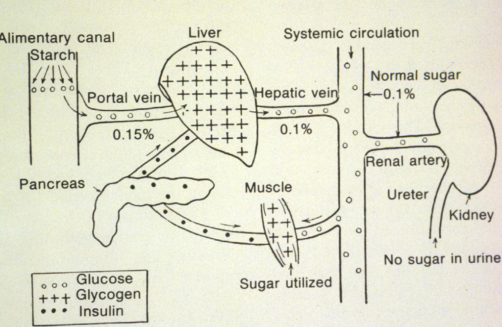
74 Sugar and protein

75 Sugar and protein
76

77 Glycogen in hepatocytes
 24-hour fast (0.9% glycogen)")
78 Dietary differences in amount of glycogen in hepatocytes 2-hour fast (8.2% glycogen) 24-hour fast (0.9% glycogen)
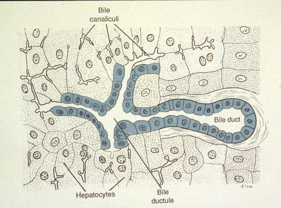
79 BILE CANALICULI
80
81 diseases


82 SURFACE SPECIALIZATIONS OF EPITHELIA TRANSCYTOSIS TO GET ANTIBODIES INTO SECRETIONS
83 Bile canaliculus Four + compounds that are deposited/secreted into this space. a. Cholesterol b. EGF c. insulin d. IgA also bile salts and BILIRUBIN
84
85
86
87 BILE CANALICULI
88 BILE duct
89
90 GALLBLADDER & BILE DUCTS FUNCTION BILIARY TRACT ORGANIZATION OF GALLBLADDER EPITHELIUM CONNECTIVE TISSUE HISTOPHYSIOLOGY
91
92
93
94
95
96
97

98 SALIVARY GLANDS FUNCTION HISTOLOGICAL ORGANIZATION ACINUS = FUNCTIONAL UNIT SEROUS MUCOUS MIXED
99

100 ACINUS = FUNCTIONAL UNIT SEROUS MUCOUS
101 SALIVA HELPS PREVENTS INFECTIONS CONTAINS SECRETED IGA
102 SALIVA HELPS PREVENTS INFECTIONS CONTAINS SECRETED IGA CONTAINS LACTOFERIN - BIND UP IRON NEEDED FOR BACTERIA DIVISION
103 SALIVA HELPS PREVENTS INFECTIONS CONTAINS SECRETED IGA CONTAINS LACTOFERIN - BIND UP IRON NEEDED FOR BACTERIA DIVISION CONTAINS LYSOSOME THAT KILLS BACTERIA

104 SALIVA HELPS PREVENTS INFECTIONS CONTAINS SECRETED IGA CONTAINS LACTOFERIN - BIND UP IRON NEEDED FOR BACTERIA DIVISION CONTAINS LYSOSOME THAT KILLS BACTERIA CONSTANTLY WASHES MOUTH TO DISLODGE AND SWEEP BACTERIA DOWN GI TRACT

105 ACINUS = FUNCTIONAL UNIT SEROUS MUCOUS MIXED
106

107 DUCTS OF SALIVARY GLANDS INTERCALATED STRIATED
108
109
110

111 PANCREAS FUNCTION 1. EXOCRINE 2. ENDOCRINE HISTOLOGICAL ORGANIZATION, EXOCRINE PORTION 1. ACINI 2. DUCTS ENDOCRINE PORTION ISLETS OF LANGERHANS HISTOPHYSIOLOGY
112
113
114
115
116
117
118
119
120
121 34218
Digestive system L 4. Lecturer Dr. Firdous M. Jaafar Department of Anatomy/Histology section
 Digestive system L 4 Lecturer Dr. Firdous M. Jaafar Department of Anatomy/Histology section objectives 1-Describe the structure of liver. 2-Define liver lobule, and identify its zones. 3-Define portal
Laboratory exercises for abdominal organs
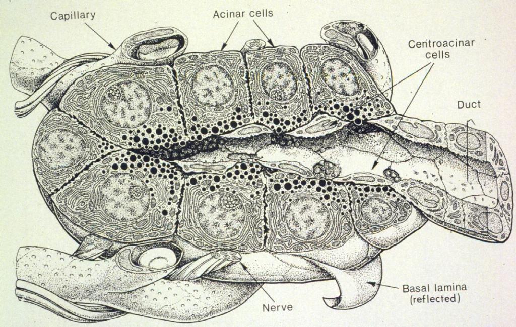
 Laboratory exercises for abdominal organs Slide #77 (C007- H- 107A). Pancreas, dog. pancreatic islets CENTROACINAR CELLS ARE THE BEGINNING CELLS OF THE INTERCALATED DUCTS THAT DRAIN THE SECRETORY ACINI
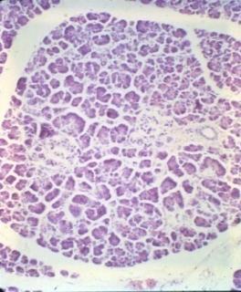
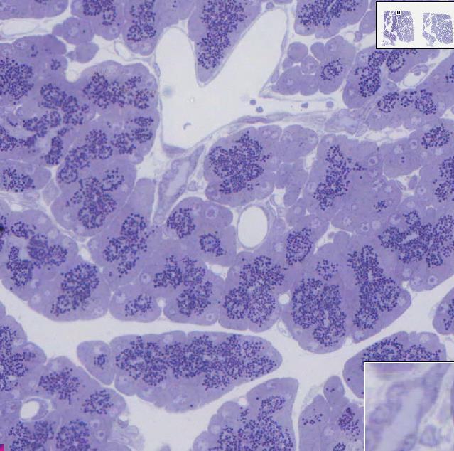
Slide 154: Pancreas, H&E
 Slide 154: Pancreas, H&E the pancreas, located adjacent to the duodenum, is a mixed exocrine and endocrine gland; it is usually readily identifiable by the presence of the interspersed endocrine pancreatic
Paneth Cells. Road Map to the Finish. No Review this Friday. Today 11/29 Finish digestion/accessory organs. Wednesday 12/1 Immune System I
 Road Map to the Finish No Review this Friday Today 11/29 Finish digestion/accessory organs Wednesday 12/1 Immune System I Paneth Cells - base of intestinal glands -! large -! intense acidophilic granules
Chapter 12 The Digestive Glands
 Chapter 12 The Digestive Glands Lyu Zhengmei Department of Histology and Embryology, Anhui Medical University Components of digestive glands large salivary glands, pancreas, liver, gallbladder. These organs
Organs Associated with the Digestive Tract. Dr. Emad I H Shaqoura M.D, M.Sc. Anatomy Faculty of Medicine, IUG March, 2016
 Organs Associated with the Digestive Tract Dr. Emad I H Shaqoura M.D, M.Sc. Anatomy Faculty of Medicine, IUG March, 2016 2 Salivary Glands Salivary Glands Major 90% of saliva Minor 10% of saliva Parotid
Chapter 18 Liver and Gallbladder
 Chapter 18 Liver and Gallbladder 解剖學科徐淑媛 本堂重點 1. Liver : functions & histology 2. Gallbladder Physiology Liver Produce circulating plasma proteins Vitamin Iron Degradation Metabolism Bile manufacture (exocrine)
Anatomy of the liver and pancreas
 Anatomy of the liver and pancreas Prof. Abdulameer Al-Nuaimi E-mail: a.al-nuaimi@sheffield.ac.uk abdulameerh@yahoo.com Liver Aorta Pulm. Trunk Rt. At, Duct. Art. Lt. Ven. Rt. Ven. Internal Posterior
HISTOLOGY VIRTUAL LABORATORY GASTROINTESTINAL SYSTEM
 HISTOLOGY VIRTUAL LABORATORY GASTROINTESTINAL SYSTEM LIP (Slides GI 1, 2) Identify the outer portion lined by stratified squamous (keratinized) epithelium. Note the hair follicles and sebaceous glands
Chapter 14: The Digestive System
 Chapter 14: The Digestive System Digestive system consists of Muscular tube (digestive tract) alimentary canal Accessory organs teeth, tongue, glandular organs 6 essential activities 1. 2. 3. 4. 5. 6.
The Digestive System. Chapter 25
 The Digestive System Chapter 25 Introduction Structure of the digestive system A tube that extends from mouth to anus Accessory organs are attached Functions include Ingestion Movement Digestion Absorption
DIGESTIVE. CHAPTER 17 Lecture: Part 1 Part 2 BIO 212: ANATOMY & PHYSIOLOGY II
 BIO 212: ANATOMY & PHYSIOLOGY II CHAPTER 17 Lecture: DIGESTIVE Part 1 Part 2 Dr. Lawrence G. Altman www.lawrencegaltman.com Some illustrations are courtesy of McGraw-Hill. SMALL INTESTINE DUODENUM > JEJUNUM
Digestive System 7/15/2015. Outline Digestive System. Digestive System
 Digestive System Biology 105 Lecture 18 Chapter 15 Outline Digestive System I. Functions II. Layers of the GI tract III. Major parts: mouth, pharynx, esophagus, stomach, small intestine, large intestine,
PRACTICAL ROADMAP. GLANDS AFFECTING LIFESTYLE WJ van der Spuy & T Tshabalala
 PRACTICAL ROADMAP GLANDS AFFECTING LIFESTYLE WJ van der Spuy & T Tshabalala GLANDS AFFECTING LIFESTYLE Submandibular gland (salivary gland) Liver Pancreas Hypophysis (pituitary gland) Thyroid Suprarenal
Histology Lab. looking at microscopic pictures of tissues, for more information use Junqueira book and you can use BlueHistolgy website
 Done By: Aseel Twaijer & Laith Sorour Histology Lab *These notes help in differentiating tissues and you must read them while looking at microscopic pictures of tissues, for more information use Junqueira
Digestive System Module 6: Accessory Organs in Digestion: The Liver, Pancreas, and Gallbladder
 Connexions module: m49293 1 Digestive System Module 6: Accessory Organs in Digestion: The Liver, Pancreas, and Gallbladder Donna Browne Based on Accessory Organs in Digestion: The Liver, Pancreas, and
1. Approximately 21 ft. long: duodenum (one ft.), jejunum (eight ft.), and ileum (twelve ft.)
, jejunum (eight ft.), and ileum (twelve ft.)") IV. Small Intestines A. General features and functions 1. Approximately 21 ft. long: duodenum (one ft.), jejunum (eight ft.), and ileum (twelve ft.) 2. Functions: move forward chyme, continue digestion,
IV. Small Intestines A. General features and functions 1. Approximately 21 ft. long: duodenum (one ft.), jejunum (eight ft.), and ileum (twelve ft.) 2. Functions: move forward chyme, continue digestion,
The Digestive System. Chapter 16. Introduction. Overview of Digestive System. Histological Organization. Movement and Mixing of Digestive Materials
 The Digestive System Chapter 16 Introduction Structure of the digestive system A tube that extends from mouth to anus Accessory organs are attached Functions include Ingestion Movement Digestion Absorption
Gastrointestinal Anatomy and Physiology. Bio 219 Napa Valley College Dr. Adam Ross
 Gastrointestinal Anatomy and Physiology Bio 219 Napa Valley College Dr. Adam Ross Functions of digestive system Digestion Breakdown of food (chemically) using enzymes, acid, and water Absorption Nutrients,
The Digestive System. What is the advantage of a one-way gut? If you swallow something, is it really inside you?
 The Digestive System What is the advantage of a one-way gut?! If you swallow something, is it really inside you? Functions and Processes of the Digestive System: Move nutrients, water, electrolytes from
MICROSCOPIC STRUCTURE OF LIVER, GALLBLADDER, GALL DUCTS, AND PANCREAS OVERVIEW OF DEVELOPMENT OF THE ALIMENTARY CANAL
 Lecture 2 ESS_3rd semester MICROSCOPIC STRUCTURE OF LIVER, GALLBLADDER, GALL DUCTS, AND PANCREAS OVERVIEW OF DEVELOPMENT OF THE ALIMENTARY CANAL MICROSCOPIC STRUCTURE OF LIVER - is the largest gland of
Epithelia will be discussed according to the following scheme: Type Number of layers Shape Line drawing. Squamous Cuboidal Columnar
 Epithelia Epithelia will be discussed according to the following scheme: Type Number of layers Shape Line drawing Simple Squamous Cuboidal Columnar Covering and Lining epithelium Pseudostratified Stratified
Glandular Epithelium. Dr. Hersh Abdul Ham-Karim BVM&S, PG Dip, MSc and PhD
 Glandular Epithelium Dr. Hersh Abdul Ham-Karim BVM&S, PG Dip, MSc and PhD Glandular Epithelium Groups of surface cells differentiate, proliferate, and penetrate underlying connective tissue. Their main
Gastric Contrac,le Ac,vity. Regula,on of Gastric Emptying
 Gastric Contrac,le Ac,vity Figure 23.18 Regula,on of Gastric Emptying Gastric emptying is regulated by: Neural enterogastric reflex Hormonal (enterogastrone) mechanisms In the presence of gastric gastrin
Lab 8: Digestive System
 BIOL 221 A&P II Lab 8: Digestive System Become familiar with the gross anatomy of the digestive system (Exercise 38) using the models, Fig. 38.1 (Activity 1), and the rat. Recognize and know the functions
Histology 3. We will continue talking about a few things from last lecture, starting with M cells:
 Histology 3 This is the last Histology lecture in the GI system. Enjoy! There are some extra notes listed as footnotes. We will continue talking about a few things from last lecture, starting with M cells:
Tissue: The Living Fabric: Part A
 PowerPoint Lecture Slides prepared by Janice Meeking, Mount Royal College C H A P T E R 4 Tissue: The Living Fabric: Part A Tissues Groups of cells similar in structure and function Types of tissues Epithelial
Nutrition. Autotrophs. plants, some protists & bacteria producers
 Nutrition Autotrophs plants, some protists & bacteria producers Nutrition Heterotrophs animals, fungi, some protists & bacteria consumers Animal Nutrition Most obtain food by ingestion take in their food
The Digestive System
 The Digestive System Identify the Structure and Function. Mesentery of the Large Intestine The mesentery functions to connect the visceral organs to the abdominal wall. Identify the Structure. Nasal Cavity
Glandular Epithelium. Dr. Heba Kalbouneh Associate Professor of Anatomy and Histology
 Glandular Epithelium Dr. Heba Kalbouneh Associate Professor of Anatomy and Histology Glands Glandular epithelia are tissues formed by cells specialized to produce secretion. Secretion: if substances produced
DIGESTIVE SYSTEM ALIMENTARY CANAL / GI TRACT & ACCESSORY ORGANS. Mar 16 10:34 PM
 DIGESTIVE SYSTEM ALIMENTARY CANAL / GI TRACT & ACCESSORY ORGANS Mar 16 10:34 PM 1 I. Digestive System Functions > Ingestion the taking in of food > Propulsion movement caused by force > Digestion breakdown
The stomach is formed of three parts: -
 The stomach is formed of three parts: - (a) CARDIAC STOMACH: - It receives the oesophagus through Cardiac aperture guarded by a cardiac sphincter which prevents regurgitation of food. (b) FUNDIC PART:
Bio 322 Human Anatomy Objectives for the laboratory exercise Digestive System
 Bio 322 Human Anatomy Objectives for the laboratory exercise Digestive System Required reading before beginning this lab: Saladin, KS: Human Anatomy 5 th ed (2017) Chapter 24 For this lab you will use
GI Histology Lab 1. Prepared by: Zeina Kalaji
 GI Histology Lab 1 Prepared by: Zeina Kalaji Lip ORAL MUCOSA -Arrow shows labial salivary glands in the submucosa. VERMILLION transitional zone. SKIN Stratified Squamous epithelium, keratinized -Arrow
Large Intestine. The large intestine consists of a mucosal membrane with no folds except in its distal (rectal) portion
 portion") GI Histology 3 Large Intestine The large intestine consists of a mucosal membrane with no folds except in its distal (rectal) portion No villi are present in this portion of the intestine The intestinal
GI Histology 3 Large Intestine The large intestine consists of a mucosal membrane with no folds except in its distal (rectal) portion No villi are present in this portion of the intestine The intestinal
Principles of Anatomy and Physiology
 Principles of Anatomy and Physiology 14 th Edition CHAPTER 24 The Digestive System Introduction The purpose of this chapter is to Identify the anatomical components of the digestive system as well as their
Pancreas Fox Chapter 18 part 2 (also Chapter 19.3 & 19.4)
") Vert Phys PCB3743 Pancreas Fox Chapter 18 part 2 (also Chapter 19.3 & 19.4) T. Houpt, Ph.D. Anatomy of Digestive System Peristalsis Stomach and Acid Secretion Liver and Bile Secretion Pancreas and pancreatic
Vert Phys PCB3743 Pancreas Fox Chapter 18 part 2 (also Chapter 19.3 & 19.4) T. Houpt, Ph.D. Anatomy of Digestive System Peristalsis Stomach and Acid Secretion Liver and Bile Secretion Pancreas and pancreatic
The Digestive System and Body Metabolism Premedical Biology
 The Digestive System and Body Metabolism Premedical Biology Copyright 2003 Pearson Education, Inc. publishing as Benjamin Cummings The Digestive System and Body Digestion Metabolism Breakdown of ingested
Lab activity manual - Histology of the digestive system. Lab activity 1: esophagus stomach - small intestines
 Lab activity manual - Histology of the digestive system Jeanne Adiwinata Pawitan Prerequisite: Histology of the 4 basic tissues In this module we learn about the histology of the digestive system, from
Topics and aims. Introduction. Metabolism and Excretion
 Topics and aims Introduction This section contains instructions that are applicable to all material, irrespective of your specific course. Please take note and make sure to comply. Failure to comply could
Chapter 9. The digestive system. Glossary. Louise McErlean
 Chapter 9 The digestive system Louise McErlean Glossary Absorption Process whereby the products of digestion move into the blood or lymph fluid. Acini glands Produce pancreatic juice. Amylase Carbohydrate
Prepared By Student. Dania Abed Al-majeed. Rahma Raad Hanna. Balqees Mohammed Aasim. Dania Hisham. Rasha Rafiee
 Prepared By Student Rahma Raad Hanna Balqees Mohammed Aasim Dania Hisham Dania Abed Al-majeed Rasha Rafiee Epithelia Epithelia can be derived from ectoderm, mesoderm or endoderm -ectoderm gives rise to
Glandular Epithelium. Dr. Heba Kalbouneh Assistant Professor of Anatomy and Histology
 Glandular Epithelium Dr. Heba Kalbouneh Assistant Professor of Anatomy and Histology Glands Gla dular epithelia are tissues for ed y ells spe ialized to produ e se retio. Secretion: if substances produced
Sheet #6. Dr. Heba Kalbouneh. Dr. Heba Kalbouneh. Dr. Heba Kalbouneh
 Sheet #6 Dr. Heba Kalbouneh Dr. Heba Kalbouneh Dr. Heba Kalbouneh Ducts - In large glands, as you go away from the secretory unit, the duct becomes larger and the lining epithelium becomes thicker (from
LIVER & SPLEEN. Color index: Slides.. Important..Notes..Extra..
 LIVER & SPLEEN Color index: Slides.. Important..Notes..Extra.. Objectives: By the end of this lecture, the student should be able to describe: 1. The histological structure of liver with special emphasis
Tissues. tissue = many cells w/ same structure and function. cell shape aids function tissue shape aids function. Histology = study of tissues
 Tissues tissue = many cells w/ same structure and function cell shape aids function tissue shape aids function Histology = study of tissues 4 types of tissues Epithelial coverings contact openings Connective
NOTES: The Digestive System (Ch 14, part 2)
") NOTES: The Digestive System (Ch 14, part 2) PANCREAS Structure of the pancreas: The pancreas produces PANCREATIC JUICE that is then secreted into a pancreatic duct. The PANCREATIC DUCT leads to the The
NOTES: The Digestive System (Ch 14, part 2) PANCREAS Structure of the pancreas: The pancreas produces PANCREATIC JUICE that is then secreted into a pancreatic duct. The PANCREATIC DUCT leads to the The
Objectives. Describe the cells of the GI tract and their function. Differentiate between different parts of the GI tract
 GI Histology 1 Objectives Describe the cells of the GI tract and their function Describe the histological features of each part of the GI tract. Differentiate between different parts of the GI tract Appreciate
Gastrointestinal Tract
 CTO Lab #5 GI TRACT & GLANDS; ENDOCRINE SYSTEM Page 1 Gastrointestinal Tract Slide 126 This section through the esophagus shows the characteristic layers of the gastrointestinal tract. Examine the non-keratinized
Connective tissue The Digestive System
 Connective tissue The Digestive System Part 1 Structure of digestive system Functions Basic Structure of the Alimentary Canal Wall Tube is made up of four layers: 1. Mucosa 2. Submucosa 3. Muscularis externa
- Digestion occurs during periods of low activity - Produces more energy than it uses. - Mucosa
 Introduction Digestive System Chapter 29 Provides processes to break down molecules into a state easily used by cells - A disassembly line: Starts at the mouth and ends at the anus Digestive functions
Epithelial Tissue. Functions include: 1. Protection 4. Absorption 2. Secretion 5. Filtration 3. Sensory reception
 Tissues There are 4 primary tissue types in the human body: 1. Epithelial (covering/lining) 2. Connective (support) 3. Muscle (movement) 4. Nervous (control) Epithelium Epithelial Tissue Covers the surface
An overview of the digestive system. mouth pharynx esophagus stomach small intestine large intestine rectum anus
 An overview of the digestive system mouth pharynx esophagus stomach small intestine large intestine rectum anus Why GIT? What are the main steps in the digestive process? Ingestion intake of food via the
Small intestine. Small intestine
 General features Tubular organ longest part; 5-6 m most of chemical digestion absorption of nutrients reabsorption of H2O occurs. Two structural features; maximize the lumenal surface area villi microvilli
Chapter 26 The Digestive System
 Chapter 26 The Digestive System Digestive System Gastroenterology is the study of the stomach and intestine. Digestion Catabolism Absorption Anabolism The actions of the digestive system are controlled
ANATOMY & PHYSIOLOGY ONLINE COURSE - SESSION 13 THE DIGESTIVE SYSTEM
 ANATOMY & PHYSIOLOGY ONLINE COURSE - SESSION 13 THE DIGESTIVE SYSTEM The digestive system also known as the alimentary canal or gastrointestinal tract consists of a series of hollow organs joined in a
NURSE-UP DIGESTIVE SYSTEM AKA G.I. SYSTEM
 NURSE-UP DIGESTIVE SYSTEM AKA G.I. SYSTEM The digestive system is used for breaking down food into nutrients which then pass into the circulatory system and are taken to where they are needed in the body.
Understandings, Applications & Skills
 D.2 Digestion Understandings, Applications & Skills Statement D.2.U1 Nervous and hormonal mechanisms control the secretion of digestive juices. D.2.U2 Exocrine glands secrete to the surface of the body
3/20/2007 Page Mechanisms of Drug Action. The Liver and Metabolism September 30, 2005
 3/20/2007 Page 1 20.201 Mechanisms of Drug Action The Liver and Metabolism September 30, 2005 Distribution of Chemicals to Liver 3/20/2007 Page 2 Chemicals entering blood are distributed in the general
(b) Stomach s function 1. Dilution of food materials 2. Acidification of food (absorption of dietary Fe in small intestine) 3. Partial chemical digest
 Stomach s function 1. Dilution of food materials 2. Acidification of food (absorption of dietary Fe in small intestine) 3. Partial chemical digest") (1) General features a) Stomach is widened portion of gut-tube: between tubular and spherical; Note arranged of smooth muscle tissue in muscularis externa. 1 (b) Stomach s function 1. Dilution of food
(1) General features a) Stomach is widened portion of gut-tube: between tubular and spherical; Note arranged of smooth muscle tissue in muscularis externa. 1 (b) Stomach s function 1. Dilution of food
Connective tissue The Digestive System
 Connective tissue The Digestive System Part 1 Structure of digestive system Functions Basic Structure of the Alimentary Canal Wall Tube is made up of four layers: 1. Mucosa 2. Submucosa 3. Muscularis externa
Sphincters heartburn diaphragm The Stomach gastric glands pepsin, chyme The Small Intestine 1-Digestion Is Completed in the Small Intestine duodenum
 Sphincters are muscles that encircle tubes and act as valves. The tubes close when the sphincters contract and they open when the sphincters relax. When food or saliva is swallowed, the sphincter relaxes
Anatomy & Physiology Revealed Instructions. 1. From the Module dropdown menu, chose the 12. Digestive system.
 #10 - Objectives: Examine the histology of selected body organs using Anatomy & Physiology Revealed software and microscope slides. Be able to identify each organ and the specific structures indicated
Histology of the liver, and the biliary system
 Histology of the liver, and the biliary system Dr. Zsuzsanna Tóth Semmelweis University Department of Anatomy, Histology and Embryology http://www.mgtowforums.com/forums/lads-night-inn/3969-give-your-liver-art.html
2. Epithelial Tissues Dr. Manal Othman
 Biology-232 GENERAL HISTOLOGY 2. Epithelial Tissues Dr. Manal Othman Anatomy Department CMMS, AGU HISTOLOGY: w Study of the structure and function of tissues and organs at the microscopic levels. w Tissues
- Digestion occurs during periods of low activity - Produces more energy than it uses. 3 Copyright 2016 by Elsevier Inc. All rights reserved.
 Introduction Digestive System Chapter 29 Provides processes to break down molecules into a state easily used by cells - A disassembly line: Starts at the mouth and ends at the anus Digestive functions
Bio & 241 A&P Unit 1 / Lecture 3
 Bio & 241 A&P Unit 1 / Lecture 3 Tissues All body tissues arise from three fundamental embryonic tissues. Endoderm: forms epithelial tissues lining internal organs such as the GI tract Mesoderm: connective
KRISHNA TEJA PHARMACY COLLEGE HUMAN ANATOMY AND PHYSIOLOGY. DIGESTIVE SYSTEM Dr.B.Jyothi
 KRISHNA TEJA PHARMACY COLLEGE HUMAN ANATOMY AND PHYSIOLOGY DIGESTIVE SYSTEM Dr.B.Jyothi Prof, Dept. Of Pharmacology KTPC The Digestive System Food undergoes six major processes: 1. Ingestion : process
Epithelium. Four primary tissue types:
 Epithelium Four primary tissue types: Epithelial (covering) Connective (support) Nervous (control) Muscular (movement) Smooth muscle Cardiac muscle Skeletal muscle 1 Epithelial Tissue Features Epithelial
5 Dr. Heba Kalbouneh
 5 Dr. Heba Kalbouneh Glandular epithelium Gland: Is a collection of epithelial cells the secrets a certain product, like: proteins, lipids and carbohydrates. Secretion : A certain material that is produced
Oral cavity Lab exercises
 Oral cavity Lab exercises Slide #190 (GT-1-32). Oral cavity, goat. large conical buccal papillae stratified squamous epithelium keratinized or non-keratinized no muscularis mucosae connective tissue represents
MICROSTRUCTURES SMALL INTESTIN LARGE INTESTIN PANCREAS LIVER GALLBLADDER SALIVARY GLANDS ADRENALS THYROID AND PARATHYROID GLANDS
 MICROSTRUCTURES SMALL INTESTIN LARGE INTESTIN PANCREAS LIVER GALLBLADDER SALIVARY GLANDS ADRENALS THYROID AND PARATHYROID GLANDS HUMAN ANATOMY: MICROSTRUCTURES CLASSIFICATION: LOCATION AND BOUNDARIES,
Dr. Abeer.c.Yousif. Histology -2 nd stage. What is histology?
 What is histology? Histology is the science of microscopic anatomy of cells and tissues, in Greek language Histo= tissue and logos = study and it's tightly bounded to molecular biology, physiology, immunology
Midterm 2 is Tuesday 5/28/13
 Business Reminder: No class Monday (Memorial Day) Midterm 2 is Tuesday 5/28/13 Optional review session tomorrow @ 5pm Homework due in Lab 1. PreLab 8 (1pt) 2. Replace a Missing Assignment (4 pts) Homework
Histology Notes -Part 1: Epithelial Tissues
 Introduction Group of cells w/ similar structure & function = TISSUE Four Basic Tissue Types 1. Epithelial-covers 2. Connective-supports 3. Muscular*-produces movement (will discuss in the muscular system
Tissues. tissue = many cells w/ same structure and function. cell shape aids its function tissue shape aids its function
 Tissues tissue = many cells w/ same structure and function cell shape aids its function tissue shape aids its function Histology = study of tissues 4 types of tissues Epithelial coverings contact openings
terminal portion of the main pancreatic duct (like the the common bile duct) will have longitudinally and circularly arranged smooth muscle which is
 will have longitudinally and circularly arranged smooth muscle which is") Chapter 14 Accessory Organs of the Small Intestine 14.1. The Pancreas The pancreas has both an exocrine and an endocrine nature. These two functions are handled by histologically distinct regions of the
Chapter 14 Accessory Organs of the Small Intestine 14.1. The Pancreas The pancreas has both an exocrine and an endocrine nature. These two functions are handled by histologically distinct regions of the
ORGANS OF THE DIGESTIVE SYSTEM
 ORGANS OF THE DIGESTIVE SYSTEM OBJECTIVES: 1. List and describe the major activities of the digestive system. 2. Identify and give the functions of the organs in and along the digestive tract. MAJOR ACTIVITIES
The Digestive System Laboratory
 The Digestive System Laboratory 1 The Digestive Tract The alimentary canal is a continuous tube stretching from the mouth to the anus. Liver Gallbladder Small intestine Anus Parotid, sublingual, and submaxillary
Physiology Unit 4 DIGESTIVE PHYSIOLOGY
 Physiology Unit 4 DIGESTIVE PHYSIOLOGY In Physiology Today Functions Motility Ingestion Mastication Deglutition Peristalsis Secretion 7 liters/day! Exocrine/endocrine Digestion Absorption Digestion of
General Structure of Digestive Tract
 Dr. Nabil Khouri General Structure of Digestive Tract Common Characteristics: Hollow tube composed of a lumen whose diameter varies. Surrounded by a wall made up of 4 principal layers: Mucosa Epithelial
Tissues Review 4 type
 Tissues Review 4 type Tissues Definition: a group of closely associated cells that perform related functions and are similar in structure Between cells: nonliving extracellular material Four basic types
Two main groups Alimentary canal continuous coiled hollow tube Accessory digestive organs
 Digestion Breakdown of ingested food Absorption of nutrients into the blood Metabolism Production of cellular energy (ATP) Constructive and degradative cellular activities Two main groups Alimentary canal
The gallbladder. Bile secretion:
 The gallbladder is a thin walled green muscular sac on the inferior surface of the liver. The gallbladder stores bile that is not immediately needed for digestion and concentrates it. When the muscular
Ch 7 Nutrition in humans
 Ch 7 Nutrition in humans Think about (Ch 7, p.2) 1. The stomach churns food into smaller pieces physically. The stomach wall secretes proteases to chemically digest proteins. It also releases hydrochloric
(A) Diarrhea. (B) Stomach cramps. (C) Dehydration due to excess fluid loss. (D) A, B, and C are correct. (E) Only answer B is correct.
 Diarrhea. (B) Stomach cramps. (C) Dehydration due to excess fluid loss. (D) A, B, and C are correct. (E) Only answer B is correct.") Human Anatomy - Problem Drill 21: The Digestive System Question No. 1 of 10 1. A 26-year-old male is treated in the emergency department for severe gastrointestinal disturbance. Which of the following
Human Anatomy - Problem Drill 21: The Digestive System Question No. 1 of 10 1. A 26-year-old male is treated in the emergency department for severe gastrointestinal disturbance. Which of the following
Histology = the study of tissues. Tissue = a complex of cells that have a common function
 { EPITHELIAL TISSUE Histology = the study of tissues Tissue = a complex of cells that have a common function The Four Primary Tissue Types: Epithelium (epithelial tissue) covers body surfaces, lines body
Practical Histology o
 Practical Histology o 1.. Contents: Histology of the : Stomach Esophagus Small intestine Large intestine Liver Gallbladder Exocrine pancreas Spleen GNT Block Things you need to know before the exam : o
12 Digestive system 4 Liver & Pancreas
 12 Digestive system 4 Liver & Pancreas 12-001 Liver Longitudinal section Transverse section 12-01. Scheme showing the 3-dimensional structure of a hepatic lobule. ( Scheme ) Interlobular bile duct, artery
Includes mouth, pharynx, esophagus, stomach, small intestine, large intestine, rectum, anus. Salivary glands, liver, gallbladder, pancreas
 Chapter 14 The Digestive System and Nutrition Digestive System Brings Nutrients Into the Body The digestive system includes Gastrointestinal (GI) tract (hollow tube) Lumen: space within this tube Includes
Digestive system. Dr. Sami Zaqout. IUG
 Digestive system Digestive system Digestive tract Associated glands Oral cavity Salivary glands Esophagus Liver Stomach Pancreas Small and large intestines Rectum and anus General Structure of the Digestive
Exercise. Digestive System. Digestive system function. 1. Define the following terms: a. Chemical digestionb. Mechanical digestionc.
 Exercise 7 The Digestive System NAME: DATE: INSTRUCTOR: SECTION: Digestive system function 1. Define the following terms: a. Chemical digestionb. Mechanical digestionc. Ingestiond. Digestione. Absorptionf.
Basic Tissue Types and Functions
 Tissues Histology Basic Tissue Types and Functions 1) Epithelial tissue covering 2) Connective tissue support 3) Muscle tissue movement 4) Nervous tissue control Epithelial Tissue 1) Covers a body surface
ANATOMY AND BASIC FUNCTION OF THE ENDOCRINE GLANDS
 ANATOMY AND BASIC FUNCTION OF THE ENDOCRINE GLANDS Know these endocrine organs of the cat: thymus, thyroid, pancreas, adrenal glands, ovaries, and testes. Review and know microslides, hormones, and structures
Epithelial Lecture Test Questions
 Epithelial Lecture Test Questions 1. Which of the following free surfaces lack(s) epithelia: a. lung alveoli (air sacs) b. hard palate c. joint cavities d. abdominal cavity e. salivary gland ducts 2. Which
5. March 23, Seminar: Relationship between structure and function of the liver. Practical class: Gastro-intestinal system, part 3.
 Katedra i Zakład Histologii i Embriologii Centrum Biostruktury Warszawski Uniwersytet Medyczny 2014/2015 SECOND SEMESTER HISTOLOGY & EMBRYOLOGY 6 years MD PROGRAM SEMINARS & PRACTICAL CLASSES SPRING SEMESTER
Biology. Dr. Khalida Ibrahim
 Dr. Khalida Ibrahim Biology Histology: Histology: is the study of the tissues of the body. Tissue: group of similar cells combined to perform a common function. The human body is composed of only 4 basic
BIO 139 ANATOMY AND PHYSIOLOGY II
 BIO 139 ANATOMY AND PHYSIOLOGY II THE DIGESTIVE SYSTEM LAB ANALOGY PAGES 248-265 MARY CATHERINE FLATH, Ph.D. DIGESTIVE ORGANS ALIMENTARY CANAL MOUTH PHARYNX ESOPHAGUS STOMACH SMALL INTESTINE LARGE INTESTINE
BIO 139 ANATOMY AND PHYSIOLOGY II. THE DIGESTIVE SYSTEM LAB ANALOGY PAGES MARY CATHERINE FLATH, Ph.D.
 BIO 139 ANATOMY AND PHYSIOLOGY II THE DIGESTIVE SYSTEM LAB ANALOGY PAGES 248-265 MARY CATHERINE FLATH, Ph.D. DIGESTIVE ORGANS ALIMENTARY CANAL MOUTH PHARYNX ESOPHAGUS STOMACH SMALL INTESTINE LARGE INTESTINE
The Digestive system
 The Digestive system The GI tract (gastrointestinal tract) Mouth Pharynx Esophagus Stomach Small intestine Large intestine Anus The accessory digestive organs Supply secretions contributing to the breakdown