RENAL NEOPLASMS: NEW ENTITIES & DIFFICULT DIAGNOSES
|
|
|
- Aileen Boone
- 5 years ago
- Views:
Transcription
1 RENAL NEOPLASMS: NEW ENTITIES & DIFFICULT DIAGNOSES Cristina Magi-Galluzzi, MD, PhD Professor of Pathology Director of Anatomic Pathology
2 Kidney Tumors American Cancer Society Cancer Facts and Figures Estimated new cases: 61,560 - Estimated deaths of disease: 14,080 5 and 10 year relative survival rates: - 72% 62% 126% increase in incidence over last 50 years ~60% incidental; 2/3 diagnosed at local stage (5-year relative survival rate 92%) 2% associated with inherited syndromes

3 Renal Epithelial Neoplasms Several subtypes of renal neoplasms with unique histological features impacting prognosis and treatment have been described 2016 WHO classification based on: Histology & IHC Molecular features Cytogenetic findings Renal cell tumours Clear cell renal cell carcinoma Multilocular cystic renal neoplasm of low malignant potential Papillary renal cell carcinoma Hereditary leiomyomatosis and renal cell carcinomaassociated renal cell carcinoma Chromophobe renal cell carcinoma Collecting duct carcinoma Renal medullary carcinoma MiT family translocation renal cell carcinomas Succinate dehydrogenase-deficient renal cell carcinoma Mucinous tubular and spindle cell carcinoma Tubulocystic renal cell carcinoma Acquired cystic disease-associated renal cell carcinoma Clear cell papillary renal cell carcinoma Renal cell carcinoma, unclassified Papillary adenoma Oncocytoma
4 Renal Cell Tumors WHO Tubulocystic renal cell carcinoma - Acquired cystic disease - associated renal cell carcinoma - Clear cell papillary renal cell carcinoma - MiT family translocation renal cell carcinomas - Hereditary leiomyomatosis and RCC - associated RCC - Succinate dehydrogenase deficiency- renal cell carcinoma - Thyroid-like follicular RCC - ALK translocation RCC - RCC with prominent (angio)leiomyomatous stroma - Multilocular cystic renal neoplasm of low malignant potential - Oncocytic PRCC is not a distinct entity - Hybrid oncocytic chromophobe RCC (subtype of CHRCC) - Epithelioid and epithelial cystic AML are variants of AML - MEST and CN-considered as spectrum of one entity - Synovial sarcoma was placed within sarcoma group New entities included in WHO 2016 Emerging/provisional entities in WHO 2016 Modifications from WHO 2004 to WHO 2016


5 Tubulocystic Renal Cell Carcinoma Rare tumor; M:F=7:1 Incidental finding in ½ Wide variation is size Well circumscribed,cystic Spongy bubble wrap
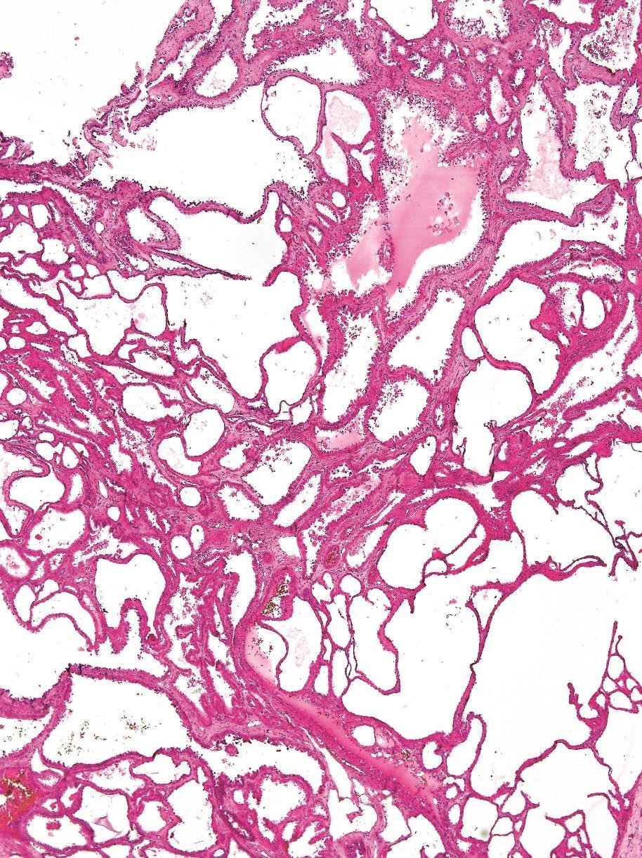
6 Tubulocystic Renal Cell Carcinoma


7 Tubulocystic Renal Cell Carcinoma Microscopic features: Cysts/tubules lined by single layer of cuboidal cells with eosinophilic to oncocytic cytoplasm Hobnail morphology Enlarged nuclei with prominent nucleoli Yang et al, AJSP, 2008

 prominent nucleoli")
8 Tubulocystic Renal Cell Carcinoma IHC and cytogenetic profile: EMA, vimentin, PAX-8 + CK8, CK18, CK19 + CD10, AMACR, CK7 + HMWCK Clinical outcome: Most present at low stage Low potential for metastasis (~10%, to bone and liver) prominent nucleoli hobnail 8


9 Tubulocystic Renal Cell Carcinoma Some cases may display focal papillary pattern
10 Tubulocystic Renal Cell Carcinoma Clinical outcome: Most present at low stage (limited to kidney) Low potential for metastasis (~10%, to bone and liver) F/U available in 11/13 [mean 27 (1-90) months]: - 10 NED; 1 AWD (PRCC) Differential diagnosis: - Cystic nephroma (CN)/Mixed epithelial and stromal tumor (MEST) - Papillary RCC - Metastasis - Collecting duct carcinoma
11 End Stage Renal Disease (ESRD) ESRD patients are more prone to kidney neoplasm: % ESRD patients with ACKD are even more prone to develop carcinoma (incidence 3-7%, a risk ~100 times that of general population) 40% of tumors are classic clear cell, papillary or chromophobe RCC Majority (60%) are represented by 2 new subtypes: - Acquired cystic disease-associated RCC (36-46%) - Clear cell (tubulo)papillary RCC of end stage kidneys (23%) Tickoo et al. AJSP 2006
 Tickoo et al. AJSP 2006; Sule et al.")
12 Acquired cystic disease-associated RCC (ACD-associated RCC) Occurs in kidneys with ACKD Often multifocal and bilateral Size range 1-8 cm Well-circumscribed; some have focally calcified capsule Hemorrhage and necrosis are common Many seem to arise in a cyst All patients on dialysis (8-11years) Tickoo et al. AJSP 2006; Sule et al. AJSP 2005


13 ACD-associated RCC Most common patterns: tubulocystic and papillary; solid, solid-alveolar, microcystic, macrocystic are also known tubulocystic pattern papillary pattern alveolar pattern


14 ACD-associated RCC Inter or intracellular microlumen formation leading to cribriform architecture Eosinophilic cells with large nuclei prominent nucleoli Frequent presence of intratumoral oxalate crystals
15 ACD-associated RCC IHC and cytogenetic profile: CK7, HMWCK, CAIX AMACR, CD57, vinculin + CD10 + (proximal tubular cell differentiation) Clinical outcome: - Relatively good prognosis (low grade, stage, metastatic rate than sporadic RCC) - Mostly indolent tumors - Has metastatic potential CK7 AMACR
16 Acquired Cystic Disease-Associated RCC: IHC and differential diagnosis PAX-8 CAIX CD10 HMWCK CK7 AMACR CD117 ACD-RCC Oncocytoma ESC-RCC HLRCC SDH-deficient RCC CK7 AMACR

; most cases have at least a partial")
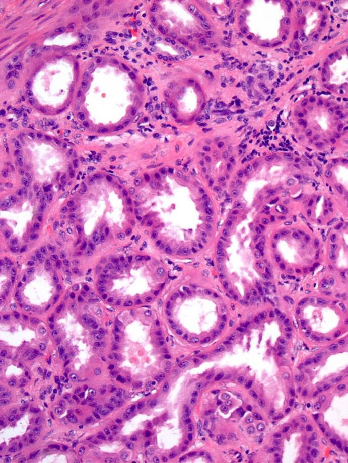
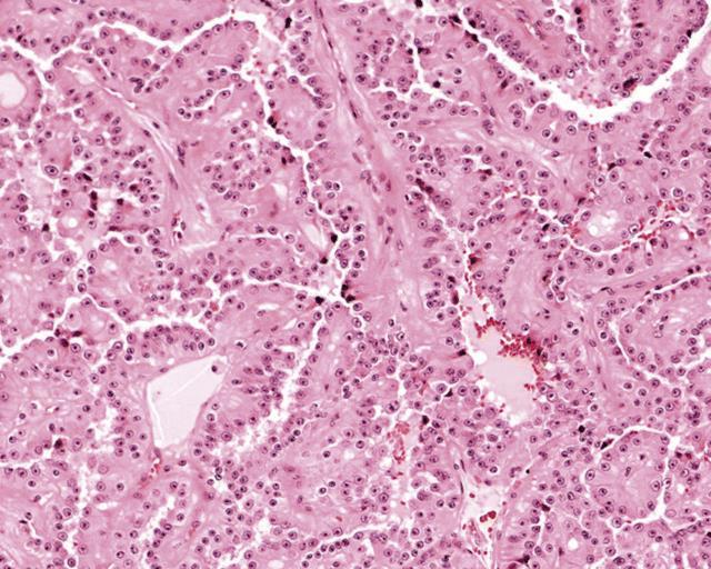
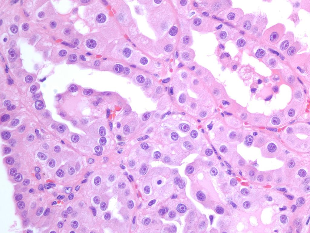
17 Clear cell papillary renal cell carcinoma Low-stage, low-grade tumor Both in ACKD patients and sporadic 4 th most common type of RCC Usually present in 6 th decade Well circumscribed small tumors Frequently cystic (up to 90%) with a thick circumferential capsule (>70%); most cases have at least a partial capsule


18 Clear cell papillary renal cell carcinoma Tubulopapillary architecture with compressed tubules lined by clear cells Short papillae arising from cyst wall, lined by a single layer of cells with clear cytoplasm Prominent fibrotic stroma


19 Clear cell papillary renal cell carcinoma 19


20 Tubules/Acini Solid tubules Clear cell papillary renal cell carcinoma Branching acini 20 Clear cell ribbon

21 Clear cell papillary renal cell carcinoma Linear arrangement of nuclei away from basal aspect of cells Lacks vascular pattern typical of CCRCC
 + AMACR, TFE3 CD10 usually - No LOH of 3p; no VHL")

22 Clear cell papillary renal cell carcinoma IHC and cytogenetic profile: CK7, HMWCK + CAIX (cup-like) + AMACR, TFE3 CD10 usually - No LOH of 3p; no VHL mutation/methylation No chr. 7/17 trisomy or chr. Y loss Prognosis: Favorable; no reported cases of metastasis or death Most cases are stage pt1 CK7 AMACR 22


23 CCRCC with features of CCPRCC Williamson et al. AJSP 2015; Dhakal et al. AJSP 2016
24 MiT family translocation renal cell carcinomas Members of MiTF/TFE transcription factor family - MiTF, TFE3, TFEB, TFEC Xp11 translocation carcinoma (TFE3 gene fusions) t(6;11)(p21;q12) carcinoma (alpha-tfeb gene fusion) Both types of translocation result in overexpression of fusion gene products Activate transcription of similar genes in vitro Translocation RCC express proteins normally driven by MiTF which are not expressed in other RCCs - Melanocytic markers - Cathepsin K (Martignoni et al. Mod Pathol 2009)
25 Xp11 translocation renal cell carcinoma Gene fusions involving TFE3 transcription factor Typically young patients; range: yrs (median 22 yrs) Comprise most of pediatric RCC; 1-2% of adult RCC 10-15% of cases are association with prior chemotherapy Stage and age predict outcome Fusion partners Tumor Age Translocation ASPL-TFE3* RCC 1-75 t(x;17)(p11.2;q25) PRCC-TFE3 RCC 2-69 t(x;1)(p11.2;q21) SFPQ-TFE3 RCC 5-68 t(x;1)(p11.2;p34) NonO-TFE3 RCC 39 inv(x)(p11.2;q12) CLTC-TFE3 RCC 14 t(x;17)(p11.2;q23)
] - papillary structures lined with clear cells - abundant clear to eosinophilic cytoplasm - discrete cytoplasm membrane - prominent")
26 Xp11 translocation renal cell carcinoma ASPL-TFE3 RCC [t(x;17)(p11.2;q25)] - papillary structures lined with clear cells - abundant clear to eosinophilic cytoplasm - discrete cytoplasm membrane - prominent nucleoli - psammoma bodies associated with hyaline nodules are often present
] - nested pattern with clear to eosinophilic cytoplasm - less abundant cytoplasm - less")
27 Xp11 translocation renal cell carcinoma PRCC-TFE3 RCC [(X;1)(p11.2;q21)] - nested pattern with clear to eosinophilic cytoplasm - less abundant cytoplasm - less prominent psammoma bodies and hyaline nodules
28 ASPL-TFE3 vs. PRCC-TFE3 RCC Feature Nodal metastasis Distant mets at presentation ASPL-TFE3 (X;17) 75% (24/32) 20% (8/40) Cathepsin K IHC 0 (0/8) PRCC-TFE3 (X;1) 35% (5/14) 0 (0/26) 86% (12/14) p value p=0.02 p=0.02 p= Ellis CL et al. Mod Pathol. 2014;27:875-86

29 Xp11 translocation renal cell carcinoma IHC and cytogenetic profile: Vimentin, CD10, RCC + CK7 - Pan-CK, AE1/3, CAM5.2, EMA: weakly/focally + Chromosomal translocation detected by: - Nuclear labeling for TFE3 protein by IHC - TFE3 rearrangements by FISH TFE3 Magers et al. Arch Pathol Lab Med 2015

30 Xp11 RCC confirmed by TFE3 break-apart FISH
")

31 t(6;11)(p21;q12) renal cell carcinoma Alpha-TFEB Gene Fusion 49 genetically confirmed cases Ages 3-68 years (median 31) Female predominance Size usually up to 10 cm 4 metastases/tumor related deaths in limited follow up PAX8+ Often Cytokeratin -

32 t(6;11) renal cell carcinoma with biphasic pattern

33 t(6;11) RCC Resembling Xp11 translocation RCC!


34 t(6;11) renal cell carcinoma IHC profile: HMB45+, Melan A+ Mostly cathepsin K+, PAX-8 + OSCAR, AE1/AE3, CAM5.2 focally + EMA Chromosomal translocation detected by: - Nuclear labeling for TFEB protein by IHC - TFEB rearrangements by FISH Smith NE et al. AJSP 2014;38: CK TFEB

35 t(6;11) RCC confirmed by TFEB break-apart FISH AJSP 2012;136:


36 t(6;11) RCC confirmed by TFEB break-apart FISH 36
")
 0% CCRCC, PRCC, ChRCC 0%")
37 Cathepsin K IHC distinguishes translocation RCC from other RCC t(6;11) RCC 100% PRCC-TFE3 RCC (X;1) 86% ASPL-TFE3 RCC (X;17) 0% CCRCC, PRCC, ChRCC 0% t(6;11) Cathepsin K


38 TFEB amplified renal cell carcinoma Emerging entity Most cases show predominant papillary architecture with high-grade features and oncocytic phenotype Potentially aggressive subtypes of RCC Argani P et al. AJSP 2016; 40; Skala SL et al. Mod Pathol 2018; Gupta S. Mod Pathol 2017
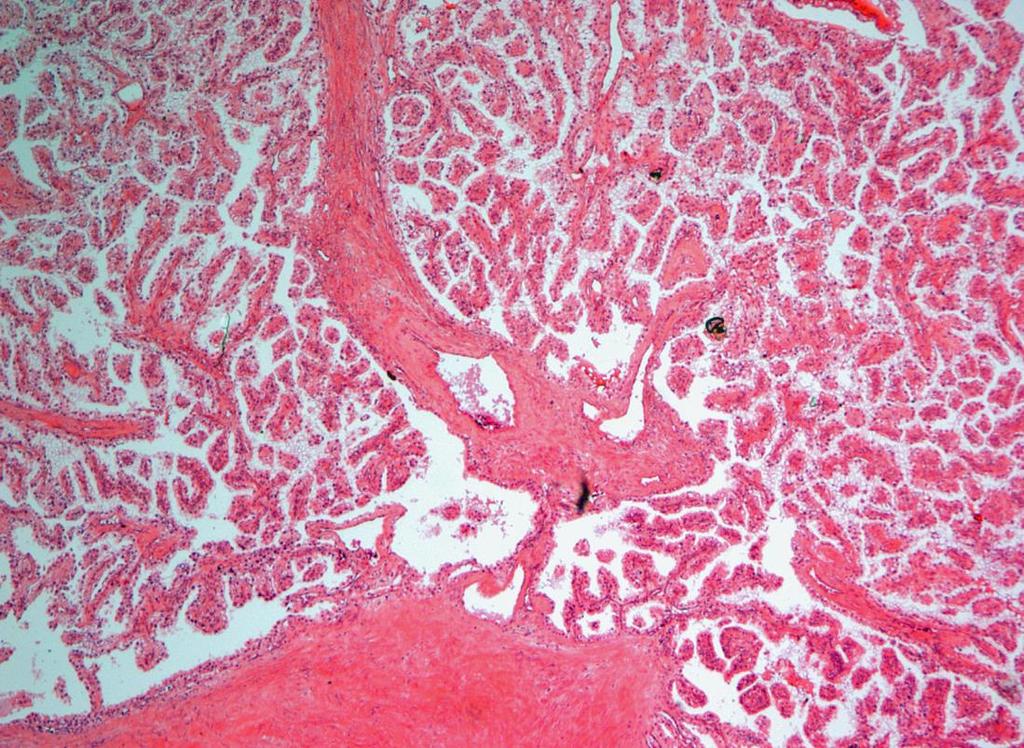
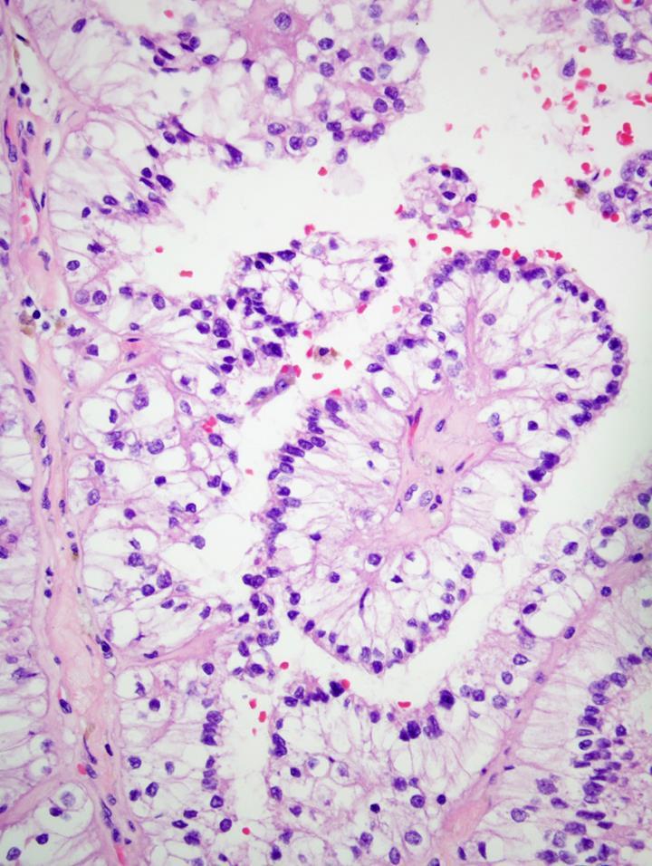
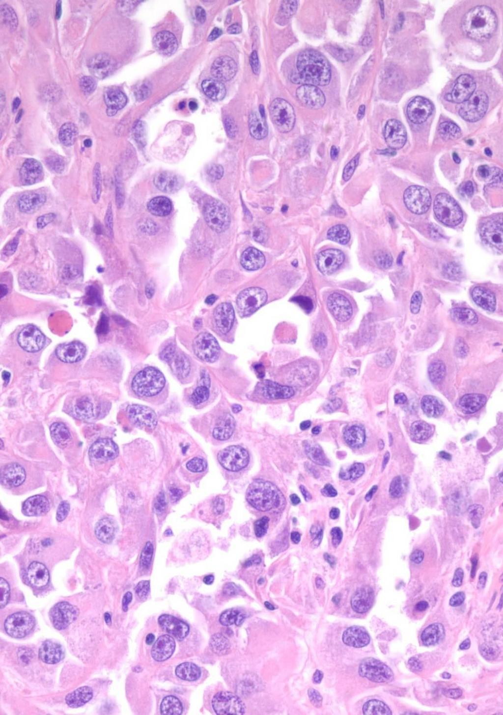
39 Hereditary leiomyomatosis and renal cell carcinoma associated renal cell carcinoma Autosomal dominant syndrome Germline-inactivating mutation in FH gene (1q42.3-1q43), encoding enzyme fumarate hydratase Cutaneous and uterine smooth muscle tumors Unilateral renal tumors (4 th decade) resembling type 2 papillary RCC: - papillary, tubulopapillary, tubular, solid patterns - papillae lined by tall cells with abundant eosinophilic cytoplasm - enlarged nuclei with a prominent eosinophilic nucleolus surrounded by a clear halo High stage at presentation, poor clinical outcomes Merino et al, AJSP 2007


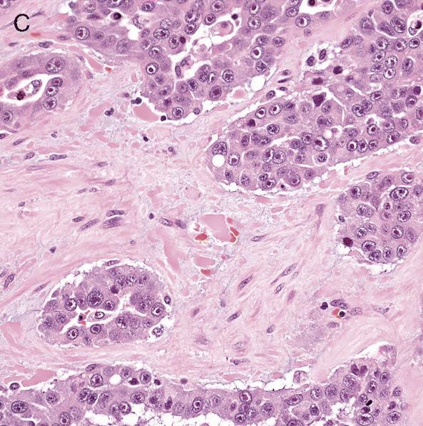
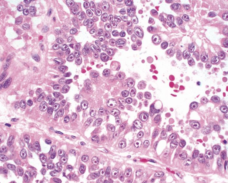
40 HLRCC Merino MJ et al. AJSP
41 HLRCC 41


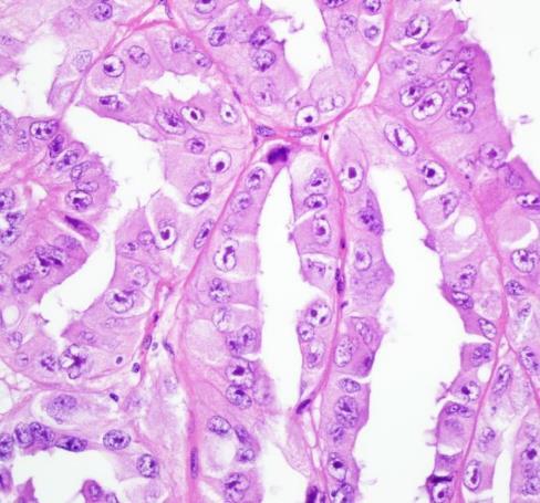
42 HLRCC CAIX AMACR Variable IHC: CK7, CAIX AMACR, CD10 + CK7 CD10 Courtesy Dr. J Lopez, Bilbao


")
43 FH-deficient RCC sensitive & specific specific, not sensitive fumarate hydratase (FH) 2-succinocysteine (2SC)
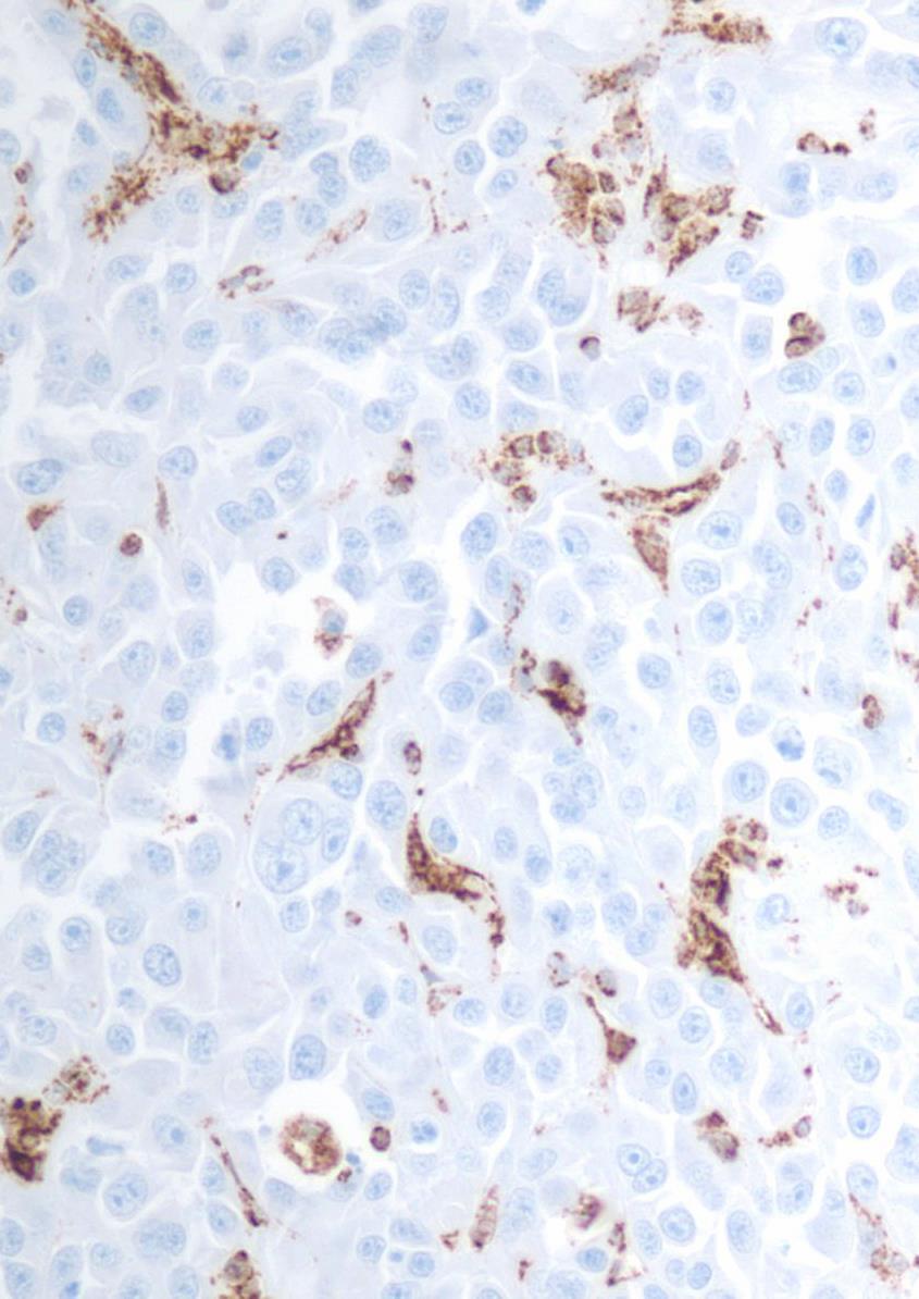
44 FH-deficient RCC FH
45 Hereditary leiomyomatosis and renal cell carcinoma associated renal cell carcinoma Clinical features definitional for HLRCC: Multiple biopsy-proven cutaneous piloleiomyomas or 2 of the following minor criteria: - Surgical treatment for symptomatic uterine leiomyomas before age 40 - Type 2 papillary RCC before age 40-1 st degree family member who meets these criteria
: A frequent morphologic pattern of")
 RCCs with TC-PD features are enriched for")

46 Tubulocystic carcinoma with poorly differentiated foci (TC-PD): A frequent morphologic pattern of FH-deficient RCC (Smith SC et al. AJSP 2016) RCCs with TC-PD features are enriched for FH deficiency FH-deficient RCC proposed term for tumors with suggestive morphology and IHC where genetic confirmation is unavailable
47 Succinate dehydrogenase (SDH) - deficient RCC Germline mutations of genes encoding SDH subunits result in hereditary syndromes: - Pheochromocytoma/paraganglioma - GIST - Pituitary adenomas - RCC SDH-deficient RCC ~30% multifocal or bilateral Solid or focally cystic growth Uniform cytology; eosinophilic flocculent cytoplasm, intracytoplasmic vacuolations and inclusions

48 SDH - deficient RCC 48

49 SDH - deficient RCC 49

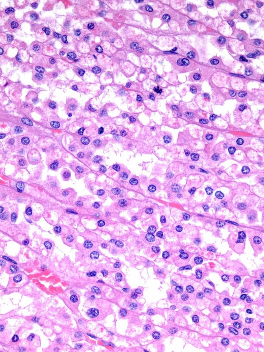
50 SDH - deficient RCC

51 Succinate dehydrogenase - deficient RCC 51
 SDHA-deficient RCC (2 cases): - genetic alteration likely somatic SDHB Kumar R et al. BMC Urol.")
52 Succinate Dehydrogenase (SDH) Deficient - RCC: IHC and differential diagnosis SDH-deficient RCC SDHB PAX-8 EMA CD10 CK20 CK7 CD * *stains intratumoral mast cells Mutation analysis: - double-hit inactivation of SDH-related genes (75% involve SDHB) SDHA-deficient RCC (2 cases): - genetic alteration likely somatic SDHB Kumar R et al. BMC Urol. 2018

53 Succinate dehydrogenase (SDH) - deficient RCC Mostly low stage, low grade May behave aggressively: - high nuclear grade - tumor necrosis - sarcomatoid differentiation 11% metastatic rate at longterm F-U Genetic evaluation of 1 st - degree relatives may be considered Gill et al. AJSP 2014

54 A distinctive low-grade oncocytic FH-deficient RCC, morphologically reminiscent of SDH-deficient RCC (Smith SC et al. Histopathology 2017; Li Y et al. Histopathology Novel form of FH deficient RCC with low grade oncocytic morphology, in 4 males (aged years) Solid, nested and focally tubular architecture; uniform polygonal cells; flocculent, vacuolated eosinophilic cytoplasm, scattered inclusions, fine chromatin, inconspicuous nucleoli FH deficient with retained SDHB expression

55 Low-grade oncocytic FH-deficient RCC 55

56 Low-grade oncocytic FH-deficient RCC 56
57 Emerging/provisional renal cell carcinomas WHO 2016
 Some are variants of CCRCC or CCPRCC Sporadic or associated with tuberous sclerosis")

58 Renal cell carcinoma associated with prominent angio(leiomyomatous) stroma Renal angiomyoadenomatous tumour (RAT) Some are variants of CCRCC or CCPRCC Sporadic or associated with tuberous sclerosis Branching tubules and papillae lined by clear/granular cells surrounded by abundant vascular and smooth muscle stroma CK7 +, CD10 + AMACR No 3p deletion; no trisomy 7 or 17 Michal et al. Ann Diagn Pathol 2000
59 ALK translocation renal cell carcinoma Chromosomal rearrangements involving anaplastic lymphoma kinase gene (ALK) at 2p23 result in fusion with various partner genes
60 ALK translocation renal tumors VCL-ALK VCL-ALK VCL-ALK TPM3-ALK; ELM4-ALK ALK rearrang. STRN-ALK Age (yrs) , 53 61, 59 33, 38 Sex M M M F, F M, M F, M Ethnicity African- American African- American African- American Japanese - Japanese Sickle cell Yes Yes Yes No No No Follow-up 9 mo, alive 21 mo, alive 19 mo, alive 2 y alive, 3 y alive Dead, 4 y; 1.4 y 9 y, 15 y nodal recurrence; 27 y alive; 3 mo alive w distant mets Size ; 2.5, ; 5.0 <4.0; 4.5 Location Medulla Medulla Pelvis Cortex - Medulla to cortex Pattern Author Diffuse sheet-like, polygonal cells Debelenko et al Solid, spindleshaped cells Marino- Enriquez et al Sheets of spindledpolygonal cells Smith et al Papillary, tubular and cribriform; eosinoph, clear cell Sugawara et al Papillary, cleareosinophilic cytoplasm Sukov et al Papillary, cribriform, solid; eosinophilic cytoplasm; signetring cell, rhabdoid Kusano et al. 2016
 and ALK Patients age: 6, 6 and 16 yrs VCL-ALK RCCs have distinctive")

61 ALK rearrangement in sickle cell trait-associated RCC 3 RCC harboring a t(2;10)(p23;q22) translocation resulting in fusion of vinculin (VCL) and ALK Patients age: 6, 6 and 16 yrs VCL-ALK RCCs have distinctive morphology: polygonal to spindle cells with abundant eosinophilic cytoplasm and frequent intracytoplasmic lumina Marino-Enriquez et al. Genes Chromosomes Cancer 2011; Debelenko et al. Mod Pathol 2011; Smith et al. AJSP 2014


62 TPM3-ALK and EML4-ALK fusion in RCC Micropapillary Papillary with mucin production Rhabdoid Sugawara et al. Cancer 2012 Courtesy of Kengo Takeuchi, Tokyo and Naoto Kuroda, Kochi
 treatment??? Sukov et al.")
63 ALK- rearrangement and copy number gain in adult renal tumors ALK rearrangements (<1%) ALK rearrangement was associated with poor outcome ALK copy number gain (10%) In CCRCC, ALK copy number gain was associated with tumor size, nuclear grade, and worse 10-yr cancer-specific survival Any benefit from ALK inhibitor (crizotinib) treatment??? Sukov et al. Mod Pathol 2012
64 Eosinophilic, solid, and cystic (ESC) - RCC Sporadic and in patients with tuberous sclerosis (TS) Sporadic tumors all in Solid and macrocystic or only solid appearance Microscopically: - Cysts lined by cells with pronounced hobnail arrangement - Cells with voluminous eosinophilic cytoplasm, prominent granular cytoplasmic stippling - Round to oval nuclei with prominent nucleoli Indolent behavior: 13/14 patients without disease progression after 2 to 138 months (mean 53) Trpkov at al. AJSP 2016

65 Eosinophilic, Solid, and Cystic Renal Cell Carcinoma 65


66 Eosinophilic, Solid, and Cystic Renal Cell Carcinoma 66

67 Eosinophilic, Solid, and Cystic Renal Cell Carcinoma pronounced hobnail arrangement 67

68 Eosinophilic, Solid, and Cystic Renal Cell Carcinoma cytoplasmic stippling 68

69 Eosinophilic, Solid, and Cystic Renal Cell Carcinoma 69
70 Eosinophilic, solid, and cystic (ESC) - RCC: IHC and differential diagnosis PAX-8 CAIX CD10 CK20 CK7 AMACR CD117 ACD-RCC patchy - Oncocytoma HLRCC SDH-deficient RCC ACD-associated RCC
 4/10 ESC were")
71 Eosinophilic solid and cystic renal cell carcinomas have metastatic potential Li Y et al. Histopathology 2018; McKenney J et al. Histopathology y/o female with multifocal ESC RCC with liver and lung metastases 69 y/o female with 15 cm ESC RCC with hilar lymph node metastasis (see pic) 4/10 ESC were multifocal (one bilateral) 4/10 ESC occurred in males
72 Take Home Message Renal tumors are not a single histologic, clinical or molecular entity Current classification integrates genetic based information with conventional histology and IHC Discovery of genetic markers will enable more precise categorization of RCCs possibly leading to better risk stratification and more appropriate therapies
73 THANK YOU!
Disclosure. Relevant Financial Relationship(s) None. Off Label Usage None MFMER slide-1
 None. Off Label Usage None MFMER slide-1") Disclosure Relevant Financial Relationship(s) None Off Label Usage None 2013 MFMER slide-1 Case Presentation A 43 year old male, with partial nephrectomy for a right kidney mass 2013 MFMER slide-2 2013
Disclosure Relevant Financial Relationship(s) None Off Label Usage None 2013 MFMER slide-1 Case Presentation A 43 year old male, with partial nephrectomy for a right kidney mass 2013 MFMER slide-2 2013
Various hereditary, acquired and neoplastic conditions can lead to cyst formation in the kidney.
 Dr. Fatima AlAl-Hashimi Hashimi,, MD, FRCPath Salmaniya Medical Complex, Bahrain Various hereditary, acquired and neoplastic conditions can lead to cyst formation in the kidney. The most frequently encountered
Renal tumours: use of immunohistochemistry & molecular pathology. Dr Lisa Browning John Radcliffe Hospital Oxford
 Renal tumours: use of immunohistochemistry & molecular pathology Dr Lisa Browning John Radcliffe Hospital Oxford Renal tumours: the use of immunohistochemistry & molecular pathology Classification of RCC
What s New in Pathology of Genitourinary Tumors. Jiaoti Huang, MD, PhD Department of Pathology Duke University School of Medicine
 What s New in Pathology of Genitourinary Tumors Jiaoti Huang, MD, PhD Department of Pathology Duke University School of Medicine Kidney Tumors Multilocular cystic renal neoplasm of low malignant potential
Spectrum of Preneoplastic and Neoplastic Cystic Lesions of the Kidney in Adult. by dr. Banan Burhan Mohammed Lecturer in Pathology Department
 Spectrum of Preneoplastic and Neoplastic Cystic Lesions of the Kidney in Adult by dr. Banan Burhan Mohammed Lecturer in Pathology Department Various hereditary, acquired, and neoplastic conditions can
ACCME/Disclosures. M31078/07 Ondřej Hes 4/13/2016
 M31078/07 Ondřej Hes Department of Pathology Charles University and University Hospital Plzeň Bioptická laboratoř Plzeň Czech Republic ACCME/Disclosures The USCAP requires that anyone in a position to
DIAGNOSTIC SLIDE SEMINAR: PART 1 RENAL TUMOUR BIOPSY CASES
 DIAGNOSTIC SLIDE SEMINAR: PART 1 RENAL TUMOUR BIOPSY CASES Dr. Andrew J. Evans MD, PhD, FACP, FRCPC Consultant in Genitourinary Pathology University Health Network, Toronto, ON Case 1 43 year-old female,
Enterprise Interest Nothing to declare
 Enterprise Interest Nothing to declare Biopsy diagnosis of renal tumors. Current applications Ondřej Hes Department of Pathology Charles University and University Hospital Plzeň Czech Republic Dealing
Updated Classification of Renal cell carcinoma
 Updated Classification of Renal cell carcinoma Suchin Worawichawong, M.D., FRCPath (Thailand) Department of Pathology, Ramathibodi Hospital, Mahidol University Incidence: Global: 338,000 new cases, 130,000
Molecular genetics and immunohistochemistry characterization of uncommon and recently described renal cell carcinomas
 Review Article Molecular genetics and immunohistochemistry characterization of uncommon and recently described renal cell carcinomas Qiu Rao 1 *, Qiu-Yuan Xia 1 *, Liang Cheng 2, Xiao-Jun Zhou 1 1 Department
IMMUNOPROFILES OF THE MAJOR RENAL NEOPLASMS (%staining)
") Stain Clear Cell Papillary IMMUNOPROFILES OF THE MAJOR RENAL NEOPLASMS (%staining) Chromophobe Collecting Duct Carcinom a Sarcomatoid Xp11 Translocat ion Dr Jon Oxley See also www.jonoxley.com Page 1 MTSCC
Stain Clear Cell Papillary IMMUNOPROFILES OF THE MAJOR RENAL NEOPLASMS (%staining) Chromophobe Collecting Duct Carcinom a Sarcomatoid Xp11 Translocat ion Dr Jon Oxley See also www.jonoxley.com Page 1 MTSCC
Case 1 PLEASE TURN OFF YOUR CELL PHONES 3/28/2017. Disclosure of Relevant Financial Relationships. Disclosure of Relevant Financial Relationships
 PLEASE TURN OFF YOUR CELL PHONES Disclosure of Relevant Financial Relationships USCAP requires that all planners (Education Committee) in a position to influence or control the content of CME disclose
Diagnostic accuracy of percutaneous renal tumor biopsy May 10 th 2018
 Diagnostic accuracy of percutaneous renal tumor biopsy May 10 th 2018 Dr. Tzahi Neuman Dep.Of Pathology Hadassah Medical Center Jerusalem, Israel, (tneuman@hadassah.org.il) Disclosure: 1 no conflicts of
04/10/2018. What s new in renal tumor pathology what s important and why. Prognostic factors in RCC
 25th Annual Seminar in Pathology Pittsburgh, PA, April 26-29, 2018 What s new in renal tumor pathology what s important and why Kiril Trpkov, MD FRCPC Department of Pathology and Laboratory Medicine kiril.trpkov@cls.ab.ca
AGGRESSIVE VARIANTS OF PAPILLARY THYROID CARCINOMA DIAGNOSIS AND PROGNOSIS
 AGGRESSIVE VARIANTS OF PAPILLARY THYROID CARCINOMA DIAGNOSIS AND PROGNOSIS PAPILLARY THYROID CARCINOMA Clinical Any age Microscopic to large Female: Male= 2-4:1 Radiation history Lymph nodes Prognosis
ACCME/Disclosures ALK FUSION-POSITIVE MESENCHYMAL TUMORS. Tumor types with ALK rearrangements. Anaplastic Lymphoma Kinase. Jason L.
 Companion Meeting of the International Society of Bone and Soft Tissue Pathology The Evolving Concept of Mesenchymal Tumors ALK FUSION-POSITIVE MESENCHYMAL TUMORS Jason L. Hornick, MD, PhD March 13, 2016
Renal Neoplasia: Diagnostic Problems and Recently Recognized Entities
 Renal Neoplasia: Diagnostic Problems and Recently Recognized Entities John N. Eble, M.D. Department Pathology Indiana University, Indianapolis, IN Holger Moch, M.D. Department Pathology University Hospital
RENAL EPITHELIAL NEOPLASMS: IS THERE A ROLE OF IMMUNOSTAINS IN DIAGNOSIS?
 RENAL EPITHELIAL NEOPLASMS: IS THERE A ROLE OF IMMUNOSTAINS IN DIAGNOSIS? John C. Cheville, M.D. Mayo Clinic and Mayo Foundation Rochester, MN The majority of renal epithelial neoplasms are diagnosed on
Case presentations 04/10/ th Annual Seminar in Pathology Pittsburgh, PA, April 26-29, 2018
 25th Annual Seminar in Pathology Pittsburgh, PA, April 26-29, 2018 Case presentations Kiril Trpkov, MD FRCPC Department of Pathology and Laboratory Medicine kiril.trpkov@cls.ab.ca 45 y/o male, pink tumor,
Normal thyroid tissue
 Thyroid Pathology Overview Normal thyroid tissue Normal thyroid tissue with follicles filled with colloid. Thyroid cells form follicles, spheres of epithelial cells (always single layered in health, usually
Pathology Mystery and Surprise
 Pathology Mystery and Surprise Tim Smith, MD Director Anatomic Pathology Medical University of South Carolina Disclosures No conflicts to declare Some problem cases Kidney tumor Scalp tumor Bladder tumor
POORLY DIFFERENTIATED, HIGH GRADE AND ANAPLASTIC CARCINOMAS: WHAT IS EVERYONE TALKING ABOUT?
 POORLY DIFFERENTIATED, HIGH GRADE AND ANAPLASTIC CARCINOMAS: WHAT IS EVERYONE TALKING ABOUT? AGGRESSIVE THYROID CANCERS PAPILLARY CARCINOMA CERTAIN SUBTYPES POORLY DIFFERENTIATED CARCINOMA HIGH GRADE DIFFERENTIATED
Synonyms. Nephrogenic metaplasia Mesonephric adenoma
 Nephrogenic Adenoma Synonyms Nephrogenic metaplasia Mesonephric adenoma Definition Benign epithelial lesion of urinary tract with tubular, glandular, papillary growth pattern Most frequently in the urinary
the urinary system pathology Dr. Fairoz A Eltorgman
 the urinary system pathology Dr. Fairoz A Eltorgman Tumors of the renal pelvis & kidney Benign tumors of the renal pelvis: Hemangioma Leiomyoma Malignant tumors: Transitional cell carcinoma Squamous cell
How does histology alter treatment? Cora N. Sternberg, MD, FACP Department of Medical Oncology San Camillo and Forlanini Hospitals Rome, Italy
 How does histology alter treatment? Cora N. Sternberg, MD, FACP Department of Medical Oncology San Camillo and Forlanini Hospitals Rome, Italy Targeting VHL/HIF in Clear Cell RCC VHL Bevacizumab (Antibody)
Kidney Case 1 SURGICAL PATHOLOGY REPORT
 Kidney Case 1 Surgical Pathology Report February 9, 2007 Clinical History: This 45 year old woman was found to have a left renal mass. CT urography with reconstruction revealed a 2 cm medial mass which
Case year old female presented with asymmetric enlargement of the left lobe of the thyroid
 Case 4 22 year old female presented with asymmetric enlargement of the left lobe of the thyroid gland. No information available relative to a prior fine needle aspiration biopsy. A left lobectomy was performed.
Salivary Glands 3/7/2017
 Salivary Glands 3/7/2017 Goals and objectives Focus on the entities unique to H&N Common board type facts Information for your future practice Salivary Glands Salivary Glands Major gland. Paratid. Submandibular.
Case 4 Diagnosis 2/21/2011 TGB
 Case 4 22 year old female presented with asymmetric enlargement of the left lobe of the thyroid gland. No information available relative to a prior fine needle aspiration biopsy. A left lobectomy was performed.
Lesions Mimicking Adenoid Cystic Carcinoma. Diagnostic Problems in Salivary Gland Pathology An Update 5/29/2009
 Diagnostic Problems in Salivary Gland Pathology An Update Lesions Mimicking Adenoid Cystic Carcinoma Stacey E. Mills, M.D. W.S. Royster Professor of Pathology Director of Surgical and Cytopathology University
Mammary analogue secretory carcinoma of salivary gland A case report of new entity
 Case Report Mammary analogue secretory carcinoma of salivary gland A case report of new entity Vaibhav Bhika Bari 1*, Sandhya Unmesh Bholay 2 1 Assistant Professor, 2 Associate Professor Rajiv Gandhi Medical
Unknown Slides Conference
 Unknown Slides Conference Jae Y. Ro, MD, PhD Weill Medical College of Cornell Univ. The Methodist Hospital, and UT MD Anderson Cancer Center Houston, TX November 9, 2013 Amman, Jordan 25 th Congress of
CYSTIC TUMORS OF THE KIDNEY JOHN N. EBLE, M.D. CYSTIC NEPHROMA
 Page 1 CYSTIC TUMORS OF THE KIDNEY JOHN N. EBLE, M.D. Department of Pathology & Laboratory Medicine Phone (317) 274-4806 Medical Science A-128 FAX: (317) 278-2018 635 Barnhill Drive jeble @iupui.edu Indianapolis,
Lecture: Dr Ming Zhou, Cleveland Clinic, Cleveland, Ohio, USA Uncommon renal tumour: Morphologic and molecular characteristics
 Australasian Division of the International Academy of Pathology Limited 35 th Annual Scientific Meeting June 4-6, 2010 Venue: Sydney Convention & Exhibition Centre, Darling Harbour, NSW International Society
International Journal of Pharma and Bio Sciences CHROMOPHOBE VARIANT OF RENAL CELL CARCINOMA MASQUARDING AS RENAL ONCOCYTOMA ON CYTOLOGY.
 Case Report Pathology International Journal of Pharma and Bio Sciences ISSN 0975-6299 CHROMOPHOBE VARIANT OF RENAL CELL CARCINOMA MASQUARDING AS RENAL ONCOCYTOMA ON CYTOLOGY. DR.MAMATHA K*, DR. ARAKERI
Ovarian Clear Cell Carcinoma
 Ovarian Clear Cell Carcinoma Rouba Ali-Fehmi, MD Professor of Pathology The Karmanos Cancer Institute, Wayne State University School of Medicine 50 year old woman with chief complaint of shortness of breath
Updates in Urologic Pathology WHO Made Those Changes?! Peyman Tavassoli Pathology Department BC Cancer Agency
 Updates in Urologic Pathology WHO Made Those Changes?! Peyman Tavassoli Pathology Department BC Cancer Agency World Health Organization Available in Feb 2016 Frame work for reporting Major contributing
4/12/2018. MUSC Pathology Symposium Kiawah Island April 18, Jesse K. McKenney, MD
 MUSC Pathology Symposium Kiawah Island April 18, 2018 Jesse K. McKenney, MD 1 Urothelial Carcinoma with Alternative Differentiation 2 Urothelial Carcinoma with Alternative Differentiation Recognition as
THE PATHOLOGY OF COMMON RENAL TUMORS. Victor E. Reuter, M.D Memorial Sloan Kettering Cancer Center
 THE PATHOLOGY OF COMMON RENAL TUMORS Victor E. Reuter, M.D Memorial Sloan Kettering Cancer Center A Practical Approach to Genitourinary Pathology Firenze, Italy May, 2016 Disclosures: none 1970 WHO classification:
Case Report Pediatric Papillary Renal Cell Carcinoma in a Horseshoe Kidney: A Case Report with Review of the Literature
 Case Reports in Pathology Volume 2015, Article ID 841237, 4 pages http://dx.doi.org/10.1155/2015/841237 Case Report Pediatric Papillary Renal Cell Carcinoma in a Horseshoe Kidney: A Case Report with Review
04/09/2018. Salivary Gland Pathology in the Molecular Era Old Friends, Old Foes, & New Acquaintances
 Salivary Gland Pathology in the Molecular Era Old Friends, Old Foes, & New Acquaintances Jennifer L. Hunt, MD, MEd Aubrey J. Hough Jr, MD, Endowed Professor of Pathology Chair of Pathology and Laboratory
Basement membrane in lobule.
 Bahram Memar, MD Basement membrane in lobule. Normal lobule-luteal phase Normal lobule-follicular phase Lactating breast Greater than 95% are adenocarcinomas in situ carcinomas and invasive carcinomas.
Case: The patient is a 24 year- old female who was found to have multiple mural nodules within the antrum. Solid and cystic components were noted on
 Case: The patient is a 24 year- old female who was found to have multiple mural nodules within the antrum. Solid and cystic components were noted on imaging. There is no significant past medical history.
Pathology of Renal Neoplasms: Recent Advances
 Pathology of Renal Neoplasms: Recent Advances Jae Y. Ro, M.D., Ph.D. The Methodist Hospital Weill Medical College of Cornell University, MD Anderson Cancer Center, Houston, Texas Ewha Womans University
57th Annual HSCP Spring Symposium 4/16/2016
 An Unusual Malignant Spindle Cell Lesion to Involve the Breast Erinn Downs-Kelly, D.O. Associate Professor of Pathology University of Utah & ARUP Laboratories No disclosures Case 39 y/o female with no
2 to 3% of All New Visceral Cancers Peak Incidence is 6th Decade M:F = 2:1 Grossly is a Bright Yellow, Necrotic Mass with a Pseudocapsule
 GENITOURINARY PATHOLOGY Kathleen M. O Toole, M.D. Renal Cell Carcinoma 2 to 3% of All New Visceral Cancers Peak Incidence is 6th Decade M:F = 2:1 Grossly is a Bright Yellow Necrotic Mass Grossly is a Bright
3/24/2017 DENDRITIC CELL NEOPLASMS: HISTOLOGY, IMMUNOHISTOCHEMISTRY, AND MOLECULAR GENETICS. Disclosure of Relevant Financial Relationships
 DENDRITIC CELL NEOPLASMS: HISTOLOGY, IMMUNOHISTOCHEMISTRY, AND MOLECULAR GENETICS Jason L. Hornick, M.D., Ph.D. Director of Surgical Pathology and Immunohistochemistry Brigham and Women s Hospital Professor
Intrarenal Extension. sinus
 Intrarenal Extension into sinus Document Capsular Penetration sinus 16 Pediatric Renal Tumor Staging Stage I Limited to Kidney & Completely Resected Intact Renal Capsule No Previous Rupture or Biopsy Renal
Pathology of the Thyroid
 Pathology of the Thyroid Thyroid Carcinoma Arising from Follicular Cells 2015-01-19 Prof. Dr. med. Katharina Glatz Pathologie Carcinomas Arising from Follicular Cells Differentiated Carcinoma Papillary
What is New in the 2015 WHO Lung Cancer Classification? Zhaolin Xu, MD, FRCPC, FCAP
 What is New in the 2015 WHO Lung Cancer Classification? Zhaolin Xu, MD, FRCPC, FCAP Professor, Dept of Pathology, Dalhousie University, Canada Pulmonary Pathologist and Cytopathologist, QEII HSC Senior
21/07/2017. Hobnail endothelial cells are not the same as epithelioid endothelial cells
 UPDATE IN CUTANEOUS VASCULAR S DERMATOPATHOLOGY SESSION BELFAST PATHOLOGY JUNE 21/2017 Dr E Calonje St John s Institute of Dermatology, London, United Kingdom THE FAMILY OF VASCULAR S WITH EPITHELIOID
RCC in ADPKD / CKD / ESRD
 RCC in ADPKD / CKD / ESRD FOIU 2018 David A. Goldfarb, MD,FACS Professor of Surgery, Cleveland Clinic Lerner College of Medicine Glickman Urological and Kidney Institute Cleveland Clinic, Cleveland, Ohio
USCAP 2012: Companion Meeting of the AAOOP. Update on lacrimal gland neoplasms: Molecular pathology of interest
 USCAP 2012: Companion Meeting of the AAOOP Vancouver BC, Canada, March 17, 2012 Update on lacrimal gland neoplasms: Molecular pathology of interest Valerie A. White MD, MHSc, FRCPC Department of Pathology
Oncocytic-Appearing Salivary Gland Tumors. Oncocytic, Cystic, Mucinous, and High Grade Salivary Gland Tumors SALIVARY GLAND FNA: PART II
 William C. Faquin, MD, PhD Professor of Pathology Harvard Medical School Director of Head and Neck Pathology Massachusetts Eye and Ear Massachusetts General Hospital SALIVARY GLAND FNA: PART II Oncocytic,
Case 2. Dr. Sathima Natarajan M.D. Kaiser Permanente Medical Center Sunset
 Case 2 Dr. Sathima Natarajan M.D. Kaiser Permanente Medical Center Sunset History 24 year old male presented with a 3 day history of right flank pain, sharp in nature Denies fever, chills, hematuria or
CASE year old male with a PET avid nodule in the left adrenal gland
 CASE 1 55 year old male with a PET avid nodule in the left adrenal gland Case 1 Adrenal gland parenchyma partly replaced by a spindle cell tumour with mild nuclear pleomorphism Atypical mitoses present
Enterprise Interest Nothing to declare
 Enterprise Interest Nothing to declare Diagnoses one would not like to miss in soft tissue pathology early in your career Marta Sbaraglia, MD Department of Pathology Hospital of Treviso University of Padua
RENAL CELL CARCINOMA 2 to 3% of All New Visceral Cancers Peak Incidence is 6th Decade M:F = 2:1 Grossly is a Bright Yellow, Necrotic Mass with a Pseud
 GENITOURINARY PATHOLOGY Kathleen M. O Toole Toole, M.D. RENAL CELL CARCINOMA 2 to 3% of All New Visceral Cancers Peak Incidence is 6th Decade M:F = 2:1 Grossly is a Bright Yellow, Necrotic Mass with a
Urinary Bladder: WHO Classification and AJCC Staging Update 2017
 Urinary Bladder: WHO Classification and AJCC Staging Update 2017 Houston Society of Clinical Pathologists 58 th Annual Spring Symposium Houston, TX April 8, 2017 Jesse K. McKenney, MD Classification
ACCME/Disclosures. Cribriform Lesions of the Prostate. Case
 Cribriform Lesions of the Prostate Ming Zhou, MD, PhD Departments of Pathology and Urology New York University Langone Medical Center New York, NY Ming.Zhou@NYUMC.ORG ACCME/Disclosures The USCAP requires
Case 1. ACCME/Disclosure. Clinical History. Dr. Mulligan has nothing to disclose
 Breast Evening Specialty Conference USCAP, 2016 Case 1 Anna Marie Mulligan University Health Network, Toronto University of Toronto ACCME/Disclosure Dr. Mulligan has nothing to disclose Clinical History
An Alphabet Soup of Thyroid Neoplasms
 Overall Objectives An Alphabet Soup of Thyroid Neoplasms Lester D. R. Thompson www.lester-thompson.com What is the current management of papillary carcinoma? What are the trends and what can we do differently?
Enterprise Interest None
 Enterprise Interest None What are triple negative breast cancers? A synopsis of their histological patterns Ian Ellis Molecular Medical Sciences, University of Nottingham Department of Histopathology,
Diplomate of the American Board of Pathology in Anatomic and Clinical Pathology
 A 33-year-old male with a left lower leg mass. Contributed by Shaoxiong Chen, MD, PhD Assistant Professor Indiana University School of Medicine/ IU Health Partners Department of Pathology and Laboratory
Mody. AIS vs. Invasive Adenocarcinoma of the Cervix
 Common Problems in Gynecologic Pathology Michael T. Deavers, M.D. Houston Methodist Hospital, Houston, Texas Common Problems in Gynecologic Pathology Adenocarcinoma in-situ (AIS) of the Cervix vs. Invasive
Case Scenario 1: Thyroid
 Case Scenario 1: Thyroid History and Physical Patient is an otherwise healthy 80 year old female with the complaint of a neck mass first noticed two weeks ago. The mass has increased in size and is palpable.
Case Report Morphologic heterogeneity and markers of renal cell carcinoma with t(6; 11)(p21; q12): a case report and literature review
(p21; q12): a case report and literature review") Int J Clin Exp Pathol 2017;10(12):11766-11770 www.ijcep.com /ISSN:1936-2625/IJCEP0065006 Case Report Morphologic heterogeneity and markers of renal cell carcinoma with t(6; 11)(p21; q12): a case report
Int J Clin Exp Pathol 2017;10(12):11766-11770 www.ijcep.com /ISSN:1936-2625/IJCEP0065006 Case Report Morphologic heterogeneity and markers of renal cell carcinoma with t(6; 11)(p21; q12): a case report
Follicular Derived Thyroid Tumors
 Follicular Derived Thyroid Tumors Jennifer L. Hunt, MD, MEd Aubrey J. Hough Jr, MD, Endowed Professor of Pathology Chair of Pathology and Laboratory Medicine University of Arkansas for Medical Sciences
Note: The cause of testicular neoplasms remains unknown
 - In the 15- to 34-year-old age group, they are the most common tumors of men. - Tumors of the testis are a heterogeneous group of neoplasms that include: I. Germ cell tumors : 95%; all are malignant.
Current insights in renal cell cancer pathology
 Urologic Oncology: Seminars and Original Investigations 26 (2008) 225 238 Review article Current insights in renal cell cancer pathology Vito Mancini, M.D. a, *, Michele Battaglia, M.D. a, Pasquale Ditonno,
carcinoma with multilocular cystic structures in an elderly patient: a case report
 Int J Clin Exp Pathol 2016;9(3):3987-3992 www.ijcep.com /ISSN:1936-2625/IJCEP0020826 Case Report Xp11.2/TFE3 translocation-associated renal cell carcinoma with multilocular cystic structures in an elderly
ACCME/Disclosures. Diagnosing Mesothelioma in Limited Tissue Samples. Papanicolaou Society of Cytopathology Companion Meeting March 12 th, 2016
 Diagnosing Mesothelioma in Limited Tissue Samples Papanicolaou Society of Cytopathology Companion Meeting March 12 th, 2016 Sanja Dacic, MD, PhD University of Pittsburgh ACCME/Disclosures GENERAL RULES
EDUCATIONAL CASES E1 & E2. Natasha Inglis 20/03/15
 EDUCATIONAL CASES E1 & E2 Natasha Inglis 20/03/15 CASE E1 79 year old female Rectum. Altemeier operation Histology Superficial erosions and mucosal congestion volcano lesion and pseudomembrane formation
Pathological Classification of Hepatocellular Carcinoma
 3 rd APASL Single Topic Conference: HCC in 3D Pathological Classification of Hepatocellular Carcinoma Glenda Lyn Y. Pua, M.D. HCC Primary liver cancer is the 2 nd most common cancer in Asia HCC is the
ARTHUR PURDY STOUT SOCIETY COMPANION MEETING: DIFFICULT NEW DIFFERENTIAL DIAGNOSES IN PROSTATE PATHOLOGY. Jonathan I. Epstein.
 1 ARTHUR PURDY STOUT SOCIETY COMPANION MEETING: DIFFICULT NEW DIFFERENTIAL DIAGNOSES IN PROSTATE PATHOLOGY Jonathan I. Epstein Professor Pathology, Urology, Oncology The Reinhard Professor of Urological
Case 1. Clinical history
 Case 1 Case 1 Clinical history 17-month-old boy with a kidney tumor found during routine childhood care program. CT scan showed a solid mass. Chemotherapy was given for 4 weeks using actinomycin D and
Surgical Pathology Evening Specialty Conference USCAP 2015
 Surgical Pathology Evening Specialty Conference USCAP 2015 John R. Goldblum, M.D. Chairman, Department of Pathology, Cleveland Clinic Professor of Pathology, Cleveland Clinic Lerner College of Medicine
Update in Salivary Gland Pathology. Benjamin L. Witt University of Utah/ARUP Laboratories February 9, 2016
 Update in Salivary Gland Pathology Benjamin L. Witt University of Utah/ARUP Laboratories February 9, 2016 Objectives Review the different appearances of a selection of salivary gland tumor types Establish
THYMIC CARCINOMAS AN UPDATE
 THYMIC CARCINOMAS AN UPDATE Mark R. Wick, M.D. University of Virginia Medical Center Charlottesville, VA CARCINOMA OF THE THYMUS General Clinical Features No apparent gender predilection Age range of 35-75
!! 2 to 3% of All New Visceral Cancers.!! Peak Incidence is 6th Decade!! M:F = 2:1
 !! Kathleen M. O Toole, M.D.!! 2 to 3% of All New Visceral Cancers!! Peak Incidence is 6th Decade!! M:F = 2:1!! Grossly is a Bright Yellow, Necrotic Mass with a Pseudocapsule 1 !!Conventional RCC! Clear
Case Based Learning Program
 Case Based Learning Program The Department of Urology Glickman Urological & Kidney Institute Cleveland Clinic Case Number 5 CBULP 2010 001 Case Based Urology Learning Program Editor: Associate Editor:
Hereditary Leiomyomatosis and Renal Cell Carcinoma Variant of Reed s Syndrome - A Rare Case Report
 American Research Journal of Urology Volume 1, Issue 1, pp:26-30 Case Hereditary Leiomyomatosis and Renal Cell Carcinoma Variant of Reed s Syndrome - A Rare Case Manas Babu, Devesh Bansal, Sony Mehta,
Tumors of kidney and urinary bladder
 Tumors of kidney and urinary bladder Overview of kidney tumors Benign and malignant Of the benign: papillary adenoma -cortical -small (0.5cm) -in 40% of population -clinically insignificant The most common
Papillary Lesions of the Breast A Practical Approach to Diagnosis. (Arch Pathol Lab Med. 2016;140: ; doi: /arpa.
 Papillary Lesions of the Breast A Practical Approach to Diagnosis (Arch Pathol Lab Med. 2016;140:1052 1059; doi: 10.5858/arpa.2016-0219-RA) Papillary lesions of the breast Span the spectrum of benign,
Prostate Pathology: Prostate Carcinoma, variants and Gleason Grading (Part 1)
") Prostate Pathology: Prostate Carcinoma, variants and Gleason Grading (Part 1) Jae Y. Ro, MD, PhD June 7, 2012 Ten Leading Cancer Types for the Estimated New Cancer Cases and Deaths By Sex, United States,
Prostate Pathology: Prostate Carcinoma, variants and Gleason Grading (Part 1) Jae Y. Ro, MD, PhD June 7, 2012 Ten Leading Cancer Types for the Estimated New Cancer Cases and Deaths By Sex, United States,
Unusual Variants of Bladder Cancer Cristina Magi-Galluzzi, MD, PhD
 Unusual Variants of Bladder Cancer Cristina Magi-Galluzzi, MD, PhD Director of Genitourinary Pathology, Professor of Pathology, Lerner College of Medicine Cleveland Clinic Objectives Update on variants
Thyroid master class. Thyroid Fine needle aspiration cytology and liquid-based techniques: Hologic and Becton Dickinson
 Thyroid master class Thyroid Fine needle aspiration cytology and liquid-based techniques: Hologic and Becton Dickinson Principle of LBC Collection of cells in liquid medium Immediate fixation Processor-prepared
THE FOLLICULAR VARIANT OF PAPILLARY THYROID CARCINOMA AND NIFTP
 THE FOLLICULAR VARIANT OF PAPILLARY THYROID CARCINOMA AND NIFTP FOLLICULAR VARIANT OF PAPILLARY CARCINOMA HISTORICAL PERSPECTIVE FOLLICULAR VARIANT OF PAPILLARY CARCINOMA 1960 described by Dr. Stuart Lindsay
FNA of Thyroid. Toward a Uniform Terminology With Management Guidelines. NCI NCI Thyroid FNA State of the Science Conference
 FNA of Thyroid NCI NCI Thyroid FNA State of the Science Conference Toward a Uniform Terminology With Management Guidelines Thyroid Thyroid FNA Cytomorphology NCI Thyroid FNA State of the Science Conference
Evening Specialty Conference: Cytopathology
 : Cytopathology N. Paul Ohori, M.D. University of Pittsburgh Medical Center Disclosure of Relevant Financial Relationships Disclosure of Relevant Financial Relationships USCAP requires that all planners
Newer soft tissue entities
 Newer soft tissue entities Examples among fibroblastic tumors Turku, May 6, 2010 Markku Miettinen, M.D. AFIP, Washington, DC Fibroblastic neoplasms Solitary fibrous tumor /Hemangiopericytoma Low-grade
Renal Cell Carcinoma: Genetics & Imaging Srinivasa R Prasad University of Texas San Antonio
 Renal Cell Carcinoma: Genetics & Imaging Srinivasa R Prasad University of Texas HSC @ San Antonio No financial disclosures Acknowledgements Dr. Peter Choyke, NIH My Gurus @ MIR, MGH 2004 WHO Taxonomy of
Immunohistochemical Diagnosis of Renal Neoplasms. Luan D. Truong, MD; Steven S. Shen, MD, PhD
 Immunohistochemical Diagnosis of Renal Neoplasms Luan D. Truong, MD; Steven S. Shen, MD, PhD N Context. Histologic diagnosis of renal neoplasm is usually straightforward by routine light microscopy. However,
Select problems in cystic pancreatic lesions
 Disclosure Select problems in cystic pancreatic lesions Five Prime Therapeutics shareholder Adicet Bio shareholder Bristol-Meyer Squibb advisory board grace.kim@ucsf.edu Pancreatic cystic lesions Intraductal
5/22/2017. An Aggressive Nasopharyngeal Tumor. Case History
 An Aggressive Nasopharyngeal Tumor Head & Neck/Endocrine Evening Specialty Conference Martin Bullock, MD, FRCPC Dalhousie University, Halifax, Nova Scotia Case History 52-year-old male, 6 month history
Anaplastic Large Cell Lymphoma (of T cell lineage)
") Anaplastic Large Cell Lymphoma (of T cell lineage) Definition T-cell lymphoma comprised of large cells with abundant cytoplasm and pleomorphic, often horseshoe-shaped nuclei CD30+ Most express cytotoxic
Anaplastic Large Cell Lymphoma (of T cell lineage) Definition T-cell lymphoma comprised of large cells with abundant cytoplasm and pleomorphic, often horseshoe-shaped nuclei CD30+ Most express cytotoxic
Self assessment case. Dr Saleem Taibjee Dorset County Hospital, Dorchester
 Self assessment case Dr Saleem Taibjee saleemtaibjee@gmail.com Dorset County Hospital, Dorchester Clinical details 34-year-old man: Shave excision Skin tag / papilloma left thigh The best diagnosis is:
Renal Masses in Patients with Known Extrarenal Primary Primary Cancer Primary Primary n Met Mets s RCC Beni L mphoma Lung Breast Others
 The Importance of Stuart G. Silverman, MD, FACR Professor of Radiology Harvard ard Medical School Director, Abdominal Imaging and Intervention Brigham and Women s Hospital Boston, MA The Importance of
3/27/2017. Pulmonary Pathology Specialty Conference. Disclosure of Relevant Financial Relationships. Clinical History:
 Pulmonary Pathology Specialty Conference Saul Suster, M.D. Medical College of Wisconsin Disclosure of Relevant Financial Relationships USCAP requires that all planners (Education Committee) in a position
59 yo male with past medical history of prostate carcinoma, presented with upper abdominal pain
 December 2016 59 yo male with past medical history of prostate carcinoma, presented with upper abdominal pain Contributed by: Divya Sharma, MD. Fellow, Gastrointestinal Pathology, Department of Pathology