MODIFIED ULTRA FAST PAP STAIN IN CYTOLOGY IN COMPARISION WITH REGULAR PAP STAIN AND MGG. Dissertation submitted in
|
|
|
- Bethany Evans
- 5 years ago
- Views:
Transcription


1 MODIFIED ULTRA FAST PAP STAIN IN CYTOLOGY IN COMPARISION WITH REGULAR PAP STAIN AND MGG Dissertation submitted in Partial fulfillment of the regulations required for the award of M.D.Degree in PATHOLOGY-BRANCH III THE TAMILNADU Dr. M.G.R. MEDICAL UNIVERSITY CHENNAI, TAMILNADU April 2017
2 DECLARATION I solemnly declare that this dissertation entitled MODIFIED ULTRA FAST PAP STAIN IN CYTOLOGY IN COMPARISION WITH REGULAR PAP STAIN AND MGG was done by me in the Department of Pathology, Coimbatore Medical College, Coimbatore during the period of July 2015 to June 2016 under the guidance and supervision of DR.V.PRABA, MD., Associate professor, Department of Pathology, Coimbatore Medical College, Coimbatore. This dissertation is submitted to the Tamilnadu Dr.M.G.R. Medical University, Chennai towards the partial fulfillment of the requirement for the award of M.D., Degree in Pathology. Place: Date: Dr. P. SELVI
3 CERTIFICATE This is to certify that the dissertation entitled MODIFIED ULTRA FAST PAP STAIN IN CYTOLOGY IN COMPARISION WITH REGULAR PAP STAIN AND MGG STAIN is a record of bonafide work done by Dr.P.SELVI., Post Graduate student in the Department of Pathology, Coimbatore Medical College and Hospital, Coimbatore under the guidance and supervision of Dr. V.PRABA, M.D., Associate Professor, Department of Pathology, Coimbatore Medical College and Hospital, Coimbatore in partial fulfillment of the regulations of Tamilnadu Dr.M.G.R. Medical University, Chennai towards the award of M.D.Degree (Branch III) in Pathology. Guide Dr.V.PRABA, M.D Associate professor, Department of pathology, Coimbatore Medical College, Coimbatore. Dr. C. Lalitha, M.D., Professor and Head, Department of Pathology, Coimbatore Medical College Coimbatore. Dr. Edwin Joe, M.D., B.L., Dean, Coimbatore Medical College, Coimbatore.
4
5 ACKNOWLEDGEMENT To begin with, I thank the almighty God for bestowing his blessing on me in this dissertation a successful one. I wish to thank the beloved Dean Dr.A. EDWIN JOE,M.D,B.L, Coimbatore Medical College and hospital, Coimbatore for permitting me to conduct this study. It s a great pleasure to express my humble gratitude to the most respectable teacher and my guide Dr. C.Lalitha. M.D., Professor and Head of the Department, Department of Pathology, Coimbatore Medical College, Coimbatore for her guidance and support. This dissertation bears her valuable suggestions and highly professional advice. I wish to express my gratitude and sincere thanks to Dr. A. Arjunan. M. D., for his guidance and support. I express my gratitude and sincere thanks to my guide Dr.V.PRABA.M.D., Associate Professor, Department of Pathology, Coimbatore Medical College, Coimbatore. I owe my gratitude Dr. A. Dhanalakshmi M. D., and Dr.G.SThiriveni Balajji M.D., for their timely advice and suggestion through the course of my work.
6 I thank all Assistant Professors and Tutors of Department of Pathology, Coimbatore Medical College, Coimbatore for their opinion and encouragement. I thank Department of Medicine, Surgery and Department of Surgical Oncology Coimbatore Medical College, Coimbatore for providing clinical cases, valuable support and guidance which made this dissertation Possible. I thank my family, husband Dr. A.Prabakaran and child P.Tharunikaa for their extensive support. I thank all lab technicians working in Department of Pathology, Coimbatore Medical College, Coimbatore.
7 Digital Receipt This receipt acknowledges that Turnitin received your paper. Below you will f ind the receipt inf ormation regarding your submission. The f irst page of your submissions is displayed below. Submission author: Assignment title: Submission title: File name: File size: Page count: Word count: Character count: Submission date: Submission ID: Md Pathology P.SELVI plagiarism MODIFIED ULTRA FAST PAP STAIN rol docx 1.87M 80 10,700 59, Sep :08PM Co pyright Turnitin. All rights reserved.
8
9 TABLE OF CONTENTS S.NO TITLE PAGE NO 1. INTRODUCTION 1 2. AIM AND OBJECTIVES 3 3. REVIEW OF LITERATURE 4 4. MATERIALS AND METHODS OBSERVATIONS AND RESULTS DISCUSSION SUMMARY CONCLUSION BIBLIOGRAPHY 10. ANNEXURES: I. PROFORMA II. LIST OF ABBREVIATIONS III. CONSENT FORM IV. KEY TO MASTER CHART V. MASTER CHART
10 LIST OF TABLES S.NO TITLE PAGE NO 1. Age distribution of cases studied Association of gender with study cases Association of gender with study cases Mean 57 Quality index of study Groups 4. Associations of Clinical Variables with Study 58 Subjects in Thyroid 5. Association of Clinical Variables with Study 60 Subjects in Breast 6. Association of Clinical Variables with Study 62 Subjects in Lymph Node 7. Association of PAP QI with study cases Association of MGG quality Index with Study 65 Cases 9. Association of MUFP quality Index with 66 Study Cases 10. Association of Background with Study cases 67
11 11 Association of overall staining with Study 68 cases 12 Association of cell morphology with study 69 cases 13 Association of Nuclear Characteristics with 70 Study cases 14 Association of Cytoplasmic details with 71 Study cases 15 Association of Air drying artifacts with 72 Study cases 16 Quality index of various organs in Shinde s 76 study and present study 17 Quality index of various organs in 76 Priyanka s study
12 LIST OF GRAPHS S.NO TITLE PAGE NO 1. Case Distribution Age distribution Association of gender with study cases Mean Quality index of study Groups Association of PAP QI with study cases Association of MGG QI with study cases 7. Association of MUFP QI with study cases 8. Association of Background with study cases 9. Association of overall staining with study cases 10. Association of Cell Morphology with study cases 11. Association of Nuclear Characteristics with Study cases 12. Association of Cytoplasmic details with Study cases 13. Association of Air drying artifacts with Study cases
13 LIST OF FIGURES FIGURES NUMBER TITLE 1. NODULAR GOITER-MUFP STAIN (10X & 40X) 2. NODULAR GOITER-PAP STAIN (10X & 40X) 3. NODULAR GOITER-MGG STAIN (40X) 4. HASHIMOTOS THYROIDITIS-MUFP STAIN (10X & 40X) 5. HASHIMOTO S THYROIDITIS-PAP STAIN (40X) 6. HASHIMOTO S THYROIDITIS-MGG STAIN (40X) 7. FIBROADENOMA BREAST -MUFP STAIN (40X) 8. FIBROADENOMA BREAST -PAP STAIN (10X & 40X) 9. FIBROADENOMA BREAST-MGG STAIN (40X) 10. DUCTAL CARCINOMA BREAST -MUFP STAIN (40X) 11. DUCTAL CARCINOMA BREAST -PAP STAIN (40X) 12. DUCTAL CARCINOMA BREAST -MGG STAIN (40X 13. TUBERCULOUS LYMPHADENITIS-MUFP STAIN (40X) 14. TUBERCULOUS LYMPHADENITIS -PAP STAIN (40X) 15. TUBERCULOUS LYMPHADENITIS -MGG STAIN, 40X SQUAMOUS CELL CARCINOMA SECONDARIES IN LYMPH NODE-MUFP STAIN, 40X SQUAMOUS CELL CARCINOMA SECONDARIES IN LYMPH NODE-PAP STAIN, 40X SQUAMOUS CELL CARCINOMA SECONDARIES IN LYMPH NODE-MGG STAIN, 40X
14 INTRODUCTION
15 INTRODUCTION The papanicolaou staining technique is a polychromatic staining method elaborated by George N Papanicolaou, who is considered to be the father of cytology in 1942 and further developed by him in 1954 and in Pap stain is used to differentiate the cells in smear preparations of various body fluids, gynecological smears and fine needle aspiration material from various organs. There has been a lot of controversy as to whether wet-fixed smears stained with Papanicolaou stain or air-dried smears stained with Romanowsky s stain are better. In fact, both are complementary, but Pap staining permit better assessment of nuclear features and are preferred by many histopathologists. Quick diagnosis of FNAC plays an important role in efficient medical practice. The need for minimal turnaround time for assessing FNA smears has encouraged innovations in staining technique that require lesser staining time with unequivocal cell morphology. Few rapid stains available these days include MGG stain, Diff quick stain and toluidine blue stain. However many cytopathologists prefer the transparent, traditional, crisp nuclear features offered by 95% 1
16 ethanol fixed PAP stain rather than air dried smears stained by Romanowsky stain. To overcome this, ultrafast papanicolaou stain was introduced by yang and Alvarez in 1994 which is a hybrid of romanowsky stain and pap stain. It not only reduces the time for pap stain to 90 seconds, but also enhances the quality. Pap stain requires ethanol for fixation. Ethanol is expensive and laboratory needs a license for acquiring ethanol in bulk quantity. 2
17 AIM & OBJECTIVES
18 AIM AND OBJECTIVES AIM: Aim of our study is to assess the use of modified ultra-fast pap stain in fine needle aspiration cytology of various organs in comparison with the standard conventional pap stain and MGG stain. OBJECTIVES: To compare the results of MUFP, Routine pap and May Grunwald Giesma (MGG) stain. To assess the quality of MUFP stain and to find the advantages of the same over routine stains used in cytology. To find a cost-effective method which can be adopted in our laboratory. To compare the quality of staining procedures used on air dried smears (MGG and MUFP) over the wet fixed smears (Pap stain) To assess the utility and applicability of these stains in cytomorphological study in FNAC of thyroid, breast, and lymph nodes. 3
19 REVIEW OF LITERATURE
20 REVIEW OF LITERATURE Cytology started as a revolutionary idea of looking at imprints of cut surface of tumors at postmortem. It has evolved through many new methods of procuring, fixing and staining cells. Its main attribute lies in its ability to allow prompt, accurate assessment of cell changes on material taken with minimally invasive procedure and processing. 1 eras; The evolution of cytopathology occurred through four overlapping a) : Early history. b) : Development and expansion of exfoliative cytology in the USA and elsewhere. c) : Consolidation of cytopathology as a discipline and the parallel developments of population screening and FNA cytology. d) : Maturation of cytopathology as a discipline and its integration with new technology (1985 to the present day). 1 In the early historical era microscopic observations of normal and abnormal human cells either in exfoliated or in imprints or scrapes were steadily and independently recorded throughout the nineteenth century. 2,3 4
21 By first decade of twentieth century, exfoliative cancer cells had been described in all types of specimens. 4 In 1922, professor LS Dudgeon used cytology at St. Thomas Hospital for the diagnosis of a wide variety of neoplastic and inflammatory diseases from imprints of surgical specimens. 5 Dudgeon considered that the stained films were much nicer to examine than paraffin sections. 6 At the same time in USA, FNA cytology was developing and the first series on aspiration of neoplasms was published from memorial Hospital for cancer and allied diseases in New York City. 7 A second era of cytopathology began in 1941 with the publication of The diagnostic value of vaginal smears in carcinoma of uterus by George N. Papanicolaou, an anatomist, and Herbert F. Traut, a gynaecologist. 8 Papanicolaou contribution to this field was two-fold; he recognized the importance of wet fixation of cytological specimens and he systematically began to accumulate examples of cancer cells in vaginal smears, culminating in his paper New Cancer Diagnosis. 9 Concurrently with the development of cervical screening the cytological method of cancer diagnosis began to be more widely applied to the respiratory, alimentary and urinary tracts as well as to the serous cavities and the central nervous system. 1 5
22 The era of consolidation was heralded by two publications: the first issue of Acta cytologica in the oldest journal devoted exclusively to cytopathology; and in by the publication of Diagnostic cytology and its Histopathologic bases by Leopold G.Koss in association with Grace R. Durfe. 10 In the last 60 years there is an explosion in the literature of cytopathology, with thousands of articles and scores of books written on the subject. 1 Population based cervical screening is now practiced to a greater or lesser extent in almost all countries of the developed world. 1 The imperial cancer Research Fund Coordinating Committee on cervical screening made the statement in 1984 that with the exception of non smoking, cervical cytology screening offers the only major proved public health measure for significantly reducing the burden of disease, 11 its introduction was highly controversial and has remained so at every stage of its development. By 1986, there was sufficient evidence from an international multicentre analysis to show that 5 yearly and 3-yearly screening reduced the risk of invasive cancer by 84% and 93% respectively, while little additional benefit was achieved by annual screening. 12 6
23 The impetus of the development of cytopathology as we know it today resulted from the painstaking research of papanicolaou in the USA. Thus papanicolaou is justly referred to as the Father of cytopathology. 13 PAPANICOLAOU STAIN For the routine diagnostic cytology, the papanicolaou stain is recommended. The use of the papanicolaou stain results in well-stained nuclear chromatin, differential cytoplasmic counterstaining, and cytoplasmic transparency. 14 Although, originally developed for interpretation for gynaecological specimen, Papanicolaou staining is now commonly used to facilitate the accurate detection and interpretation of abnormal cells from variety of sources. NUCLEAR STAINS 15 : Haematoxylin: Haematoxylin - most widely used nuclear stain. It is extracted from logwood campeachy wood). The Haematoxylin campechianum is a tree that has been scientifically cultivated in Jamaica since Freshly cut wood is colorless. It becomes dark reddish-brown when exposed to atmospheric oxygen. Haematoxylin is not a dye, only the 7
24 oxidation product of hematoxylin-hematein is a weak anionic dye. Oxidation of hematoxylin achieved naturally by exposing the solutions to atmospheric oxygen or by using oxidizing agents such as sodium iodate, mercuric oxide, and potassium permanganate. Oxidation process is called ripening. Solutions should always contain some unoxidized hematoxylin, since complete or over oxidation leads to a breakdown of the solution and the loss of good staining. Oxidized hematoxylin (hematein) has little affinity for tissue but becomes a strong dye with a particular affinity for nuclei when combined with a metallic mordant. In some solutions of hematoxylin, the oxidizer also serves as the mordant. For (e.g);iron hematoxylin,but it is not stable. To achieve stability, the mordant should not oxidize the solution. Ammonium or potassium aluminum sulfate, phosphotungstic acid, and phosphomolybdic acid are mordants. The mordant-dye combination is called lake. most commonly used hematoxylin lakes are combinations of hematein with either aluminum or iron. The routine nuclear stains should be called aluminum hematoxylins, or more properly aluminum hemateins, since aluminium is a mordant. 8
25 More selective nuclear staining achieved by adding either an excess of acid or an excess of aluminum. Formulas for some of the aluminum hematoxylins follow, Harris Hematoxylin Hematoxylin g Absolute ethyl alcohol ml Ammonium aluminium sulfate g Distilled water ml Mercuric oxide g Gill s hematoxylin (Gill et al modified) Preparation of solution Hematoxylin 2 g Sodium iodate 0.2 g Aluminum sulfate 17.6 g Distilled water 750 ml Ethylene glycol (ethandiol) 250 ml Glacial acetic acid 20 ml 9
26 Ehrlich s Hematoxylin Hematoxylin.. 2gm 95% Alcohol.. 100ml Distilled water.. 100ml Glycerol.. 100ml Ammonium or Potassium Aluminium sulphate.. 3gm Glacial acetic acid.. 10m It is used as progressive or Regressive staining. In progressive staining the reaction is stopped once the desired staining intensity is achieved. In Regressive staining, longer time is required to over stain the tissue before the stain is selectively removed in acid alcohol (1% hydrochloric acid in 70% ethanol). CYTOPLASMIC STAINS 15 : Counterstain is mostly used as eosin. The staining of eosin is best at a ph of 4.6 to
27 Used properly, three shades of pink can be obtained with eosin alone; erythrocytes, collagen, and the cytoplasm of muscle or epithelial cells should stain with different shades or intensities of pink. Eosin solution Eosin Y (1% aqueous solution) ml 95% ethyl alcohol ml Acetic acid, glacial.. 4.0ml 2.Eosin-Phloxine B Eosin Y (1% aqueous solution) ml Phloxine B (1% aqueous solution) ml 95% alcohol ml Acetic acid, glacial.. 5.0ml There are multiple acid dyes also used to provide differential counterstaining and Cytoplasmic transparency. - Orange G6 (OG 6). - Eosin azure 36 (EA 36 or EA 50) -contains light green, Eosin and bismark brown. 11
28 MODIFICATION OF PAPANICOLAOU STAIN 14 : Modification of the original papanicolaou stain (1942) was published by Dr.Papanicolaou in 1954 and Papanicolaou Technique I: Uses Harris hematoxylin regressively. Papanicolaou Technique II: Described for urinary and gastric preparations,uses hematoxylin progressively. Other modifications include Gill s modification, Miller s modification, Saccomanno s modification for carbowax fixed smears and Durfee s modification for urine sediment smears. FIXATIVES 14 : Rapid fixation of smears is needed to preserve cytological details of cells. For many years the fixative of choice for gynecologic and other smear preparations was the one recommended by Papanicolaou, a solution contain equal parts of ether and 95% of ethyl alcohol. Subsequently, it is was abandoned, since the ether present in the pap stain is a fire hazard. Ninety-five percent ethyl alcohol (ethanol) is now used as a fixative by many laboratories, with excellent results. Smears should remain in 95% ethanol fixative for a minimum of 15 minutes prior to staining. To obtain ethanol without federal taxation, a license is required. 12
29 EQUIVALENT CONCENTRATIONS OF SEVERAL ALCOHOLS FOR PURPOSES OF CELL FIXATION 100% Methanol 95% Ethanol 95% Denatured alcohol 80%propanol 80%Isopropanol WET FIXATION 14,16 : Wet fixation is traditional method of fixation in which smears are immediately kept in fixative before air-drying of smears. The disadvantages of wet fixed smears are air-drying artifacts, hemorrhagic background and cell loss during fixation. To overcome these disadvantages, in 1988, Chan and Kung reported that air-dried smears can be rehydrated by immersing the smears in 0.9% Nacl for 30sec which is used in MUFP. 13
30 FINE NEEDLE ASPIRATION CYTOLOGY: FNA was first introduced in Sweden by Franzen, a haematologist & oncologist tried the Romanowsky staining methods for cytology. 17 The technique was further developed by Soderstrom, Fox and also by Lopes Cardoso, Von Haam, Crepinko and Hauptmann. 18 In the UK, FNA was pioneered by a surgeon, John webb, who was given enthusiastic support by some of the renowed cytopathologists of the time. 19 The technique also became popular in the USA after a long interval since its early use in the 1930s. The present day focus of FNA cytology is on obtaining a satisfactory specimen on which a reliable diagnosis can be made and therefore, that the aspirate sample should provide true reflection of the disease process. 20 FNA is now established as the first line investigation of mass lesions where-ever they occur in the body
31 ADVANTAGES OF FNA 20 : It is a minimally invasive procedure. economical. The technique is relatively painless, produces a quick result and is The method is applicable to easily palpable lesions. New radiological techniques are now available for internal imaging of organs and also for FNA of deep seated impalpable structures. It can be done as OP procedure, so hospital stay can be avoided. Diagnosis can be obtained within minutes rather than days Mostly accurate in many situation, hence it is used as essential preoperative /pretreatment investigation Risk of complication will be less is an additional advantage that allows FNA cytology to be performed as an OP procedure. Cells obtained by FNA can be manipulated in a variety of ways useful for ultra structural study, Immunocytochemistry, gene rearrangement, morphometry, image analysis and DNA analysis. 15
32 In routine practice, success of FNAC depends on 1. Samples from the representative lesion. 2. Adequate number of cells and other tissue components. 3. Smear making and processing of the samples. 4. Accompanied by correct clinical /radiological information. LIMITATIONS OF FNAC FNA cytology has its own limitations. Scanty samples and loss of histological architecture leads to difficulty in interpretation of FNA smears based on morphology. There is a risk of complications of FNA. The overall morbidity and mortality to FNA has been estimated in several studies and the risk of death is approximately 1 in Serious complications have been reported such as major hemorrhage after FNA of lung, liver and kidney; septicemia after prostate aspiration; bile peritonitis following needling of liver; and acute peritonitis resulting from pancreatic aspiration. However, such complications are very rare. Review of literature shows, the risk is increased due to multiple passes, larger needles, and absence of normal parenchyma covering the lesion
33 Contraindications of deep seated aspiration- It include anticoagulant therapy and Intrinsic bleeding problems because it produce increase the risk of bruising and hemorrhage. Intractable cough and poor respiratory function are absolute contraindications to transthoracic FNA. Aspirates of unsuspected hydatid cyst carry the potential risk of anaphylactic shock resulting from rupture and is best avoided. FNA of phaeochromocytomas is contraindicated for fear of inducing a hypertensive crisis. 20 NEED FOR RAPID ASSESSMENT IN CYTOLOGY: Quick diagnosis of fine-needle aspiration cytology (FNAC) plays an important role in efficient medical practice. 22 Rapid assessment of FNA smears has become increasingly popular due to the global trend in reducing health care costs. The goal is minimal time for hospitalization and the fastest possible turnaround time for test results. 23 Immediate examination of the aspirates for adequacy, while the patient remains in the biopsy suite, reduces the number of inadequate samples and decreases the number of needle passes performed
34 The need for minimal turnaround time for assessing fine needle aspiration smears has encouraged innovations in staining technique that require lesser staining time with unequivocal cell morphology. 25 TWO FUNDAMENTALLY DIFFERENT METHODS ARE USED FOR ROUTINE FIXATION AND STAINING OF CYTOLOGIC SPECIMENS. Romanowsky-type stains (e.g., Wright s, May- Grunwald Giemsa, Diff-Quick) are based on air-drying. 26 The trichrome papanicolaou and bichrome Hematoxylin and Eosin are based on wet-fixation. Many cytopathologists prefer the transparent traditional, crisp nuclear features offered by 95%, ethanol fixed papanicolaou stain than nuclear features (opacity of nuclei, nuclear enlargement etc.) offered by air-dried Romanowsky stains. 25 MODIFIED ULTRAFAST PAPANICOLAOU STAIN (MUFP): To overcome the disadvantages of both Romanowsky and pap stains, Yang and Alverez in 1995 suggested an ultrafast Papanicolaou (UFP) stain, which is a hybrid of Romonwsky and Pap stains, and requires only 90 seconds. 18
35 It involves rehydration of air dried smears, fixation in alcoholic formalin and subsequent pap staining except that the duration of each step is shortened. 25 ULTRAFAST PAPANICOLAOU STAIN Normal saline - 30 seconds 2. 95% Ethanol (optional), for storage/transport 3. Alcoholic formalin - 10 seconds 4. Water - 6 slow dips 5. Richard Allan Hematoxylin 2 2 slow dips 6. Water - 6 slow dips 7. 95% Ethanol - 6 slow dips 8. Richard-Allan Cytostain - 4 slow dips 9. 95% Ethanol - 6 slow dips % Ethanol - 6 slow dips 11. Xylene - 10 slow dips 12. Mount and coverslip 19
36 Earlier rapid papanicolaou stains (Kline s rapid and Tao s rapid) are identical to the routine stain except that the duration of each step is shortened. The problem with rapid papanicolaou stain is four fold: 1. Both the cytoplasm and nucleus lose much cellular detail from inadequate fixation. 2. Since FNA samples are inherently bloody, the tumor cells are often covered with ubiquitous RBC s, this is particularly annoying with papanicolaou stain as RBC s stain orange. 3. The wet-fixed cells are much smaller than air-dried cells; 4. Loss of wet fixed cells during processing. 23 To overcome the first problem, fixative is changed from 95% ethanol to 4% formaldehyde in 65% formalin. 23 Alcoholic formalin differentiates RNA from DNA in subsequent staining because of acidic PH (PH-5). It renders the nucleoli red and colours more vibrant. 25 The last three problems can be overcome by chan and kung s rehydration of air-dried smears. Air drying allows the cells to stick firmly 20
37 to the glass slide and the rehydration in normal saline allows RBCs to hemolyse, unmasking the cells for morphologic analysis. 23 The chief limitation of ultrafast papanicolaou stain, is that Richard Allan Haematoxylin (RA-H) and Richard Allan cytostain (RA-C), used in the staining procedure are not universally available. 25 MM Kamal and MM Munshi (2000) made two modifications in the ultrafast pap stain. First, Instead of Richard Allan Haematoxlyin (RA-H), Gill s Haematoxlyin is used. Second, modification was instead of Richard Allan cytostain (RA- C) which is an Alcoholic mixture of orange G, Eosin Y, Light Green and Aniline blue, they used EA modified which is an alcoholic mixture of Eosin Y, light green, Phosphotungstic acid and glacial acetic acid. 25 As orange G was omitted from the staining solution in Modified ultrafast pap stain, orange discoloration was no longer a problem. Modified ultrafast papanicolaou stain can be used for tissues where chances of cytoplasmic keratinization is negligible. 21
38 MODIFIED ULTRAFAST PAPANICOLAOU STAIN: 0.9% Normal saline (30sec) Alcoholic formalin (10 sec) 6 dips in tap water Gill s Haematoxylin (30 sec) 6 dips in tap water 95% Alcohol (6 dips) EA Modified (15 sec) 95% Alcohol (6 dips) 100% Alcohol (6 dips) Xylene (6 dips) Modified ultrafast papanicolaou (MUFP) is useful in rapid assessment of adequacy of smears and for intra operative FNA consultations. 25,27 Priyanka and Sudhamani et al. showed that Harri s hematoxylin gives good staining as much as Gill s hematoxylin in MUFP
39 A study was conducted by Junko Maruta and Hironobu Hashimoto et al., in 2002 showed the applicability of modified ultrafast stain for quick diagnosis of thyroid diseases. Two specimens from each of 251 thyroid aspirations (120 malignant and 131 benign) were prepared using the modified ultrafast stain and the standard papanicolaou stain. The sensitivities of cytologic diagnosis in specimens stained by standard papanicolaou method and the modified ultrafast method were 95.0% and 93.3% respectively, and the specifities were 99.2% and 97.7% respectively. 22 Another study done by Grace C.H.Yang and Doreen et all in 2001 on ultrasound guided FNA of thyroid showed that MUFP high lighting the orphan Annie-eyed clear nuclei, helped to differentiate follicular variant of papillary thyroid Carcinoma from follicular neoplasms. 28 MM Kamal and MM Munsi et al., done a study on Efficacy of modified ultrafast papanicolaou stain for Breast aspirates in In this study smears from FNA from 100 breasts lumps were stained by the MUFP stain. Eighty six breast aspirates are adequate for interpretation. Smears showed transparent cells with crisp nuclear features, equal to and even better than the conventional papanicolaou stain, in a blood free background
40 Shinde and Ajita et al., done a study on Application of Modified ultrafast papanicolaou stain in cytology of various organs in In their study, Group-I included 40 FNAC smears of various organs. In each case, three smears were prepared and stained by MUFP, Papanicolaou, and MGG stains. For assessment of MUFP stain, scores were given on four parameters; background of smears, overall staining pattern, cell morphology and nuclear staining. Quality index was calculated from ratio of score achieved to maximum possible score. Diagnosis made by MUFP stain was compared with standard stains. The diagnosis was correct except in three cases of metastatic squamous cell carcinoma. Hence, it was concluded that MUFP stain is useful for rapid diagnosis by FNAC, but is not useful for squamous cell lesions. 29 This is because Orange G is not being used in this method. Luciano.B Lemos and Mithra Baliga in 1997 did a one year study in Fine Needle Aspiration and concluded that ultrafast papanicolaou stain is particularly useful in diagnosing squamous carcinoma because of the bright orange staining it imparts to keratinizing squamous carcinoma cells, which is an important consideration in the diagnosis of many head and neck carcinomas as well as metastatic carcinoma
41 Kenji Bando and Reiji Haba et al., done a study on Utility of Immediate cytologic Diagnosis of Lung masses using ultrafast papanicolaou stain in In this study out of 503 cases investigated, the results of immediate cytology using ultrafast pap stain were positive in 348 cases and negative in 153 cases. The study concluded that immediate cytology can be implemented fairly easily in any hospital, and is superior technique for obtaining high diagnostic accuracy. 32 Priyanka Choudhary and Sudhamani S et al., did a study on Comparison of MUFP with the standard rapid papanicolaou stain in cytology of various organs in In this study a total of 100 FNAC cases were studied by random sampling. Two smears were prepared for each case and stained by both the MUFP and the rapid pap stain. Scores were given and the quality index was calculated, followed by the statistical analysis. The cases included lymph node (43), thyroid (25), breast (23), salivary gland (02), and soft tissues (07). Scores were given on four parameters: Background of smears, Overall staining pattern, Cell morphology and nuclear staining. Quality index was calculated from the ratio of score achieved to the maximum score possible. The study concluded that quality index of 25
42 MUFP smears was better compared to the rapid pap stain in all the organs, and was statically significant. 33 Another study was conducted by Shuji Bandoh and Jiro Fujita et al., in 2013 on Diagnostic Accuracy and safety of Flexible Bronchoscopy with Multiplanar Reconstruction images (MPR) and Ultrafast Papanicolaou Stain (UFP). This study includes one hundred consecutive patients with solitary pulmonary nodule who underwent bronchoscopy with multiplanar reconstruction and MUFP stain. The total diagnostic accuracy of bronchoscopy in the MPR and UFP group (91%) was significantly higher compared with the historical control group (58%) [P<0.05]. The conclusion of the study was that combined use of MPR image and UFP during flexible bronchoscopy improved diagnostic accuracy and safety in evaluating solitary pulmonary nodules. 34 M.Kamal and Madhura M.Kulkarni et al., in 2011 did a study to find out the efficacy of the ultrafast papanicolaou staining technique for immediate cytologic diagnosis and to check specimen adequacy during radiologically guided FNAC procedure. In this study Group I included 238 out patient FNACs, groups II included 59 radiologically guided FNACs and group III included 50 cases of intraoperative cytology. 26
43 Overall diagnosis was possible in 297(85.6%) cases. Only 8 (2.3%) cases could not be diagnosed due to staining difficulties. The overall concordance rate was 98%. The conclusion of the study was UFP staining technique is an accurate and reliable method for rapid cytology reporting. It significantly reduces total turnaround time of the test result, thereby it is cost-effective both for the patient and the hospital
44 BREAST Although most countries in Europe continue to perform fine needle aspiration biopsy (FNB) as their first choice in the investigation of breast lesions in both screening and symptomatic populations, the use of core needle biopsy (CNB) is increasing. 73 A preoperative diagnosis of FNA breast offers several advantages: 1. Saving of time and relieves the patient s anxiety by immediate diagnosis. 2. A definitive treatment can be planned and discussed with the patient in early. 3. If cancer is confirmed, additional imaging studies (bone scan, liver scan, etc.) Can be done preoperatively to determine stage. 4. With the triple test assessment, many benign conditions can be confidently diagnosed by FNB or CNB and surgery avoided. 5. It is cost-effective and allowing one-step definitive surgery including lymph node sampling in malignant cases. 6. The need for frozen section diagnosis is reduced. The place of FNB and CNB in the investigative sequence While FNB of a palpable breast lump should generally be preceded by mammographic and/or ultrasonographic examination, as the 28
45 radiological findings help to select the most appropriate area to be biopsied, FNB may be performed as the first-line investigation, especially in symptomatic and screening populations. 74,75 The main purpose of FNB or CNB of breast lumps is to confirm cancer preoperatively and to avoid unnecessary surgery in specific benign conditions. The role of FNB in the assessment of a breast lump includes: 1. The diagnosis of simple cysts, 2. The investigation of suspected recurrence or metastasis in cases of previously diagnosed cancer, 3. The confirmation of inoperable, locally advanced cancer, 4. The preoperative confirmation of clinically suspected cancer, 5. The investigation of any palpable lump, clinically benign or malignant, as a guide to clinical management, 6. The ability to obtain tumor cells for special analysis and research, (e.g.) hormone receptor studies, DNA analysis, immunohistochemistry, cell kinetics and molecular studies. Accuracy of diagnosis in FNB and CNB However, in our experience the presence of malignant cells on FNB in a palpable mass yields invasive carcinoma at excision in 29
46 approximately 98% and thus the addition of a CNB only adds additional information in a few cases..78 Core biopsy is needed for fibrotic or collagenous lesion and suspected invasive lobular carcinoma, as these lesions can be paucicellular on FNB. 79 The reported sensitivities, specificities and positive and negative predictive values for FNB vary depending upon, insufficiency of the samples (as positive, negative or excluded) and atypical samples are categorized as (positive or negative). When insufficent samples and atypical and benign findings are presumed to be negative, sensitivities range from 43.8% to 95%, specificities from 89.8% to 100%, positive predicitive values from 76.2% to 100% and negative predicitive values from 46.3% to 98.8%. If insufficient samples are excluded, sensitivities and specificities improve to a range of 58.3% to 100% and 55% to 100%, respectively, with a slight change in the negative predictive value to between 46.6% and 98.6%. The aim should be sensitivity is not less than 95% and this can be achieved by increasing experience. Sensitivity is lower for low-grade 30
47 carcinomas (invasive and in situ), for lobular carcinoma, and for very small and very large cancers. 80,81 The unsatisfactory sample If samples are not obtained by experienced pathologist or if Smear preparation is not done by technical staff and given to the laboratory. If the lesion is fibrous in nature with low cellularity, sclerosed FA, desmoplastic ca, or hypertrophic fatty tissue smears can be of low cellularity.a smear with low cellularity must be analysed based on clinical findings and the consistency of the lesion felt by biopsy needle passes. Poorly prepared smears with crush or drying artifacts or with cells trapped in clotted blood should be rejected as unsatisfactory. Overall 20% of samples will have scanty cell content. It includes microcalcifications. However, unsatisfactory samples are mainly occur in benign lesions, and malignant microcalcifications yield much more satisfactory sample. 31
48 Standardized reporting of FNB and CNB samples and quality assurance A national conference conducted by the National Institute of Health in the USA collected the views of many pathologists regarding details required on requestion forms, methods of sampling, cytologic subcategories and details to be furnished in the cytology report. Five categories of FNB reports are recommended: Benign, atypical/indeterminate, suspicious/ probably malignant, malignant and unsatisfactory. The benign category is again subdivided into benign specific and benign NOS. This includes specific diagnosis such as cyst, fibroadenoma, fat necrosis, etc.in benign NOS, means simply non-neoplastic breast lesion, for that radiologic and clinical follow-up is mandatory. Complications Complications are uncommon. Hematomas are very rare. Vasovagal reactions may occur. Pneumothorax is rare one but most important complication,occur in thin patients when the FNA done in medial breast or axilla is sampled. 32
49 THYROID For the past five or six decades, fine needle aspiration (FNA) cytology of the thyroid has been increasingly utilized for the investigation of thyroid lesions. 82 The prevalence of thyroid nodules is 4 8% in Western populations. 83 The prevalence of malignancy in solitary cold nodules ranges from 10% to 44.7%. Simplicity, diagnostic accuracy and most of all cost effectiveness, have given FNA the status of the first-line diagnostic test in the preoperative evaluation of thyroid lesions. With increasing experience, FNA has been shown to be able to categorise many benign and malignant lesions and thereby guide therapeutic protocols. It is also useful in the diagnosis and monitoring of autoimmune thyroid lesions, especially in clinically equivocal cases and cases where biochemical and immunological parameters are normal or marginally abnormal. The main indications of FNA in thyroid lesions are the following: 33
50 1. Evaluation of solitary thyroid nodules (with a view to distinguish benign from malignant), 2. Evaluation of diffuse thyroid lesions (with a view to distinguish inflammatory/autoimmune lesions from nodular goiter), 3. Confirmation and categorization of clinically obvious thyroid malignancy (especially anaplastic carcinomas that may require preoperative palliative treatment, and Lymphoma and metastatic malignancy where surgery is usually not indicated), 4. To obtain material for ancillary tests/prognostic parameters, 5. Evaluation of lesions detected initially by imaging, measuring cm in diameter with features suspicious of malignancy.86 FNA has been shown to be the safest and most accurate of diagnostic tools in thyroid lesions, 87 with a sensitivity as high as 93.4%, a positive predictive value of malignancy of 98.6%, and a specificity of 74.9%. The accuracy of FNA is distinctly higher in centers where not only the interpretation but the needling too is carried out by the pathologist. Ultrasonography (US), thyroid function tests, antibody profiles and FNA, used in conjunction in selected cases, complement one another. 34
51 US-guided FNA of thyroid is useful, especially in cystic and multinodular lesions harboring malignancy. Several studies have compared the accuracy and complications of core needle biopsy with that of FNA that has increased adequacy rate but reduced sensitivity, especially for papillary carcinoma. Combination of core needle biopsy with FNA increases diagnostic accuracy but the problem of distinguishing benign and malignant follicular neoplasms remains. In general, safety and ease of use of FNA outweigh the slight increase in accuracy achieved by core needle biopsy. Nomenclature used in reporting Reporting of thyroid FNA specimens should follow a standard format that is clinically relevant in order to direct management. At the National Cancer Institute sponsored thyroid state of the science conference in Bethesda in October, 2007, consensus was reached regarding indications, pre-fna requirements, FNA techniques, diagnostic terminology, etc. The Bethesda System reporting terminology includes six categories: non-diagnostic, benign, atypia of undetermined origin, Follicular neoplasm (FN/) suspicious of FN, suspicious for malignancy and malignant. 88 Every category carries with it the implied risk for 35
52 malignancy. Each category should be further qualified as to the possible pathological entity. If an indeterminate diagnosis is being made due to features suspicious but not diagnostic of a neoplasm, we suggest the revised Papanicolaou system of reporting 89 which is simple and easily reproducible with the following six categories that are useful in triaging patients for either clinical follow-up or surgery. Unsatisfactory, Benign, Atypical cellular lesion, Follicular neoplasm, Suspicious for malignancy, Positive for malignancy. Accuracy and limitations of cytodiagnosis In experienced hands, and in situations where the pathologist performs the needling, cytology can be a very sensitive tool with sensitivity and specificity of up to 94% and 98% for the diagnosis of malignant lesions and nearly 90% accuracy rates for the identification of malignancy if follicular lesions are excluded. 90,91 36
53 Cytologic diagnosis is generally accurate in thyroiditis, usual type of Papillary Carcinoma PC, medullary carcinoma (MC), anaplastic carcinoma (AC) and high-grade lymphoma. False negatives generally occur in cystic lesions harboring malignancy, in low-grade or intermediate-grade lymphomas occurring in a background of Hashimoto s thyroiditis (HT), in AC with necrosis, in focal involvement of the gland by thyroiditis and in cases with dual pathology where the dominant non-neoplastic lesion overlies or obscures a small carcinoma. 92,93 False negatives have been shown to be minimized by using USguided FNA. The false positive rate can be reduced further by excluding indeterminate follicular lesions. Complications There are no contraindications to thyroid FNA. Local hemorrhage may be caused by needling, occasionally causing a hematoma in the anterior neck. 94 that in turn may cause airway compression 95 Carotid hematoma is an extremely rare complication.96 Transient vocal cord paralysis 97 37
54 Acute transient goiter, 98 acute suppurative thyroiditis, 99 Needling may convert a hot nodule to a cold one and vice versa, therefore scans (and in general, all noninvasive investigations) should be done before FNA. Hemorrhage, necrosis or infarction caused by needling may occasionally obscure the histological pattern of thyroid neoplasms. Cellular and vascular granulation tissue of organizing hematoma or necrosis can mimic sarcoma or angiomatous tumors. Fibrosis, papillary hyperplasia, calcification, cholesterol clefts, vascular thrombosis and capsular distortion simulating invasion are other worrisome histological alterations that occasionally follow needling
55 LYMPH NODE Lymphadenopathy is a commonly encountered clinical problem which has a multitude of causes. The commonest cause of peripheral lymphadenopathy is a non-specific reactive hyperplasia in which the underlying etiology is infrequently found (probably an asymptomatic inflammatory process). In general practice, less than 1% of patients with peripheral lymphadenopathy have a malignant process. In comparison, retroperitoneal or intra-abdominal lymphadenopathy is usually malignant. In contrast, in young patients, intrathoracic lymphadenopathy is often associated with infectious mononucleosis, sarcoidosis and tuberculosis. The likelihood of malignant disease as a cause of peripheral lymphadenopathy increases over the age of 40 years, nodes over 2 cm in size, firm or matted nodes and non-tender/non-painful node. 59 Fine needle biopsy (FNB) offers the alternative of an immediate, preliminary, although not always specific diagnosis with little trauma and cost, thus providing ample information for further management
56 The place of FNA in the investigative sequence As a rule, cytological examination of FNB smears can determine whether lymphadenopathy is due to reactive hyperplasia, infection, metastatic malignancy or malignant lymphoma. In order to make the most rational use of fine needle aspiration cytology (FNAC), clinicians and pathologists alike must understand and accept that the aims and purpose of FNB of peripheral lymph nodes at the primary or community level are different from those at the secondary or specialist level. At the primary level, FNAC is used as a triage to distinguish between cases of lymphadenopathy with a high or a low level of suspicion of significant disease by the simplest, least invasive and least costly method. only. This preliminary assessment is based on routine cytologic smears At the specialist/secondary level, the role of FNB is to provide material for further cytomorphologic analysis and for ancillary studies. 40
57 The aim is to arrive at a definitive diagnosis and lymphoma typing making full use of the armamentarium of ancillary laboratory techniques. This also applies to most abnormal lymph nodes in deep sites. Lymph nodes clinically suspected of metastatic malignancy constitute one of the commonest indications for FNB. In patients without a previous malignant diagnosis, not only can metastatic malignancy be confirmed by FNB, but clues to the nature and site of the primary can also be given in most cases. In addition, a specific diagnosis by FNB of disseminated carcinoma of the prostate, breast, ovary and thyroid, germ cell tumors and neuroendocrine carcinoma should be pursued since treatment is available. The value of FNB in the investigation of suspected lymphoma can be summarized as follows: 1. At the community/general practitioner level, a preliminary cytological diagnosis suggests appropriate management/referral and further investigations without delay. 2. A representative node can be selected for surgical biopsy by FNB sampling of multiple nodes. The biggest or the most easily 41
58 accessible node is not always the most suitable and may show only reactive change. 3. If a suspicion of lymphoma is known beforehand, any surgical tissue specimen will be used to ensure a complete immunologic investigation and the preparation of imprint smears to provide additional cytonuclear detail. 4. Other biopsies bone marrow, liver, spleen can be coordinated with node excision, saving time and additional anesthesia. 5. In patients with advanced intra-abdominal or mediastinal lymphoma without involvement of superficial nodes, FNB combined with FCM and clinical and radiological assessment of the extent of disease may be a sufficient basis for therapeutic decisions. Alternatively, a CNB, with or without FNB, can be used to obtain more tissue for ancillary studies, a greatly expanded immunopanel and to give some idea of tissue and immunoarchitecture. 61,62 Surgical intervention, with its risk of morbidity, to obtain tissue for histologic examination can be avoided. 6. Suspected recurrent or residual disease in patients with previously confirmed lymphoma can be substantiated by FNB alone. 63 Any 42
59 change in the type or grade of lymphoma will also usually be recognized. Since the recurrent tumor may be the only sign of disease by which the response to systemic therapy can be monitored, it is best left intact. 7. At the secondary/specialist level, an accurate lymphoma subtype may be provided when supported by a range of ancillary studies and by appropriate expertise. Accuracy of diagnosis The accuracy of FNAC of lymph nodes in the diagnosis of metastatic malignancy is influenced by many factors such as the size and site of the node, fibrosis, necrosis, previous irradiation and the number of punctures made. Diagnostic sensitivity is occasionally limited by the fact that small metastatic deposits, metastases confined to the subcapsular sinus and single-cell metastases can be missed. However, early micrometastases rarely produce significant lymph node enlargement and if a lymph node is palpable it is likely to contain enough tumor tissue to be detectable by FNB. The diagnostic sensitivity of metastatic and recurring malignancy reported in the literature is usually above 95%. 64,65 43
60 Failure to obtain a representative sample is responsible for most false-negative diagnoses. Thus, although a negative cytological report makes malignancy unlikely, it is not singularly diagnostic, and if the lymphadenopathy does not show signs of regression within a month or two, FNB should be repeated or a node should be excised for histology. Diagnostic specificity for malignancy, on the other hand, is high. False-positive diagnoses are rare, if particular caution is observed in the interpretation of smears from nodes in fields of previous irradiation and in the presence of necrosis. Diagnostic accuracy not only depends on the aspirate being representative, but also very much on the quality of the cytological preparations. Diagnostic sensitivity has generally been found to be lower for lymphoma than for metastatic malignancy. In an extensive review of the literature, two-thirds of the 30 studies reviewed, in which FNB was supplemented by immunophenotyping, diagnostic sensitivity was over 80% and specificity over 90%. 44
61 As a rule, FNB samples from malignant lymphoma are very cellular and can be used for ancillary studies. 68 The FNB provides superior cytomorphology and is an excellent source for flow cytometry cell suspensions. This combined approach can increase diagnostic accuracy, assist in classification and reduce the number of insufficient samples. 69,70 Complications: Significant complications do not occur. Post-aspiration hematoma or necrosis is rare. To date, septic complications or tumor implantation in the needle track have not been reported following FNB of lymph nodes. Technical considerations Both reactive nodes and nodes involved by metastatic malignancy or lymphoma are highly cellular and moderately vascular tissues. Sufficient material is therefore easily obtained using gauge needles, except sometimes in the presence of fibrosis or necrosis. 71,72 FNB without aspiration has been used routinely for several years in some institutions. It has the advantage over the traditional aspiration of giving the operator a more direct and sensitive feeling of the consistency of tissues through the needle. This is helpful when small or deep nodes 45
62 are biopsied. Non-aspiration also results in less admixture with blood. An abundance of blood adversely affects cell fixation and tends to cause shrinkage and distortion of cells. If aspiration is used, multiple rapid biopsies from different points of entry are preferable to multiple passes in different directions, in order to obtain a representative and adequate sample without too much blood. It is not easy to make perfect direct smears from samples of lymphoid tissue and this takes considerable practice. An air-dried smear has to dry quickly for optimal fixation and has to be made thin and even. The smearing pressure must be well balanced to obtain a thin smear and at the same time avoiding crush artifacts. If the aspirate is bloody or thinned by a large amount of lymph fluid, cells need to be concentrated and separated as much as possible from the fluid. A wetfixed smear must be fixed immediately to minimize drying artifacts. 59 While air-dried MGG or Diff-Quik-stained smears are essential for the evaluation of cytoplasm and background elements, alcohol fixation and staining with H&E or Pap is helpful in assessing nucleoli and chromatin pattern. Whenever possible, both air-dried and wet-fixed smears should be made of each FNB sample as they may provide complementary information
63 MATERIALS & METHODS


64 MATERIALS AND METHODS This prospective study was carried out in Department of pathology, Coimbatore medical college, Coimbatore. During July 2015 to June Study includes fine needle aspiration from lesions of various organs i.e., thyroid, breast, and lymph nodes. Total number of cases studied Thyroid - 40 Breast - 40 Lymph node 20 Graph No: 1 Case distribution Case Distribution [N=100] Lymphnode 20% Breast 40% Thyroid 40% 47
65 PROCEDURE FOR SMEAR PREPARATION AND FIXATION: The Fine needle aspiration was done in our hospital by standard technique. A minimum 3 smears were obtained on glass slides. Out of which 1 smear was fixed in 95% ethanol for a minimum of 15 minutes. These smears were stained for pap. The remaining 2 smears were air dried out of which one was stained by MGG stain and other smear was rehydrated with normal saline and subsequently fixed in alcoholic formalin and stained by MUFP stain. Inclusion Criteria Fine needle aspirations from thyroid, breast, and lymph node lesions done in central laboratory, Department of Pathology, Coimbatore medical college, Coimbatore. Exclusion Criteria: 1. Fine needle aspiration from other organs 2. Cervical cytology 3. Inadequate material on FNAC 48
66 STAINING PROCEDURES OF PAP, MGG AND MUFP PAPANICOLAOU METHOD REAGENTS REQUIRED: 1. Harri s Haematoxylin (Without acetic acid) 2. Orange G 6 (OG 6). 0.5 Orange G in 95% alcohol 100 ml Phosphotungstic acid 0.15g. 3. Eosin azure 36 (EA 36 OR EA 50) 0.5 Light green SF yellow in 95% alcohol 45ml 0.5% Bismark brown in 95% alcohol 10 ml 0.5% Eosin Y in 95% alcohol 45 ml Phosphotungstic acid Saturated aqueous lithium carbonate 0.2 g 1 drop 49
67 TECHNIQUE: 1. Smears are fixed (while still moist) in 95% alcohol 15 mints. 2. Rinse in distilled water. 3. Stain with Harri s haematoxylin for 4 mints. 4. Wash in tap water for 1-2 mints. 5. Differentiate with acid alcohol (25% HCL in 70% alcohol). 6. Blue in tap water or 1.5% sodium bicarbonate. 7. Rinse the smears in distilled water. 8. Transfer to 70% alcohol and then 95% alcohol for a few seconds. 9. Stain with O G6 for 1-2 minutes. 10. Rinse in 3 changes of 95% alcohol for a few seconds. 11. Stain in EA 50 for 3 5 minutes. 12. Rinse in 3 changes of 95% alcohol for a few seconds. MAY GRUNWALD GIEMSA STAIN (MGG) Stock Solution of MGG: 0.5 grams of MGG powder dissolved in 100 ml of methanol. Stock Solution of Giemsa: 0.75 grams of Giemsa Powder dissolved in 100 ml of methanol. 50
68 Working Solution of MGG: Two parts of Stock Solution of MGG and one part of methanol. Working Solution of Giemsa: One part of Stock Solution of Giemsa and nine parts of distilled water. Staining Technique: Stain with Working Solution of MGG in 1-2 minutes. Dilution with Working Solution of Giemsa 10 minutes and wash in tap water and dry. REAGENTS USED IN MUFP STAIN: 1. Normal Saline 2. Alcoholic Formalin- 3 liters of alcoholic formalin was prepared by the following (ph 5) 40% Formalin-300 ml 95% Alcohol-2053ml Distilled water-647ml 3. Gill s Hematoxylin 4. 95% Alcohol % Alcohol 6. Tap water 7. EA Xylene 51
69 MODIFIED ULTRAFAST PAPANICOLAOU STAIN (MUFP) Air dried smears Hydration with Normal saline (30sec) Alcoholic Formalin (10 secs) Tap water (6 dips) 1% Gills hematoxylin (30 secs) Tap water (6 dips) 95% Ethanol (6 dips) EA-50,15 sec (4 dips) 95% Ethanol (6 dips) 100% alcohol (6 dips) Xylene (10 dips) 52
70 SCORING SYSTEM USED IN ASSESSMENT OF STAINING: PARAMETER SCORE=1 SCORE=2 SCORE=3 Background Hemorrhage Clean Overall staining Poor Average Good Cell morphology Nuclear Characteristics Cytoplasmic details Air drying artifacts Poorly Preserved Smudgy Chromatin Moderately Preserved Moderately Crisp Chromatin Unsatisfactory Suboptimal >50% <50% 0% Well Preserved Crisp Chromatin Optimal The maximum score was 17 for a single case, it was consider into account of all the six parameters, The Quality Index was calculated as the ratio of actual score obtained to the maximum score possible. Quality Index= actual score obtained /maximum score(17) compared. Quality Index for each of the three stains of the three organs was 53
71 OBSERVATION AND RESULTS
72 OBSERVATION AND RESULTS Table No: 1 Age distribution of cases studied Age Distribution Cases Age Thyroid Breast Lymph node Total < > Total
73 Graph No: 2 Age distribution Age Distribution [N=100][p>0.05] 35% 30% 25% 20% 15% 10% 5% 0% < >70 Thyroid 13% 20% 33% 23% 8% 5% 0% Breast 15% 15% 18% 30% 15% 5% 3% Lymph node 0% 10% 20% 25% 15% 20% 10% Case distribution in thyroid is maximum in 31-40(33%) age group and (23%) years. Case distribution in lymph node is maximum in (25%) age group and (20%) and 61-70(20%) years. Case distribution in breast is maximum in (30%) age group and 31-40(18%) years. 55

74 Table No: 2 Association of gender with study cases Cases Gender Thyroid Breast Lymph node Total Male Female Total Graph No.3 Association of gender with study cases Association of Gender with study cases [N=100][p<0.001] 100% 80% 60% 40% 20% 0% Thyroid Breast Lymph node Female 98% 95% 55% Male 3% 5% 45% Gender distribution of patients is significant. Thyroid lesions are more common in females (98%). Breast lesions are more common in females (95%). Lymph node lesions are also common in females (95%). 56
75 Table No: 3 Mean Quality index of study Groups 95% CI for Mean Clinical Diagno Mean SD Lower Upper Minimum Maximum Sig PAP Thyroid Breast <0.001 Lymph nod MGG Thyroid Breast <0.001 Lymph nod MUFP Thyroid Breast >0.05 Lymph nod Graph No: 4 Mean Quality index of study Groups Mean Quality Index of study Groups Thyroid Breast Lymph node Thyroid Breast Lymph node Thyroid Breast PAP MGG MUFP Lymph node Mean In thyroid maximum Quality Index score was seen in MUFP stain followed by pap and MGG stain. In breast maximum Quality Index score was seen in MUFP stain followed by Pap and MGG stain. In lymph Node maximum Quality Index score was seen in MUFP stain followed by Pap and MGG stain. 57
76 Before calculating mean statistics were applied first to specific organ and then inter organ comparision was calculated. Table No: 4 Associations of Clinical Variables with Study Subjects in Thyroid Cases [n=40] PAP MGG MUFP Sig Background n % n % n % Hemorrhagic 30 75% 31 78% 0 0% Clean 10 25% 9 23% % <0.001 Overall staining Poor 0 0% 1 3% 0 0% Average 21 53% 36 90% 7 18% <0.001 Good 19 48% 3 8% 33 83% Cell morphology Poorly preserved 0 0% 2 5% 0 0% Moderately Preserved 14 35% 35 88% 5 13% <0.001 Well preserved 26 65% 3 8% 35 88% Nuclear characteristics Smudgy Chromatin 0 0% 8 20% 0 0% Mod crisp Chromatin 4 10% 29 73% 15 38% <0.001 Crisp chromatin 36 90% 3 8% 25 63% Cytoplasmic details Unsatisfactory 0 0% 0 0% 0 0% Sub-optimal 20 50% 4 10% 9 23% <0.001 Optimal 20 50% 36 90% 31 78% Air drying artifacts >50% 0 0% 0 0% 0 0% <50% 27 68% 7 18% 7 18% 0% 13 33% 33 83% 33 83% <
77 FNAC OF 40 THYROID LESIONS YIELDED FOLLOWING RESULTS Clean background was seen in 40(100%) cases of MUFP cases and in 10(25%) cases of Pap. Hemorrhagic background was seen in 31(78%) cases of MGG. 83% of MUFP cases showed good overall staining. 48% of Pap and 8% of MGG cases showing good overall staining. 90% of MGG cases showed average overall staining. Well preserved cell morphology was seen in in 88% of MUFP, 65% of Pap and 8% of MGG cases. Nuclear characteristics of crisp chromatin was seen in 63% of MUFP cases 90% of Pap and 8% of MGG cases. 90% of MGG stain showed moderately crisp chromatin. 90% of MGG stain showed optimal cytoplasmic details, 78% of MUFP and 50% of Pap stain showed optimal cytoplasmic details. 50% of Pap cases showed sub-optimal cytoplasmic details. Least air drying artifacts were seen in MGG (83%) and air dried rehydrated smears like MUFP(83%). 59
78 Table No: 5 Association of Clinical Variables with Study Subjects in Breast Cases [n=40] PAP MGG MUFP Sig Background n % n % n % Hemorrhagic 20 50% 32 80% 0 0% Clean 20 50% 8 20% % <0.001 Overall staining Poor 0 0% 2 5% 0 0% Average 11 28% 36 90% 6 15% <0.001 Good 29 73% 2 5% 34 85% Cell morphology Poorly preserved 0 0% 3 8% 0 0% Moderately Preserved 14 35% 31 78% 7 18% <0.001 Well preserved 26 65% 6 15% 33 83% Nuclear characteristics Smudgy Chromatin 0 0% 3 8% 0 0% Mod crisp Chromatin 5 13% 32 80% 13 33% <0.001 Crisp chromatin 35 88% 5 13% 27 68% Cytoplasmic details Unsatisfactory 0 0% 0 0% 0 0% Sub-optimal 15 38% 4 10% 7 18% <0.01 Optimal 25 63% 36 90% 33 83% Air drying artifacts >50% 0 0% 0 0% 0 0% <50% 23 58% 5 13% 7 18% 0% 17 43% 35 88% 33 83% <
79 FNAC OF 40 BREAST LESIONS YIELDED FOLLOWING RESULTS: Clean background was seen in 100% of MUFP and 50% of Pap stain. 80% of MGG stain showed hemorrhagic background. 85%of MUFP and 73% of Pap stain showed good overall staining. 36 cases (90%) of MGG showed average overall staining. Well preserved cell morphology seen in 83% of MUFP and 65% of pap stained smears. 78% of MGG cases showed moderately preserved cell morphology. Crisp nuclear chromatin was seen in 88% of Pap and 68 % of MUFP cases. 80% of MGG stain showed moderately crisp nuclear characteristics. 90% of MGG, 83% of MUFP and (63%) of pap stain showed optimal cytoplasmic details. 88% of MGG 83% of MUFP and 43% of Pap cases showed no air drying artifacts. 61
80 Table No: 6 Association of Clinical Variables with Study Subjects in Lymph Node Cases [n=20] PAP MGG MUFP Sig Background n % n % n % Hemorrhagic 12 60% 16 80% 0 0% Clean 8 40% 4 20% % <0.001 Overall staining Poor 0 0% 0 0% 0 0% Average 7 35% 16 80% 3 15% <0.001 Good 13 65% 4 20% 17 85% Cell morphology Poorly preserved 0 0% 0 0% 0 0% Moderately Preserved 3 15% 19 95% 2 10% <0.001 Well preserved 17 85% 1 5% 18 90% Nuclear characteristics Smudgy Chromatin 0 0% 2 10% 0 0% Mod crisp Chromatin 2 10% 17 85% 3 15% <0.001 Crisp chromatin 18 90% 1 5% 17 85% Cytoplasmic details Unsatisfactory 0 0% 0 0% 0 0% Sub-optimal 6 30% 0 0% 2 10% <0.05 Optimal 14 70% % 18 90% Air drying artifacts >50% 0 0% 0 0% 0 0% <50% 6 30% 2 10% 4 20% 0% 14 70% 18 90% 16 80% >
81 FNAC OF 20 LYMPH NODE LESIONS YIELDED FOLLOWING RESULTS: 100% of MUFP and 40% of pap stain showed clean background. 80% of MGG cases showed hemorrhagic background. 85% of MUFP and 65% of Pap cases showed good overall staining. 80% of MGG cases showed average overall staining. 95% of MUFP and 85% of Pap cases showed well preserved cell morphology. 95% MGG cases showed moderately preserved cell morphology. Crisp nuclear chromatin was seen in 90% of Pap and 85% of MUFP stain. 85% of MGG cases showed moderately crisp nuclear characteristics. 100% of MGG, 90% of MUFP and 70% of Pap cases showed optimal cytoplasmic details. 90% of MGG,80% of MUFP and70% of Pap stain showed no air drying artifacts. 63

82 RESULTS OF SPECIFIC STAIN IN DIFFERENT ORGANS: Table No: 7 Association of PAP QL with study cases Cases PAP QI Thyroid Breast Lymph node Total < Total P value of PAP score is <0.001 Mean Quality index score of Pap stain is maximum for lymph node, followed by breast and thyroid. Graph No: 5 Association of PAP QL with study cases Association of PAP QI with study cases [N=100][p<0.001] 120% 100% 80% 60% 40% 20% 0% Thyroid Breast Lymph node < % 0% 0% % 100% 100% 64

83 Table No: 8 Association of MGG quality Index with Study Cases Cases MGG QI Thyroid Breast Lymph node Total < Total Mean Quality index score of MGG stain is maximum for lymph node,breast and followed by thyroid. Graph No: 6 Association of MGG QL with study cases 65
84 Table No: 9 Association of MUFP quality Index with Study Cases Cases MUFP QI Thyroid Breast Lymph node Total < Total Mean Quality index score of MUFP stain is maximum for lymph node followed by thyroid and breast. Graph No: 7 Association of MUFP QI with study cases Association of MUFP QI with study cases [N=100][p>0.05] 120% 100% 80% 60% 40% 20% 0% Thyroid Breast Lymph node <0.80 0% 0% 0% % 100% 100% 66
85 Table No: 10 Association of Background with Study cases Cases Background PAP MGG MUFP Hemorrhagic Clean Total Graph No: 8 Association of Background with study cases Association of Background with study cases [N=100][p<0.05] 120% 100% 80% 60% 40% 20% 0% PAP MGG MUFP Hemorrhagic 62% 79% 0% Clean 38% 21% 100% 67
86 Table No: 11 Association of overall staining with Study cases Cases Overall staining PAP MGG MUFP Poor Average Good Total Graph No: 9 Association of overall staining with study cases 100% 90% 80% 70% 60% 50% 40% 30% 20% 10% 0% Association of Overall staining with study cases [N=100][p<0.05] PAP MGG MUFP Poor 0% 3% 0% Average 39% 88% 16% Good 61% 9% 84% 68
87 Table No: 12 Association of cell morphology with study cases Cases Cell morphology PAP MGG MUFP Poorly preserved Moderately Preserved Well preserved Total Graph No: 10 Association of Cell Morphology with study cases Assoication of Cell Morphology with study cases [N=100][p<0.05] 100% 90% 80% 70% 60% 50% 40% 30% 20% 10% 0% PAP MGG MUFP Poorly preserved 0% 5% 0% Moderately Preserved 31% 85% 14% Well preserved 69% 10% 86% 69
88 Table No: 13 Association of Nuclear Characteristics with Study cases Cases Nuclear characteristics PAP MGG MUFP Smudgy Chromatin Mod crisp Chromatin Crisp chromatin Total Graph No: 11 Association of Nuclear Characteristics with Study cases Association of Nuclear Characteristics with study cases [N=100][p<0.05] 100% 90% 80% 70% 60% 50% 40% 30% 20% 10% 0% PAP MGG MUFP Smudgy Chromatin 0% 13% 0% Mod crisp Chromatin 11% 78% 31% Crisp chromatin 89% 9% 69% 70
89 Table No: 14 Association of Cytoplasmic details with Study cases Cases Cytoplasmic details PAP MGG MUFP Unsatisfactory Sub-optimal Optimal Total Graph No: 12 Association of Cytoplasmic details with Study cases Assoication of Cytoplasmic details with study cases [N=100][p<0.05] 100% 90% 80% 70% 60% 50% 40% 30% 20% 10% 0% PAP MGG MUFP Unsatisfactory 0% 0% 0% Sub-optimal 41% 8% 18% Optimal 59% 92% 82% 71
90 Table No: 15 Association of Air drying artifacts with Study cases Cases Air drying artifacts PAP MGG MUFP >50% <50% % Total Graph No: 13 Association of Air drying artifacts with Study cases 100% 90% 80% 70% 60% 50% 40% 30% 20% 10% 0% Association of Air drying artifacts with study cases [N=100][p<0.05] PAP MGG MUFP >50% 0% 0% 0% <50% 56% 14% 18% 0% 44% 86% 82% 72
91 STATISTICAL ANALYSIS
92 STATISTICAL ANALYSIS: The data are reported as the mean +/- SD or the median, depending on their distribution. Frequencies are expressed in percentages. ANOVA was used to assess the quantitative variables. The chi square test & Fisher Exact test were used assess differences in categ oric variables between groups. A p value of <0.05 using a two-tailed test was taken as being of significance for all statistical tests. All data were analysed with a statistical software package. (SPSS, version 16.0 for windows) 73
93 COLOUR PLATES

")
 PAP")
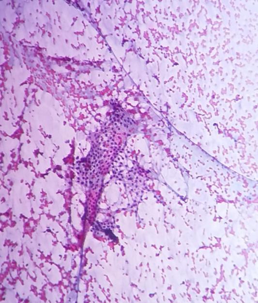
94 THYROID FIGURE: 1 NODULAR GOITER- MUFP STAIN (40X) NODULAR GOITER- MUFP STAIN (10X) FIGUARE: 2 NODULAR GOITER-PAP STAIN (40X) NODULAR GOITER-PAP STAIN (10X)
")

95 FIGUARE: 3 NODULAR GOITER-MGG STAIN (40X) FIGUARE: 4 HASHIMOTO S THYROIDITIS-MUFP STAIN (40X)


96 FIGUARE: 5 HASHIMOTO S THYROIDITIS-PAP STAIN (40X) FIGUARE: 6 HASHIMOTO S THYROIDITIS-MGG STAIN (40X)

")
 PAP STAIN")
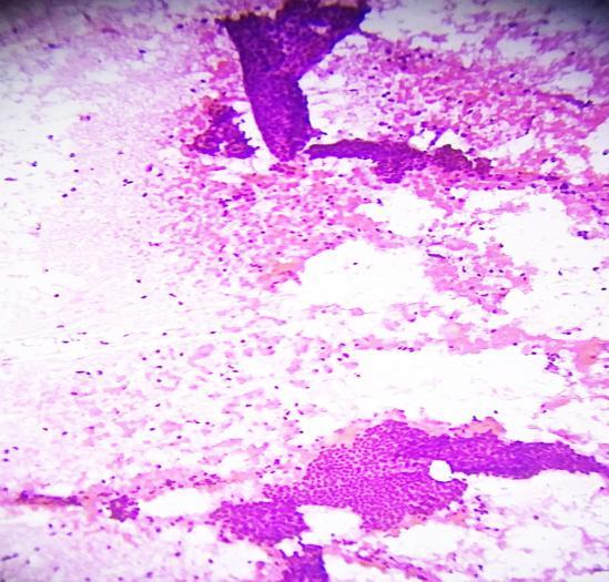
97 BREAST FIGUARE: 7 FIBROADENOMA BREAST MUFP STAIN (40X) FIBROADENOMA BREAST MUFP STAIN (10X) FIGUARE: 8 FIBROADENOMA BREAST PAP STAIN (40X) FIBROADENOMA BREAST PAP STAIN (10X)
")

98 FIGUARE: 9 FIBROADENOMA BREAST MGG STAIN (40X) FIGUARE: 10 DUCTAL CARCINOMA BREAST -MUFP STAIN (40X)


99 FIGUARE: 11 DUCTAL CARCINOMA BREAST -MUFP STAIN (40X) FIGUARE: 12 DUCTAL CARCINOMA-MGG STAIN (40x)

 FIGUARE: 14")
100 LYMPH NODE FIGUARE: 13 TUBERCULOUS LYMPHADENITIS-MUFP STAIN (40X) FIGUARE: 14 TUBERCULOUS LYMPHADENITIS-PAP STAIN (40X)
")

101 FIGUARE: 15 TUBERCULOUS LYMPHADENITIS-MGG STAIN (40X) FIGUARE: 16 SQUAMOUS CELL CARCINOMA SECONDARIES IN LYMPH NODE, MUFP STAIN, 40X


102 FIGUARE: 17 SQUAMOUS CELL CARCINOMA SECONDARIES IN LYMPH NODE, MUFP STAIN, 40X FIGUARE: 18 SQUAMOUS CELL CARCINOMA SECONDARIES IN LYMPH NODE, MGG STAIN, 40X
Update on Thyroid FNA The Bethesda System. Shikha Bose M.D. Associate Professor Cedars Sinai Medical Center
 Update on Thyroid FNA The Bethesda System Shikha Bose M.D. Associate Professor Cedars Sinai Medical Center Thyroid Nodules Frequent occurrence Palpable: 4-7% of adults Ultrasound: 10-31% Majority benign
Dr. Issraa Ali Hussein
 CLINICAL 09888888;rCYTOLOGY Dr. Issraa Ali Hussein objectives Define diagnostic cytology (clinical cytology). Explain the differences between histopathology and cytopathology. Recognize the methods for
Cytological evaluation of effusion fluid with cell block technique and cytology smears among Sudanese patients
 EUROPEAN ACADEMIC RESEARCH Vol. IV, Issue 3/ June 2016 ISSN 2286-4822 www.euacademic.org Impact Factor: 3.4546 (UIF) DRJI Value: 5.9 (B+) Cytological evaluation of effusion fluid with cell block technique
Cytology for the Endocrinologist. Nicole Massoll M.D
 Cytology for the Endocrinologist Nicole Massoll M.D Objectives Discuss slide preperation Definitions of adequacy ROSE (Rapid On-Site Evaluation) Thyroid Cytology Adequacy Nicole Massoll M.D. University
Evolution of Pap Stain
 Biomedical Research and Therapy 2016, 3(2): 490-500 ISSN 2198-4093 www.bmrat.org DOI 10.7603/s40730-016-0006-8 REVIEW Kalyani Raju Professor of Pathology, Sri Devaraj Urs Medical College, Sri Devaraj Academy
The role of the cytologist in breast cancer screening
 The role of the cytologist in breast cancer screening I.Seili-Bekafigo, MD, PhD Clinical cytologist KBC Rijeka Croatian Society for Clinical Cytology Fine needle aspiration (FNA, FNAB, FNAC) Fine needle
Fellowship in Cytopathology Department of Pathology. All India Institute of Medical Sciences (AIIMS) Jodhpur, Rajasthan, India
 Jodhpur, Rajasthan, India") Fellowship in Cytopathology Department of Pathology All India Institute of Medical Sciences (AIIMS) Jodhpur, Rajasthan, India Syllabus for Fellowship in Cytopathology: FNAC Direct, Guided, EUS Exfoliative
Fellowship in Cytopathology Department of Pathology All India Institute of Medical Sciences (AIIMS) Jodhpur, Rajasthan, India Syllabus for Fellowship in Cytopathology: FNAC Direct, Guided, EUS Exfoliative
Review of Literatures
 Review of Literatures Fine needle biopsy was popular in the Scandinavian countries some four decades ago. Though FNAC for any palpable tumor was first introduced in America in the 1920s by Martin, Ellis
Asthana A et al: Comparison of the routine Papanicolaou staining technique
 Original Article Comparison of the routine Papanicolaou staining technique with the rapid, economic, acetic acid, Papanicolaou (REAP) technique Asthana A 1, Singh AK 2 1 Dr Abhilasha Asthana BDS, MDS Senior
Ranu RoyBiswas 1*, Chandi C. Paral 2 ; Ramprasad Dey 3 and Subhash C Biswas 3
 ORIGI NAL ARTICLE Al Ameen J Med Sci (20 0 8 )1 (2 ):9 9-1 0 3 Rapid Economic, Acetic Acid, Papanicolaou Stain (REAP) - Is it suitable alternative to standard PAP stain? Ranu RoyBiswas 1*, Chandi C. Paral
Ultrasound-Guided Fine-Needle Aspiration of Thyroid Nodules: New events
 Ultrasound-Guided Fine-Needle Aspiration of Thyroid Nodules: New events Sandrine Rorive, M.D., PhD. Erasme Hospital - Université Libre de Bruxelles (ULB) INTRODUCTION The assessment of thyroid nodules
Objectives. Salivary Gland FNA: The Milan System. Role of Salivary Gland FNA 04/26/2018
 Salivary Gland FNA: The Milan System Dr. Jennifer Brainard Section Head Cytopathology Cleveland Clinic Objectives Introduce the Milan System for reporting salivary gland cytopathology Define cytologic
A Study of Thyroid Swellings and Correlation between FNAC and Histopathology Results
 International Journal of Current Microbiology and Applied Sciences ISSN: 2319-7706 Volume 6 Number 4 (2017) pp. 265-269 Journal homepage: http://www.ijcmas.com Original Research Article https://doi.org/10.20546/ijcmas.2017.604.030
Repeat Thyroid Nodule Fine-Needle Aspiration in Patients With Initial Benign Cytologic Results
 Anatomic Pathology / REPEAT THYROID FINE-NEEDLE ASPIRATION Repeat Thyroid Nodule Fine-Needle Aspiration in Patients With Initial Benign Cytologic Results Melina B. Flanagan, MD, MSPH, 1 N. Paul Ohori,
INTRA-OPERATIVE CYTOLOGY AND FROZEN SECTIONS OF BREAST LESIONS: A COMPARISON FROM A SAUDI TEACHING HOSPITAL
 Bahrain Medical Bulletin, Volume 18, Number 1, March 1996 INTRA-OPERATIVE CYTOLOGY AND FROZEN SECTIONS OF BREAST LESIONS: A COMPARISON FROM A SAUDI TEACHING HOSPITAL Ammar C.Al-Rikabi, MD,MRCPath,FIAC*
Almost any suspected tumor can be aspirated easily and safely. Some masses are more risky to aspirate including:
 DOES THIS PATIENT HAVE CANCER? USING IN-HOUSE CYTOLOGY TO HELP YOU MAKE THIS DIAGNOSIS. Joyce Obradovich, DVM, Diplomate, ACVIM (Oncology) Animal Cancer & Imaging Center, Canton, Michigan Almost every
CPC 4 Breast Cancer. Rochelle Harwood, a 35 year old sales assistant, presents to her GP because she has noticed a painless lump in her left breast.
 CPC 4 Breast Cancer Rochelle Harwood, a 35 year old sales assistant, presents to her GP because she has noticed a painless lump in her left breast. 1. What are the most likely diagnoses of this lump? Fibroadenoma
Thyroid Pathology: It starts and ends with the gross. Causes of Thyrophobia. Agenda. Diagnostic ambiguity. Treatment/prognosis disconnect
 Thyroid Pathology: It starts and ends with the gross Jennifer L. Hunt, MD, MEd Aubrey J. Hough Jr, MD, Endowed Professor of Pathology Chair of Pathology and Laboratory Medicine University of Arkansas for
DOWNLOAD ENTIRE DOCUMENT FROM
 PREVIEW ONLY 1 Atlas on Bethesda system for reporting Thyroid Cytology PREVIEW ONLY 2 OVERVIEW 1. Indications and goal of thyroid FNA 2. Contraindications 3. Procurement of cell sample 4. Staining methods
"Atypical": Criteria and
 "Atypical": Criteria and Controversies Esther Rossi MD PhD MIAC Division of Anatomic Pathology and Cytology Catholic University of Sacred Heart Rome, Italy CASE HISTORY In 2015, 45 y/o woman underwent
Diagnostic accuracy fine needle aspiration cytology of thyroid gland lesions.
 International Journal of Pharmaceutical Science Invention ISSN (Online): 2319 6718, ISSN (Print): 2319 670X Volume 3 Issue 4 April 2014 PP.05-10 Diagnostic accuracy fine needle aspiration cytology of thyroid
Role of fine needle aspiration cytology and cytohistopathological co-relation in thyroid lesions: experience at a tertiary care centre of North India
 International Journal of Research in Medical Sciences Chandra S et al. Int J Res Med Sci. 2016 Oct;4(10):4552-4556 www.msjonline.org pissn 2320-6071 eissn 2320-6012 Research Article DOI: http://dx.doi.org/10.18203/2320-6012.ijrms20163328
IBCM 2, April 2009, Sarajevo, Bosnia and Herzegovina
 Preoperative diagnosis and treatment planning in breast cancer The pathologist s perspective L. Mazzucchelli Istituto Cantonale di Patologia Locarno, Switzerland IBCM 2, 23-25 April 2009, Sarajevo, Bosnia
Interpretation of Breast Pathology in the Era of Minimally Invasive Procedures
 Shahla Masood, M.D. Professor and Chair Department of Pathology and Laboratory Medicine University of Florida College of Medicine Jacksonville Medical Director, UF Health Breast Center Chief of Pathology
Thyroid master class. Thyroid Fine needle aspiration cytology and liquid-based techniques: Hologic and Becton Dickinson
 Thyroid master class Thyroid Fine needle aspiration cytology and liquid-based techniques: Hologic and Becton Dickinson Principle of LBC Collection of cells in liquid medium Immediate fixation Processor-prepared
Relationship of Cytological with Histopathological Examination of Palpable Thyroid Nodule
 Relationship of Cytological with Histopathological Examination of Palpable Thyroid Nodule NAUSHEEN HENNA 1, SHAHZAD AHMED FAKHAR 2, NAVEED AKHTER 3, MUHAMMAD MASOOD AFZAL 4, KHIZER AFTAB AHMAD KHAN 5,
Palpable Breast Lesions Cytomorphological Analysis and Scoring System with Histopatholgical Correlation
 IOSR Journal of Dental and Medical Sciences (IOSR-JDMS) e-issn: 2279-0853, p-issn: 2279-0861.Volume 15, Issue 10 Ver. III (October. 2016), PP 25-29 www.iosrjournals.org Palpable Breast Lesions Cytomorphological
Study of Fine Needle Aspiration Cytology of Breast Lump: Correlation of Cytologically Malignant Cases with Their Histological Findings
 Study of Fine Needle Aspiration Cytology of Breast Lump: Correlation of Cytologically Malignant Cases with Their Histological Findings Touhid Uddin Rupom 1, Tamanna Choudhury 2, Sultana Gulshana Banu 3
Disclosures. Parathyroid Pathology. Objectives. The normal parathyroid 11/10/2012
 Disclosures Parathyroid Pathology I have nothing to disclose Annemieke van Zante MD/PhD Assistant Professor of Clinical Pathology Associate Chief of Cytopathology Objectives 1. Review the pathologic features
Providence Medford Medical Center Pathology Department
 Providence Medford Medical Center Pathology Department Anatomic pathology services including histology, cytology and autopsies are offered through Providence Medford Medical Center Pathology Department.
Keywords: papillary carcinoma, Hurthle, FNAC, follicular pattern.
 bü z ÇtÄ TÜà väx Efficacy of Fine Needle Aspiration Cytology in the Diagnosis of Thyroid Swellings in Red Sea State, Sudan 1 Abstract Background: Fine needle aspiration cytology (FNAC) is a safe, easy
Abstract. Introduction. Salah Abobaker Ali
 Sensitivity and specificity of combined fine needle aspiration cytology and cell block biopsy versus needle core biopsy in the diagnosis of sonographically detected abdominal masses Salah Abobaker Ali
Background to the Thyroid Nodule
 William C. Faquin, MD, PhD Professor of Pathology Harvard Medical School Director of Head and Neck Pathology Massachusetts Eye and Ear Massachusetts General Hospital THYROID FNA: PART I Background to the
FNA of Thyroid. Toward a Uniform Terminology With Management Guidelines. NCI NCI Thyroid FNA State of the Science Conference
 FNA of Thyroid NCI NCI Thyroid FNA State of the Science Conference Toward a Uniform Terminology With Management Guidelines Thyroid Thyroid FNA Cytomorphology NCI Thyroid FNA State of the Science Conference
Thyroid nodules - medical and surgical management. Endocrinology and Endocrine Surgery Manchester Royal Infirmary
 Thyroid nodules - medical and surgical management JRE Davis NR Parrott Endocrinology and Endocrine Surgery Manchester Royal Infirmary Thyroid nodules - prevalence Thyroid nodules common, increase with
The role of Electron Microscopy in the study of cytologic specimens. Elba A. Turbat-Herrera, MD
 The role of Electron Microscopy in the study of cytologic specimens. Elba A. Turbat-Herrera, MD Louisiana State University Health Sciences Center Shreveport, LA, USA Introduction The field of Cytology
Salivary Gland Cytology: A Clinical Approach to Diagnosis and Management of Atypical and Suspicious Lesions
 Salivary Gland Cytology: A Clinical Approach to Diagnosis and Management of Atypical and Suspicious Lesions W.C. Faquin, M.D., Ph.D. Massachusetts General Hospital Harvard Medical School, USA Marc Pusztaszeri,
International Journal of Health Sciences and Research ISSN:
 International Journal of Health Sciences and Research www.ijhsr.org ISSN: 2249-9571 Original Research Article Utility of Modified Cell Block Technique in Cases of Pleural Effusion Suspected of Malignancy
THYROID CYTOLOGY THYROID CYTOLOGY FINE-NEEDLE-ASPIRATION ANCILLARY TESTS IN THYROID FNA
 ANCILLARY TESTS IN THYROID FNA Prof. Fernando Schmitt Department of Pathology and Oncology, Medical Faculty of Porto University Head of Molecular Pathology Unit, IPATIMUP General-Secretary of the International
FINE NEEDLE ASPIRATION CYTOLOGY: A DIAGNOSTIC TOOL IN LYMPHADENOPTHY
 Int. J. LifeSc. Bt & Pharm. Res. 2014 Mahesh S Patil and Anand A S, 2014 Research Paper ISSN 2250-3137 www.ijlbpr.com Vol. 3, No. 1, January 2014 2014 IJLBPR. All Rights Reserved FINE NEEDLE ASPIRATION
performed to help sway the clinician in what the appropriate diagnosis is, which can substantially alter the treatment of management.
 Hello, I am Maura Polansky at the University of Texas MD Anderson Cancer Center. I am a Physician Assistant in the Department of Gastrointestinal Medical Oncology and the Program Director for Physician
AUDIT OF FNA CYTOLOGY, ADEQUACY AND REPORTING AT THE DERRIFORD HOSPITAL ONE STOP HEAD & NECK LUMP CLINIC
 AUDIT OF FNA CYTOLOGY, ADEQUACY AND REPORTING AT THE DERRIFORD HOSPITAL ONE STOP HEAD & NECK LUMP CLINIC Dr Michael Ghisel Foundation Trainee Dr Tim Bracey Consultant Pathologist What is it? The Derriford
The Bethesda System for Reporting Thyroid Cytopathology, Laila Khazai 11/4/17
 The Bethesda System for Reporting Thyroid Cytopathology, 2017 Laila Khazai 11/4/17 In Summary No major changes for cytologists. The clinical team is faced with different risk of malignancies (ROM) associated
Diagnostic Value of EBUS-TBNA in Various Lung Diseases (Lymphoma, Tuberculosis, Sarcoidosis)
") Diagnostic Value of EBUS-TBNA in Various Lung Diseases (Lymphoma, Tuberculosis, Sarcoidosis) Sevda Sener Cömert, MD, FCCP. SBU, Kartal Dr.Lütfi Kırdar Training and Research Hospital Department of Pulmonary
Diagnostic Value of EBUS-TBNA in Various Lung Diseases (Lymphoma, Tuberculosis, Sarcoidosis) Sevda Sener Cömert, MD, FCCP. SBU, Kartal Dr.Lütfi Kırdar Training and Research Hospital Department of Pulmonary
Evaluation and Management of Thyroid Nodules. Nick Vernetti, MD, FACE Palm Medical Group Las Vegas, Nevada
 Evaluation and Management of Thyroid Nodules Nick Vernetti, MD, FACE Palm Medical Group Las Vegas, Nevada Disclosure Consulting Amgen Speaking Amgen Objectives Understand the significance of incidental
Thyroid Neoplasm. ORL-Head and neck Surgery 2014
 In The Name of God Thyroid Neoplasm ORL-Head and neck Surgery 2014 Malignant Neoplasm By age 90, virtually everyone has nodules Estimates of cancer prevalence at autopsy 4% to 36% Why these lesions are
CN 925/15 History. Microscopic Findings
 CN 925/15 History 78 year old female. FNA indeterminate lesion right thyroid lobe. Previous THY1C (UK) Bethesda category 1 cyst fluid. Ultrasound showed part solid/cystic changes, indeterminate in nature
The Frozen Section: Diagnostic Challenges and Pitfalls
 The Frozen Section: Diagnostic Challenges and Pitfalls William C. Faquin, M.D., Ph.D. Director, Head and Neck Pathology Massachusetts General Hospital & Massachusetts Eye and Ear Infirmary Harvard Medical
Pitfalls in thyroid tumor pathology. Prof.Valdi Pešutić-Pisac MD, PhD
 Pitfalls in thyroid tumor pathology Prof.Valdi Pešutić-Pisac MD, PhD Too many or... Tumour herniation through a torn capsule simulating capsular invasion fibrous capsule with a sharp discontinuity, suggestive
GOALS AND OBJECTIVES CYTOPATHOLOGY
 GOALS AND OBJECTIVES CYTOPATHOLOGY LEVEL: PGY2, PGY4, PGY5 The 1st block in PGY2 is an introductory in nature and is followed by two more blocks in PGY-4 (please, see core rotation for PGY4 below) and
INTRODUCTION TO PATHOLOGICAL TECHNIQUES. 1. Types of routine biopsy procedures 2. Special exams (IHC, FISH)
") INTRODUCTION TO PATHOLOGICAL TECHNIQUES 1. Types of routine biopsy procedures 2. Special exams (IHC, FISH) Biopsy-Indications Diffuse/multifocal lesions (neoplastic, inflammatory, etc) Etiology of the
INTRODUCTION TO PATHOLOGICAL TECHNIQUES 1. Types of routine biopsy procedures 2. Special exams (IHC, FISH) Biopsy-Indications Diffuse/multifocal lesions (neoplastic, inflammatory, etc) Etiology of the
MEDICAL POLICY Gene Expression Profiling for Cancers of Unknown Primary Site
 POLICY: PG0364 ORIGINAL EFFECTIVE: 04/22/16 LAST REVIEW: 07/26/18 MEDICAL POLICY Gene Expression Profiling for Cancers of Unknown Primary Site GUIDELINES This policy does not certify benefits or authorization
Diseases of the breast (1 of 2)
") Diseases of the breast (1 of 2) Introduction A histology introduction Normal ducts and lobules of the breast are lined by two layers of cells a layer of luminal cells overlying a second layer of myoepithelial
Diseases of the breast (1 of 2) Introduction A histology introduction Normal ducts and lobules of the breast are lined by two layers of cells a layer of luminal cells overlying a second layer of myoepithelial
Thyroid Nodule. Disclosure. Learning Objectives P A P A P A 3/18/2014. Nothing to disclose.
 Thyroid Nodule Evaluating the patient with a thyroid nodule and some management options. Miguel V. Valdez PA C Disclosure Nothing to disclose. Learning Objectives Examination of thyroid gland Options for
Volume 2 Issue ISSN
 Volume 2 Issue 3 2012 ISSN 2250-0359 Correlation of fine needle aspiration and final histopathology in thyroid disease: a series of 702 patients managed in an endocrine surgical unit *Chandrasekaran Maharajan
Presentation material is for education purposes only. All rights reserved URMC Radiology Page 1 of 98
 Presentation material is for education purposes only. All rights reserved. 2011 URMC Radiology Page 1 of 98 Radiology / Pathology Conference February 2011 Brooke Koltz, Cytopathology Resident Presentation
Cytopathological evaluation of various thyroid lesions based on Bethesda system for reporting thyroid lesions
 International Journal of Research in Medical Sciences Khadatkar AS et al. Int J Res Med Sci. 2017 Apr;5(4):1339-1343 www.msjonline.org pissn 2320-6071 eissn 2320-6012 Original Research Article DOI: http://dx.doi.org/10.18203/2320-6012.ijrms20170901
FNA Thyroid Cytology Structured Reporting Proforma
 FNA Thyroid Cytology Structured Reporting Proforma Mandatory questions (i.e. protocol standards) are in bold (e.g. S1.03). Family name Given name(s) Date of birth S1.02 Clinical details Male Female Intersex/indeterminate
Role of FNAC in Evaluation of Neck Masses
 Original Article Elmer Press Role of FNAC in Evaluation of Neck Masses Mohd Hazmi Mohamed a, c, Shahrul Hitam b, Sushil Brito-Mutunayagam b, Mohd Razif Mohamad Yunus a Abstract Background: Despite fine
Imaging in breast cancer. Mammography and Ultrasound Donya Farrokh.MD Radiologist Mashhad University of Medical Since
 Imaging in breast cancer Mammography and Ultrasound Donya Farrokh.MD Radiologist Mashhad University of Medical Since A mammogram report is a key component of the breast cancer diagnostic process. A mammogram
Enterprise Interest None
 Enterprise Interest None Risk stratification of salivary gland lesions on cytology based on the proposed Milan System for reporting salivary gland cytopathology: A pilot study Kartik Viswanathan, M.D.,
Ultrasound Guided Fine Needle Aspiration Biopsy of Retroperitoneal Masses
 Ultrasound Guided Fine Needle Aspiration Biopsy of Retroperitoneal Masses Ahmad SS *, Akhtar K +, Akhtar SS +, Nasir A #, Khalid M **, Mansoor T ++ Abstract The study was undertaken to evaluate the reliability
PAAF vs Core Biopsy en Lesiones Mamarias Case #1
 5/19/2014 PAAF vs Core Biopsy en Lesiones Mamarias Case #1 Fine Needle Aspiration Cytology of Breast: Correlation with Needle Core Biopsy 64-year-old woman Mass in breast Syed Hoda, MD CD31 Post-Radiation
Cytyc Corporation - Case Presentation Archive - March 2002
 FirstCyte Ductal Lavage History: 68 Year Old Female Gail Index: Unknown Clinical History: Negative Mammogram in 1995 6 yrs. later presents with bloody nipple discharge Subsequent suspicious mammogram Suspicious
Thyroid Nodules: Understanding FNA Cytology (The Bethesda System for Reporting of Thyroid Cytopathology) Shamlal Mangray, MB, BS
 Shamlal Mangray, MB, BS") Thyroid Nodules: Understanding FNA Cytology (The Bethesda System for Reporting of Thyroid Cytopathology) Shamlal Mangray, MB, BS Attending Pathologist Rhode Island Hospital, Providence, RI DISCLOSURE:
Thyroid Nodules: Understanding FNA Cytology (The Bethesda System for Reporting of Thyroid Cytopathology) Shamlal Mangray, MB, BS Attending Pathologist Rhode Island Hospital, Providence, RI DISCLOSURE:
Papillary Lesions of the Breast A Practical Approach to Diagnosis. (Arch Pathol Lab Med. 2016;140: ; doi: /arpa.
 Papillary Lesions of the Breast A Practical Approach to Diagnosis (Arch Pathol Lab Med. 2016;140:1052 1059; doi: 10.5858/arpa.2016-0219-RA) Papillary lesions of the breast Span the spectrum of benign,
5/3/2017. Ahn et al N Engl J Med 2014; 371
 Alan Failor, M.D. Clinical Professor of Medicine Division of Metabolism, Endocrinology and Nutrition University of Washington April 20, 2017 No disclosures to report 1. Appropriately evaluate s in adult
Cleveland Clinic Laboratories. Anatomic Pathology
 Cleveland Clinic Laboratories Anatomic Pathology OUR MISSION Cleveland Clinic Laboratories contributes to excellent patient care by providing high-quality, comprehensive laboratory testing and patient-focused
Ultrasound-guided FNA Biopsy. American Thyroid Association 2017
 Ultrasound-guided FNA Biopsy American Thyroid Association 2017 Robert A. Levine, MD, FACE, ECNU Thyroid Center of New Hampshire Geisel School of Medicine at Dartmouth University Disclosures: No relevant
Management guideline for patients with differentiated thyroid cancer. Teeraporn Ratanaanekchai ENT, KKU 17 October 2007
 Management guideline for patients with differentiated thyroid Teeraporn Ratanaanekchai ENT, KKU 17 October 2007 Incidence (Srinagarind Hospital, 2005, both sex) Site (all) cases % 1. Liver 1178 27 2. Lung
Introduction: Ultrasound guided Fine Needle Aspiration: When and how
 International Course of Thyroid Ultrasonography and minimally invasive procedure 7-8 October 2016 University of Pisa, Italy Introduction: Ultrasound guided Fine Needle Aspiration: When and how Teresa Rago
PAPILLARY THYROID CARCINOMA PRESENTING AS A LATERAL NECK MASS MASS. Dr. Pamela Hanson DO PGY3
 PAPILLARY THYROID CARCINOMA PRESENTING AS A LATERAL NECK MASS MASS Dr. Pamela Hanson DO PGY3 MK CASE PRESENTATION 28 yo Female presented to the ENT Clinic in October 2016, with the complaint of chronic
Evaluation of Neck Mass. Disclosure. Learning Objectives 3/24/2014. Karen T. Pitman MD, FACS Banner MDACC, Gilbert AZ. Nothing to disclose
 Evaluation of Neck Mass Karen T. Pitman MD, FACS Banner MDACC, Gilbert AZ Nothing to disclose Disclosure Learning Objectives 1. Describe a systematic method to evaluate a patient with a neck mass 2. Select
To evaluate the role of sputum in the diagnosis of lung cancer in south Indian population
 International Journal of Research in Medical Sciences Muniyappa M et al. Int J Res Med Sci. 2014 May;2(2):545-550 www.msjonline.org pissn 2320-6071 eissn 2320-6012 Research Article DOI: 10.5455/2320-6012.ijrms20140534
QUALITY CONTROL IN CYTOLOGY
 27 QUALITY CONTROL IN CYTOLOGY 27.1 INTRODUCTION Cytopathologists are concerned about and committed to quality assurance and quality control in their laboratories. These practices include, among others,
RE-AUDIT OF THYROID FNA USING THE THY GRADING SYSTEM AND HISTOLOGY AT SUNDERLAND ROYAL HOSPITAL, 2011
 Audit: RE-AUDIT OF THYROID FNA USING THE THY GRADING SYSTEM AND HISTOLOGY AT SUNDERLAND ROYAL HOSPITAL, 2011 Auditors: Dr Lena Wilkinson SpR Histopathology Dr. Debra Milne Consultant Histocytopathologist
Suspicious Cytologic Diagnostic Category in Endoscopic Ultrasound-Guided FNA of the Pancreas: Follow-Up and Outcomes
 Suspicious Cytologic Diagnostic Category in Endoscopic Ultrasound-Guided FNA of the Pancreas: Follow-Up and Outcomes Evan A. Alston, MD 1 ; Sejong Bae, PhD 2 ; and Isam A. Eltoum, MD, MBA 1 BACKGROUND:
Metachronic solitary breast metastasis from renal cell carcinoma: case report
 Metachronic solitary breast metastasis from renal cell carcinoma: case report Abstract We describe the case of a patient with solitary and metachronic breast metastasis, 3 years after nephrectomy for renal
Goals and Objectives for Cytopathology Rotation
 Goals and Objectives for Cytopathology Rotation Level: PGY3, PGY4, PGY5 The 1st block in PGY3 is an introductory in nature and is followed by three more blocks in PGY-4 (please, see core rotation for PGY4
SPECIMEN PREPARATION AND ADEQUACY OF THE MATERIAL
 SPECIMEN PREPARATION AND ADEQUACY OF THE MATERIAL Guido FADDA, MD, MIAC Head, Cytopathology Section Department of Anatomic Pathology and Laboratory Medicine Agostino Gemelli School of Medicine and Hospital
VDx: Unlocking Complex Diagnostics
 VDx: Unlocking Complex Diagnostics VDx now offers PARR testing in-house on formalin-fixed tissue Complicated Case? Is this cat s chronic lymphocytic enteritis really chronic IBD or is this early small
Thyroid Cytopathology: Weighing In The Bethesda System
 Thyroid Cytopathology: Weighing In The Bethesda System V8 Conflicts No financial consideration Bias Work in the Canadian environment where litigation is less Thyroid cytology is often referred in by small
TBSRTC 1- Probabilistic approach and Relationship to Clinical Algorithms
 The Benefits of a Uniform Reporting System for Thyroid Cytopathology BETHESDA REPORTING SYSTEM Prof. Fernando Schmitt Department of Pathology and Oncology, Medical Faculty of Porto University Head of Molecular
ROSE in EUS guided FNA of Pancreatic Lesions
 ROSE in EUS guided FNA of Pancreatic Lesions Guy s Hospital, London, 16 April 2018 Laxmi Batav Imperial College NHS Trust Imperial College NHS Trust Cytology Workload Cervical Cytology 57,500 (decreases
ISPUB.COM. A Kochhar, G Duggal, K Singh, S Kochhar INTRODUCTION MATERIAL AND METHOD
 ISPUB.COM The Internet Journal of Pathology Volume 13 Number 2 Spectrum Of Cytological Findings In Patients With Lymphadenopathy In Rural Population Of Southern A Kochhar, G Duggal, K Singh, S Kochhar
FY 2015 BCCCP Procedure Code Reference Chart
 Procedure Reference Chart 77057 G0202 77055 G0206 77056 G0204 Screening mammography, bilateral (two view film study of each breast) Screening mammography producing direct digital image, bilateral, all
Cytological study of palpable breast lumps with their histological correlation in a tertiary care hospital
 Original Research Article Cytological study of palpable breast lumps with their histological correlation in a tertiary care hospital Sunita Mistry 1*, Jignasha Patel 2, Kamlesh Shah 3, Ajit Patel 4 1 Assistant
FINE NEEDLE ASPIRATION (FNAC) AS A DIAGNOSTIC TOOL IN PAEDIATRIC LYMPHADENOPATHY.
 AS A DIAGNOSTIC TOOL IN PAEDIATRIC LYMPHADENOPATHY.") IJCRR Vol 06 issue 01 Section: Healthcare Category: Research Received on: 16/10/13 Revised on: 18/11/13 Accepted on: 20/12/13 FINE NEEDLE ASPIRATION (FNAC) AS A DIAGNOSTIC TOOL IN PAEDIATRIC Heming Agrawal,
IJCRR Vol 06 issue 01 Section: Healthcare Category: Research Received on: 16/10/13 Revised on: 18/11/13 Accepted on: 20/12/13 FINE NEEDLE ASPIRATION (FNAC) AS A DIAGNOSTIC TOOL IN PAEDIATRIC Heming Agrawal,
Cervical Lymph Nodes
 Cervical Lymph Nodes Diana Gaitini, MD Unit of Ultrasound, Department of Medical Imaging Rambam Medical Center and Faculty of Medicine Technion, Israel Institute of Technology Haifa, Israel Learning Targets
Case year female. Routine Pap smear
 Case 1 57 year female Routine Pap smear Diagnosis? 1. Atypical glandular cells of unknown significance (AGUS) 2. Endocervical AIS 3. Endocervical adenocarcinoma 4. Endometrial adenocarcinoma 5. Adenocarcinoma
Contributions to Anatomic Pathology, over the years
 Contributions to Anatomic Pathology, over the years Anatomic Pathology, part 1 G.B. Morgagni Xavier Bichat Rudolf Wirchow Anatomic Pathology, part 1 Anatomic pathology materials: morphological samples
Cytopathology. Robert M Genta Pathologie Clinique Université de Genève
 Cytopathology Robert M Genta Pathologie Clinique Université de Genève Learning objectives At the end of this hour you will know: 1. What cytopathology is 2. How specimens are collected, processed, and
Journal of Diagnostic Pathology 2011 (6); 1: Leading Article
; 1: Leading Article") Leading Article Beyond the horizon of current thyroid cytology reporting in Sri Lanka... Lokuhetty MDS Thyroid enlargement is a commonly encountered clinical problem among Sri Lankan patients, be it diffuse
Leading Article Beyond the horizon of current thyroid cytology reporting in Sri Lanka... Lokuhetty MDS Thyroid enlargement is a commonly encountered clinical problem among Sri Lankan patients, be it diffuse
Thyroid follicular neoplasms in cytology. Ulrika Klopčič Institute of Oncology, Department of Cytopathology, Ljubljana, Slovenia
 Thyroid follicular neoplasms in cytology Ulrika Klopčič Institute of Oncology, Department of Cytopathology, Ljubljana, Slovenia Lecture overview importance of FNAB in assessing thyroid lesions follicular
Building On The Best A Review and Update on Bethesda Thyroid 2017
 Building On The Best A Review and Update on Bethesda Thyroid 2017 Syed Z. Ali, MD, FRCPath, FIAC Professor of Pathology and Radiology The Johns Hopkins Hospital, Baltimore, Maryland USA TBSRTC Diagnostic
Case Scenario 1: Thyroid
 Case Scenario 1: Thyroid History and Physical Patient is an otherwise healthy 80 year old female with the complaint of a neck mass first noticed two weeks ago. The mass has increased in size and is palpable.
system and the Bethesda system applied for reporting thyroid cytopathology
 Original Research Article A comparative study between conventional system and the Bethesda system applied for reporting thyroid cytopathology M. Mamatha 1*, S. Chandra Sekhar 2, H. Sandhya Rani 3, S. Sandhya
JMSCR Vol 06 Issue 01 Page January 2018
 www.jmscr.igmpublication.org Impact Factor 5.84 Index Copernicus Value: 71.58 ISSN (e)-2347-176x ISSN (p) 2455-0450 DOI: https://dx.doi.org/10.18535/jmscr/v6i1.126 Study of Fine Needle Aspiration Cytology
Research Article Correlation of Fine Needle Aspiration Cytology with Histopathology in the Diagnosis of Solitary Thyroid Nodule
 SAGE-Hindawi Access to Research Thyroid Research Volume 2010, Article ID 379051, 5 pages doi:10.4061/2010/379051 Research Article Correlation of Fine Needle Aspiration Cytology with Histopathology in the
XIII CONGRESSO NAZIONALE Roma, 7-9 novembre NODULO TIROIDEO: Agoaspirato o Core Needle Biopsy?
 XIII CONGRESSO NAZIONALE NODULO TIROIDEO: Agoaspirato o Core Needle Biopsy? Anna Crescenzi Policlinico Universitario Campus Bio-Medico Roma Indeterminate lesions are heterogeneous The gray zone CONSERVATIVE