What s Your Diagnosis?
|
|
|
- Blaze Horn
- 6 years ago
- Views:
Transcription
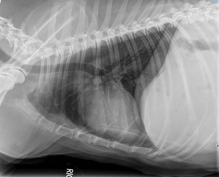
1 What s Your Diagnosis? Courtney S. Wait Signalment: 11 year old FS Labrador Retriever Presenting Complaint/History: The patient presented to the referring DVM for inappetance, vomiting, lethargy, and anorexia. Bloodwork and physical examination at the rdvm showed azotemia, anemia, and a palpable abdominal mass. She was referred to KSU VHC Small Animal Medicine service after 5 days of clinical symptoms. Physical Exam Findings: -Temperature: 100.2º F -Pulse: Strong -Heart Rate: 70 bpm -Respiratory Rate: 25 bpm -Weight: 40.4 kg -Mucus Membranes: pale pink and moist -CRT: <2 sec -Moderate dental tartar bilaterally -Abdominal palpation: mass palpable in the cranial abdomen, tense and painful -Bilaterally enlarged lymph nodes (prescapular, axillary, popliteal) -Several firm subcutaneous masses palpable over the entire body Bloodwork (noted from rdvm): Regenerative anemia Azotemia (BUN: 103, Crt: 5.9) Elevated phosphate Elevated ALP and ALK Diagnostic Plan: Step 1: Thoracic Radiographs (3 view)

2

3 Radiographic Findings: Three-view thorax: The cardiac silhouette, pulmonary vasculature, and pulmonary parenchyma are within normal limits. There is a rounded soft tissue opacity dorsal to the second and third sternebra, consistent in location with sternal lymph node. In the limited view of the cranial abdomen, the gastric axis is severely displaced caudal dorsally. There are multiple irregularly-shaped gas foci superimposed with the enlarged liver. Radiographic Impressions: 1. Cranial abdominal mass effect with gas, is consistent with liver, and has differentials of necrotic neoplasm or abscess. 2. Sternal lymphadenopathy is either metastatic or reactive. Step 2: Abdominal Radiographs
4

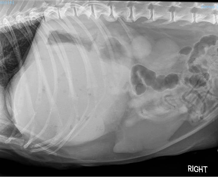
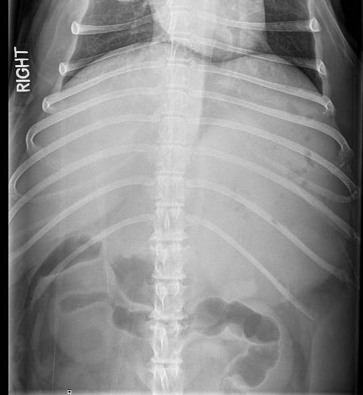
5 Abdominal Radiograph Findings: The gastric axis is severely displaced caudal dorsally. There are multiple irregularlyshaped gas foci superimposed with the enlarged rounded liver. Impressions: 1. Cranial abdominal mass effect with gas, is consistent with liver, and has differentials of necrotic neoplasm or abscess. Step 3: Abdominal Ultrasound Ultrasound Findings: Abdomen: There was a very large complex hepatic mass measuring greater than 15 cm dimension in the left cranial abdomen. There were cystic and large irregularly shaped fluid filled regions throughout this mass, and a very large amount of punctate and small gas foci, diffusely. There was no normal left sided hepatic parenchyma appreciated. Right-sided hepatic parenchyma was diffusely hyperechoic with poorly defined hypoechoic nodules measuring up to approximately 1 cm. There was an irregularly shaped, poorly defined, very mildly hypoechoic 1.2 cm nodule in the spleen. There was a thin hypoechoic rim measuring approximately 1-2 mm thick surrounding the left and right renal cortices. The right kidney was irregularly marginated, and contained a few anechoic through
6 transmitting cysts that measured up to 2 cm diameter. There was suspended particulate debris in the urinary bladder. The left adrenal gland measured 8.6 mm thick, and there is a heterogeneous mass in the caudal pole of the right adrenal gland that measured 2.5 cm thick and 3.3 cm in length. There was a scant amount of anechoic peritoneal effusion. The gastrointestinal tract was within normal limits. Fine needle aspirates of the hepatic mass obtained without immediate complication. Ultrasound Impressions: 1. Large hepatic complex mass is most consistent with necrotizing neoplasia, with much less consideration given to a primary hepatic abscess. 2. Diffuse hepatic changes have differentials of nodular hyperplasia and less likely neoplasia. 3. Splenic nodule may represent neoplasia or hyperplasia. 4. Right adrenal nodule is most consistent with a primary or metastatic neoplasia. 5. Hypoechoic rim surrounding the left and right kidneys has differentials of neoplasia, interstitial/glomerulonephritis or a normal variation. 6. Mild left adrenomegaly may be neoplastic or hyperplastic. 7. Urinary bladder proteinaceous or cellular debris. 8. Scant peritoneal effusion is in exudate, neoplastic, or hemorrhage. Step 4: Fine Needle Aspirate of Hepatic Mass Findings: Moderate to marked neutrophilic inflammation with a mild macrophagic component Marked necrosis Evidence of previous hemorrhage (hematoidin crystals) Outcome Because the patient had severe renal failure, surgery under general anesthesia was not recommended. Due to the expense of treatment and the high likelihood of neoplastic disease, the patient was sent to the referring veterinarian to be humanely euthanized. Discussion Radiographic gas opacity within the liver is a rare occurrence in dogs and cats. There are three ways gas may enter the liver: ascension through the biliary system, ascension through the portal venous system, or primary gas production through liver abscessation. Gas within the portal vessels may occur as a result of severe necrotizing gastritis or enteritis, gastrointestinal ulceration, distention, trauma, or interventional procedures. When gas enters the portal vessels, a linear branching radiolucent appearance (similar
7 to an air bronchogram) may be visible. Gas in or around the gallbladder occurs with emphysematous cholecystitis, or may also be seen after surgery of the duodenum or biliary system. Hepatic abscesses often contain anaerobic bacteria, which produce gas opacities within the hepatic parenchyma. Based on the radiographic pattern of gas foci within the liver and severe hepatomegaly, hepatic neoplasia with secondary necrosis and abscessation was the primary differential in this case. Hepatic abscessation can be cause by several etiologies including hematogenous spread, direct extension from pancreatitis or peritonitis, traumatic penetrating wounds, and ischemic damage to the liver. In this patient s case, the abscessation was cause by ischemic damage. Primary and metastatic neoplasia in the liver causes destruction of normal vascular supply and disruption of normal phagocytic defenses. This allows bacteria within the liver to proliferate and cause central necrosis. Some of the bacteria are gas-forming organisms, and they create radiolucent areas within the hepatic parenchyma. These areas appear as irregularly stipled or mottled gas patterns. However, not all hepatic abscesses contain gas and if it is not seen radiographically, hepatic abscesses cannot be ruled out. Typically, the treatment or any hepatic abscess is drainage and removal of the affected tissue. Supportive fluid therapy, systemic antibiotics, and treatment of any underlying disease process is also warranted. Preemptively, the antibiotic course should be a cephalosporin or penicillin combined with an aminoglycoside for broad spectrum coverage. Ideally, antibiotic therapy should be based off of culture and sensitivity.the procedure of choice in dogs is an abdominal exploratory so that all organs may be assessed for infection. Hepatic lobectomies may be necessary. Complications include abscess rupture, abdominal contamination, and septic peritonitis. References: Grooters, Amy M., Robert G. Sherding, and Susan E. Johnson. "Hepatic Abscesses in Dogs." Compendium on Continuing Education for the Practicing Veterinarian Article 17.6 (1995): Web. Grooters, Amy M., Robert G. Sherding, David S. Biller, and Susan E. Johnson. "Hepatic Abscesses Associated With Diabetes Mellitus in Two Dogs." Journal of Veterinary Internal Medicine 8.3 (1994): Wiley Online Library. Web. 17 June Larson, Martha Moon, and Donald E. Thrall. "Chapter 37: The Liver and Spleen." Textbook of Veterinary Diagnostic Radiology. 6th ed. St. Louis: Saunders Elsevier, Print. Schwarz, Leslie A., Dominique G. Penninck, and Cynthia Leveille-Webster. "Hepatic Abscesses In 13 Dogs: A Review Of The Ultrasonographic Findings, Clinical Data And Therapeutic Options." Veterinary Radiology Ultrasound 39.4 (1998): PubMed. Web. 17 June 2017.
8
What s your diagnosis?
 What s your diagnosis? Signalment: 9 year old MC 2.7 kg Papillion Presenting Complaint: Presented for work up of anorexia and vomiting History: He had presented to cardiology for work up of a grad IV/VI
What s Your Diagnosis?
 What s Your Diagnosis? Signalment: 5 year old MC Belgian Malinois Presenting Complaint: Perineal hernia as well as not eating or defecating History: The patient presented to the KSU VHC on 7/28/2018 for
What s Your Diagnosis? Sara Alves, Class of Signalment: 9-year-7-month old female spay American Miniature Eskimo dog
 What s Your Diagnosis? Sara Alves, Class of 2018 Signalment: 9-year-7-month old female spay American Miniature Eskimo dog Presenting Complaint: The patient presented on 5/30/17 with signs of lethargy and
What s Your Diagnosis??? Renée Fahrenholz, Class of 2012
 Renée Fahrenholz, Class of 2012 What s Your Diagnosis??? Signalment Emma, a 9 year old, Female, Spayed, Domestic Short Haired Feline Presenting Complaint Weight loss, vomited the morning of her visit,
What s Your Diagnosis? Signalment: Species: Canine Breed: Golden Retriever Sex: Female (spayed) Date of Birth: 04/01/99
 Date of Birth: 04/01/99") What s Your Diagnosis? Signalment: Species: Canine Breed: Golden Retriever Sex: Female (spayed) Date of Birth: 04/01/99 Presenting Complaint: Acute onset of lethargy Vomited twice (partially digested food)
What s Your Diagnosis? Signalment: Species: Canine Breed: Golden Retriever Sex: Female (spayed) Date of Birth: 04/01/99 Presenting Complaint: Acute onset of lethargy Vomited twice (partially digested food)
Signalment: Gidget, 12 year old, female spayed, Scottish Terrier, 10.7 kg
 Signalment: Gidget, 12 year old, female spayed, Scottish Terrier, 10.7 kg Presenting Complaint: Gidget presented after having elevated liver enzymes, patchy alopecia and PU/PD. History: Gidget had been
What s your diagnosis? Malori Marotz. Squirt, an 8month old mix breed puppy. History:
 What s your diagnosis? Malori Marotz Squirt, an 8month old mix breed puppy History: The owner obtained squirt at 12 weeks of age. The owner reported that Squirt was passing soft stools lately and he is
What s Your Diagnosis? Allison Crow, Class of 2014
 What s Your Diagnosis? Allison Crow, Class of 2014 Signalment: 13 year old male castrated mixed breed dog History: The patient presented to the rdvm for pain in the hind end, weakness and neck stretching
Calvin 9 year old NM DLH. Dr. Norman Ackerman Memorial Radiography Case Challenge
 September 2014 Dr. Norman Ackerman served the University of Florida, College of Veterinary Medicine with distinction as Professor of Radiology from 1979 to 1994. A concerned teacher of veterinary students
What s Your Diagnosis? Jessica Eisenbarth. Signalment: Jazz is a female intact 2 year old German Shorthaired Pointer.
 What s Your Diagnosis? Jessica Eisenbarth Signalment: Jazz is a female intact 2 year old German Shorthaired Pointer. Presenting complaint: Jazz was presented to the K-State emergency service on August
GENERAL ABDOMINAL IMAGING PERITONEAL SPACE, PANCREAS, & SPLEEN
 GENERAL ABDOMINAL IMAGING PERITONEAL SPACE, PANCREAS, & SPLEEN VMB 960 March 25, 2013 REFERENCE Chapters 35-36 Pages 650-678 Chapter 37 Pages 694-701 Chapter 3 Pages 38-49 OBJECTIVES Radiography and Ultrasound
DIAGNOSTIC IMAGING: LIVER DISEASE
 Vet Times The website for the veterinary profession https://www.vettimes.co.uk DIAGNOSTIC IMAGING: LIVER DISEASE Author : Abby Caine Categories : Vets Date : February 1, 2010 ABBY CAINE reviews both established
GENERAL ABDOMINAL IMAGING PERITONEAL SPACE, PANCREAS, & SPLEEN. VMB 960 March 25, 2013
 GENERAL ABDOMINAL IMAGING PERITONEAL SPACE, PANCREAS, & SPLEEN VMB 960 March 25, 2013 REFERENCE Chapters 35-36 Pages 650-678 Chapter 37 Pages 694-701 Chapter 3 Pages 38-49 OBJECTIVES Radiography and Ultrasound
What s Your Diagnosis? Catherine Donewald, Class of 2016
 What s Your Diagnosis? Catherine Donewald, Class of 2016 Signalment: 9 ½ year old, male castrate Greyhound dog History: The patient presented to referring veterinarian with a history of decreased energy
What is Your Diagnosis?
 What is Your Diagnosis? Izabela Ragan, Class of 2014 Signalment Species: Canine Breed: English Bulldog Sex: Male castrated Date of birth: 04/14/11 Presenting Complaint Dog was presented for vomiting and
Ultrasonography of Peritoneal and Retroperitoneal Spaces and Abdominal Lymph Nodes
 IMAGING Ultrasonography of Peritoneal and Retroperitoneal Spaces and Abdominal Lymph Nodes Clifford R. Berry, DVM, DACVR; Elizabeth Huyhn, DVM; and Danielle Mauragis, CVT University of Florida Welcome
Close window to return to IVIS. in collaborazione con RICHIESTO ACCREDITAMENTO. organizzato da certificata ISO 9001:2000
 in collaborazione con Close window to return to IVIS RICHIESTO ACCREDITAMENTO SOCIETÀ CULTURALE ITALIANA VETERINARI PER ANIMALI DA COMPAGNIA SOCIETÀ FEDERATA ANMVI organizzato da certificata ISO 9001:2000
GENERAL DIAGNOSTIC IMAGING IN SMALL ANIMAL ONCOLOGY
 GENERAL DIAGNOSTIC IMAGING IN SMALL ANIMAL ONCOLOGY Jantra Ngosuwan Suran, DVM, Dipl. ACVR, Cert Clin Res University of Pennsylvania, School of Veterinary Medicine 3900 Delancey St, Philadelphia, PA 19104
Proceedings of the World Small Animal Veterinary Association Sydney, Australia 2007
 Proceedings of the World Small Animal Sydney, Australia 2007 Hosted by: Next WSAVA Congress THE LAST GASP II: LUNGS AND THORAX David Holt, BVSc, Diplomate ACVS University of Pennsylvania School of Veterinary
Proceedings of the 34th World Small Animal Veterinary Congress WSAVA 2009
 www.ivis.org Proceedings of the 34th World Small Animal Veterinary Congress WSAVA 2009 São Paulo, Brazil - 2009 Next WSAVA Congress : Reprinted in IVIS with the permission of the Congress Organizers IMAGING
Abdominal Ultrasound
 Abdominal Ultrasound Imaging Control Buttons Depth The organ imaged should take up 3/4 of the screen Frequency = Penetration Use high frequencies (harmonics) for fluid filled and superficial structures
Job Task Analysis for ARDMS Abdomen Data Collected: June 30, 2011
 Job Task Analysis for ARDMS Abdomen Data Collected: June 30, 2011 Reported: Analysis Summary for: Abdomen Examination Survey Dates 06/13/2011-06/26/2011 Invited Respondents 6,000 Surveys with Demographics
Proceedings of the 34th World Small Animal Veterinary Congress WSAVA 2009
 www.ivis.org Proceedings of the 34th World Small Animal Veterinary Congress WSAVA 2009 São Paulo, Brazil - 2009 Next WSAVA Congress : Reprinted in IVIS with the permission of the Congress Organizers MANAGEMENT
Principles of Surgical Oncology. Winnie Achilles Tierklinik Hollabrunn Lastenstrasse Hollabrunn
 Principles of Surgical Oncology Winnie Achilles Tierklinik Hollabrunn Lastenstrasse 2 2020 Hollabrunn boexi@gmx.de The first surgery provides the best chance for a cure in an animal with a tumor Clinical
Appendix 5. EFSUMB Newsletter. Gastroenterological Ultrasound
 EFSUMB Newsletter 87 Examinations should encompass the full range of pathological conditions listed below A log book listing the types of examinations undertaken should be kept Training should usually
EUROPEAN ASSOCIATION OF VETERINARY DIAGNOSTIC IMAGING EUROPEAN COLLEGE OF VETERINARY DIAGNOSTIC IMAGING
 EISAGOGIKO EUROPEAN ASSOCIATION OF VETERINARY DIAGNOSTIC IMAGING EUROPEAN COLLEGE OF VETERINARY DIAGNOSTIC IMAGING ARISTOTLE UNIVERSITY OF THESSALONIKI SCHOOL OF VETERINARY MEDICINE SECTION OF RADIOLOGY
Case Discussion Splenic Abscess
 Case Discussion Splenic Abscess Personal Data Gender: male Birth Date: 1928/Mar/06th Allergy: Mefenamic Smoking: 0.5 PPD for 55 years Alcohol: negative (?) 4 Months Ago Abdominal pain: epigastric area
What s Your Diagnosis? Signalment: Species: Ferret, Mustela putorius furo Sex: Female Spayed Date of Birth: 03/01/02 History of Adrenal Disease
 What s Your Diagnosis? Signalment: Species: Ferret, Mustela putorius furo Sex: Female Spayed Date of Birth: 03/01/02 History of Adrenal Disease Presenting Complaint: Diarrhea; Acute Dyspnea. For a couple
Imaging the pancreas CONTINUING EDUCATION
 Imaging the pancreas The diagnosis of pancreatic disease can be challenging but has been improved in recent years with the introduction and advancement of various diagnostic tests. Tim Trevail BVetMed
A CASE OF HEPATIC CYST AND HEPATIC LOBE TORSION IN A CHOW-CHOW MALE
 Scientific Works. Series C. Veterinary Medicine. Vol. LXIII (1) ISSN 2065-1295; ISSN 2343-9394 (CD-ROM); ISSN 2067-3663 (Online); ISSN-L 2065-1295 A CASE OF HEPATIC CYST AND HEPATIC LOBE TORSION IN A CHOW-CHOW
Inflammation Laboratory 3 Emphasis: Chronic inflammation and healing. Shannon Martinson: VPM 152: April 2013
 Inflammation Laboratory 3 Emphasis: Chronic inflammation and healing Shannon Martinson: http://people.upei.ca/smartinson VPM 152: April 2013 Example A Reproductive tract and colon/rectum from a sheep Previous
COMPANY OR UNIVERSITY
 CONTRIBUTOR NAME Daniel Heinrich, DVM CONTRIBUTOR EMAIL dheinric@umn.edu COAUTHORS Jed Overmann, DVM, DACVP; Davis Seelig DVM, PhD, DACVP & Matthew Sturos, DVM COMPANY OR UNIVERSITY University of Minnesota
Appendix 9: Endoscopic Ultrasound in Gastroenterology
 Appendix 9: Endoscopic Ultrasound in Gastroenterology This curriculum is intended for clinicians who perform endoscopic ultrasonography (EUS) in gastroenterology. It includes standards for theoretical
Bowel Obstruction. Donald E Thrall, DVM, PhD Ross University School of Veterinary Medicine Basseterre, St. Kitts
 Bowel Obstruction Donald E Thrall, DVM, PhD Ross University School of Veterinary Medicine Basseterre, St. Kitts Bowel Obstruction Considered commonly Important for patient that we be correct Seasoned radiologists
Melanoma Case Scenario 1
 Melanoma Case Scenario 1 History and physical 11/5/16 Patient is a single, 48-year-old male in good health who presented to his primary physician for a yearly physical exam during which a 3.4 x 2.8 x 1.5
Abdominal ultrasound:
 Abdominal ultrasound: Non-traumatic acute abdomen Wittanee Na-ChiangMai, MD Department of Radiology ChiangMai University 26/04/2017 Contents Technique of examination Normal anatomy Emergency conditions
Concepts in Small Animal Thoracic Radiology Thoracic Radiology
 Concepts in Small Animal Thoracic Radiology + Radiology of the Pleural Space VMB 960 2/21/2011 Optimizing Image Quality Inherent subject contrast Thorax has high inherent subject contrast c/f abdomen Primarily
Normal Sonographic Anatomy
 hapter 2:The Liver DUNSTAN ABRAHAM Normal Sonographic Anatomy Homogeneous, echogenic texture (Figure 2-1) Measures approximately 15 cm in length and 10 12.5 cm anterior to posterior; measurement taken
General Abdominal Radiography
 General Abdominal Radiography Tony Pease, DVM, MS Assistant Professor of Radiology North Carolina State University Objectives Acquisition of radiographs Abdominal radiographic anatomy Radiographic patterns
Melanoma Case Scenario 1
 Melanoma Case Scenario 1 History and physical 11/5/16 Patient is a single, 48-year-old male in good health who presented to his primary physician for a yearly physical exam during which a 3.4 x 2.8 x 1.5
1/13/2014. Proper Radiographs. Proper Radiographs. A Review of Pulmonary Patterns
 Live Webinar A Review of Pulmonary Patterns Sofija R. Liles, DVM, DACVR Proper Radiographs Which views? One lateral plus ventrodorsal (at least) Left lateral is best for thorax Three views for full metastatic
Proceedings of the 34th World Small Animal Veterinary Congress WSAVA 2009
 www.ivis.org Proceedings of the 34th World Small Animal Veterinary Congress WSAVA 2009 São Paulo, Brazil - 2009 Next WSAVA Congress : Reprinted in IVIS with the permission of the Congress Organizers DIFFUSE
Canine Cutaneous Melanoma
 Canine Cutaneous Melanoma By Elizabeth Downing Clinical Advisor: Dr. Angharad Waite, VMD Basic Science Advisor: Dr. Cheryl Balkman, DVM, DACVIM Senior Seminar Paper Cornell University College of Veterinary
Australasian Association of Veterinary Diagnostic Imaging
 AAVDI Newsletter Christmas 2010 Image courtesy of the Daily Mail We hope are members are enjoying the festive season and looking forward to the New Year. I am hoping Santa is going to fit a nice new Lumimed
Pathology of the Liver and Biliary Tract 5 Diseases of the Biliary Tract. Shannon Martinson, March 2017
 Pathology of the Liver and Biliary Tract 5 Diseases of the Biliary Tract Shannon Martinson, March 2017 http://people.upei.ca/smartinson/ OUTLINE Normal anatomy & function Hepatobiliary injury and responses
Imaging the Urogenital System
 maging the Urogenital System Tony Pease, DVM, MS, DACVR Assistant Professor of Radiology North Carolina State University Reading Thrall Chapters 42-46 Prostate Gland Not visible radiographically in normal
Imaging in breast cancer. Mammography and Ultrasound Donya Farrokh.MD Radiologist Mashhad University of Medical Since
 Imaging in breast cancer Mammography and Ultrasound Donya Farrokh.MD Radiologist Mashhad University of Medical Since A mammogram report is a key component of the breast cancer diagnostic process. A mammogram
Pathology of the Liver and Biliary Tract 5 Diseases of the Biliary Tract. Shannon Martinson, April 2016
 Pathology of the Liver and Biliary Tract 5 Diseases of the Biliary Tract Shannon Martinson, April 2016 http://people.upei.ca/smartinson/ OUTLINE Normal anatomy & function Hepatobiliary Injury and responses
Abdomen Sonography Examination Content Outline
 Abdomen Sonography Examination Content Outline (Outline Summary) # Domain Subdomain Percentage 1 2 3 Anatomy, Perfusion, and Function Pathology, Vascular Abnormalities, Trauma, and Postoperative Anatomy
ABDOMINAL RADIOLOGY UNDERSTANDING
 ABDOMINAL RADIOLOGY UNDERSTANDING CACVT 2017 SPRING CONFERENCE - GREENWOOD VILLAGE, CO Amy Newfield, CVT, VTS (ECC) BluePearl Massachusetts - Waltham, MA INTRODUCTION As a technician you will likely be
HISTOPATHOLOGY. Shannon Martinson
 HISTOPATHOLOGY Shannon Martinson March 2013 Case #1 History: 8 year old beagle Neck pain for the past couple of weeks Paresis, followed by paralysis developed over the past few days Gross Description courtesy
Ultrasonographic and Clinical Studies on Benign Prostatic Hyperplasia in Dogs
 Theriogenology Insight: 6(1): 67-72, April, 2016 DOI Number: 10.5958/2277-3371.2016.00009.7 Ultrasonographic and Clinical Studies on Benign Prostatic Hyperplasia in Dogs K. Rajkumar* and C. Ansarkamran
RELATIONSHIP BETWEEN PROSTATOMEGALY, PROSTATIC MINERALIZATION, AND CYTOLOGIC DIAGNOSIS
 RELATIONSHIP BETWEEN PROSTATOMEGALY, PROSTATIC MINERALIZATION, AND CYTOLOGIC DIAGNOSIS CHRISTINA A. BRADBURY, JODI L. WESTROPP, RACHEL E. POLLARD Canine prostatic disease is commonly evaluated with abdominal
The Focused Assessment with Sonography for Trauma, (FAST) procedure.
 procedure.") The Focused Assessment with Sonography for Trauma, (FAST) procedure. ROBERT H. WRIGLEY Professor Veterinary Diagnostic Imaging University of Sydney Veterinary Teaching Hospital Professor Emeritus Colorado
The Focused Assessment with Sonography for Trauma, (FAST) procedure. ROBERT H. WRIGLEY Professor Veterinary Diagnostic Imaging University of Sydney Veterinary Teaching Hospital Professor Emeritus Colorado
Early Klebsiella pneumoniae Liver Abscesses associated with Pylephlebitis Mimic
 Early Klebsiella pneumoniae Liver Abscesses associated with Pylephlebitis Mimic Hepatocellular Carcinoma Chih-Hao Shen, MD 3, Jung-Chung Lin, MD, PhD 2, Hsuan-Hwai Lin, MD 1, You-Chen Chao, MD 1, and Tsai-Yuan
Case Study: #3: Gallbladder Carcinoma?
 Case Study: #3: Gallbladder Carcinoma? By: Megan Wyatt K. SON Wyatt 225 2B1 RDMS, RVT Patient: Male 85 YOA Caucasian Indication: Elevated Alkaline Phosphatase History Annual physical showed elevated alkaline
My Patient Has Abdominal Pain PoCUS of the Biliary Tract and the Urinary Tract
 My Patient Has Abdominal Pain PoCUS of the Biliary Tract and the Urinary Tract Objectives PoCUS for Biliary Disease PoCUS for Renal Colic PoCUS for Urinary Retention Biliary Disease A patient presents
Histiocytic Neoplasms of the Dog and Cat
 Histiocytic Neoplasms of the Dog and Cat V.E. Valli DVM Histiocytic and Dendritic Cell Populations Both lineages are bone marrow derived. Macrophages are part of the innate immune system that are phagocytic
Radiology of hepatobiliary diseases
 GI cycle - Lecture 14 436 Teams Radiology of hepatobiliary diseases Objectives 1. To Interpret plan x-ray radiograph of abdomen with common pathologies. 2. To know the common pathologies presentation.
HOW TO DEAL WITH THOSE ABNORMAL LIVER ENZYMES David C. Twedt DVM, DACVIM Colorado State University Fort Collins, CO
 HOW TO DEAL WITH THOSE ABNORMAL LIVER ENZYMES David C. Twedt DVM, DACVIM Colorado State University Fort Collins, CO The identification of abnormal liver enzymes usually indicates liver damage but rarely
Endocrine Lab. Heather Fenton VPM 222 November
 Endocrine Lab Heather Fenton VPM 222 November 27 2012 Case 1: Nursery pig Case 1: Nursery pig Description: There are multifocal round (approximately 1cm diameter) firm lesions within the adrenal gland
Mast Cell Tumors in Dogs
 Mast Cell Tumors in Dogs 803-808-7387 www.gracepets.com These notes are provided to help you understand the diagnosis or possible diagnosis of cancer in your pet. For general information on cancer in pets
Almost any suspected tumor can be aspirated easily and safely. Some masses are more risky to aspirate including:
 DOES THIS PATIENT HAVE CANCER? USING IN-HOUSE CYTOLOGY TO HELP YOU MAKE THIS DIAGNOSIS. Joyce Obradovich, DVM, Diplomate, ACVIM (Oncology) Animal Cancer & Imaging Center, Canton, Michigan Almost every
What s Your Diagnosis?
 Claire Legallet What s Your Diagnosis? Signalment: Species: Canine Breed: Catahoula Sex: Female Intact Age at presentation: 6 months Presenting Complaint: Chronic intermittent bloody diarrhea and vomiting
Pediatric Hepatobiliary, Pancreatic & Splenic US
 Pediatric Hepatobiliary, Pancreatic & Splenic US Susan J. Back, MD Department of Radiology, The Children s Hospital of Philadelphia No Disclosures Objectives Normal Abnormal: cases and US advances Objectives
1. Referral. 2. Clinical Evaluation
 VCAWLAspecialty.com 1. Referral Moose, a 13-year-old Labrador Retriever, first came to the Internal Medicine Department at for evaluation of a 1 month history of progressive hacking/retching, increased
Disorders of Cell Growth & Neoplasia. Histopathology Lab
 Disorders of Cell Growth & Neoplasia Histopathology Lab Paul Hanna April 2010 Case #84 Clinical History: 5 yr-old, West Highland White terrier. skin mass from axillary region. has been present for the
Lymphoma. Types of Lymphoma. Clinical signs
 Lymphoma Lymphoma is a tumour originating from lymphoid tissue, either nodal (lymph ) or extranodal (thymus, spleen, mucosa, conjunctiva, or skin-associated lymphoid tissue). It is one of the most common
US in non-traumatic acute abdomen. Lalita, M.D. Radiologist Department of radiology Faculty of Medicine ChiangMai university
 US in non-traumatic acute abdomen Lalita, M.D. Radiologist Department of radiology Faculty of Medicine ChiangMai university Sagittal Orientation Transverse (Axial) Orientation Coronal Orientation Intercostal
Surgery of the spleen and liver an introduction Liver Fossum chapter 21. Liver diagnostics. Liver diagnostics
 Surgery of the spleen and liver an introduction 2017 11 30 Liver Fossum chapter 21 For diagnostics Biopsy of the liver! Portosystemic shunts the principle The Gallbladder disease and treatment Always biopsy
TRIAGE AND INITIAL ASSESSMENT. Elisa A. Rogers CVT, VTS(ECC) MJR Veterinary Hospital University of Pennsylvania Philadelphia Pa
 MJR Veterinary Hospital University of Pennsylvania Philadelphia Pa") TRIAGE AND INITIAL ASSESSMENT Elisa A. Rogers CVT, VTS(ECC) MJR Veterinary Hospital University of Pennsylvania Philadelphia Pa Triage and Initial Assessment An emergency can be described as any situation
TRIAGE AND INITIAL ASSESSMENT Elisa A. Rogers CVT, VTS(ECC) MJR Veterinary Hospital University of Pennsylvania Philadelphia Pa Triage and Initial Assessment An emergency can be described as any situation
Pathology of the Hematopoietic System. Case studies
 Pathology of the Hematopoietic System Case studies Shannon Martinson, September 2015 Signalment: 9 yr-old MC cat Case Study 1 History: Cat had been anorexic and developed bleeding in the eyes Physical
Surgical Treatment of special Tumours. Winnie Achilles Tierklinik Hollabrunn Lastenstrasse Hollabrunn
 Surgical Treatment of special Tumours Winnie Achilles Tierklinik Hollabrunn Lastenstrasse 2 2020 Hollabrunn boexi@gmx.de Hepatocellular Tumours Hepatocellular Carcinoma, hepatocellular adenoma, and hepatoblastoma
Canine Histiocytic Disorders DR. MEREDITH GAUTHIER, DVM DACVIM (ONCOLOGY) OCTOBER 29, 2015
 OCTOBER 29, 2015") Canine Histiocytic Disorders DR. MEREDITH GAUTHIER, DVM DACVIM (ONCOLOGY) OCTOBER 29, 2015 Canine Histiocytes! Cells derived from CD34+ stem cells and blood monocytes! Macrophages! Dendritic cells (DC)!
Canine Histiocytic Disorders DR. MEREDITH GAUTHIER, DVM DACVIM (ONCOLOGY) OCTOBER 29, 2015 Canine Histiocytes! Cells derived from CD34+ stem cells and blood monocytes! Macrophages! Dendritic cells (DC)!
Kidney Case 1 SURGICAL PATHOLOGY REPORT
 Kidney Case 1 Surgical Pathology Report February 9, 2007 Clinical History: This 45 year old woman was found to have a left renal mass. CT urography with reconstruction revealed a 2 cm medial mass which
Respiratory Pathology Lab 2: Lung. Shannon Martinson,
 Respiratory Pathology Lab 2: Lung Shannon Martinson, 2017 http://people.upei.ca/smartinson/ Case 1 Signalment: 9 month old DSH cat History: Poor doer with stunted growth One month of lethargy one day the
Radiographic Positioning. Small Animal Abdominal Radiography. Lecture Outline. Matthew Paek, VMD, MS, DACVR
 Small Animal Abdominal Radiography Matthew Paek, VMD, MS, DACVR Email: Matthew.Paek@SynergyVIP.com 7/30/2018 1 Lecture Outline Radiographic technique Introduction to systematic review and principles of
Renal masses - the role of diagnostic imaging
 Renal masses - the role of diagnostic imaging Poster No.: C-2471 Congress: ECR 2015 Type: Educational Exhibit Authors: V. Rai#; Bjelovar/HR Keywords: Cysts, Cancer, Structured reporting, Ultrasound, MR,
Proceedings of the World Small Animal Veterinary Association Sydney, Australia 2007
 Proceedings of the World Small Animal Veterinary Association Sydney, Australia 2007 Hosted by: Australian Small Animal Veterinary Association (ASAVA) Australian Small Animal Veterinary Association (ASAVA)
Inflammation Laboratory 2. Shannon Martinson: VPM 152: March 2012
 Inflammation Laboratory 2 Shannon Martinson: http://people.upei.ca/smartinson VPM 152: March 2012 Reminder - Creating a Morphologic Diagnosis for Inflammatory Lesions Organ and Process Exudate Distribution
Pathology of the Hematopoietic System - Lab.
 Pathology of the Hematopoietic System - Lab http://people.upei.ca/smartinson/ Shannon Martinson, September 2015 Case #1 Signalment: 96 kg gilt History: Pig from minimal disease herd. Sudden death Case
Diagnostics of free abdominal fluid
 Diagnostics of free abdominal fluid Bert Jan Reezigt, DVM Swedish specialist in diseases of dogs and cats May 2018 Contents Radiography Ultrasound Fluid analysis Why is this important for surgeons? Key
Case Scenario 1: Thyroid
 Case Scenario 1: Thyroid History and Physical Patient is an otherwise healthy 80 year old female with the complaint of a neck mass first noticed two weeks ago. The mass has increased in size and is palpable.
Describing and interpreting gross lesions. Prepared for VPM 4600, May 2018; Shannon Martinson
 Describing and interpreting gross lesions Prepared for VPM 4600, May 2018; Shannon Martinson How to Describe (and Interpret) Lesions Step 1 Step 2 Step 3 Step 4 Look at the specimen: Is it normal or abnormal
PATHOLOGY OF LIVER & BILIARY TRACT. Lecture 5. Idiopathic & proliferative conditions; diseases of the biliary tract
 PATHOLOGY OF LIVER & BILIARY TRACT Lecture 5 Idiopathic & proliferative conditions; diseases of the biliary tract Enrique Aburto Winter 2015 IX. Diseases of uncertain origin Equine serum hepatitis Idiopathic
EFSUMB EUROPEAN FEDERATION OF SOCIETIES FOR ULTRASOUND IN MEDICINE AND BIOLOGY Building a European Ultrasound Community
 MINIMUM TRAINING REQUIREMENTS FOR THE PRACTICE OF MEDICAL ULTRASOUND IN EUROPE Appendix 9: Endoscopic Ultrasound in Gastroenterology This curriculum is intended for clinicians who perform endoscopic ultrasonography
DIAGNOSTIC ULTRASOUND D R. E R I C A J O H N S O N
 DIAGNOSTIC ULTRASOUND D R. E R I C A J O H N S O N ULTRASOUND BASICS Medical ultrasound machines generate and receive ultrasound waves Ultrasound waves are emitted from the peizolectric crystals of the
Sex: 女 Age: 51 Occupation: 無 Admission date:92/07/22
 Sex: 女 Age: 51 Occupation: 無 Admission date:92/07/22 Chief complaint Unknown fever for one month Hand tremor and left huge renal tumor was noted Present illness Suffered from fever for one month, hand
Acute flank pain in children: Imaging considerations
 Acute flank pain in children: Imaging considerations Carlos J. Sivit MD Rainbow Babies and Children s Hospital Case Western Reserve School of Medicine Flank pain Results from distention of ureter or renal
Pancreas Case Scenario #1
 Pancreas Case Scenario #1 An 85 year old white female presented to her primary care physician with increasing abdominal pain. On 8/19 she had a CT scan of the abdomen and pelvis. This showed a 4.6 cm mass
Chief complaint. A mass at right chest
 Chief complaint A mass at right chest Present illness This 1-year-5-month-old girl had a mass at right side chest since one month ago. flat and not tender at first In the recent 2 days, the mass enlarged
Imaging the Urinary Tract
 Imaging the Urinary Tract Laura Armbrust, DVM, DACVR Gregory F. Grauer, DVM, MS, DACVIM Kansas State University Radiographic and ultrasound imaging in addition to history, physical examination, and clinicopathologic
Pulmonary Patterns & Correlated Pathology
 Pulmonary Patterns & Correlated Pathology Russell Tucker, DVM, DACVR Washington State University College of Veterinary Medicine Objective: correlate radiographic findings of common lung diseases to actual
Guidelines, Policies and Statements D5 Statement on Abdominal Scanning
 Guidelines, Policies and Statements D5 Statement on Abdominal Scanning Disclaimer and Copyright The ASUM Standards of Practice Board have made every effort to ensure that this Guideline/Policy/Statement
TUMOR,NEOPLASM. Pathology Department, Zhejiang University School of Medicine,
 TUMOR,NEOPLASM Pathology Department, Zhejiang University School of Medicine, 马丽琴,maliqin198@zju.edu.cn The points in this chapter What is a neoplasm (conception) Morphology of neoplasm Macroscopy of Neoplasm
Contents. Basic Ultrasound Principles and Terminology. Ultrasound Nodule Characteristics
 Contents Basic Ultrasound Principles and Terminology Basic Ultrasound Principles... 1 Ultrasound System... 2 Linear Transducer for Superficial Images and Ultrasound-Guided FNA... 3 Scanning Planes... 4
Inflammation of the Prostate (Prostatitis) and Prostatic Abscess
 and Prostatic Abscess") Customer Name, Street Address, City, State, Zip code Phone number, Alt. phone number, Fax number, e-mail address, web site Inflammation of the Prostate (Prostatitis) and Prostatic Abscess Basics OVERVIEW
Customer Name, Street Address, City, State, Zip code Phone number, Alt. phone number, Fax number, e-mail address, web site Inflammation of the Prostate (Prostatitis) and Prostatic Abscess Basics OVERVIEW
5/21/2013 TAMU # TAMU # TAMU #203505
 TAMU #203505 Sig: 14 yr M(n) Shih Tzu CC: Abdominal pain HPI: Began two days ago, has vomited once PU-PD for one week Normal appetite/body weight PE: No significant abnormalities TAMU #203505 PCV = 24%
Decision-Making in the Acute Management of Blunt and Penetrating Wounds Mary Somerville, DVM, DACVS
 Decision-Making in the Acute Management of Blunt and Penetrating Wounds Mary Somerville, DVM, DACVS Providing the best quality care and service for the patient, the client, and the referring veterinarian.
CT abdomen and pelvis
 CT abdomen and pelvis General indications: Assessment of vague abdominal symptoms (pain, colics,distenstion,...) Varifecation of a lesion discovered by other diagnostic modalities as US, barium,ivp, Staging