Pitfalls in the diagnosis of well-differentiated hepatocellular lesions
|
|
|
- Ferdinand McLaughlin
- 6 years ago
- Views:
Transcription


1 2013 Colorado Society of Pathology Pitfalls in the diagnosis of well-differentiated hepatocellular lesions Sanjay Kakar, MD University of California, San Francisco Outline Hepatocellular adenoma: new WHO classification HCA vs. focal nodular hyperplasia HCA vs. well-differentiated HCC Benign hepatocellular lesions: pre-1990 vs. HCA Focal nodular hyperplasia Hepatocellular adenoma Focal nodular hyperplasia Hepatocellular adenoma Central scar Present Absent Fibrous septa Typically present Typically absent Nodular architecture Ductular reaction Present Generally prominent Absent Absent Clonality Polyclonal Monoclonal Telangiectatic Benign hepatocellular lesions: 1990s Focal nodular hyperplasia Telangiectatic Hepatocellular adenoma 1

2 Telangiectatic Hepatocellular adenoma The French Revolution Clonality studies: Monoclonal Protein profiling: cluster with adenomas Imaging: resembles adenoma Reclassified as telangiectatic adenoma Ductular reaction Fibrous septa with dystrophic arterioles Telangiectasia, inflammatory infiltrate Paradis, Gastroenterology, 2004 Benign hepatocellular lesions: pre-2008 Focal nodular hyperplasia Hepatocellular adenoma Telangiectatic or variant adenoma Hepatology. 2006;43: HCA: WHO classification 2010 HNF-1α inactivated activated HNF-1α mutation mutation JAK/STAT pathway Women, OC use Familial Marked steatosis, no atypia HCC rare 40% in men Androgens Pseudoacinar, cytologic atypia, small cell change Women (OCs), men Obesity, diabetes Inflammation, sinusoidal dilatation, ductular reaction HCC rare HNF-1α HNF-1α Mutation ve IL-6 receptor signalling Mutation negative Mutation ve Non-inflammatory Mutation-negative cases : Similar to telangiectatic adenoma Non-inflammatory: no inflammation or sinusoidal dilatation 2

 CRP+ SAA+ Glutamine synthetase () Nuclear β-")
: no known defining")
 -C reactive protein (CRP)")

3 Hepatocellular adenoma The French Revolution 2006: part 2 Hepatology ;46:740-8 HCA: immunohistochemistry HNF-1α Fatty acid binding protein (FABP) FABP negative C-reactive protein (CRP) Serum amyloid associated protein (SAA) CRP+ SAA+ Glutamine synthetase () Nuclear β- catenin Diffuse HNF1α Women Steatosis Atypia, risk of HCC: minimal Fatty acid binding protein absent Unclassified (5-10%): no known defining features HNF-1α -Fatty acid binding protein (FABP) HCA: immunohistochemistry Glutamine synthetase () -C reactive protein (CRP) -Serum amyloid associated protein (SAA) Unclassified No defining features 40% men Cytologic atypia, frequent association with HCC Nuclear translocation of FABP negative Nuclear Diffuse CRP+ SAA+ 3
 Nuclear beta-catenin Diffuse -C reactive protein (CRP) -Serum amyloid associated")
, men")
: no known")

4 Normal liver: perivenular -activated adenoma: diffuse HCA: genotype-phenotype HNF1- -Fatty acid binding protein (FABP) FABP negative Beta-catenin -Beta-catenin -Glutamine synthetase () Nuclear beta-catenin Diffuse -C reactive protein (CRP) -Serum amyloid associated protein (SAA) CRP+ SAA+ hepatocellular adenoma SAA in inflammatory adenoma Inflammation Sinusoidal dilatation Ductules Pseudo-PT HCA: WHO classification 2010 HNF-1α inactivated activated 35-50% 40-50% 10% Women, OC use Marked steatosis, no atypia Women (OCs), men Obesity, diabetes Inflammation, sinusoidal dilatation, ductular reaction 40% in men Androgens, glycogen storage disease Pseudoacinar, small cell change HCC rare HCC rare HCC 40% FABP negative SAA positive CRP positive Nuclear Diffuse Unclassified (5-10%): no known defining features HNF-1α inactivated HCA classification: immunohistochemistry L-FABP SAA CRP negative negative membranous perivascular cytoplasmic positive membranous perivascular activated cytoplasmic negative nuclear diffuse Unclassified cytoplasmic negative membranous perivascular 4
 was")


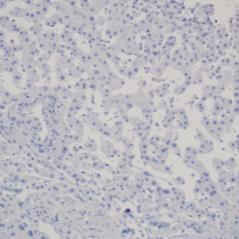
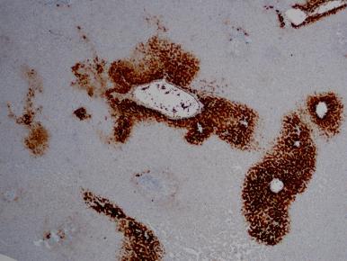
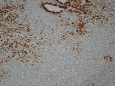
5 Case 1: Obese 42/F with multiple (>15) liver lesions; largest (4 cm) was biopsied L-FABP L-FABP in lesion L-FABP in normal liver SAA Diagnosis HNF1α-inactivated hepatocellular adenoma Hepatic adenomatosis By definition, >10 adenomas Young women Most are HNF1α-inactivated or inflammatory Pathogenesis Obesity, less strong association with OCs Germ line HNF1α mutations Management Resection Conservative with annual surveillance Conservative with annual surveillance Management of HCA Tumor characteristics Women: solitary HCA >5 cm Men (?women>50 years): all cases activated Glycogen storage disease Solitary HCA <5 cm Multiple HCAs or adenomatosis, depending on size 5


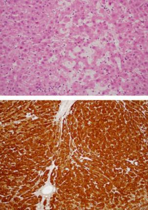
6 Case 2: 53/M, 6 cm liver mass SAA positive diffuse Questions membranous, diffuse Does this represent activation? SAA+ and diffuse Is this inflammatory HCA or activated HCA? Beta-catenin: membranous : diffuse 15% of activated HCA Most have β- catenin mutation Nuclear focal or below threshold : diffuse SAA positive HCA 10% have activation Diagnosis HCA with activation 6


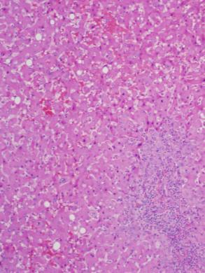
7 Immunostaining variations in HCA Variation Interpretation Membranous, diffuse activation SAA positive, diffuse, + nuclear Nuclear in few tumor cells LFABP patchy; negative in some areas Morphology not typical of inflammatory HCA, SAA positive HCA by morphology, SAA negative activation activation Does not support HNF1αinactivated HCA HCA HCA Outline Hepatocellular adenoma: new WHO classification HCA vs. focal nodular hyperplasia HCA vs. well-differentiated HCC HCA vs. : clinical significance Focal nodular hyperplasia Non-neoplastic No surgery in most cases Large, symptomatic, atypical features Hepatocellular adenoma Neoplastic Surgery if high risk features: Male, size >5 cm Risk of hemorrhage, associated HCC Case 3 32 year old woman on OCs Ultrasound for workup of abdominal pain 5 cm liver mass, suggestive of or inflammatory HCA For For inflammatory HCA Imaging favors Aberrant arterioles in fibrous septa Ductular reaction Sinusoidal dilatation Inflammation Inflammation Arterioles Few ductules Sinusoidal dilatation 7
 Glutamine synthetase () Moderate to strong")


8 or inflammatory HCA Immunostain HCA Serum amyloid A (SAA) Glutamine synthetase () Moderate to strong activated: diffuse Others: Perivascular/patchy Absent/focal Map-like : map-like pattern Diagnosis: Focal nodular hyperplasia SAA Histologic feature Sinusoidal dilatation HCA p value 18% 83% <0.001 Inflammation 40% 60% 0.08 Steatosis 21% 57% Fibrous bands 90% 26% <0.001 Ductular reaction 83% 43% <0.001 Joseph/Kakar, USCAP meeting 2011 vs. IHCA: challenges in interpretation Map-like patterns on needle biopsies Map-like vs. diffuse pattern Pseudo map-like pattern SAA in and adjacent liver Use of CRP in addition to SAA : map-like : normal 8


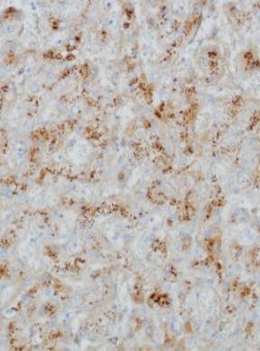
9 Case 4: 40F with 6 cm liver mass : Focal map-like pattern in SAA : map-like vs diffuse HCA with SAA+ : perivascular and patchy expanded staining : pseudo map-like Joseph/Kakar, USCAP


")

10 : Pseudo map-like Confined to periphery Patchy and variable Broad hepatocyte groups with uniform strong staining absent Case 5: 37F with 4 cm liver mass HE stain SAA+ with map-like and SAA+ SAA+ in adjacent liver SAA SAA Role of CRP SAA SAA and CRP in IHA and IHA SAA+ (>50%) CRP+ (>50%) 80% 5-10% 100% 20-25% Joseph/Kakar/Ferrell, Mod Pathol


11 -like lesion Morphology and staining similar to classic Adjacent to tumors Cirrhosis Vascular abnormalities Metastatic adenocarcinoma Metastatic adenocarcinoma : Map-like HCC vs. hepatocellular adenoma Case 6 * 60/M with diabetes and renal failure CT showed a 3.9 cm mass, concerning for HCC or metastatic carcinoma, but appearance was nonspecific and can represent hepatocellular adenoma 11
 - + Small")


12 Reticulin stain International Working Party Adenoma WD-HCC Wide plates (> 3) - + Small cell change - + Cytologic atypia - -/+ Reticulin Normal Fragmented Ferrell et al, Hepatology 1995 Combined immunostaining HSP70, and GPC-3: HCA vs. HCC Glutamine synthetase HSP 70 Study Lagana et al, Appl Immunohistochem Mol Morph, 2012 Nguyen/Kakar, USCAP, 2012 Results HSP70, GPC-3 <50% sensitivity 100% specific 80% sensitivity 50% specific HSP70 in 13% of HCA Overall limited utility Case 6: 58/M with 5 cm hepatic mass: biopsy 58/M with 5 cm liver mass: resection 12


")


13 Reticulin stain HCA or HCC For HCA For HCC Thin cell plates Lack of cytologic atypia Intact reticulin Age >50 years Male gender 61/M with 3.0 cm liver mass (3 yrs later) Well-differentiated HCC Beta-catenin FISH CEP1 gain. CEP8 gain Kakar/Ferrell, Histopathol, 2009, FISH by JP Grenert, UCSF 13

 Is activated HCA malignant?")
 Immunostaining Nuclear Diffuse Well-differentiated hepatocellular neoplasm with atypical")
14 activated HCA, or activated HCC Morphology* HCC* Cytogenetics** Atypia: 70% Concurrent /follow-up: 40% Chromosomal changes: 60% * B Sage, Hepatol 2008 ** Evason/Kakar, Human Pathol 2012 What makes a tumor malignant? Source Webster Medical Dictionary Stedman Medical Dictionary Dorland Medical dictionary Robbins Pathology Definition Ability to invade local tissues Ability to spread to distant sites (metastasize) Is activated HCA malignant? High risk factors Local invasion (recurrence) Metastasis Pathologic features Supportive evidence Yes Yes Yes/no Yes Focal atypical morphology Pseudoacinar Small cell change Thick plates Reticulin loss Age/gender Male gender Older age (>50 yrs) Immunostaining Nuclear Diffuse Well-differentiated hepatocellular neoplasm with atypical features, HCC or activated hepatocellular adenoma HCC arising in adenoma Most activated hepatocellular tumors can be diagnosed as HCC with careful attention to morphology and reticulin staining pattern, especially in resection specimens. * 14


15 Reticulin HCC in adenoma Minimum stains Adenoma Stoot, HPB, 2010 studies from Size >5 cm <5 cm Presence of HCC 68/1635= 4% >95% ~4% Stain Reticulin SAA Interpretation Loss: HCC Diffuse: activation Map-like: Other patterns: HCA HCA Well-differentiated hepatocellular lesion Stain Reticulin SAA Stain LFABP Beta-catenin CRP Utility HCC HCA Map-like: Diffuse: Beta-catenin Utility Glypican-3, HSP70 HCC HNF1-alpha inactivated HCA Beta-catenin Imaging features Central scar Present Absent Contrast CT enhancement Early homogenous HCA Heterogeneous and persistent MRI TI-weighted Hypointense Hyperintensity MRI T2-weighted Hypointense Strong hyperintensity 15
16 Feature Pitfall Approach SAA staining in peritumoral liver in needle biopsy that missed the lesion SAA negative in a lesion that shows typical features of IHA SAA positive in a lesion that shows typical features of Misinterpreted as evidence of IHA, SAA+ is not specific for IHA. especially when other peritumoral Interlobular bile ducts and absence of features like inflammation, sinusoidal diffuse CD34 staining can help in dilatation and ductular reaction are also confirming that the biopsy comprises present non-neoplastic liver. Misinterpreted as absolute evidence against IHA Misinterpreted as IHA Perivenous and patchy in a lesion that Misinterpreted as map-like pattern of shows typical features of IHA Diffuse staining Pseudo-map like pattern Misinterpreted as map-like pattern of Misinterpreted as map-like pattern of SAA can be negative in 5-10% of IHA. Imaging and absence of map-like staining is needed to confirm IHA in these cases. Focal SAA can be seen in 15% of. Map-like confirms irrespective of SAA. This staining is common in IHA. It is distinguished from map-like pattern by (i) lack of anastamosing pattern of staining. (ii) staining is heterogeneous and weak compared to homogeneous strong staining in map-like pattern. Diffuse is seen in beta-cateninactivated tumors and does not have areas of periseptal sparing of map-like pattern. Most also show nuclear beta-catenin. This is seen at the periphery of IHA and 10% of. When seen in biopsies, it is more likely to be. Diagnosis on needle biopsy may remain indeterminate if imaging and SAA are not helpful. : map-like SAA: negative or positive Compatible with vs. IHCA : resembles map-like, but not typical SAA: negative Morphology and imaging Not typical of Indeterminate : perivascular and/or patchy SAA positive IHCA SAA negative Morphology and imaging Consider CRP Likely SAAnegative IHCA : diffuse SAA: positive activated IHCA 16
Atypical Well differentiated Hepatocellular Neoplasms Cruising through the maze of criteria, terminology and risk assessment
 2016 Hans Popper Companion Meeting Atypical Well differentiated Hepatocellular Neoplasms Cruising through the maze of criteria, terminology and risk assessment Disclosure Dr. Kakar has nothing to Disclose
O Farrell Legacy UPDATE ON WHO NOMENCLATURE. World Health Organization, 2010 DISCLOSURES WITH EMPHASIS ON PROBLEM HEPATOCELLULAR TUMORS
 O Farrell Legacy UPDATE ON WHO NOMENCLATURE WITH EMPHASIS ON PROBLEM HEPATOCELLULAR TUMORS Linda Ferrell, MD University of California San Francisco Vice Chair, Director of Surgical Pathology World Health
PATHOLOGY OF LIVER TUMORS
 PATHOLOGY OF LIVER TUMORS Pathobasic, 31.05.2016 WHO Classification Approach to a Liver Mass Lesion in a patient with chronic liver disease? Lesion in a patient without chronic liver disease? Malignant
Evaluation of Liver Mass Lesions. American College of Gastroenterology 2013 Regional Postgraduate Course
 Evaluation of Liver Mass Lesions American College of Gastroenterology 2013 Regional Postgraduate Course Lewis R. Roberts, MB ChB, PhD Division of Gastroenterology and Hepatology Mayo Clinic College of
Jesse Civan, M.D. Medical Director, Jefferson Liver Tumor Center
 Liver Tumors Jesse Civan, M.D. Medical Director, Jefferson Liver Tumor Center Differential Diagnosis Malignant Metastatic from non-hepatic primary Hepatocellular carcinoma Cholangiocarcinoma Biliary cystcarcinoma
Hepatocellular Adenomas: Genetics & Imaging Update 2017
 No financial disclosures Hepatocellular Adenomas: Genetics & Imaging Update 2017 Srinivasa Prasad MD The UT MD Anderson Cancer Center Aims & Objectives To provide a current update on genetics & molecular
HEPATOCYTE SPECIFIC CONTRAST MEDIA: WHERE DO WE STAND?
 HEPATOCYTE SPECIFIC CONTRAST MEDIA: WHERE DO WE STAND? Andrew T. Trout, MD @AndrewTroutMD Disclosures No relevant disclosures Outline Review of hepatocyte specific contrast media Review of hepatocellular
Hepatocellular adenomas (HCAs) are uncommon primary benign tumours. They are constantly monoclonal tumours.
 are uncommon primary benign tumours. They are constantly monoclonal tumours.") Hepatocellular adenoma: Evaluation with contrast enhanced ultrasound, MRI And Correlation with pathologic and phenotypic classification. About 26 lesions. Poster No.: C-1561 Congress: ECR 2011 Type: Scientific
Hepatocellular adenoma: Evaluation with contrast enhanced ultrasound, MRI And Correlation with pathologic and phenotypic classification. About 26 lesions. Poster No.: C-1561 Congress: ECR 2011 Type: Scientific
activated hepatocellular adenoma
 pissn 2287-2728 eissn 2287-285X Liver Pathology Clinical and Molecular Hepatology 2013;19:185-189 5.2 3.6 cm sized, homogeneous, green colored hepatic mass was observed on the cut section (Fig. 1). The
Invited Re vie W. Analytical histopathological diagnosis of small hepatocellular nodules in chronic liver diseases
 Histol Histopathol (1 998) 13: 1077-1 087 http://www.ehu.es/histoi-histopathol Histology and Histopathology Invited Re vie W Analytical histopathological diagnosis of small hepatocellular nodules in chronic
Mesenchymal Tumors. Cavernous Hemangioma (CH) VASCULAR TUMORS MESENCHYMAL TUMORS OF THE LIVER: WHAT S NEW AND UNUSUAL (MY PERSPECTIVE)
 VASCULAR TUMORS MESENCHYMAL TUMORS OF THE LIVER: WHAT S NEW AND UNUSUAL (MY PERSPECTIVE)") Mesenchymal Tumors MESENCHYMAL TUMORS OF THE LIVER: WHAT S NEW AND UNUSUAL (MY PERSPECTIVE) CURRENT ISSUES IN ANATOMIC PATHOLOGY MAY 23, 2014 Linda Ferrell, MD, UCSF Focus on Vascular Tumors Benign and
Mesenchymal Tumors MESENCHYMAL TUMORS OF THE LIVER: WHAT S NEW AND UNUSUAL (MY PERSPECTIVE) CURRENT ISSUES IN ANATOMIC PATHOLOGY MAY 23, 2014 Linda Ferrell, MD, UCSF Focus on Vascular Tumors Benign and
Mesenchymal Tumors MESENCHYMAL TUMORS OF THE LIVER: WHAT S NEW AND UNUSUAL (MY PERSPECTIVE)
") MESENCHYMAL TUMORS OF THE LIVER: WHAT S NEW AND UNUSUAL (MY PERSPECTIVE) CURRENT ISSUES IN ANATOMIC PATHOLOGY MAY 23, 2014 Linda Ferrell, MD, UCSF Mesenchymal Tumors Focus on Vascular Tumors Benign and
MESENCHYMAL TUMORS OF THE LIVER: WHAT S NEW AND UNUSUAL (MY PERSPECTIVE) CURRENT ISSUES IN ANATOMIC PATHOLOGY MAY 23, 2014 Linda Ferrell, MD, UCSF Mesenchymal Tumors Focus on Vascular Tumors Benign and
MRI OF FOCAL LESIONS IN
 Introduction MRI OF FOCAL LESIONS IN THE NON-CIRRHOTIC LIVER Ivan Pedrosa M.D. Ph.D. Associate Professor of Radiology and Advanced Imaging Research Center University of Texas Southwestern. Dallas, TX Incidental
Outline. Hepatocellular Carcinoma Histologic variants. HCC: Histologic variants
 2018 Park City AP Update Hepatocellular Carcinoma Histologic variants Sanjay Kakar, MD University of California, San Francisco Outline Histologic variants of HCC Morphologic and Immunohistochemical pitfalls
Intrahepatic cholangiocarcinoma Histologic spectrum, novel markers and molecular assays
 2018 Current Issues in Surgical Pathology Summary (not actual lecture) Intrahepatic cholangiocarcinoma Histologic spectrum, novel markers and molecular assays Sanjay Kakar, MD University of California,
Benign liver tumors : Diagnosis and management
 4th International Hepatology Conference 2016 HEPATOLOGY SOCIETY, DHAKA, BANGLADESH Benign liver tumors : Diagnosis and management Pr Laurence Chiche Hepato biliary surgery and transplantation Bordeaux,
Liver Specialty Evening Conference. Matthew M. Yeh, MD, PhD Professor of Pathology Adjunct Professor of Medicine University of Washington, Seattle
 Liver Specialty Evening Conference Matthew M. Yeh, MD, PhD Professor of Pathology Adjunct Professor of Medicine University of Washington, Seattle Case History A 65 year-old man presents with abdominal
Financial Disclosure
 Benign Liver Masses Adil Abdalla, MBBS Creighton University-CHI Health August 25, 2018 Financial Disclosure Nothing to disclose Financial Disclosure 1 Objectives To assess patients with benign liver tumors
Liver Tumors. Prof. Dr. Ahmed El - Samongy
 Liver Tumors Prof. Dr. Ahmed El - Samongy Objective 1. Identify the most important features of common benign liver tumors 2. Know the risk factors, diagnosis, and management of hepatocellular carcinoma
Alastair Burt Newcastle University
 Alastair Burt Newcastle University Benign Hepatocellular adenoma 8170/0 Focal nodular hyperplasia Malignancy-associated and premalignant lesions Large cell change (formerly dysplasia ) Small cell change
Hepatocellular adenomas are benign liver tumors,
 Case Report Hepatobiliary & Pancreatic Diseases International Pigmented well-differentiated hepatocellular neoplasm with β-catenin mutation Lara Neves Souza, Rodrigo Bronze de Martino, Richard Thompson,
Disclosure. Relevant Financial Relationship(s) None. Off Label Usage None MFMER slide-1
 None. Off Label Usage None MFMER slide-1") Disclosure Relevant Financial Relationship(s) None Off Label Usage None 2013 MFMER slide-1 Case Presentation A 43 year old male, with partial nephrectomy for a right kidney mass 2013 MFMER slide-2 2013
Disclosure Relevant Financial Relationship(s) None Off Label Usage None 2013 MFMER slide-1 Case Presentation A 43 year old male, with partial nephrectomy for a right kidney mass 2013 MFMER slide-2 2013
Papillary Lesions of the Breast A Practical Approach to Diagnosis. (Arch Pathol Lab Med. 2016;140: ; doi: /arpa.
 Papillary Lesions of the Breast A Practical Approach to Diagnosis (Arch Pathol Lab Med. 2016;140:1052 1059; doi: 10.5858/arpa.2016-0219-RA) Papillary lesions of the breast Span the spectrum of benign,
Pathological Classification of Hepatocellular Carcinoma
 3 rd APASL Single Topic Conference: HCC in 3D Pathological Classification of Hepatocellular Carcinoma Glenda Lyn Y. Pua, M.D. HCC Primary liver cancer is the 2 nd most common cancer in Asia HCC is the
Disclosures. Parathyroid Pathology. Objectives. The normal parathyroid 11/10/2012
 Disclosures Parathyroid Pathology I have nothing to disclose Annemieke van Zante MD/PhD Assistant Professor of Clinical Pathology Associate Chief of Cytopathology Objectives 1. Review the pathologic features
Select problems in cystic pancreatic lesions
 Disclosure Select problems in cystic pancreatic lesions Five Prime Therapeutics shareholder Adicet Bio shareholder Bristol-Meyer Squibb advisory board grace.kim@ucsf.edu Pancreatic cystic lesions Intraductal
CTA/MRA of Pediatric Hepatic Masses Radiology-Pathology Correlation
 Acta Radiológica Portuguesa, Vol.XVIII, nº70, pág. 41-50, Abr.-Jun., 2006 CTA/MRA of Pediatric Hepatic Masses Radiology-Pathology Correlation Marilyn J. Siegel Mallinckrodt Institute of Radiology, Washington
Differential diagnosis of HCC
 Hepatocellular Carcinoma Quest for an Ideal Immunohistochemical Panel Sanjay Kakar, MD UCSF Differential diagnosis of HCC Hepatocellular lesions Adenoma, FNH, HG dysplasia Adenocarcinoma CholangioCA, metastasis
ACCME/Disclosures. Cribriform Lesions of the Prostate. Case
 Cribriform Lesions of the Prostate Ming Zhou, MD, PhD Departments of Pathology and Urology New York University Langone Medical Center New York, NY Ming.Zhou@NYUMC.ORG ACCME/Disclosures The USCAP requires
Salivary Gland FNA ATYPICAL : Criteria and Controversies
 Salivary Gland FNA ATYPICAL : Criteria and Controversies W.C. Faquin, M.D., Ph.D. Director, Head and Neck Pathology Massachusetts General Hospital Massachusetts Eye and Ear Infirmary Harvard Medical School
A 60-year old Man with Left Jaw Mass. Simon Chiosea, MD University of Pittsburgh medical Center 3/15/2016
 ACCME/Disclosures The USCAP requires that anyone in a position to influence or control the content of CME disclose any relevant financial relationship WITH COMMERCIAL INTERESTS which they or their spouse/partner
Prostate Immunohistochemistry. Literature Interpretation: Caveats. Must be aware of staining pattern of antibody in the relevant tissue
 IHC Interpretation: General Principles (1) Prostate Immunohistochemistry Murali Varma Cardiff, UK wptmv@cf.ac.uk Sarajevo Nov 2013 Must be aware of staining pattern of antibody in the relevant tissue Nuclear/cytoplasmic/membranous
04/10/2018. Intraductal Papillary Neoplasms Of Breast INTRADUCTAL PAPILLOMA
 Intraductal Papillary Neoplasms Of Breast Savitri Krishnamurthy MD Professor of Pathology Deputy Division Head The University of Texas MD Anderson Cancer Center 25 th Annual Seminar in Pathology Pittsburgh,
CT & MRI of Benign Liver Neoplasms Srinivasa R Prasad
 CT & MRI of Benign Liver Neoplasms Srinivasa R Prasad No financial disclosures Acknowledgements Many thanks to Drs. Heiken, Narra & Menias (MIR) Dr. Sahani (MGH) for sharing images Benign Liver Tumors:
Neoplasia part I. Dr. Mohsen Dashti. Clinical Medicine & Pathology nd Lecture
 Neoplasia part I By Dr. Mohsen Dashti Clinical Medicine & Pathology 316 2 nd Lecture Lecture outline Review of structure & function. Basic definitions. Classification of neoplasms. Morphologic features.
Hepatic adenoma (HA) is an uncommon, benign
 is an uncommon, benign") Review Article Update on the New Classification of Hepatic Adenomas Clinical, Molecular, and Pathologic Characteristics Sadhna Dhingra, MD; M. Isabel Fiel, MD Context. Hepatic adenoma is an uncommon, benign,
Review Article Update on the New Classification of Hepatic Adenomas Clinical, Molecular, and Pathologic Characteristics Sadhna Dhingra, MD; M. Isabel Fiel, MD Context. Hepatic adenoma is an uncommon, benign,
How to Approach a Medical Liver Biopsy. 9 th Bryan Warren School of Pathology Pancreatic and Liver Pathology. Sarajevo, 6 th -7 th November 2015
 1 A Brief Introduction to the Liver Sessions 9 th Bryan Warren School of Pathology Pancreatic and Liver Pathology Sarajevo, 6 th -7 th November 2015 Stefan Hübscher, Institute of Immunology & Immunotherapy,
A Triple Stain of Reticulin, Glypican-3, and Glutamine Synthetase
 A Triple Stain of Reticulin, Glypican-3, and Glutamine Synthetase A Useful Aid in the Diagnosis of Liver Lesions Benjamin J. Swanson, MD, PhD; Martha M. Yearsley, MD; William Marsh, MD; Wendy L. Frankel,
Basic patterns of liver damage what information can a liver biopsy provide and what clinical information does the pathologist need?
 Basic patterns of liver damage what information can a liver biopsy provide and what clinical information does the pathologist need? Rob Goldin r.goldin@imperial.ac.uk Fatty liver disease Is there fatty
ACG Clinical Guideline: Diagnosis and Management of Focal Liver Lesions
 ACG Clinical Guideline: Diagnosis and Management of Focal Liver Lesions Jorge A. Marrero, MD, 1 Joseph Ahn, MD, FACG, 2 K. Rajender Reddy, MD, FACG 3 1 University of Texas at Southwestern, Dallas, Texas,
Malignant Focal Liver Lesions
 Malignant Focal Liver Lesions Other Than HCC Pablo R. Ros, MD, MPH, PhD Departments of Radiology and Pathology University Hospitals Cleveland Medical Center Case Western Reserve University Pablo.Ros@UHhospitals.org
Hepatocellular neoplasia - Recent developments
 22 November 2018 Hepatocellular neoplasia - Recent developments Dina G. Tiniakos Institute of Cellular Medicine, Faculty of Medical Sciences, Newcastle University, UK Dept of Pathology, Aretaieion Hospital,
Interesting Cases from Liver Tumor Board. Jeffrey C. Weinreb, M.D.,FACR Yale University School of Medicine
 Interesting Cases from Liver Tumor Board Jeffrey C. Weinreb, M.D.,FACR Yale University School of Medicine jeffrey.weinreb@yale.edu Common Liver Diseases Hemangioma Cyst FNH Focal Fat/Sparing THID Non-Cirrhotic
Essentials of Clinical MR, 2 nd edition. 65. Benign Hepatic Masses
 65. Benign Hepatic Masses Pulse sequences acquired for abdominal MRI typically consist of fast acquisition schemes such as single-shot turbo spin echo (i.e. HASTE) and gradient echo schemes such as FLASH
A 53 year-old woman with a lung mass, right hilar mass and mediastinal adenopathy.
 November 2015 Case of the Month A 53 year-old woman with a lung mass, right hilar mass and mediastinal adenopathy. Contributed by: Rasha Salama, M.D., IU Department of Pathology and Laboratory Medicine
British Liver Transplant Group Pathology meeting September Leeds cases
 British Liver Transplant Group Pathology meeting September 2014 Leeds cases Leeds Case 1 Male 61 years Liver transplant for HCV cirrhosis with HCC in January 2014. Now raised ALT and bilirubin,? acute
Essentials of Clinical MR, 2 nd edition. 73. Urinary Bladder and Male Pelvis
 73. Urinary Bladder and Male Pelvis Urinary bladder carcinoma is best locally staged with MRI. It is important however to note that a thickened wall (> 5 mm) is a non-specific finding seen in an underfilled
Clinical and Molecular Approach to Using Thyroid Needle Biopsy for Nodular Disease
 Clinical and Molecular Approach to Using Thyroid Needle Biopsy for Nodular Disease Robert L. Ferris, MD, PhD Department of Otolaryngology/Head and Neck Surgery and Yuri E. Nikiforov, MD, PhD Division of
Raga Ramachandran, MD, PhD Assistant Professor and Director of Medical Education, UCSF Pathology
 Variants of Hepatocellular Carcinoma: Practical Issues Raga Ramachandran, MD, PhD Assistant Professor and Director of Medical Education, UCSF Pathology raga.ramachandran@ucsf.edu A full copy of the presentation
Workup of a Solid Liver Lesion
 Workup of a Solid Liver Lesion Joseph B. Cofer MD FACS Chief Quality Officer Erlanger Health System Affiliate Professor of Surgery UTHSC-Chattanooga I have no financial or other relationships with any
ACCME/Disclosures ALK FUSION-POSITIVE MESENCHYMAL TUMORS. Tumor types with ALK rearrangements. Anaplastic Lymphoma Kinase. Jason L.
 Companion Meeting of the International Society of Bone and Soft Tissue Pathology The Evolving Concept of Mesenchymal Tumors ALK FUSION-POSITIVE MESENCHYMAL TUMORS Jason L. Hornick, MD, PhD March 13, 2016
Neoplasms of the Canine, Feline and Lemur Liver:
 Neoplasms of the Canine, Feline and Lemur Liver: Classification and Prognosis Annual Seminar of the French Society of Veterinary Pathology John M. Cullen VMD PhD DACVP North Carolina State University Primary
Detection and Characterization of Hepatocellular Carcinoma by Imaging
 CLINICAL GASTROENTEROLOGY AND HEPATOLOGY 2005;3:S136 S140 Detection and Characterization of Hepatocellular Carcinoma by Imaging OSAMU MATSUI Department of Imaging Diagnosis and Interventional Radiology,
Follicular Derived Thyroid Tumors
 Follicular Derived Thyroid Tumors Jennifer L. Hunt, MD, MEd Aubrey J. Hough Jr, MD, Endowed Professor of Pathology Chair of Pathology and Laboratory Medicine University of Arkansas for Medical Sciences
5/21/2018. Difficulty in Underdiagnosing Prostate Cancer. Diagnosis of Prostate Cancer. Evaluation of Prostate Cancer and Atypical on Needle Biopsy
 Evaluation of Prostate Cancer and Atypical on Needle Biopsy Jonathan I. Epstein Difficulty in Underdiagnosing Prostate Cancer Limited tissue on needle biopsy (1 cm. x
LIVER IMAGING TIPS IN VARIOUS MODALITIES. M.Vlychou, MD, PhD Assoc. Professor of Radiology University of Thessaly
 LIVER IMAGING TIPS IN VARIOUS MODALITIES M.Vlychou, MD, PhD Assoc. Professor of Radiology University of Thessaly Hepatocellular carcinoma is a common malignancy for which prevention, screening, diagnosis,
Linda Ferrell, MD Distinguished Professor Vice Chair Director of Surgical Pathology Dept of Pathology
 Linda Ferrell, MD Distinguished Professor Vice Chair Director of Surgical Pathology Dept of Pathology Nonalcoholic steatohepatitis and Fatty Liver Disease Liver manifestations of the obesity epidemic Changes
Special stains in liver pathology
 Current Issues in Surgical Pathology 2014 Special stains in liver pathology Which, why, how Really? Sanjay Kakar, MD University of California, San Francisco Outline Which stains Why the stain is done How
A 42-year-old woman with a liver mass
 April 2016 Case of the Month A 42-year-old woman with a liver mass Contributed by: Natalia I. Rush, MD, Resident Physician, Indiana University School of Medicine, Department of Pathology and Laboratory
PITFALLS IN THE DIAGNOSIS OF MEDICAL LIVER DISEASE WITH TWO CONCURRENT ETIOLOGIES I HAVE NOTHING TO DISCLOSE CURRENT ISSUES IN ANATOMIC PATHOLOGY 2017
 CURRENT ISSUES IN ANATOMIC PATHOLOGY 2017 I HAVE NOTHING TO DISCLOSE Linda Ferrell PITFALLS IN THE DIAGNOSIS OF MEDICAL LIVER DISEASE WITH TWO CONCURRENT ETIOLOGIES Linda Ferrell, MD, UCSF THE PROBLEM
Pigmented hepatocellular adenoma with β-catenin activation: case report and literature review
 598 Coelho R, et al., 2016; 15 (4): 598-603 CASE REPORT July-August, Vol. 15 No. 4, 2016: 598-603 The Official Journal of the Mexican Association of Hepatology, the Latin-American Association for Study
ACCME/Disclosures. Diagnosing Mesothelioma in Limited Tissue Samples. Papanicolaou Society of Cytopathology Companion Meeting March 12 th, 2016
 Diagnosing Mesothelioma in Limited Tissue Samples Papanicolaou Society of Cytopathology Companion Meeting March 12 th, 2016 Sanja Dacic, MD, PhD University of Pittsburgh ACCME/Disclosures GENERAL RULES
2014 CURRENT ISSUES IN PATHOLOGY
 2014 CURRENT ISSUES IN PATHOLOGY SPECIAL STAINS IN LIVER BIOPSY PATHOLOGY Sanjay Kakar, MD University of California, San Francisco Trichrome stain : (1) Assess degree of fibrosis. H&E stain is not reliable
Papillary Lesions of the breast
 Papillary Lesions of the breast Emad Rakha Professor of Breast Pathology The University of Nottingham Papillary lesions of the breast are a heterogeneous group of disease, which are characterised by neoplastic
What s New in Adrenal Gland Pathology. Marina Scarpelli
 What s New in Adrenal Gland Pathology Marina Scarpelli Background Histological criteria for adrenocortical proliferative lesions Immunohistochemical markers Molecular markers Histological Criteria for
NEOPLASMS OF THE THYROID PATHOLOGY OF PARATHYROID GLANDS. BY: Shifaa Qa qa
 NEOPLASMS OF THE THYROID PATHOLOGY OF PARATHYROID GLANDS BY: Shifaa Qa qa Neoplasmas of the thyroid thyroid nodules Neoplastic ---- benign, malignant Non neoplastic Solitary nodules ----- neoplastic Nodules
INTRADUCTAL LESIONS OF THE PROSTATE. Jonathan I. Epstein
 INTRADUCTAL LESIONS OF THE PROSTATE Jonathan I. Epstein Topics Prostatic intraepithelial neoplasia (PIN) Intraductal adenocarcinoma (IDC-P) Intraductal urothelial carcinoma Ductal adenocarcinoma High Prostatic
Kidney Case 1 SURGICAL PATHOLOGY REPORT
 Kidney Case 1 Surgical Pathology Report February 9, 2007 Clinical History: This 45 year old woman was found to have a left renal mass. CT urography with reconstruction revealed a 2 cm medial mass which
Note: The cause of testicular neoplasms remains unknown
 - In the 15- to 34-year-old age group, they are the most common tumors of men. - Tumors of the testis are a heterogeneous group of neoplasms that include: I. Germ cell tumors : 95%; all are malignant.
Malignant neoplasms of the gastrointestinal (GI) tract,
 tract,") Special Section First Chinese American Pathologists Association Diagnostic Pathology Course, Part II Practical Immunohistochemistry in Neoplastic Pathology of the Gastrointestinal Tract, Liver, Biliary
Special Section First Chinese American Pathologists Association Diagnostic Pathology Course, Part II Practical Immunohistochemistry in Neoplastic Pathology of the Gastrointestinal Tract, Liver, Biliary
Solitary Fibrous Tumor of the Kidney with Massive Retroperitoneal Recurrence. A Case Presentation
 246) Prague Medical Report / Vol. 113 (2012) No. 3, p. 246 250 Solitary Fibrous Tumor of the Kidney with Massive Retroperitoneal Recurrence. A Case Presentation Sfoungaristos S., Papatheodorou M., Kavouras
Disclosures. Giant Cell Rich Tumors of Bone. Outline. The osteoclast. Giant cell rich tumors 5/21/11
 Disclosures Giant Cell Rich Tumors of Bone Andrew Horvai, MD, PhD Associate Clinical Professor, Pathology This lecture discusses "off label" uses of a number of pharmaceutical agents. The speaker is describing
Recently role of non-invasive diagnostics methods
 CERTAIN ASPECTS OF NLS-DIAGNOSTICS OF LIVER FOCAL PATHOLOGY A.Y. Shvack, V.I. Nesterov, N.L. Ogluzdina This article contains information about NLS-graphy application in diagnostics of liver focal affections:
Papillary Lesions of the Breast
 Papillary Lesions of the Breast Laura C. Collins, M.D. Associate Professor of Pathology Associate Director, Division of Anatomic Pathology Beth Israel Deaconess Medical Center and Harvard Medical School
Case: The patient is a 62 year old woman with a history of renal cell carcinoma that was removed years ago. A 2.4 cm liver mass was found on CT
 Case: The patient is a 62 year old woman with a history of renal cell carcinoma that was removed years ago. A 2.4 cm liver mass was found on CT during follow- up. ALT, AST, Alk Phos and bilirubin were
BREAST PATHOLOGY. Fibrocystic Changes
 BREAST PATHOLOGY Lesions of the breast are very common, and they present as palpable, sometimes painful, nodules or masses. Most of these lesions are benign. Breast cancer is the 2 nd most common cause
CASE 1 11/1/2016 HEPATOBILIARY IMAGING CASE PRESENTATIONS DECLARATION. Dr. Chirag Patel ORGAN IMAGING yr old lady
 HEPATOBILIARY IMAGING CASE PRESENTATIONS DECLARATION No financial disclosures or affiliations with commercial organisations No discussion of investigational or off-label use of medical devices, products
Prostate cancer ~ diagnosis and impact of pathology on prognosis ESMO 2017
 Prostate cancer ~ diagnosis and impact of pathology on prognosis ESMO 2017 Dr Puay Hoon Tan Division of Pathology Singapore General Hospital Prostate cancer (acinar adenocarcinoma) Invasive carcinoma composed
Question No. Clinical Aspect Drop-Down List Response Q1 Primary indication for liver biopsy from original request form Deranged LFTs
 Appendix E1 Questionnaire A: Organizational Questionnaire Question No. Question Q1 How many image-guided/assisted liver biopsies in total were performed in your department from 1/1/2008 31/12/2008? Q2
Evening Specialty Conference Bone and Soft Tissue Pathology. Diagnostic pitfalls in bone and soft tissue pathology
 Evening Specialty Conference Bone and Soft Tissue Pathology. Case 1 Elizabeth G Demicco, MD, PhD Mount Sinai Hospital, New York Disclosure of Relevant Financial Relationships USCAP requires that all planners
Video Microscopy Tutorial 8
 Video Microscopy Tutorial 8 Common and Uncommon Lesions of the Liver Gladwyn Leiman, MD There are no disclosures necessary. Common and Uncommon Lesions in Liver FNA Gladwyn Leiman University of Vermont
Biliary tract tumors
 Short Course 2010 Annual Fall Meeting of the Korean Society for Pathologists Biliary tract tumors Joon Hyuk Choi, M.D., Ph.D. Professor, Department of Pathology, Yeungnam Univ. College of Medicine, Daegu,
Hyperplastische Polyps Innocent bystanders?
 Hyperplastische Polyps Innocent bystanders?? K. Geboes P th l i h O tl dk d Pathologische Ontleedkunde, KULeuven Content Historical Classification Relation Hyperplastic polyps carcinoma The concept cept
HEPATO-BILIARY IMAGING
 HEPATO-BILIARY IMAGING BY MAMDOUH MAHFOUZ MD PROF.OF RADIOLOGY CAIRO UNIVERSITY mamdouh.m5@gmail.com www.ssregypt.com CT ABDOMEN Indications Patient preparation Patient position Scanogram Fasting 4-6 hours
Presentation material is for education purposes only. All rights reserved URMC Radiology Page 1 of 98
 Presentation material is for education purposes only. All rights reserved. 2011 URMC Radiology Page 1 of 98 Radiology / Pathology Conference February 2011 Brooke Koltz, Cytopathology Resident Presentation
New insights into fatty liver disease. Rob Goldin Centre for Pathology, Imperial College
 New insights into fatty liver disease Rob Goldin Centre for Pathology, Imperial College r.goldin@imperial.ac.uk Prevalence of NASH Global prevalence of NAFLD is 25% with highest prevalence in the Middle
The Panel Approach to Diagnostics. Lauren Hopson International Product Specialist Cell Marque Corporation
 The Panel Approach to Diagnostics Lauren Hopson International Product Specialist Cell Marque Corporation Cell Marque Rocklin, California About Cell Marque: IVD primary antibody manufacturer Distributors
Five Most Common Problems in Surgical Neuropathology
 Five Most Common Problems in Surgical Neuropathology If the brain were so simple that we could understand it, we would be so simple that we couldn t Emerson Pugh What is your greatest difficulty in neuropathology?
Update on 2015 WHO Classification of Lung Adenocarcinoma 1/3/ Mayo Foundation for Medical Education and Research. All rights reserved.
 1 Our speaker for this program is Dr. Anja Roden, an associate professor of Laboratory Medicine and Pathology at Mayo Clinic as well as consultant in the Anatomic Pathology Laboratory and co-director of
3/27/2017. Pulmonary Pathology Specialty Conference. Disclosure of Relevant Financial Relationships. Clinical History:
 Pulmonary Pathology Specialty Conference Saul Suster, M.D. Medical College of Wisconsin Disclosure of Relevant Financial Relationships USCAP requires that all planners (Education Committee) in a position
The Frozen Section: Diagnostic Challenges and Pitfalls
 The Frozen Section: Diagnostic Challenges and Pitfalls William C. Faquin, M.D., Ph.D. Director, Head and Neck Pathology Massachusetts General Hospital & Massachusetts Eye and Ear Infirmary Harvard Medical
Liver Cancer And Tumours
 Liver Cancer And Tumours What causes liver cancer? Many factors may play a role in the development of cancer. Because the liver filters blood from all parts of the body, cancer cells from elsewhere can
Hepatobiliary and Pancreatic Malignancies
 Hepatobiliary and Pancreatic Malignancies Gareth Eeson MD MSc FRCSC Surgical Oncologist and General Surgeon Kelowna General Hospital Interior Health Consultant, Surgical Oncology BC Cancer Agency Centre
Management of Rare Liver Tumours
 Gian Luca Grazi Hepato-Biliary-Pancreatic Surgery National Cancer Institute Regina Elena Rome Fibrolamellar Carcinoma Mixed Hepato Cholangiocellular Carcinoma Hepatoblastoma Carcinosarcoma Primary Hepatic
40th European Congress of Cytology Liverpool, UK, 2-5 th October 2016
 40th European Congress of Cytology Liverpool, UK, 2-5 th October 2016 EUS FNA of abdominal organs: An approach to reporting and triage for ancillary testing Date and time: Sunday 2 nd October 2016 15.00-16.30
The Focal Hepatic Lesion: Radiologic Assessment
 The Focal Hepatic Lesion: Radiologic Assessment Kevin Kuo, Harvard Medical School Year III Our Patient: PS 67 y/o female w/ long history of alcohol use Drinking since age 18, up to one bottle of wine/day
GOBLET CELL CARCINOID. Hanlin L. Wang, MD, PhD University of California Los Angeles
 GOBLET CELL CARCINOID Hanlin L. Wang, MD, PhD University of California Los Angeles Disclosure of Relevant Financial Relationships USCAP requires that all planners (Education Committee) in a position to
GOBLET CELL CARCINOID
 GOBLET CELL CARCINOID Hanlin L. Wang, MD, PhD University of California Los Angeles Disclosure of Relevant Financial Relationships USCAP requires that all planners (Education Committee) in a position to
Objectives. Salivary Gland FNA: The Milan System. Role of Salivary Gland FNA 04/26/2018
 Salivary Gland FNA: The Milan System Dr. Jennifer Brainard Section Head Cytopathology Cleveland Clinic Objectives Introduce the Milan System for reporting salivary gland cytopathology Define cytologic
Case report Solid pseudopapillary tumor: a rare neoplasm of the pancreas
 Gastroenterology Report 2 (2014) 145 149, doi:10.1093/gastro/gou006 Advance access publication 28 February 2014 Case report Solid pseudopapillary tumor: a rare neoplasm of the pancreas Asim Shuja 1, *