Caveat sonologist Mistakes to avoid in Kidney Ultrasound
|
|
|
- Sheryl Atkins
- 6 years ago
- Views:
Transcription

1 Caveat sonologist Mistakes to avoid in Kidney Ultrasound Simon Freeman Derriford Hospital, Plymouth

2 Bear trap 1 Report: There is a 4cm solid mass arising from the left kidney likely to represent a RCC
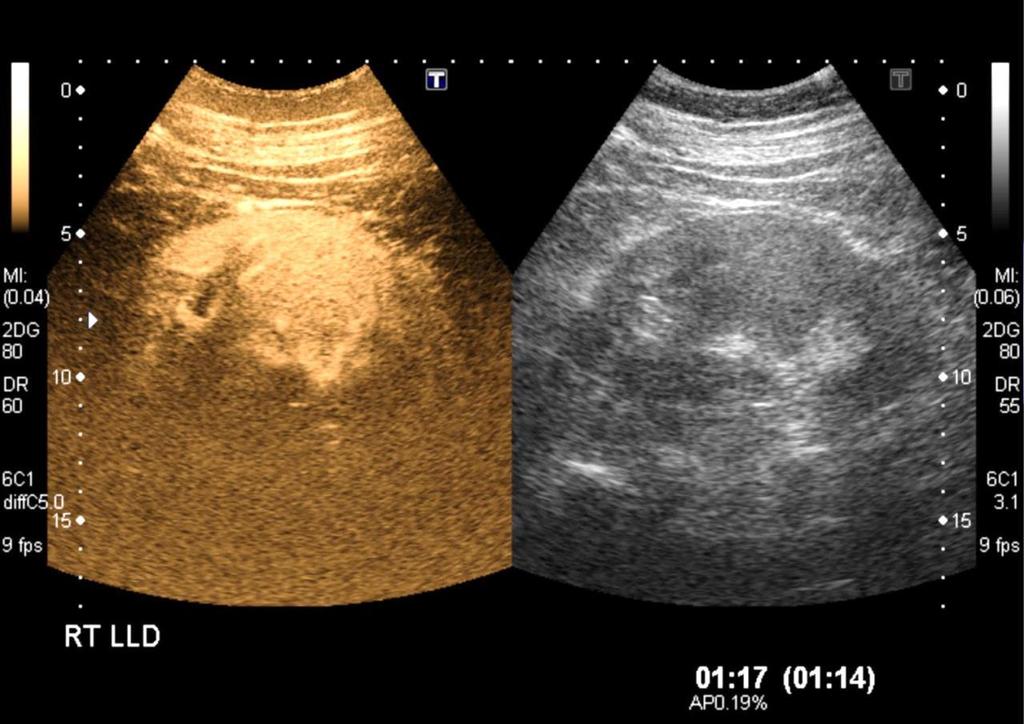
3 Is it truly a mass? Renal pseudotumours 1 Dromedary/splenic hump Hypertrophied septum of Bertin Persistent foetal lobulation Scarring After partial nephrectomy Splenorenal fusion Value of CEUS 2 (but also DMSA, CT and MRI) 1. Bhatt. American Journal of Roentgenology 2007;188: Piscaglia, F. Ultraschall in der Medizin 2012;33:33-59.


")
4 European Journal of Ultrasound (Ultraschall in der Medizin) February 2012
5
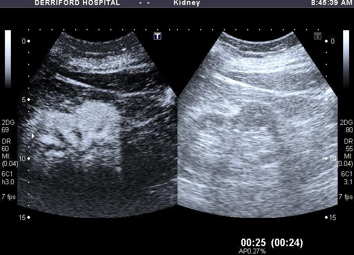
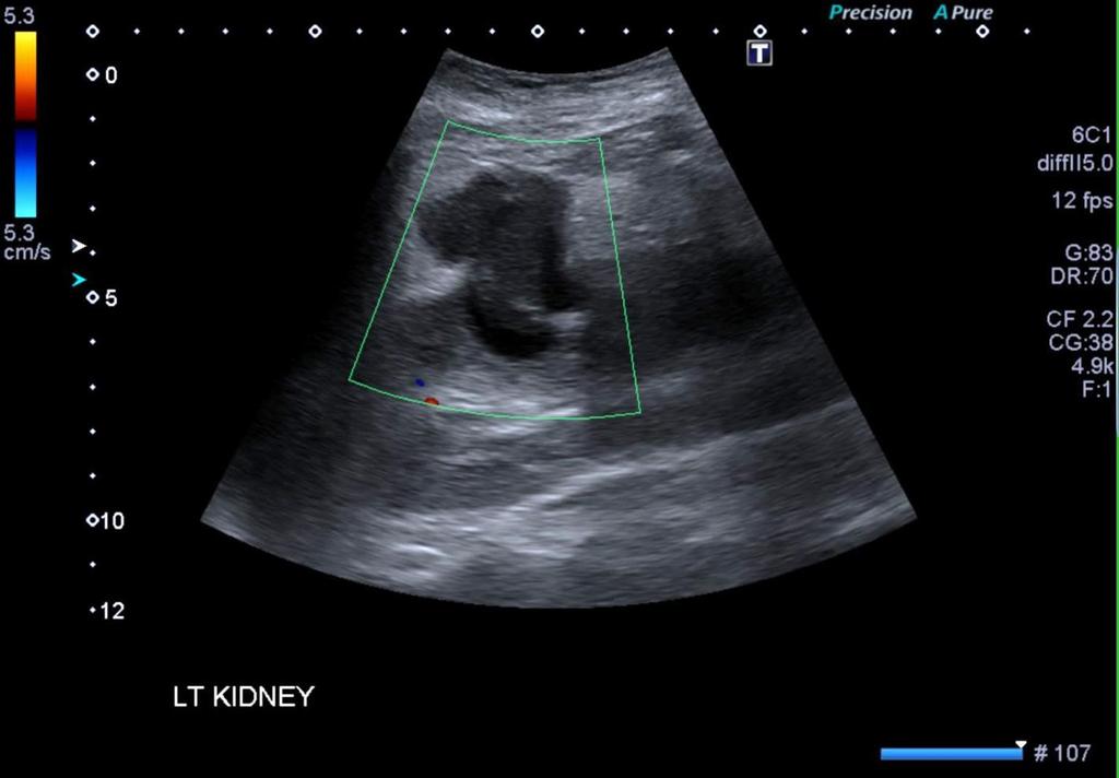
6 Report: As an incidental finding there is a column of Bertin in the left kidney
7
 Can be more than one")
 Lafortune M.")
8 Hypertrophied Column of Bertin 1744 Exupère Joseph Bertin closisins of cortex running between the papillae Present in up to half of all adults Lt>Rt, often bilateral (18%) Can be more than one Junction of upper and middle third Isoechoic (occasionally echogenic) Smooth renal contour Pyramid sometimes seen Smooth displacement of vessels Usually < 2cm If in doubt CEUS (or CT/MRI) Lafortune M. AJR 1986;146:53
9


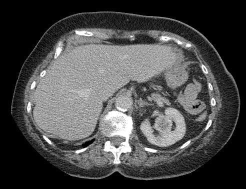
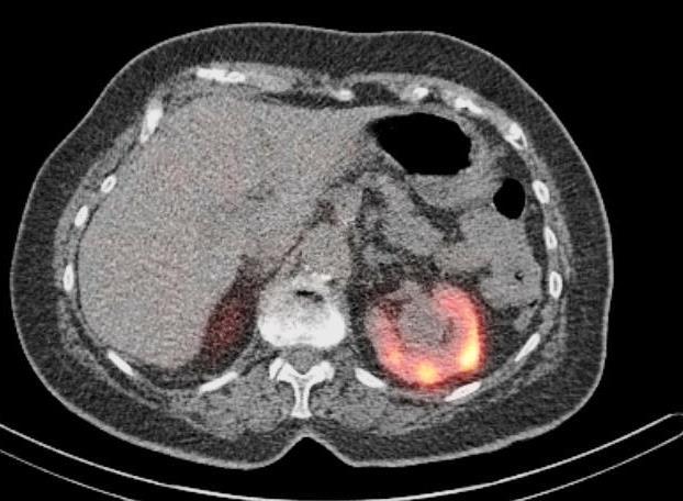
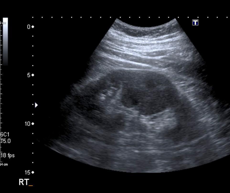
10 Bear Trap 2 Report: There is a 4.5cm solid isoechoic mass arising from the interpolar region of the right kidney, likely to represent a RCC


11 Not all masses are neoplastic: remember infection Lobar Nephronia Focal pyelonephritis May progress to abscess formation Can be difficult or impossible to differentiate from a renal malignancy on imaging Review the clinical status of the patient


12 Acute focal pyelonephritis can be echogenic, mixed echogenicity or echopoor Increased echogenicity is most common Farmer K. Clinical Radiology 2002;57:483-48
13

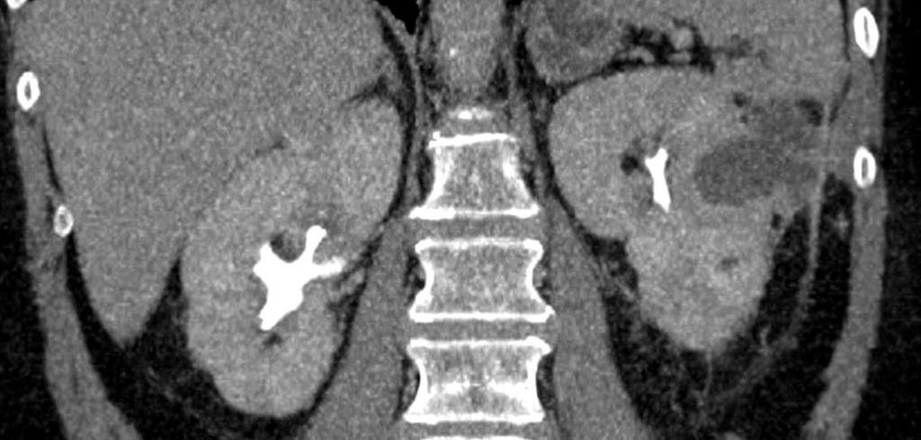
14 Report: The right kidney was difficult to identify and may be small, calcified or absent


15 Remember that gas is echogenic missing or disappearing kidney Emphysematous pyelonephritis Kidney can be difficult to find on US, CT is the imaging modality of choice. Gas forming organism Usually diabetic (90%) High mortality if not rapidly diagnosed and treated Medical management +/- percutaneous drainage. Nephrectomy if not responding Patel C. Saudi J Kidney Disease 2015;26:764
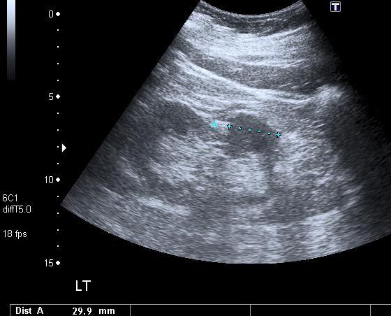
16 Bear Trap 3 Report: There is a 2cm angiomyolipoma arising from the interpolar region of the right kidney

17 Beware of the echogenic mass RCC AML

18 How do we determine echogenicity? Sidhar K. J Ultrasound Med 2016;35:311
19 Ultrasound cannot diagnose fat Approximately a third of RCC of any size are echogenic relative to parenchyma 1 Small RCCs (SRTs) are usually echogenic (77%) 1 SRTs in particular are sometimes very echogenic (equal or greater than to renal sinus fat) 2,3 Hypoechogenic rim, cystic areas and calcification favour RCC Acoustic shadowing favours AML Contrast and elastography show possible differences RCC vs AML 4 1. Foreman. Radiology 1993;188: Siegel. Radiology 1996;198: Sidhar J. Ultrasound Med 2016;35: Tan. AJR 2013;200:W369
20 Echogenic and <1cm in size Unfortunately there is no unequivocal guidance Incidentally discovered echogenic masses <1cm are so likely benign they can be ignored (no cancers in 120 cases) 1 29% of RCC less than 2cm are very hyperechoic 2 1. Itani M. J Ultrasound Med 2016;35: Sidhar K. J Ulltrasound Med 2016;35:311

21 Bottom Line: US cannot diagnose AML CT/MRI is required All non-calcified echogenic renal lesions detected with US need CT to rule our RCC 1 Clear guidance cannot be given for lesions <1cm but you should have a departmental policy 1. Farrelly C. Abdom Imaging 2008;33:44


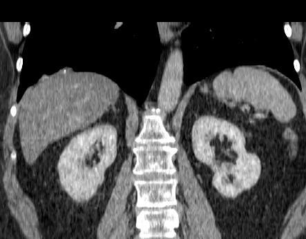
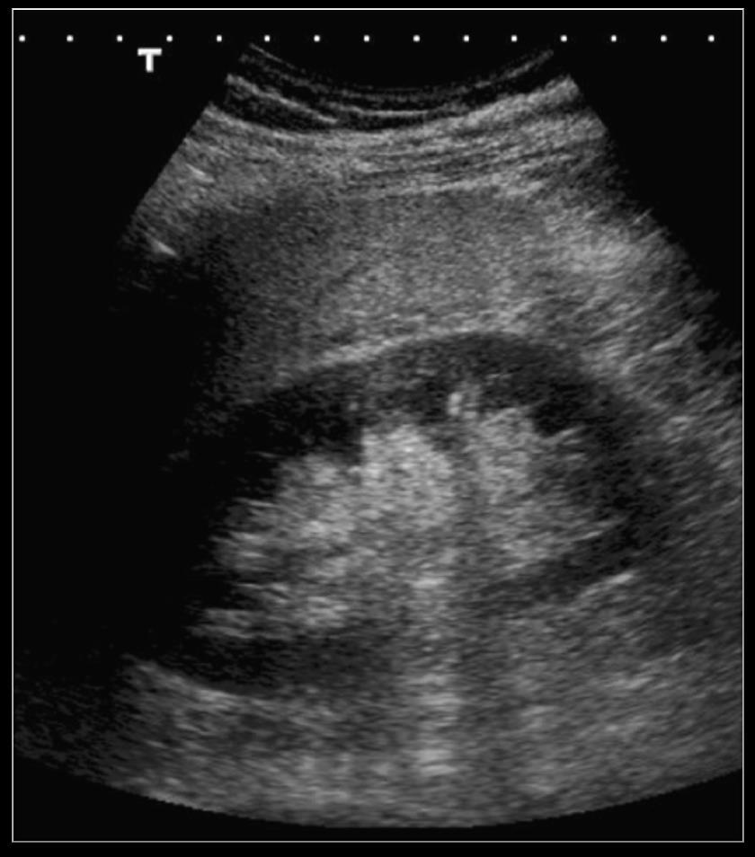
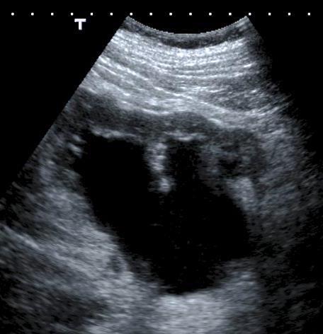
22 Bear Trap 4 Report: Both kidneys are moderately hydronephrotic, further evaluation with a CT urogram is advised

 Tarzamni MK. Cases J.")
23 Parapelvic cysts can look like hydronephrosis Parapelvic cysts Do not communicate with the collecting system Usually asymptomatic Often multiple and bilateral Do the cystic spaces communicate? Are the walls convex? Is there a renal pelvis? If in doubt an excretion study is needed (IVU, CT, MR) Tarzamni MK. Cases J. 2008;1:161
24 Bear Trap 5 Which kidney is obstructed?
25 X
26 Renal Obstruction Pelvicalyceal dilatation is not specific for obstruction 26% false positives Examples Post obstructive dilatation Bladder distension Pregnancy Reflux Some patients with obstruction will not have hydronephrosis 35% of patients will not show significant hydronephrosis in the early stages of obstruction, particularly when dehydrated


27 How can we improve accuracy? 1. Arterial Doppler RI >0.7 or Δ RI > Physiological measurement of intrarenal pressure 2. Ureteric Jets Only valuable in high grade obstruction Absent jet or continuous low velocity trickle
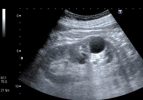
28 Bear Trap 6 Report: as an incidental finding there is a 3cm diameter simple cyst arising from the lower pole of the left kidney
 usually require further imaging CEUS or")
29 Evaluate cysts carefully Cystic RCC (Bosniak Category IV) Lymph node metastases Cysts showing more than minimal complexity (one or two fine septa or minimal calcification) usually require further imaging CEUS or CT (MRI)

30 CEUS of complex cysts CEUS more sensitive than CT in showing septal and wall enhancement CEUS equivalent or superior to CT for categorising complex renal cysts 1-5 Ultrasound tends to upstage lesions in comparison with CT but also downgrades some indeterminate lesions with no enhancement 6 CEUS can provide increased confidence that borderline lesions are benign 7 and help to triage lesions equivocal on CT/MRI 8 CEUS for Category IIF follow-up 1. Xue L-Y. Abdominal Imaging 2014;39 2. Quaia E. AJR 2008; Park BK. Eur J Radiol 2007;61 4. Clevert DA. Clinical hemorheology and microcirculation 2008;39 5. Ascenti G. Radiology 2007; Xue L-Y. Abdominal Imaging 2014;39 7. Barr RG. Radiology 2014:;71 8. Harvey CJ. Radiographics;2015;35


31 Recommended for characterisation of complex cystic masses as benign, intermediate or malignant to provide information for surgical strategy Quaia E. AJR 2008;191 Piscaglia, F., et al. Ultraschall in der Medizin 2012: 33:33 CEUS Pattern No enhancement Occasional bubble or stream of bubbles in a thin septum More complex Classification Benign Benign Malignant Adapted from: Barr RG Radiology 2014;271


32 Remember not all solid looking masses are solid. 85 yr. old woman with sudden onset flank pain


33 Bear Trap 7 Report: There are multiple bilateral renal simple cysts, this is likely to indicate polycystic kidney disease 70 year old man


34 Don t over diagnose polycystic kidney disease Beware of over diagnosis where there is no family history Renal cysts are common in older patients. 9% patients aged >70 have bilateral renal cysts. 1 Age and family history are critical 2 For ADPKD1 and at risk: Age 15-29: at least 2 cysts Age 30-59: at least 2 cysts in each kidney 60+: at least 4 cysts in each kidney In affected families of unknown genotype: 3 Age 15-39: 3 or more cysts Age 40-59: 2 or more cysts in each kidney Age 60+: at least 4 cysts in each kidney 15% of cases are new mutations. Consider when multiple cysts in a young patient, particularly if cysts also seen in liver/pancreas. Difficult diagnosis and may require genetic testing 1. Ravine D. Am J Kidney Dis 1993;22:803 2.Ravine D. Lancet 1994;343: Pei Y. JASN 2009;20:205

35 Bear Trap 8 Report: There is a large simple cyst arising from the upper pole of the left kidney

 Upper moiety prone to obstruction, lower")
36 Remember duplex kidneys Weigert-Meyer law: With complete duplication the upper moiety has an ectopic insertion medial and inferior to the lower pole moiety (often with a ureterocoele) Upper moiety prone to obstruction, lower moiety reflux

37 Bear Trap 9 Report: There is a 7mm diameter simple cyst in the lower pole of the kidney


38 Pseudoaneurysms look like cysts - don t forget the Doppler! Haematuria following kick in flank during rugby match

39 Bear Trap 10 Report: The transplant is well perfused with blood flow demonstrated in all areas. The resistance index (0.78) is near normal


40 Don t trust the autotrace! RI 0.78 RI 0.76 Automatic calculations are often wrong Does the value make sense? Look at the pattern not the number!
41 Conclusions Avoiding Bear Traps in Kidney Ultrasound 1. Know normal variants and their mimics CEUS is very useful 2. Remember infection 3. Don t diagnose AML with US 4. Pitfalls in hydronephrosis Parapelvic cysts Dilatation doesn t always mean obstruction, obstructed kidneys are not always hydronephrotic 5. Evaluate cysts meticulously 6. Don t over-diagnose ADPKD 7. Remember duplex kidneys and pseudoaneurysms 8. Don t trust the autotrace!
42
INTERDISCIPLINARY DISCUSSIONS IN LOCALISED RCC DIAGNOSIS AND SURGICAL STRATEGIES FOR ATYPICAL RENAL CYSTIC LESIONS. Maria Cova
 INTERDISCIPLINARY DISCUSSIONS IN LOCALISED RCC DIAGNOSIS AND SURGICAL STRATEGIES FOR ATYPICAL RENAL CYSTIC LESIONS Maria Cova Radiology Department University of Trieste (IT) Eleventh European International
Role of imaging in RCC. Ultrasonography. Solid lesion. Cystic RCC. Solid RCC 31/08/60. From Diagnosis to Treatment: the Radiologist Perspective
 Role of imaging in RCC From Diagnosis to Treatment: the Radiologist Perspective Diagnosis Staging Follow up Imaging modalities Limitations and pitfalls Duangkamon Prapruttam, MD Department of Therapeutic
Excretory urography (EU) or IVP US CT & radionuclide imaging
 or IVP US CT & radionuclide imaging") Excretory urography (EU) or IVP US CT & radionuclide imaging MRI arteriography studies requiring catherization or direct puncture of collecting system EU & to a lesser extent CT provide both functional
Excretory urography (EU) or IVP US CT & radionuclide imaging MRI arteriography studies requiring catherization or direct puncture of collecting system EU & to a lesser extent CT provide both functional
The Incidental Renal lesion
 The Incidental Renal lesion BACKGROUND Increase in abdominal CT/US in last 15 years Resulted in detection of many (small) renal lesions 50% > 50yrs has at least 1 lesion majority simple cysts Renal lesions
Renal masses - the role of diagnostic imaging
 Renal masses - the role of diagnostic imaging Poster No.: C-2471 Congress: ECR 2015 Type: Educational Exhibit Authors: V. Rai#; Bjelovar/HR Keywords: Cysts, Cancer, Structured reporting, Ultrasound, MR,
Fetal Renal Malformations: The Role of Ultrasound in Diagnosis & Management
 Fetal Renal Malformations: The Role of Ultrasound in Diagnosis & Management 12 weeks Alfred Abuhamad, M.D. Eastern Virginia Medical School 13 weeks 2nd trimester Medullary pyramids Renal Sinus Cortex 2nd
Obstetrics Content Outline Obstetrics - Fetal Abnormalities
 Obstetrics Content Outline Obstetrics - Fetal Abnormalities Effective February 2007 10 16% renal agenesis complete absence of the kidneys occurs when ureteric buds fail to develop Or degenerate before
Urinary system Ultrasound (Renal & Urinary bladder)
") Urinary system Ultrasound (Renal & Urinary bladder) Edited & Presented by ; Hussien A.B ALI DINAR. Msc.Phd ISRRT Associate Member Lecturer (National university) Reporting Sonographer (PHC) Objective By
Urinary system Ultrasound (Renal & Urinary bladder) Edited & Presented by ; Hussien A.B ALI DINAR. Msc.Phd ISRRT Associate Member Lecturer (National university) Reporting Sonographer (PHC) Objective By
Acute flank pain in children: Imaging considerations
 Acute flank pain in children: Imaging considerations Carlos J. Sivit MD Rainbow Babies and Children s Hospital Case Western Reserve School of Medicine Flank pain Results from distention of ureter or renal
Contrast Enhanced Ultrasound of Parenchymal Masses in Children
 Contrast Enhanced Ultrasound of Parenchymal Masses in Children Sue C Kaste, DO On behalf of Beth McCarville, MD St. Jude Children s Research Hospital Memphis, TN Overview Share St. Jude experience with
CME Article Clinics in diagnostic imaging (135)
") Medical Education Singapore Med J 2011; 52(5) : 384 CME Article Clinics in diagnostic imaging (135) Pojchamarnwiputh S, Muttarak M, Sriplakich S H 1a 1b 1c 1d Fig. 1 (a) Axial unenhanced; (b & c) delayed
Medical Education Singapore Med J 2011; 52(5) : 384 CME Article Clinics in diagnostic imaging (135) Pojchamarnwiputh S, Muttarak M, Sriplakich S H 1a 1b 1c 1d Fig. 1 (a) Axial unenhanced; (b & c) delayed
Pediatric Retroperitoneal Masses Radiologic-Pathologic Correlation
 Acta Radiológica Portuguesa, Vol.XVIII, nº 70, pág. 61-70, Abr.-Jun., 2006 Pediatric Retroperitoneal Masses Radiologic-Pathologic Correlation Marilyn J. Siegel Mallinckrodt Institute of Radiology, Washington
The role for contrast-enhanced ultrasonography outside of focal liver lesions
 The role for contrast-enhanced ultrasonography outside of focal liver lesions Paul S. Sidhu King s College Hospital, London, UK Introduction Contrast-enhanced ultrasonography (US) of focal liver lesions
Outline. Introduction to imaging modalities of the urinary system. Case base learning of common diseases in urinary tract
 Outline Introduction to imaging modalities of the urinary system Case base learning of common diseases in urinary tract Outline Introduction to imaging modalities of the urinary system Case base learning
Outline. Introduction to imaging modalities of the urinary system. Case base learning of common diseases in urinary tract
 Outline Introduction to imaging modalities of the urinary system Case base learning of common diseases in urinary tract Diagnostic Investigations in Urinary System PLAIN KUB EXCRETORY UROGRAPHY RETROGRADE
My Patient Has Abdominal Pain PoCUS of the Biliary Tract and the Urinary Tract
 My Patient Has Abdominal Pain PoCUS of the Biliary Tract and the Urinary Tract Objectives PoCUS for Biliary Disease PoCUS for Renal Colic PoCUS for Urinary Retention Biliary Disease A patient presents
Contrast-enhanced ultrasound (CEUS) in the evaluation and characterization of complex renal cysts
 in the evaluation and characterization of complex renal cysts") Contrast-enhanced ultrasound (CEUS) in the evaluation and characterization of complex renal cysts Poster No.: C-2812 Congress: ECR 2018 Type: Educational Exhibit Authors: J. A. Torres de Abreu Macedo,
Contrast-enhanced ultrasound (CEUS) in the evaluation and characterization of complex renal cysts Poster No.: C-2812 Congress: ECR 2018 Type: Educational Exhibit Authors: J. A. Torres de Abreu Macedo,
Abdominal Ultrasound. Diane Hallinen, MD. Bloodroot
 Abdominal Ultrasound Diane Hallinen, MD Bloodroot Abdominal Ultrasound Vasculature Hepatobiliary Spleen Kidney Bladder Bowel Where to put the probe? Vasculature We are going to talk about Celiac Trunk
Proceedings of the 34th World Small Animal Veterinary Congress WSAVA 2009
 www.ivis.org Proceedings of the 34th World Small Animal Veterinary Congress WSAVA 2009 São Paulo, Brazil - 2009 Next WSAVA Congress : Reprinted in IVIS with the permission of the Congress Organizers IMAGING
THYROID NODULES: THE ROLE OF ULTRASOUND
 THYROID NODULES: THE ROLE OF ULTRASOUND NOVEMBER 2017 DR. DEAN DURANT DEFINITION Thyroid nodule: Focal area within the thyroid gland with echogenicity different from surrounding parenchyma. THYROID NODULES
Kidneys and Urinary Tract Content Outline. Anatomy Coverings. Location. (Effective February 2007) (16%-24%)
 (16%-24%)") Kidneys and Urinary Tract Content Outline (Effective February 2007) (16%-24%) Anatomy Coverings true capsule perirenal fat surrounds capsule Gerota s fascia separates perirenal from extraperitoneal fat
Kidneys and Urinary Tract Content Outline (Effective February 2007) (16%-24%) Anatomy Coverings true capsule perirenal fat surrounds capsule Gerota s fascia separates perirenal from extraperitoneal fat
Sex: 女 Age: 51 Occupation: 無 Admission date:92/07/22
 Sex: 女 Age: 51 Occupation: 無 Admission date:92/07/22 Chief complaint Unknown fever for one month Hand tremor and left huge renal tumor was noted Present illness Suffered from fever for one month, hand
How To Approach Renal Masses? - Differential Diagnosis On Image
 How To Approach Renal Masses? - Differential Diagnosis On Image Poster No.: C-1646 Congress: ECR 2015 Type: Educational Exhibit Authors: A. E. A. G. Costa, A. Gomes, A. Duarte, I. Távora; Lisbon/PT Keywords:
The relationship Between The Divided Shape of Kidney and the Duplication of Ureter
 The relationship Between The Divided Shape of Kidney and the Duplication of Ureter Poster No.: C-1373 Congress: ECR 2014 Type: Authors: Keywords: DOI: Scientific Exhibit J. Lee, B. S. Cho, S. J. Kim, K.
Kidney & Urinary Tract Ultrasound. Fatina Fadel Hafez Bazaraa
 Kidney & Urinary Tract Ultrasound Fatina Fadel Hafez Bazaraa Ultrasonography Ultrasound Available Rapid Inexpensive Painless & no sedation needed No adverse effects/ complications Can be repeated Useful
US in non-traumatic acute abdomen. Lalita, M.D. Radiologist Department of radiology Faculty of Medicine ChiangMai university
 US in non-traumatic acute abdomen Lalita, M.D. Radiologist Department of radiology Faculty of Medicine ChiangMai university Sagittal Orientation Transverse (Axial) Orientation Coronal Orientation Intercostal
Normal Sonographic Anatomy
 hapter 2:The Liver DUNSTAN ABRAHAM Normal Sonographic Anatomy Homogeneous, echogenic texture (Figure 2-1) Measures approximately 15 cm in length and 10 12.5 cm anterior to posterior; measurement taken
PROFESSIONAL SKILLS 1 3RD YEAR SEMESTER 6 RADIOGRAPHY. THE URINARY SYSTEM Uz. Fatema shmus aldeen Tel
 PROFESSIONAL SKILLS 1 3RD YEAR SEMESTER 6 RADIOGRAPHY THE URINARY SYSTEM Uz. Fatema shmus aldeen Tel. 0925111552 Professional skills-2 THE URINARY SYSTEM The urinary system (review anatomy and physiology)
Kidney Case 1 SURGICAL PATHOLOGY REPORT
 Kidney Case 1 Surgical Pathology Report February 9, 2007 Clinical History: This 45 year old woman was found to have a left renal mass. CT urography with reconstruction revealed a 2 cm medial mass which
Audit of split-bolus CT urography for the investigation of haematuria over a 12 month period at two district general hospitals
 Audit of split-bolus CT urography for the investigation of haematuria over a 12 month period at two district general hospitals Poster No.: C-1349 Congress: ECR 2010 Type: Educational Exhibit Topic: Genitourinary
Imaging Ejaculatory Disorders and Hematospermia
 ATHENS 4-6 October 2018 European Society of Urogenital Radiology Imaging Ejaculatory Disorders and Hematospermia Parvati Ramchandani, MD Professor, Radiology and Surgery University of Pennsylvania Medical
Autosomal Dominant Polycystic Kidney Disease
 Case Studies [1] July 01, 2014 By Amar Udare, MBBS [2] Case History: 45-year-old female with vague pain in the abdomen. Case History: A 45-year-old female presented with vague pain in the abdomen. A USG
Abdominal Ultrasound : Aorta, Kidneys, Bladder
 Abdominal Ultrasound : Aorta, Kidneys, Bladder Nilam J. Soni, MD, MSc Associate Professor of Medicine Divisions of Hospital Medicine and Pulmonary/Critical Care Medicine Department of Medicine University
이학종분당서울대학교병원. Ultrasound in Urinary Colic
 이학종분당서울대학교병원 Ultrasound in Urinary Colic U l t r a s o u n d i n U r i n a US: Normal Kidney r y C o l i c Contents 1. 1. Definition and clinical consideration 2. 2. Pathophysiology 3. 3. US in in obstructive
Genitourinary Radiology In-Training Test Questions for Diagnostic Radiology Residents
 Genitourinary Radiology In-Training Test Questions for Diagnostic Radiology Residents March, 2013 Sponsored by: Commission on Education Committee on Residency Training in Diagnostic Radiology 2013 by American
CASE REPORT RENAL TUBERCULOSIS CAUSE OF RENAL REPLACEMENT LIPOMATOSIS : A RARE ASSOCIATION
 CASE REPORT RENAL TUBERCULOSIS CAUSE OF RENAL REPLACEMENT LIPOMATOSIS : A RARE ASSOCIATION DR ANAND AARTI 1, DR CHANDAK PRIYA 2,DR SURESH PARVATHY 3 1. PROF AND HOD, DEPARTMENT OF RADIODIAGNOSIS, GOVERNMENT
Urology An introduction to cut up DR J R GOEPEL
 Urology An introduction to cut up DR J R GOEPEL Overview Principles Individual organs Small pieces Partial resections Whole organs Data recording and data sets Principles You are working for the patient
Naif H. Alsaikhan, MD Noushin Vahdat, MD. University of California in San Diego VA San Diego Healthcare System
 Naif H. Alsaikhan, MD Noushin Vahdat, MD University of California in San Diego VA San Diego Healthcare System Goals Describe the morphologic parameters that urologists and interventional radiologists need
11/1/2014. Radiologic incidentalomas Ordering pitfalls Newer technology and applications
 Bilal Tahir, MD Gitasree Borthakur, MD Indiana University School of Medicine Department of Radiology & Imaging Sciences October 31, 2014 ACP 2014 Dr. V. Aaron Nuclear (vaaron@iupui.edu) Dr. S. Westphal
Ultrasound of the Breast BASICS FOR THE ORDERING CLINICIAN
 Ultrasound of the Breast BASICS FOR THE ORDERING CLINICIAN Breast Ultrasound Anatomy Skin Breast Parenchyma Pectoralis Fascia Pectoralis Breast Ultrasound Anatomy Indications for Breast Ultrasound Palpable
Management of the Incidental Renal Mass on CT: A White Paper of the ACR Incidental Findings Committee
 ORIGINAL ARTICLE Management of the Incidental Renal Mass on CT: A White Paper of the ACR Incidental Findings Committee Brian R. Herts, MD a, Stuart G. Silverman, MD b, Nicole M. Hindman, MD c, Robert G.
MANAGEMENT RECOMMENDATIONS
 1 MANAGEMENT RECOMMENDATIONS 1. Adrenal masses!!!!!!! page 2 2. Liver Masses!!!!!!! page 3 3. Obstetric US Soft Markers for Aneuploidy!! pages 4-6 4. Ovarian and Adnexal Cysts!!!!! pages 7-10 5. Pancreatic
Do Incidental Hyperechoic Renal Lesions Measuring Up to 1 cm Warrant Further Imaging? Outcomes of 161 Lesions
 Genitourinary Imaging Original Research Genitourinary Imaging Original Research Ankur M. Doshi 1 Abimbola Ayoola Andrew B. Rosenkrantz Doshi AM, Ayoola A, Rosenkrantz AB Keywords: angiomyolipoma, hyperechoic
Role of MDCT in Radiological evaluation of Renal Masses and its beneficial effects on patient management.
 International Journal of advances in health sciences (IJHS) ISSN 2349-7033 Vol2, Issue1, 2015, pp56-63 http://www.ijhsonline.com Research Article Role of MDCT in Radiological evaluation of Renal Masses
Case-based discussion:
 Case-based discussion: Pailin Kongmebhol, M.D. Department of Radiology Faculty of Medicine Chiang Mai University There are many guidelines for managing thyroid nodules Two important guidelines: 2015 American
Imaging findings in renal infections
 Imaging findings in renal infections Poster No.: C-0221 Congress: ECR 2013 Type: Educational Exhibit Authors: I. lópez blasco, D. Soriano Mena, R. Pastor Toledo, S. Paz Maya, A. M. Julve Parreño, J. Palmero
Renal Mass Biopsy: Needed Now More than Ever
 Renal Mass Biopsy: Needed Now More than Ever Stuart G. Silverman, MD, FACR Professor of Radiology Harvard Medical School Director, Abdominal Imaging and Intervention Brigham and Women s Hospital Boston,
Endocrinology and Metabolic Disorder Unit Regina Apostolorum Hospital
 Enrico Papini Endocrinology and Metabolic Disorder Unit Regina Apostolorum Hospital Albano Laziale, Italy The Following Faculty have provide no information regarding significant relationship with commercial
Various kinds of cystic tumor or tumor-like lesions in the kidney :radiologic-pathologic correlation.
 Various kinds of cystic tumor or tumor-like lesions in the kidney :radiologic-pathologic correlation. Poster No.: C-0299 Congress: ECR 2014 Type: Educational Exhibit Authors: S.-J. Lee, J.-H. Yoon, Y.
Appendix 5. EFSUMB Newsletter. Gastroenterological Ultrasound
 EFSUMB Newsletter 87 Examinations should encompass the full range of pathological conditions listed below A log book listing the types of examinations undertaken should be kept Training should usually
Rad Lab 4 Unknowns: Genitourinary!
 Rad Lab 4 Unknowns: Genitourinary! Peter Clarke MD! Don Di Salvo, MD! Clerkship Directors for Radiology! Harvard Medical School! Brigham and Women s Hospital! Dana Farber Cancer Institute! Case 1: 69 year
Guidelines, Policies and Statements D5 Statement on Abdominal Scanning
 Guidelines, Policies and Statements D5 Statement on Abdominal Scanning Disclaimer and Copyright The ASUM Standards of Practice Board have made every effort to ensure that this Guideline/Policy/Statement
ID data. Sex: female Age: 46y/o Birthday: 1955/10/13
 ID data Sex: female Age: 46y/o Birthday: 1955/10/13 Chief Complain Right upper quadrate abdominal tenderness for one month. Present illness (1) This 46 years old female patient was in a healthy condition
Index. Note: Page numbers of article titles are in boldface type.
 Magn Reson Imaging Clin N Am 12 (2004) 587 591 Index Note: Page numbers of article titles are in boldface type. A Adenoma(s), adrenal, gadolinium-enhanced MR imaging in, 533 534 hyperfunctioning versus
The Child With An Abdominal Mass
 The Child With An Abdominal Mass Today we are going to talk about pediatric surgery, the abdominal masses in children. Firstly we have to take a full history and make a general, local and rectal examination
Imaging characterization of renal clear cell carcinoma
 Imaging characterization of renal clear cell carcinoma Poster No.: C-0327 Congress: ECR 2011 Type: Educational Exhibit Authors: S. Ballester 1, A. Gaser 2, M. Dotta 1, M. F. CAPPA 1, F. Hammar 1 ; 1 2
Ultrasonography of acute flank pain: a focus on renal stones and acute pyelonephritis
 Ultrasonography of acute flank pain: a focus on renal stones and acute pyelonephritis Ki Choon Sim Department of Radiology, Korea University nam Hospital, Korea University College of Medicine, Seoul, Korea
Certificate in Clinician Performed Ultrasound (CCPU) Syllabus. Renal Hydronephrosis & Calculi
 Syllabus. Renal Hydronephrosis & Calculi") Certificate in Clinician Performed Ultrasound (CCPU) Syllabus Renal Hydronephrosis & Calculi Page 1 of 6 01/17 Renal Hydronephrosis and Calculi Syllabus Purpose: This unit is designed to cover the theoretical
Certificate in Clinician Performed Ultrasound (CCPU) Syllabus Renal Hydronephrosis & Calculi Page 1 of 6 01/17 Renal Hydronephrosis and Calculi Syllabus Purpose: This unit is designed to cover the theoretical
Acute renal colic Radiological investigation in patients with renal colic
 Acute renal colic Radiological investigation in patients with renal colic Mikael Hellström Professor Department of Radiology Sahlgrenska University Hospital Göteborg University 0.9-1.8/1.000 inhabitants
Malignant Focal Liver Lesions
 Malignant Focal Liver Lesions Other Than HCC Pablo R. Ros, MD, MPH, PhD Departments of Radiology and Pathology University Hospitals Cleveland Medical Center Case Western Reserve University Pablo.Ros@UHhospitals.org
Bilateral Renal Angiomyolipomas with Invasion of the Renal Vein: A Case Report
 Case Study TheScientificWorldJOURNAL (2008) 8, 145 148 TSW Urology ISSN 1537-744X; DOI 10.1100/tsw.2008.29 Bilateral Renal Angiomyolipomas with Invasion of the Renal Vein: A Case Report C. Blick, N. Ravindranath,
Pediatric Hepatobiliary, Pancreatic & Splenic US
 Pediatric Hepatobiliary, Pancreatic & Splenic US Susan J. Back, MD Department of Radiology, The Children s Hospital of Philadelphia No Disclosures Objectives Normal Abnormal: cases and US advances Objectives
Leonard M. Glassman MD
 BI-RADS The New BI-RADS Leonard M. Glassman MD FACR Former Chief of Breast Imaging American Institute for Radiologic Pathology Washington Radiology Associates, PC Breast Imaging Reporting and Data System
Congenital Pediatric Anomalies: A Collection of Abdominal Scintigraphy Findings: An Imaging Atlas
 ISPUB.COM The Internet Journal of Nuclear Medicine Volume 5 Number 1 Congenital Pediatric Anomalies: A Collection of Abdominal Scintigraphy Findings: An Imaging Atlas V Vijayakumar, T Nishino Citation
Imaging in breast cancer. Mammography and Ultrasound Donya Farrokh.MD Radiologist Mashhad University of Medical Since
 Imaging in breast cancer Mammography and Ultrasound Donya Farrokh.MD Radiologist Mashhad University of Medical Since A mammogram report is a key component of the breast cancer diagnostic process. A mammogram
CYSTIC DISEASES of THE KIDNEY. Dr. Nisreen Abu Shahin
 CYSTIC DISEASES of THE KIDNEY Dr. Nisreen Abu Shahin 1 Types of cysts 1-Simple Cysts 2-Dialysis-associated acquired cysts 3-Autosomal Dominant (Adult) Polycystic Kidney Disease 4-Autosomal Recessive (Childhood)
R adio logical investigations of urinary system
 R adio logical investigations of urinary system There are 4 main radiological Ix: 1 IVU: Intravenous urography. 2- U/S 3-CT scan 4-Radioisotope scan. Others (not frequently used): MRI, arteriography, antegrade
Chapter IV. Angionephrography in Simple Renal Cysts
 Acta Radiologica ISSN: 0001-6926 (Print) (Online) Journal homepage: http://www.tandfonline.com/loi/iaro20 Chapter IV. Angionephrography in Simple Renal Cysts To cite this article: (1957) Chapter IV. Angionephrography
A Giant Hydronephrotic Kidney with Ureteropelvic Junction Obstruction with Blunt Renal Trauma in a Boy
 A Giant Hydronephrotic Kidney with Ureteropelvic Junction Obstruction with Blunt Renal Trauma in a Boy BY JUNYA TSURUKIRI, HIDEFUMI SANO, YOSUKE TANAKA, TAKAO SATO, HIROKAZU TAGUCHI Abstract An 18-year-old
Basic of Ultrasound Physics E FAST & Renal Examination. Dr Muhammad Umer Ihsan MBBS,MD, DCH CCPU,DDU1,FACEM
 Basic of Ultrasound Physics E FAST & Renal Examination Dr Muhammad Umer Ihsan MBBS,MD, DCH CCPU,DDU1,FACEM What is Sound? Sound is Mechanical pressure waves What is Ultrasound? Ultrasounds are sound waves
CLINICS IN DIAGNOSTIC IMAGING (18)
") I R DÏOLOGICAL, CASE j CLINICS IN DIAGNOSTIC IMAGING (18) K L Chan, K W Chan, W C G Peh SINGAPORE MED J 1996; Vol 37: 536-540 CASE REPORT A one-year three-month old boy was incidentally noted to have a
I R DÏOLOGICAL, CASE j CLINICS IN DIAGNOSTIC IMAGING (18) K L Chan, K W Chan, W C G Peh SINGAPORE MED J 1996; Vol 37: 536-540 CASE REPORT A one-year three-month old boy was incidentally noted to have a
Evaluation of Liver Mass Lesions. American College of Gastroenterology 2013 Regional Postgraduate Course
 Evaluation of Liver Mass Lesions American College of Gastroenterology 2013 Regional Postgraduate Course Lewis R. Roberts, MB ChB, PhD Division of Gastroenterology and Hepatology Mayo Clinic College of
X-ray Corner. Imaging of The Pancreas. Pantongrag-Brown L
 X-ray Corner 125 Imaging of The Pancreas Modern imaging modalities commonly used in pancreas include ultrasound (US), CT, and MRI. Pancreas is a retroperitoneal organ which makes it difficult to visualize
Cystic Pancreatic Lesions: Approach to Diagnosis
 Cystic Pancreatic Lesions: Approach to Diagnosis Poster No.: R-0130 Congress: RANZCR-AOCR 2012 Type: Educational Exhibit Authors: A. AGARWAL, R. M. Mendelson; Perth/AU Keywords: Cysts, Biopsy, Endoscopy,
Abdominal ultrasound:
 Abdominal ultrasound: Non-traumatic acute abdomen Wittanee Na-ChiangMai, MD Department of Radiology ChiangMai University 26/04/2017 Contents Technique of examination Normal anatomy Emergency conditions
Renal Masses in Patients with Known Extrarenal Primary Primary Cancer Primary Primary n Met Mets s RCC Beni L mphoma Lung Breast Others
 The Importance of Stuart G. Silverman, MD, FACR Professor of Radiology Harvard ard Medical School Director, Abdominal Imaging and Intervention Brigham and Women s Hospital Boston, MA The Importance of
Abdominal Pain in Pediatric Patients Image Gently
 Abdominal Pain in Pediatric Patients Image Gently Susan D. John, M.D. Baptist Health Emergency Radiology 2017 Disclosure I have no financial relationships with a commercial entity producing healthcarerelated
X-Ray Corner. Imaging Approach to Cystic Liver Lesions. Pantongrag-Brown L. Solitary cystic liver lesions. Hepatic simple cyst (Figure 1)
") THAI J 136 Imaging Approach to Cystic Liver Lesions GASTROENTEROL 2013 X-Ray Corner Imaging Approach to Cystic Liver Lesions Pantongrag-Brown L Cystic liver lesions are common findings in daily practice
THAI J 136 Imaging Approach to Cystic Liver Lesions GASTROENTEROL 2013 X-Ray Corner Imaging Approach to Cystic Liver Lesions Pantongrag-Brown L Cystic liver lesions are common findings in daily practice
Interpreting the Thyroid Ultrasound Report
 Interpreting the Thyroid Ultrasound Report Michael Neuman, MD Radiology Specialists of the Northwest February 2, 2018 Goals Review indications for thyroid ultrasound Review the role of ultrasound in evaluation
Personal data. Age : 63 Gender : male
 Personal data Age : 63 Gender : male Chief complain No specific symptom or discomfort A hepatic mass, found by abdominal sonography of routine health exam on 88-12-08 Past history 1984-3-3 Old CVA with
Simplifying liver assessment in internal medicine
 Ultrasound Customer story Simplifying liver assessment in internal medicine Philips Affiniti ultrasound for elastography and contrast-enhanced ultrasound (CEUS) Where Sonography Institute, Uster, Switzerland
The Royal Marsden. MDT case study. Mr Alan Thompson Consultant Urological Surgeon The Royal Marsden
 MDT case study Mr Alan Thompson Consultant Urological Surgeon The Royal Marsden 2 The Royal Marsden Case history 56 year old lady from Bangladesh with 5 children Rarely seen her GP over the last 10 years
Zoltan Harkanyi M.D., Ph.D. Department of Radiology, Heim Pal Children s Hospital, Budapest, Hungary
 Zoltan Harkanyi M.D., Ph.D. Department of Radiology, Heim Pal Children s Hospital, Budapest, Hungary CEUS expereince 10 years Department of Radiology, Heim Pal Children s Hospital, Budapest US N o 1 study
Imaging of Kidney Cancer
 119 Imaging of Kidney Cancer RADIOLOGIC CLINICS OF NORTH AMERICA Radiol Clin N Am 45 (2007) 119 147 Jingbo Zhang, MD*, Robert A. Lefkowitz, MD, Ariadne Bach, MD - Detection and diagnosis - CT scan Solid
Thyroid in a Nutshell Dublin Catherine Kirkpatrick Consultant Sonographer ULHT
 Thyroid in a Nutshell Dublin 2017 Catherine Kirkpatrick Consultant Sonographer ULHT Acknowledgements Dr. Steve Colley Dr. Rhodri Evans Dr. Rhian Rhys Dr. Andrew McQueen Aims Anatomy & Physiology Incidence
Chapter 6: Genitourinary and Gastrointestinal Systems 93
 Chapter 6: Genitourinary and Gastrointestinal Systems 93 Chapter 6 Genitourinary and Gastrointestinal Systems Embryology Three sets of excretory organs or kidneys develop in human embryos: Pronephros:
Uroradiology For Medical Students
 Uroradiology For Medical Students Lesson 4: Cystography & Urethrography - Part 2 American Urological Association Review Cystography is useful in evaluating the bladder, the urethra and the competence of
MULTILOCULAR CYSTIC RENAL CELL CARCINOMA
 MULTILOCULAR CYSTIC RENAL CELL CARCINOMA Khalaf M. Al-Jader, MD* ABSTRACT Objective: Multilocular cystic renal cell carcinoma appears to be uncommon subtype of renal cell carcinoma with characteristic
The Adnexal Mass. Handout NCUS 3/18/2017 Suzanne Dixon, MD
 The Adnexal Mass Handout NCUS 3/18/2017 Suzanne Dixon, MD Objectives: Pelvic mass differential Characteristics of the normal ovary Standard terminology for ovarian masses Benign vs. malignant features
Hyperechoic renal masses
 Hyperechoic renal masses Jean-Yves Meuwly, MD Department of Diagnostic and Interventional Radiology, University Hospital Lausanne, Switzerland Department of Diagnostic and Interventional Radiology Renal
RENAL SCINTIGRAPHY IN THE 21 st CENTURY
 RENAL SCINTIGRAPHY IN THE 21 st CENTURY 99m Tc- MAG 3 with zero time injection of Furosemide (MAG 3 -F 0 ) : A Fast and Easy Protocol, One for All Indications Clinical Experience Congenital Disorders PROTOCOL
Case Report Imaging of a Renal Artery Aneurysm Detected Incidentally on Ultrasonography
 Case Reports in Radiology, Article ID 375805, 4 pages http://dx.doi.org/10.1155/2014/375805 Case Report Imaging of a Renal Artery Aneurysm Detected Incidentally on Ultrasonography Vasileios Rafailidis,
Ultrasound of soft-tissue vascular anomalies
 Ultrasound of soft-tissue vascular anomalies Oscar M. Navarro Associate Professor, University of Toronto Dept. of Diagnostic Imaging, The Hospital for Sick Children Toronto, Canada Declaration of Disclosure
Role of multi detector computed tomography (MDCT) in evaluation of renal masses
 in evaluation of renal masses") International Journal of Research in Medical Sciences Karthikeyan MA et al. Int J Res Med Sci. 2018 Mar;6(3):974-980 www.msjonline.org pissn 2320-6071 eissn 2320-6012 Original Research Article DOI: http://dx.doi.org/10.18203/2320-6012.ijrms20180625
International Journal of Research in Medical Sciences Karthikeyan MA et al. Int J Res Med Sci. 2018 Mar;6(3):974-980 www.msjonline.org pissn 2320-6071 eissn 2320-6012 Original Research Article DOI: http://dx.doi.org/10.18203/2320-6012.ijrms20180625
Sonographically Identified Echogenic Renal Masses Up to 1 cm in Size Are So Rarely Malignant They Can Be Safely Ignored
 ORIGINAL RESEARCH Sonographically Identified Echogenic Renal Masses Up to 1 cm in Size Are So Rarely Malignant They Can Be Safely Ignored Malak Itani, MD, Amit Pandya, MBBS, Ronald O. Bude, MD Received
Contents. Basic Ultrasound Principles and Terminology. Ultrasound Nodule Characteristics
 Contents Basic Ultrasound Principles and Terminology Basic Ultrasound Principles... 1 Ultrasound System... 2 Linear Transducer for Superficial Images and Ultrasound-Guided FNA... 3 Scanning Planes... 4
Characterization of Renal Cell Carcinoma Using Agent Detection Imaging: Comparison with Gray-Scale US
 Characterization of Renal Cell Carcinoma Using Agent Detection Imaging: Comparison with Gray-Scale US Byung Kwan Park, MD 1, 2 Seung Hyup Kim, MD 1 Hyuck Jae Choi, MD 1 Index terms: Contrast media Ultrasound
GU Ultrasound in First Trimester
 Fetal Renal Malformations: The Role of Ultrasound in Diagnosis & Management Outline 1. Renal Anomalies Urinary Tract Dilation Aberrant Early Development Defects Terminal Maturation Alfred Abuhamad, M.D.