Salivary gland cytology. Salivary gland cytology. Triage helps the clinician. Salivary gland tumors. Diagnostic difficulties
|
|
|
- Elfrieda Nicholson
- 6 years ago
- Views:
Transcription
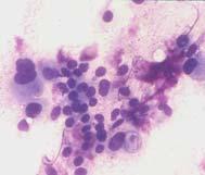
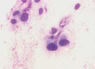
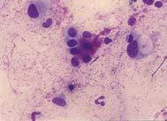
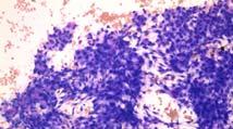
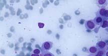
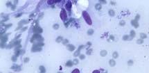
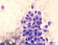
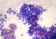
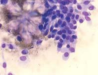
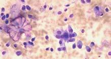
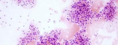
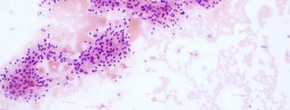
1 Salivary gland cytology Salivary Gland Cytology Pınar Fırat, MD Professor of Pathology İ.U. İstanbul Faculty of Medicine Çapa, İstanbul It is a reliable diagnostic test However, definitive subclassification may be difficult for some lesions Diagnostic accuracy differs according to the entity (e.g. high for pleomorphic adenoma, low for basal cell adenocarcinoma) Diagnostic accuracy is higher for neoplastic vs non-neoplastic lesions for low-grade vs high-grade tumors Sensitivity 77-97%, specificity % Salivary gland cytology Triage Is it a salivary gland lesion? Or arising in the adjacent tissues, lymph node? skin? soft tissue? Is the lesion neoplastic? Benign or malignant? If possible, type of neoplasm? Triage helps the clinician Non-neoplastic lesions: Surgery may not be required Systemic diseases: Different therapeutic modalities Benign tumors, low-grade malignancies: Limited surgery (superficial parotidectomy) High-grade malignancies: Extensive surgery (Facial nerve sacrifice, lymph node neck dissection may be necessary; neo-adjuvant therapy may be indicated) Inoperable patients Diagnostic difficulties Salivary gland tumors Wide spectrum of benign and malignant tumors Some are extremely rare Some are diagnosed by architecture only-invasion Overlaps in different conditions Cystic lesions (neoplasic/ non-neoplasitic) Squamous cells Hyaline stromal globules Basaloid morphology Spindle cell lesions Benign Pleomorphic adenoma Myoepithelioma Basal cell adenoma Warthin tumor Oncocytoma Malignant Acinic cell carcinoma Mucoepidermoid carcinoma Adenoid cystic carcinoma Epithelial-myoepithelial carcinoma Polymorphous low grade adenocarcinoma Salivary duct carcinoma 1


 with indistinct")
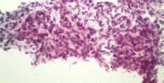
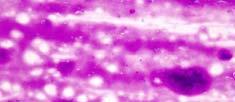
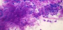
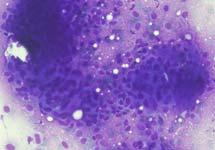
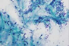
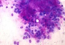
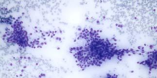
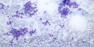
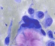
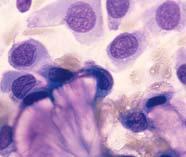
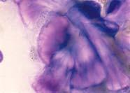
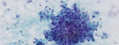
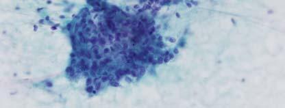
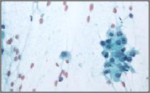
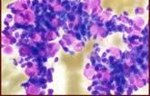
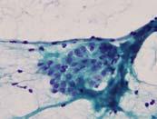
2 Tükrük bezi tümörleri Benign Pleomorfik adenom Myoepitelyoma Malign Adenoid kistik karsinom Epitelyal-myoepitelyal Bazal hücreli Myoepithelial adenom karsinom Warthin tümörü Basaloid Polimorfik düşük dereceli OnkositomOncocytic adenokarsinom Others with poligonal/cuboidal Asinik hücreli cells karsinom Cystic (squamous, mucinous) High grade Mukoepidermoid karsinom Tükrük bezi duktus karsinomu Pleomorphic adenoma Myoepithelial cells, often plasmacytoid or spindled Cohesive epithelial cells Chondromyxoid matrix - fibrillary and bright magenta (Romanowsky stains) with indistinct margins Myoepithelial cells embedded into the fibrillary matrix Fibrillary matrix 2


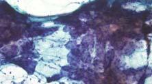
3 Pleomorphic adenoma Adenoid cystic carcinoma Globuler matrix 3

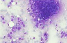
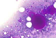
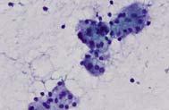
4 Cystic change Metaplasias: squamous / sebaceous Mucin in the background Atypia in pleomorphic adenoma Pitfalls in diagnosing pleomorphic adenomas : Cellular specimens with sparse or absent matrix material Lesions with focal hyaline globules/adenoid cystic-like areas Lesions with cytologic atypia Lesions with metaplastic changes, especially squamous or mucinous features Cellularity with scanty matrix 4


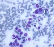
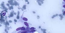
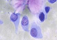
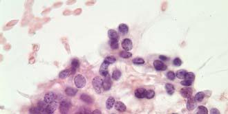
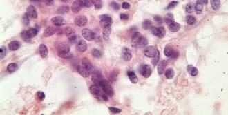
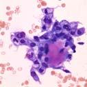
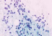
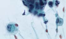
5 67y F 2cm nodular mass in the hard palate Myoepithelioma Myoepithelial cells Epitheloid, plasmacytoid, spindle cell, clear cell patterns Plasmacytoid cells NO matrix Elongated spindle cells Differential dx Pleomorphic adenoma Soft tissue lesions Leiomyoma, schwannoma, noduler fascitis Clear cell tumors If nuclear atypia, necrosis and invasion is present: Myoepithelial carcinoma Myoepitelioma - Collagenous crystals 5

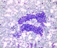
6 Spindle cell myoepithelioma Schwannom Irving Dardick, Sudha Kini, Salivary Gland Tumor Cytopathology, Pathology Images Inc., Canada, 2006 Myoepitehlial l tumors Bazaloid tumors Histology: Myoepithelial carcinoma Basal cell adenoma Basaloid cells Round-oval uniform nuclei, scanty cytoplasm, regular chromatin Varied cellularity Peripheral palisading Hyaline stroma Stick to cells, globules, basement-memb.like material Squamous metaplasia Bazal cell adenoma 6


")


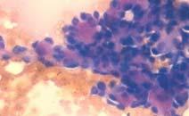
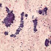
7 Basal cell adenoma Basal cell adenoma Differential diagnosis: Pleomorphic adenoma (Polymorphic, fibrillary matrix) Basal cell adenocarcinoma (nuclear atypia, mitosis, necrosis) Adenoid cystic carcinoma (Hyperchromatic irregular nucleus, coarse chromatin) Adenoid cystic carcinoma Basal cell Adenocarcinoma Painful mass or pain during the FNA Basaloid cells with dark angulated nuclei (variable nuclear atypia) Acellular hyaline matrix with sharp borders Variably sized, often large, three-dimensional hyaline spheres May be identical to BA Nuclear atypia Mitotic figures Invasion Hyaline matrix Nuclear atypia is not always present 7


8 Naked nuclei are seen in the background Solid variant of adenoid cystic carcinoma do not show abundant matrix May closely mimic basal cell tumors as the number of hyaline globules and their size increases, the diagnosis gets closer to adenoid cystic carcinoma Epitelyal-myoepitelyal karsinom Adenoid kistik karsinom Epithelial-myoepithelial carcinoma Matrix producing basaloid looking tumor Hyaline globules / myxoid matrix Cellular smears, naked nuclei in the background Dual cell population One component may dominate Epitelyal myoepitelyal Ca. 8

9 Dual cell population Epitelyal - myoepitelyal Epitelyal - myoepitelyal karsinom Polymorphous Low Grade Adenocarcinoma Minor salivary glands Branching papilla Larger amount of cytoplasm Matrix hyaline / myxoid Irving Dardick, Sudha Kini, Salivary Gland Tumor Cytopathology, Pathology Images Inc., Canada,


10 Neoplasms with basaloid cells Neoplasms producing matrix Basal cell adenoma Basal cell adenoca. Adenoid cystic carcinoma Epithelial-myoepithelial carcinoma Pleomorphic adenoma Neoplasms of the skin basal cell carcinoma pilomatrixoma Small cell carcinoma Basal cell adenoma Basal cell adenoca. Adenoid cystic carcinoma Epithelial-myoepithelial carcinoma Polymorphous low-grade adenocarcinoma Pleomorphic adenoma PA Basal cell Ad Basal cell adenoma Basal cell adenocarcinoma ACC Ep-Myo Ca Pleomorphic adenoma Adenoid cystic ca. Matrix producing, basaloid looking tumors Pattern PA BCA BCAC ACC sheets and syncytia, cells embedded in matrix cohesive clusters; + peripheral palisading; cohesive clusters; + peripheral palisading; 3-D cylinders and branching groups Cells Nuclear atypia Matrix Clinical features plasmacytoid & spindled myoepithelial cells and cuboidal epithelial cells Fibrillar chondromyxoid matrix-irregular edges Basaloid cells, round to oval or elongated nuclei Intercellular hyaline matrix; circumferential hyaline bands Basaloid cells, round to oval or elongated nuclei; +atypia Intercellular hyaline matrix; circumferential hyaline bands Background Myoepithelial cells naked nuclei naked nuclei; + necrosis Basaloid cells, maybe some myoepithelial cells, oval to angulated nuclei; mild to moderate atypia large acellular cylinders and globules of hyaline matrix surrounded by cells- sharp edges naked nuclei; + necrosis Adenoid cystic carcinoma Ki-67 Modified from William C. Faquin s hand out, USCAP,


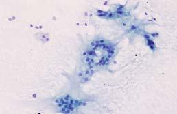
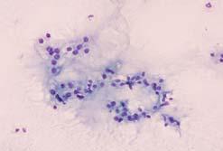
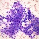
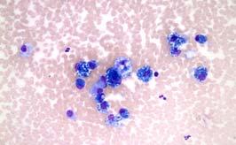
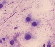
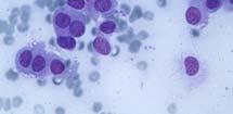
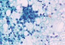
11 Histology: Basal cell adenoma 70 y, F CT: 1cm spiculated mass in the right upper lobe of the lung. PET/CT: increased FDG up-take in left parotid gland (Well circumscribed mass, 1.5cm in diameter) Never trust globules Ask the clinical features, see the nuclear atypia PET scan for salivary gland : Limited value Warthin s tumors, pleomorphic adenomas, basal cell adenomas show increased FDG uptake Warthin s tumor Oncocytes with large polygonal granular cytoplasm forming clusters/ monolayers Lymphocytes, like a lymph node Cystic background looking like necrosis Warthin Tümörü May present only by one or two components 11


12 Mast cell Onkositler Cystic lesions of the salivary glands Non-neoplastic Lenfoepitelhial cyst Retantion cyst Mucocel Branchial cyst Dermoid cyst Epidermoid cyst Neoplastic Benign Warthin tumor Pleomorphic adenoma Cystadenoma Malign Mucoepidermoid carcinoma Acinic cell carcinoma Gabrijela Kocjan, Clinical Cytopathology of the Head and Neck, cases with histopathologic follow up FNAC correctly diagnosed 25 of 36 neoplasms however..., 5 Warthin s tumors 2 squamous cell carcinomas 2 mucoepidermoid carcinomas 2 schwannomas yielded non-representative aspirates Sensitivity 70% Specificity 96% 12


13 Branchial cyst Well differentiated squamous cell carcinoma 62y, F 2 cm mass in the left parotid Oncocytoma Cellularity, isolated oncocytes 3-dimentional oncocytic groups Round uniform nucleus, prominent nucleoli, large granuler eosinophilic cytoplasm Capillary fragments within the groups NO cystic background, NO lymphocytes Oncocytoma Differential diagnosis: Noduler oncocytic hyperplasia Hypocellularity Warthin tumor Monolayers, cystic background, lymphocytes y Oncocytic carcinoma Warthin Dyscohesion, large nucleus, pleomorphism, mitosis, necrosis Acinic cell carcinoma Prominent asiner structures 13


14 Oncocytic carcinoma Irving Dardick, Sudha Kini, Salivary Gland Tumor Cytopathology, Pathology Images Inc., Canada, 2006 Acinic cell carcinoma Cellular smears of acinar cells Sheets and dyshesive crowded 3-D clusters Large polygonal cells with abundant finely vacuolated to granular cytoplasm PAS+D resistant t cytoplasmic zymogen granules Bland nuclear cytologic features Background naked nuclei + lymphocytes Acinic cell Oncocytoma 14

15 Epithelial-myoepithelial carcinoma Acinic cell carcinoma Salivary gland tissue Serous and mucinous acinar cells in grapelike clusters Admixed small tubules and/or sheets of ductal epithelium Adipose tissue 15


16 Acinic cell carcinoma Differential diagnosis: Salivary gland tissue Oncocytic tumors Clear cell tumors Mucoepidermoid carcinoma Cytomorphology depends on the grade of the tumor Mucus-secreting cells Squamous cells Intermediate cells (low N/C ratio) Mucoid background Mucoepidermoid carcinoma Low grade MEC Common cause of false-negative cytologic diagnosis, the aspirate may yield only cyst contents The epithelial cells are bland, easily be misinterpreted as histiocytes 16


17 Warthin High grade MEC Onkositom Acinic cell MEC MEC Warthin Metastatic carcinomas Salivary duct carcinoma Overtly malignant cytology Polygonal cells with abundant cytoplasm Large hyperchromatic, pleomorphic nuclei Prominent nucleoli Prominent nucleoli Sheets, clusters, papillae, and cribriform groups Background necrosis 17


18 Salivary gland tumors Epithelial cells What type? Basaloid, clear, oncocytic? Nuclear atypia? Overt malignancy? Myoepithelial l cells ( plasmocytoid/ spindle cells) Matrix production Fibrillary? Hyaline? Background Cyst content, mucin, necrosis? Main differential diagnosis. Matrix-containing lesions: Pleomorphic adenoma vs adenoid cystic carcinomaprimary Basaloid neoplasms: salivary gland Basal cell adenoma vs basal cell adenocarcinoma neoplasm! vs adenoid cystic carcinoma Oncocytic lesions: Warthin s tumor vs oncocytoma vs acinic cell carcinoma Mucinous cysts: Low-grade mucoepidermoid carcinoma vs mucocele High-grade carcinomas: Clinical Mucoepidermoid carcinoma vs salivary duct carcinoma features vs! metastatic carcinoma Spindle cell lesions: Immuno! Myoepithelial tumors vs soft tissue tumors 18


19 MILAN REPORTING SYSTEM William Faquin, MD, PhD q,, 19
FNA OF SALIVARY GLANDS: A PRACTICAL APPROACH
 FNA OF SALIVARY GLANDS: A PRACTICAL APPROACH FNA of Salivary Glands: Challenges Wide range of neoplastic and non-neoplastic lesions Cytological overlap between the different benign and malignant tumors
Salivary Gland Cytology
 Salivary Gland Cytology Diagnostic challenges and potential pitfalls Tarik M. Elsheikh, MD Professor and Medical Director Anatomic Pathology Cleveland Clinic FNA Salivary Gland Lesions Indications Distinguish
Salivary Glands 3/7/2017
 Salivary Glands 3/7/2017 Goals and objectives Focus on the entities unique to H&N Common board type facts Information for your future practice Salivary Glands Salivary Glands Major gland. Paratid. Submandibular.
Oncocytic-Appearing Salivary Gland Tumors. Oncocytic, Cystic, Mucinous, and High Grade Salivary Gland Tumors SALIVARY GLAND FNA: PART II
 William C. Faquin, MD, PhD Professor of Pathology Harvard Medical School Director of Head and Neck Pathology Massachusetts Eye and Ear Massachusetts General Hospital SALIVARY GLAND FNA: PART II Oncocytic,
Salivary gland tumor cytologic and histologic correlation: Algorithmic and risk stratification based approaches
 Salivary gland tumor cytologic and histologic correlation: Algorithmic and risk stratification based approaches Christopher C. Griffith, MD, PhD Raja R. Seethala, MD 1. Salivary gland tumor cytology: A
Objectives. Salivary Gland FNA: The Milan System. Role of Salivary Gland FNA 04/26/2018
 Salivary Gland FNA: The Milan System Dr. Jennifer Brainard Section Head Cytopathology Cleveland Clinic Objectives Introduce the Milan System for reporting salivary gland cytopathology Define cytologic
ARIZONA SOCIETY OF PATHOLOGISTS 13 TH APRIL 2013 HEAD AND NECK CYTOPATHOLOGY. F ZAHRA ALY, MD, PhD
 ARIZONA SOCIETY OF PATHOLOGISTS 13 TH APRIL 2013 HEAD AND NECK CYTOPATHOLOGY F ZAHRA ALY, MD, PhD The main areas sites amenable for cytopathology include lymph nodes, thyroid, major salivary glands especially
PRELIMINARY CYTOLOGIC DIAGNOSIS: Suspicious for Acinic Cell Carcinoma. Cell Block: Immunohistochemical Studies CYTOLOGIC DIAGNOSIS:
 1 PRELIMINARY CYTOLOGIC DIAGNOSIS: Suspicious for Acinic Cell Carcinoma. Cell Block: Immunohistochemical Studies GCDFP-15 S-100 CYTOLOGIC DIAGNOSIS: Consistent with mammary analogue secretory carcinoma.
Salivary gland Workshop Trondheim 31th may 2012
 Salivary gland Workshop Trondheim 31th may 2012 Peter Jebsen cytopathologist Oslo University Hospital Rikshospitalet Anna Bofin ass. Professor St. Olavs Hospital, Trondheim Drying artifacts Lymfocytes
Los Angeles Society Of Pathologists Dr. Shobha Castelino Prabhu
 Los Angeles Society Of Pathologists Dr. Shobha Castelino Prabhu Loma Linda University Medical Center June 12, 2007 CASE 1 76 year-old gentleman Status post right parotidectomy 1 year ago for a rare tumor
An Integrated Cytologic and Histologic Approach to the Diagnosis of Salivary Gland Tumors
 An Integrated Cytologic and Histologic Approach to the Diagnosis of Salivary Gland Tumors W.C. Faquin, M.D., Ph.D. Massachusetts General Hospital Massachusetts Eye and Ear Infirmary Boston, MA An Integrated
Lesions Mimicking Adenoid Cystic Carcinoma. Diagnostic Problems in Salivary Gland Pathology An Update 5/29/2009
 Diagnostic Problems in Salivary Gland Pathology An Update Lesions Mimicking Adenoid Cystic Carcinoma Stacey E. Mills, M.D. W.S. Royster Professor of Pathology Director of Surgical and Cytopathology University
Cytomorphological spectrum of pleomorphic adenoma with emphasis on differential diagnosis and diagnostic pitfalls
 Original Research Article DOI: 10.18231/2394-6792.2017.0102 Cytomorphological spectrum of pleomorphic adenoma with emphasis on differential diagnosis and diagnostic pitfalls Vaneet Kaur Sandhu 1,*, Navtej
Ben Witt, MD University of Utah/ARUP Laboratories Assistant Professor of Anatomic Pathology
 Ben Witt, MD University of Utah/ARUP Laboratories Assistant Professor of Anatomic Pathology Review some of the more common cytodiagnoses of the Head and Neck Establish an approach to some of the diagnostic
04/09/2018. Salivary Gland Pathology in the Molecular Era Old Friends, Old Foes, & New Acquaintances
 Salivary Gland Pathology in the Molecular Era Old Friends, Old Foes, & New Acquaintances Jennifer L. Hunt, MD, MEd Aubrey J. Hough Jr, MD, Endowed Professor of Pathology Chair of Pathology and Laboratory
Update in Salivary Gland Pathology. Benjamin L. Witt University of Utah/ARUP Laboratories February 9, 2016
 Update in Salivary Gland Pathology Benjamin L. Witt University of Utah/ARUP Laboratories February 9, 2016 Objectives Review the different appearances of a selection of salivary gland tumor types Establish
Pancreatitis: A Potential Pitfall in Endoscopic Ultrasound Guided Pancreatic FNA
 Pancreatitis: A Potential Pitfall in Endoscopic Ultrasound Guided Pancreatic FNA Jack Yang, MD Department of Pathology, Medical University of South Carolina Objectives Understand the indication of EUS
My Journey into the World of Salivary Gland Sebaceous Neoplasms
 My Journey into the World of Salivary Gland Sebaceous Neoplasms Douglas R. Gnepp Warren Alpert Medical School at Brown University Rhode Island Hospital Pathology Department Providence RI Asked to present
4/17/2015. Case 1. A 37 year old man with a 2.2 cm solitary left thyroid mass.
 Case 1 A 37 year old man with a 2.2 cm solitary left thyroid mass. Case 1 Case 1 1 Case 1: Diagnosis? A. Benign B. Atypia of undetermined significance/follicular lesion of undetermined significance C.
Outline 11/2/2017. Pancreatic EUS-FNA general aspects. Cytomorphologic features of solid neoplasms/lesions of the pancreas
 ENDOSCOPIC ULTRASOUND GUIDED-FINE NEEDLE ASPIRATION CYTOLOGY OF PANCREAS Khalid Amin M.D. Assistant Professor Department of Laboratory Medicine and Pathology University of Minnesota Outline Pancreatic
FNA of Thyroid. Toward a Uniform Terminology With Management Guidelines. NCI NCI Thyroid FNA State of the Science Conference
 FNA of Thyroid NCI NCI Thyroid FNA State of the Science Conference Toward a Uniform Terminology With Management Guidelines Thyroid Thyroid FNA Cytomorphology NCI Thyroid FNA State of the Science Conference
Educational Cases EQA November T.J. Palmer Raigmore Hospital Inverness
 Educational Cases EQA November 2013 T.J. Palmer Raigmore Hospital Inverness Case 2 Clinical Details Dob 11 February 1951 PMH: 1964 Extraction of 45 aet 13 yr 1966 Cyst between 44 and 46 enucleated 1973
Pleomorphic Adenoma: Cytologic Variations and Potential Diagnostic Pitfalls
 Pleomorphic Adenoma: Cytologic Variations and Potential Diagnostic Pitfalls Uma Handa, M.D., Neerja Dhingra, M.D., D.N.B.,* Rajan Chopra, M.D., and Harsh Mohan, M.D., M.N.A.M.S., F.I.C.P. The diverse morphological
Salivary Gland Pathology
 IN THE NAME OF GOD Salivary Gland Pathology CHAPTER 11 Dr.kheirandish Oral and maxillofacial pathology Sialadenosis Adenomatoid Hyperplasia of the Minor Salivary Glands Necrotizing Sialometaplasia Pleomorphic
Cytomorphological study of major salivary gland lesions: a 5-year experience at a tertiary care center
 Original article Cytomorphological study of major salivary gland lesions: a 5year experience at a tertiary care center Aruna S, Prathiksha Pai, Shreekant K. Kittur Department of Pathology, Belagavi Institute
Slide Seminar of the Head and Neck Session of the European Congress of Pathology Bilbao, Spain, 2018.
 Slide Seminar of the Head and Neck Session of the European Congress of Pathology Bilbao, Spain, 2018. Prof Sulen Sarioglu, MD Dokuz Eylul University Faculty of Medicine Department of Pathology Graduate
Fine needle aspiration cytology of salivary gland lesions with histopathological correlation - A two year study
 Original article Fine needle aspiration cytology of salivary gland lesions with histopathological correlation - A two year study Tessy PJ, Jayalekshmy PS, Cicy PJ, Usha Poothiode Department of Pathology,
Salivary Gland FNA ATYPICAL : Criteria and Controversies
 Salivary Gland FNA ATYPICAL : Criteria and Controversies W.C. Faquin, M.D., Ph.D. Director, Head and Neck Pathology Massachusetts General Hospital Massachusetts Eye and Ear Infirmary Harvard Medical School
Gynecologic Cytopathology: Glandular lesions
 Gynecologic Cytopathology: Glandular lesions Lin Wai Fung (MSc, MPH, CMIAC) 17/4/2014 Glandular lesions of the uterus Endocervix Endometrium Normal endocervical cells Sheets, strips well-preserved architecture:
Differential Diagnosis of Oral Masses. Palatal Lesions
 Differential Diagnosis of Oral Masses Palatal Lesions Palatal Masses Periapical Abscess Torus Palatinus Mucocele Lymphoid Hyperplasia Adenomatous Hyperplasia Benign Salivary Neoplasms Malignant Salivary
Thyroid master class. Thyroid Fine needle aspiration cytology and liquid-based techniques: Hologic and Becton Dickinson
 Thyroid master class Thyroid Fine needle aspiration cytology and liquid-based techniques: Hologic and Becton Dickinson Principle of LBC Collection of cells in liquid medium Immediate fixation Processor-prepared
PLEOMORPHIC ADENOMA ( BENIGN MIXED TUMOR )
") ( BENIGN MIXED TUMOR ) Grossly, the tumor is freely movable, solid, sometimes lobulated and occasionally cystic. If recurrent, multinodular masses are common. Histologically, within a fibrous capsule,
( BENIGN MIXED TUMOR ) Grossly, the tumor is freely movable, solid, sometimes lobulated and occasionally cystic. If recurrent, multinodular masses are common. Histologically, within a fibrous capsule,
40th European Congress of Cytology Liverpool, UK, 2-5 th October 2016
 40th European Congress of Cytology Liverpool, UK, 2-5 th October 2016 EUS FNA of abdominal organs: An approach to reporting and triage for ancillary testing Date and time: Sunday 2 nd October 2016 15.00-16.30
DISORDERS OF THE SALIVARY GLANDS Neoplasms Dr.M.Baskaran Selvapathy S IV
 DISORDERS OF THE SALIVARY GLANDS Neoplasms Dr.M.Baskaran Selvapathy S IV NEOPLASMS A) Epithelial I. Benign Pleomorphic adenoma( Mixed tumour) Adenolymphoma (Warthin s tumour) Oxyphil adenoma (Oncocytoma)
Research Article Transmucosal Fine Needle Aspiration of Oral and Pharyngeal Lesions
 International Scholarly Research Network ISRN Pathology Volume 2011, Article ID 267145, 7 pages doi:10.5402/2011/267145 Research Article Transmucosal Fine Needle Aspiration of Oral and Pharyngeal Lesions
Oncocytic carcinoma: A rare malignancy of the parotid gland
 ISPUB.COM The Internet Journal of Pathology Volume 8 Number 2 Oncocytic carcinoma: A rare malignancy of the parotid gland K Mardi, J Sharma Citation K Mardi, J Sharma.. The Internet Journal of Pathology.
1 NORMAL HISTOLOGY AND METAPLASIAS
 1 NORMAL HISTOLOGY AND METAPLASIAS, MD Anatomy and Histology 1 Metaplasias 2 ANATOMY AND HISTOLOGY The female breast is composed of a branching duct system, which begins at the nipple with the major lactiferous
Zubair W. Baloch, MD, PhD: Consultant for Veracyyte, INC Tarik M. Elsheikh, MD: Nothing to disclose
 Cytology Works shop #8 Zubair W. Baloch, MD, PhD: Consultantt for Veracyte, INC Tarik M. Elsheik kh, MD: Nothing to disclose Controversies and Diagnostic Challenges in Head and Neck Cytopathology Zubair
DOWNLOAD ENTIRE DOCUMENT FROM
 PREVIEW ONLY 1 Atlas on Bethesda system for reporting Thyroid Cytology PREVIEW ONLY 2 OVERVIEW 1. Indications and goal of thyroid FNA 2. Contraindications 3. Procurement of cell sample 4. Staining methods
Papillary Lesions of the breast
 Papillary Lesions of the breast Emad Rakha Professor of Breast Pathology The University of Nottingham Papillary lesions of the breast are a heterogeneous group of disease, which are characterised by neoplastic
(CYLINDROMA) ATLAS OF HEAD AND NECK PATHOLOGY ADENOID CYSTIC CARCINOMA
 ATLAS OF HEAD AND NECK PATHOLOGY ADENOID CYSTIC CARCINOMA") (CYLINDROMA) This malignant tumor is poorly encapsulated and while seemingly well defined within the affected gland, there is usually infiltration of surrounding tissue on closer examination. The cut surface
(CYLINDROMA) This malignant tumor is poorly encapsulated and while seemingly well defined within the affected gland, there is usually infiltration of surrounding tissue on closer examination. The cut surface
Thyroid follicular neoplasms in cytology. Ulrika Klopčič Institute of Oncology, Department of Cytopathology, Ljubljana, Slovenia
 Thyroid follicular neoplasms in cytology Ulrika Klopčič Institute of Oncology, Department of Cytopathology, Ljubljana, Slovenia Lecture overview importance of FNAB in assessing thyroid lesions follicular
Prepared By Jocelyn Palao and Layla Faqih
 Prepared By Jocelyn Palao and Layla Faqih The structure of the suspected atypical cell should always be compared to the structure of other similar, benign, cells which are present in the smears. The diagnosis
FNA Biopsy of Salivary Gland
 FNA Biopsy of Salivary Gland Richard M. DeMay, MD Professor of Pathology Director of Cytopathology The University of Chicago Objective: To describe the use of FNA Bx to diagnose salivary gland lesions
DISCUSSION: PLGA accounts for about 2% of all salivary gland tumours and occurs almost exclusively in the minor salivary glands.
 SWELLING ON THE HARD PALATE PRESENTING AS POLYMORPHOUS LOW GRADE ADENOCARCINOMA: A AND REVIEW OF LITERATURE Swapnil D. Chandekar 1, Sunita S. Dantkale 2, Rahul R. Narkhede 3, Snehal V. Chavhan 4, Khushboo
Objectives. Atypical Glandular Cells. Atypical Endocervical Cells. Reactive Endocervical Cells
 2013 California Society of Pathologists 66 th Annual Meeting San Francisco, CA Atypical Glandular Cells to Early Invasive Adenocarcinoma: Cervical Cytology and Histology Christina S. Kong, MD Associate
Departments of Pathology and *Otorhinolaryngology, Faculty of Medicine, University of Malaya, Kuala Lumpur
 Malaysian J Pathol 2002; 24(2) : 107-112 CASE REPORT Papillary cystic type of acinic cell carcinoma of parotid: fine needle aspiration cytological features of a high grade variant with oncocytic metaplasia
NODULAR CYSTIC HIDRADENOMA OVER THE GLUTEAL REGION: A RARE CYTOMORPHOLOGICAL DIAGNOSIS
 NODULAR CYSTIC HIDRADENOMA OVER THE GLUTEAL REGION: A RARE CYTOMORPHOLOGICAL DIAGNOSIS Abstract: The primary as well as metastatic tumours of the skin can be diagnosed by fine needle aspiration cytology
Pleomorphic adenoma of breast - a case report and distinction with metaplastic carcinoma D Gupta, S Agrawal, N Trivedi, A Tewari
 of breast - a case report and distinction with metaplastic carcinoma D Gupta, S Agrawal, N Trivedi, A Tewari Introduction, also known as mixed tumour, is a benign tumour which typically presents as a painless,
ISPUB.COM. Salivary duct carcinoma of parotid gland. V Kinnera, R Nandyala, M Yootla, K Mandyam INTRODUCTION CASE REPORT
 ISPUB.COM The Internet Journal of Pathology Volume 10 Number 1 V Kinnera, R Nandyala, M Yootla, K Mandyam Citation V Kinnera, R Nandyala, M Yootla, K Mandyam.. The Internet Journal of Pathology. 2008 Volume
Case year female. Routine Pap smear
 Case 1 57 year female Routine Pap smear Diagnosis? 1. Atypical glandular cells of unknown significance (AGUS) 2. Endocervical AIS 3. Endocervical adenocarcinoma 4. Endometrial adenocarcinoma 5. Adenocarcinoma
Cytyc Corporation - Case Presentation Archive - March 2002
 FirstCyte Ductal Lavage History: 68 Year Old Female Gail Index: Unknown Clinical History: Negative Mammogram in 1995 6 yrs. later presents with bloody nipple discharge Subsequent suspicious mammogram Suspicious
CINtec p16 INK4a Staining Atlas
 CINtec p16 INK4a Staining Atlas Rating Rating Positive The rating positive will be assigned if the p16 INK4a -stained slide shows a continuous staining of cells of the basal and parabasal cell layers of
Respiratory Tract Cytology
 Respiratory Tract Cytology 40 th European Congress of Cytology Liverpool, UK Momin T. Siddiqui M.D. Professor of Pathology and Laboratory Medicine Director of Cytopathology Emory University Hospital, Atlanta,
Enterprise Interest None
 Enterprise Interest None Risk stratification of salivary gland lesions on cytology based on the proposed Milan System for reporting salivary gland cytopathology: A pilot study Kartik Viswanathan, M.D.,
Workshop 2. Controversies and Diagnostic Challenges in Head and Neck Cytopathology. Zubair Baloch, MD,PhD. Veracyte, Inc: Consultant
 Workshop 2 Controversies and Diagnostic Challenges in Head and Neck Cytopathology Zubair Baloch, MD,PhD Veracyte, Inc: Consultant Tarik Elsheikh, MD There are no disclosures necessary. Controversies and
Histopathologic spectrum of salivary gland neoplasms
 Original article Histopathologic spectrum of salivary gland neoplasms Dr. Dipkana Das 1, Dr.Subhasish Saha 1, Dr V Satyanarayana 2 Name & Address of Institution: Department of Pathology, Kamineni Institute
Pancreas. Atrophy, acinar cell. Pathogenesis: Diagnostic key features:
 Pancreas Atrophy, acinar cell Pathogenesis: Decrease in number and/or size of acinar cells may be due to spontaneous or experimentally induced degenerative changes, apoptosis, or a sequel of chronic inflammation.
Disclosures. Parathyroid Pathology. Objectives. The normal parathyroid 11/10/2012
 Disclosures Parathyroid Pathology I have nothing to disclose Annemieke van Zante MD/PhD Assistant Professor of Clinical Pathology Associate Chief of Cytopathology Objectives 1. Review the pathologic features
Fine-Needle Aspiration Cytology of Low-Grade Cribriform Cystadenocarcinoma with Many Psammoma Bodies of the Salivary Gland
 The Korean Journal of Pathology 2013; 47: 481-485 CASE STUDY Fine-Needle Aspiration Cytology of Low-Grade Cribriform Cystadenocarcinoma with Many Psammoma Bodies of the Salivary Gland Ji Yun Jeong Dongbin
The Frozen Section: Diagnostic Challenges and Pitfalls
 The Frozen Section: Diagnostic Challenges and Pitfalls William C. Faquin, M.D., Ph.D. Director, Head and Neck Pathology Massachusetts General Hospital & Massachusetts Eye and Ear Infirmary Harvard Medical
Review of the AP Part II Practical Examination. Dr David Clift Co Chief Examiner
 Review of the AP Part II Practical Examination Dr David Clift Co Chief Examiner General Remarks The part II practical examination involved 15 cases which were presented with sufficient clinical data to
Mammary analogue secretory carcinoma of salivary gland A case report of new entity
 Case Report Mammary analogue secretory carcinoma of salivary gland A case report of new entity Vaibhav Bhika Bari 1*, Sandhya Unmesh Bholay 2 1 Assistant Professor, 2 Associate Professor Rajiv Gandhi Medical
Mody. AIS vs. Invasive Adenocarcinoma of the Cervix
 Common Problems in Gynecologic Pathology Michael T. Deavers, M.D. Houston Methodist Hospital, Houston, Texas Common Problems in Gynecologic Pathology Adenocarcinoma in-situ (AIS) of the Cervix vs. Invasive
Basal cell carcinoma diagnosed on Fine-Needle Aspiration Cytology A. Pathological Case Report
 Basal cell carcinoma diagnosed on Fine-Needle Aspiration Cytology A Abstract Dr. Madhuri S.Kate 1, Dr. Preeti Jain 2, Dr. Shailesh S. Patne 3 Introduction: Basal cell carcinoma (BCC) is a locally invasive
Case #1 FNA of nodule in left lobe of thyroid in 67 y.o. woman
 Challenging Cases Manon Auger M.D., F.R.C.P. (C) Professor, Department of Pathology McGill University Director, Cytopathology Laboratory McGill University it Health Center Case #1 FNA of nodule in left
A 60-year old Man with Left Jaw Mass. Simon Chiosea, MD University of Pittsburgh medical Center 3/15/2016
 ACCME/Disclosures The USCAP requires that anyone in a position to influence or control the content of CME disclose any relevant financial relationship WITH COMMERCIAL INTERESTS which they or their spouse/partner
Enterprise Interest None
 Enterprise Interest None What are triple negative breast cancers? A synopsis of their histological patterns Ian Ellis Molecular Medical Sciences, University of Nottingham Department of Histopathology,
3/28/2017. Head and Neck/Endocrine Pathology Specialty Conference Case 4 Raja R. Seethala, M.D. University of Pittsburgh Medical Center
 Head and Neck/Endocrine Pathology Specialty Conference Case 4 Raja R. Seethala, M.D. University of Pittsburgh Medical Center Disclosure of Relevant Financial Relationships Disclosure of Relevant Financial
Pathology Slides. [Pathology]
![Pathology Slides. [Pathology]](/thumbs/94/120604575.jpg "Pathology Slides. [Pathology]") Pathology Slides MedicoNotes provides real laboratory pathological slides to aid you to differentiate between different pathological structures under microscope. www.mediconotes.com Histology slides example
Pathology Slides MedicoNotes provides real laboratory pathological slides to aid you to differentiate between different pathological structures under microscope. www.mediconotes.com Histology slides example
Histopathology: Cervical HPV and neoplasia
 Histopathology: Cervical HPV and neoplasia These presentations are to help you identify basic histopathological features. They do not contain the additional factual information that you need to learn about
Performance Characteristics of Adenoid Cystic Carcinoma of the Salivary Glands in Fine-Needle Aspirates
 CAP Laboratory Improvement Programs Performance Characteristics of Adenoid Cystic Carcinoma of the Salivary Glands in Fine-Needle Aspirates Results From the College of American Pathologists Nongynecologic
3/28/2017. Disclosure of Relevant Financial Relationships. GU Evening Subspecialty Case Conference. Differential Diagnosis:
 GU Evening Subspecialty Case Conference Rajal B. Shah, M.D. VP, Medical Director, Urologic Pathology Miraca Life Sciences, Irving, Texas Clinical Associate Professor of Pathology Baylor College of Medicine,
Polymorphous Low-Grade. December 5 th, 2008
 Polymorphous Low-Grade Adenocarcinoma December 5 th, 2008 Epidemiology Represents 2 nd or 3 rd most common minor salivary gland malignancy (17-26%) 1 st mucoepidermoid carcinoma Rare in reported Asian
CPC 4 Breast Cancer. Rochelle Harwood, a 35 year old sales assistant, presents to her GP because she has noticed a painless lump in her left breast.
 CPC 4 Breast Cancer Rochelle Harwood, a 35 year old sales assistant, presents to her GP because she has noticed a painless lump in her left breast. 1. What are the most likely diagnoses of this lump? Fibroadenoma
Gross appearance of nodular hyperplasia in material obtained from suprapubic prostatectomy. Note the multinodular appearance and the admixture of
 Tiền liệt tuyến Tiền liệt tuyến Gross appearance of nodular hyperplasia in material obtained from suprapubic prostatectomy. Note the multinodular appearance and the admixture of solid and microcystic areas.
doi: /j.anl
 doi: 10.1016/j.anl.2006.07.001 Synchronous unilateral parotid gland neoplasms of three different histological types Shuho Tanaka 1, Keiji Tabuchi 1, Keiko Oikawa 1, Rika Kohanawa 1, Hideki Okubo 1, Dai
LGM International, Inc.
 Liqui-PREP TM Cytology Atlas Preface The following pictures are examples with descriptions of cytology slides processed with the Liqui-PREP TM System.. The descriptions are reviewed by Pathologists. It
BOSNIAN-TURKISH CYTOPATHOLOGY SCHOOL June 18-19, 2016 Sarajevo. Case Discussions. 60 year old woman Routine gynecologic control LBC
 BOSNIAN-TURKISH CYTOPATHOLOGY SCHOOL June 18-19, 2016 Sarajevo Case Discussions Prof Dr Sıtkı Tuzlalı Tuzlalı Pathology Laboratory 60 year old woman Routine gynecologic control LBC 1 2 Endometrial thickening
Special Course Cytomorphology II : FNA Cytology
 Special Course Cytomorphology II : FNA Cytology Thyroid Richard M. DeMay, MD Lung Kristen A. Atkins, MD Salivary Glands William C. Faquin, MD, PhD Pancreas Gregg A. Staerkel, MD Disclosure information
Salivary Gland Cytology: A Clinical Approach to Diagnosis and Management of Atypical and Suspicious Lesions
 Salivary Gland Cytology: A Clinical Approach to Diagnosis and Management of Atypical and Suspicious Lesions W.C. Faquin, M.D., Ph.D. Massachusetts General Hospital Harvard Medical School, USA Marc Pusztaszeri,
Department of General Surgery, Sri Manakula Vinayagar Medical College and Hospital, Pondicherry, India ABSTRACT
 Original Article DOI: 10.21276/APALM.1278 Cytological and Histomorphological Correlation of Salivary Gland Lesions: An Experience At Rural Tertiary Healthcare Hospital Thangam R 1, Vaishali Dhananjay Kotasthane
number Done by Corrected by Doctor Maha Shomaf
 number 16 Done by Waseem Abo-Obeida Corrected by Zeina Assaf Doctor Maha Shomaf MALIGNANT NEOPLASMS The four fundamental features by which benign and malignant tumors can be distinguished are: 1- differentiation
04/10/2018. Intraductal Papillary Neoplasms Of Breast INTRADUCTAL PAPILLOMA
 Intraductal Papillary Neoplasms Of Breast Savitri Krishnamurthy MD Professor of Pathology Deputy Division Head The University of Texas MD Anderson Cancer Center 25 th Annual Seminar in Pathology Pittsburgh,
Case # year old man with a 2 cm right kidney mass
 Case # 4. 52 year old man with a 2 cm right kidney mass Figure 1 Figure 2 Figure 3 Figure 4 Diagnosis: Negative/Non-diagnostic Normal kidney tissue Fine needle aspiration (FNA) of the kidney is performed
University Journal of Pre and Para Clinical Sciences
 ISSN 2455 2879 Volume 2 Issue 1 2016 Metaplastic carcinoma breast a rare case report Abstract : Metaplastic carcinoma of the breast is a rare malignancy with two distinct cell lines described as a breast
Proliferative Epithelial lesions of the Breast. Sami Shousha, MD, FRCPath Charing Cross Hospital & Imperial College, London
 Proliferative Epithelial lesions of the Breast Sami Shousha, MD, FRCPath Charing Cross Hospital & Imperial College, London Amman, November2013 Proliferative Epithelial Lesions of the Breast Usual type
Slide seminar. Asist. Prof. Jože Pižem, MD, PhD Institute of Pathology Medical Faculty, University of Ljubljana
 Slide seminar Asist. Prof. Jože Pižem, MD, PhD Institute of Pathology Medical Faculty, University of Ljubljana Case 5 A 57-year-old man with a dermal/subcutaneous lesion on the scalp, which was interpreted
Normal endometrium: A, proliferative. B, secretory.
 Normal endometrium: A, proliferative. B, secretory. Nội mạc tử cung Nội mạc tử cung Cyclic changes in endometrium.. Approximate relationship of useful microscopic changes. Arias-Stella reaction in endometrial
Macro- and microacinar proliferations of the prostate
 Macro- and microacinar proliferations of the prostate (with emphasis on cancer mimics) Rodolfo Montironi, MD (IT), FRCPath (UK), IFCAP (USA) Polytechnic University of Marche Region (Ancona) School of Medicine,
DIAGNOSTIC CHALLENGES Pancreas FNAB. Dr. M. Weir Oct 2017
 DIAGNOSTIC CHALLENGES Pancreas FNAB Dr. M. Weir Oct 2017 CONFLICT OF INTEREST DISCLOSURE I have not had in the past 3 years, a financial interest, arrangement or affiliation with one or more organizations
SQUAMOUS CELLS: Atypical squamous cells (ASC) - of undetermined significance (ASC-US) - cannot exclude HSIL (ASC-H)
 - of undetermined significance (ASC-US) - cannot exclude HSIL (ASC-H)") SQUAMOUS CELLS: Atypical squamous cells (ASC) - of undetermined significance (ASC-US) - cannot exclude HSIL (ASC-H) ASC refers to cytologic changes suggestive of SIL, which are qualitativley or quantitatively
SQUAMOUS CELLS: Atypical squamous cells (ASC) - of undetermined significance (ASC-US) - cannot exclude HSIL (ASC-H) ASC refers to cytologic changes suggestive of SIL, which are qualitativley or quantitatively
Salivary Gland Imaging. Mary Scanlon MD FACR October 2016
 Salivary Gland Imaging Mary Scanlon MD FACR October 2016 Objectives Recognize normal and abnormal anatomy Discuss work up, management and differential diagnosis of commonly referred clinical scenarios
Histopathological Study of Lacrimal Gland Tumors
 ORIGINAL ARTICLE Pratikkumar B. Desai 1, Ami Shah 2 1 4 th Year Resident, Pathology Department, B.J.Medical College, Civil Hospital, Ahmedabad 2 Associate Professor, M. J. Institute of Ophthalmology, Civil
Presentation material is for education purposes only. All rights reserved URMC Radiology Page 1 of 98
 Presentation material is for education purposes only. All rights reserved. 2011 URMC Radiology Page 1 of 98 Radiology / Pathology Conference February 2011 Brooke Koltz, Cytopathology Resident Presentation
encapsulated thyroid nodule with a follicular architecture and some form of atypia. The problem is when to diagnose
 Histological Spectrum of Papillary Carcinoma of Thyroid A Two Years Study Gomathi Srinivasan 1, M. Vennila 2 1 Associate Professor Pathology, Government Medical College, Omandurar Estate, Chennai 600 002
Disclosure of Relevant Financial Relationships
 Squamous entities of the thyroid: Reactive to Neoplastic Michelle D. Williams Associate Professor Dept of Pathology, Head & Neck Section University of Texas MD Anderson Cancer Center Disclosure of Relevant
3/22/2017. Disclosure of Relevant Financial Relationships. Cytomorphologic Thresholds for Classifying Thyroid FNAs as Suspicious and Positive for PTC
 Cytomorphologic Thresholds for Classifying Thyroid FNAs as Suspicious and Positive for PTC Tarik M. Elsheikh, MD Professor and Medical Director Anatomic Pathology Cleveland Clinic Laboratories Disclosure
Review and Updates of Immunohistochemistry in Selected Salivary Gland and Head and Neck Tumors
 Review and Updates of Immunohistochemistry in Selected Salivary Gland and Head and Neck Tumors. Monophasic tumors : myoepithelioma, acinic cell carcinoma, and salivary duct carcinoma. Biphasic tumors includes
ARTHUR PURDY STOUT SOCIETY COMPANION MEETING: DIFFICULT NEW DIFFERENTIAL DIAGNOSES IN PROSTATE PATHOLOGY. Jonathan I. Epstein.
 1 ARTHUR PURDY STOUT SOCIETY COMPANION MEETING: DIFFICULT NEW DIFFERENTIAL DIAGNOSES IN PROSTATE PATHOLOGY Jonathan I. Epstein Professor Pathology, Urology, Oncology The Reinhard Professor of Urological