Inflammatory and Reactive Lesions of the Breast
|
|
|
- Ethelbert Hutchinson
- 6 years ago
- Views:
Transcription
1 Inflammatory and Reactive Lesions of the Breast Laura C. Collins, M.D. Vice Chair of Anatomic Pathology Professor of Pathology Beth Israel Deaconess Medical Center and Harvard Medical School Boston, MA
2 Topic for Discussion Inflammatory lesions of the breast Mammary duct ectasia Lymphocytic mastopathy Granulomatous lesions Squamous metaplasia of lactiferous ducts (SMOLD) IgG4-related mastitis Reaction to mammary implants Mimics of inflammatory/reactive lesions
3 Topic for Discussion Inflammatory lesions of the breast Mammary duct ectasia Lymphocytic mastopathy Granulomatous lesions Squamous metaplasia of lactiferous ducts (SMOLD) IgG4-related mastitis Reaction to mammary implants Mimics of inflammatory/reactive lesions
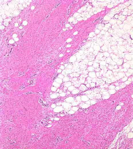
4 Mammary Duct Ectasia A misunderstood entity! Not simply ectatic ducts
5 Mammary Duct Ectasia Primarily peri- and post-menopausal women Clinical presentation Pain, nipple discharge, nipple retraction, mass May mimic carcinoma Mammography Ductal pattern of calcification, may simulate that of DCIS
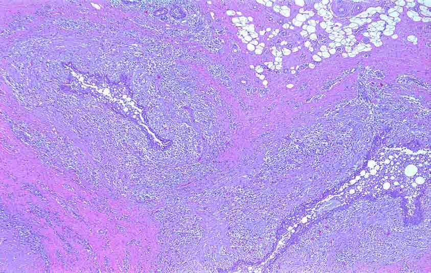
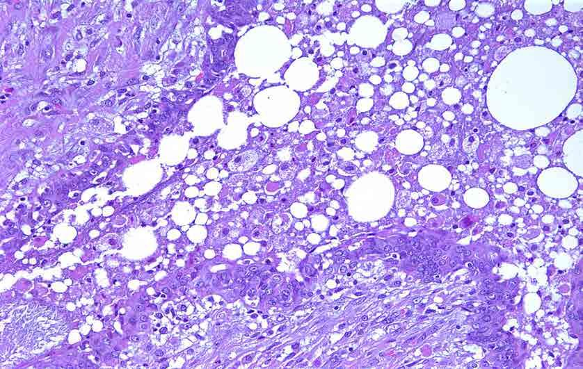
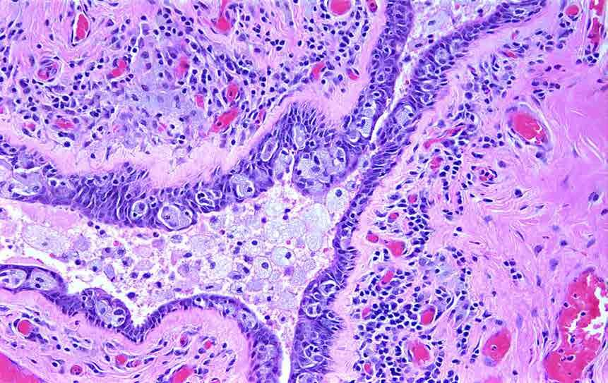
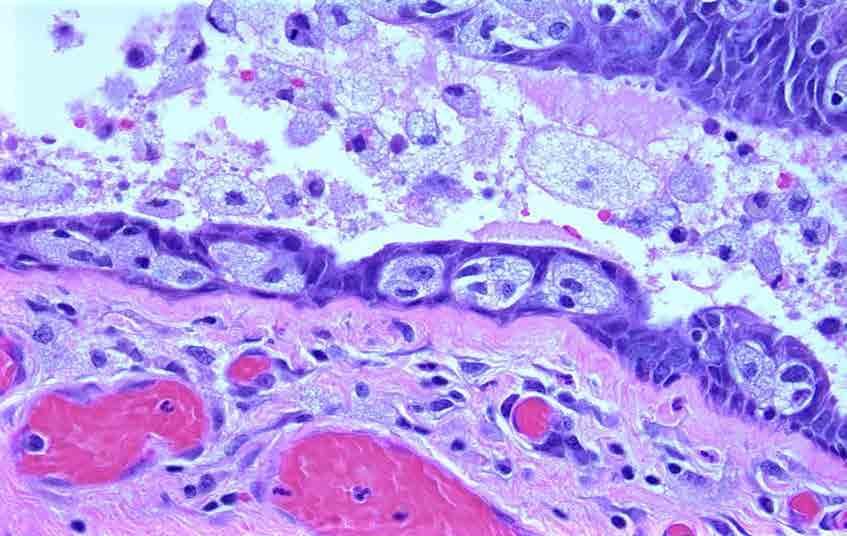
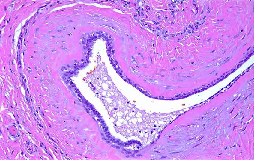
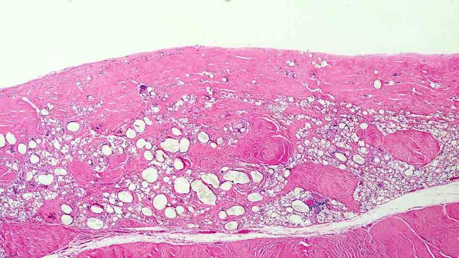
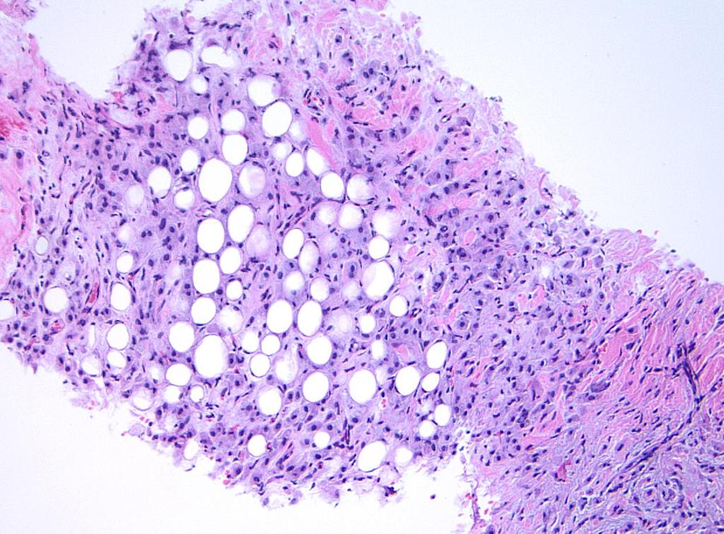
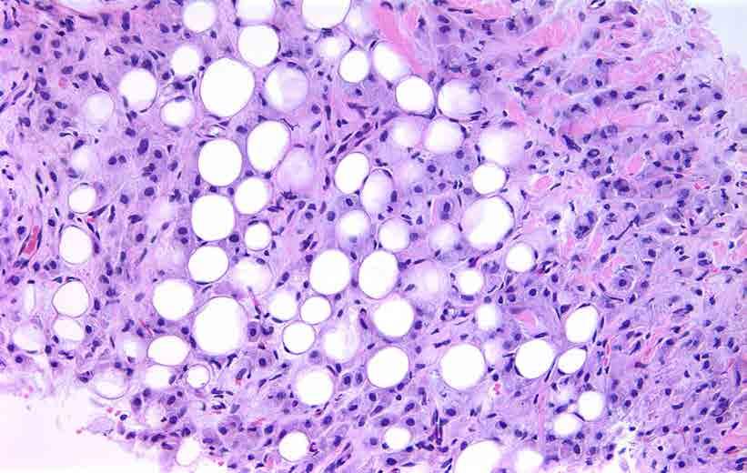
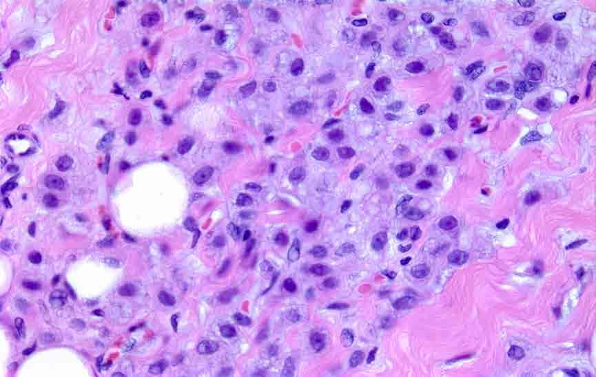
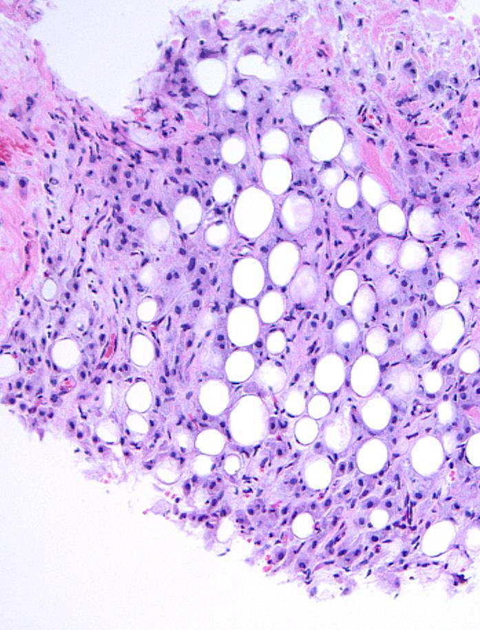
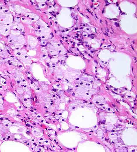
6 Mammary Duct Ectasia Initially confined to large subareolar ducts but later may involve an entire segment Gross: Dilated, thick-walled ducts filled with pasty, yellowbrown secretions (may be mistaken for DCIS with comedo necrosis) Variably fibrotic intervening stroma
7 Mammary Duct Ectasia Microscopic features: Early: inspissation of lipid-rich secretions, periductal inflammation (with prominent plasma cells), foamy histiocytes (in secretions, duct wall, epithelium), granulomatous or xanothogranulomatous features Late: Duct dilatation/ectasia, periductal fibrosis, obliteration of duct lumens
8
9
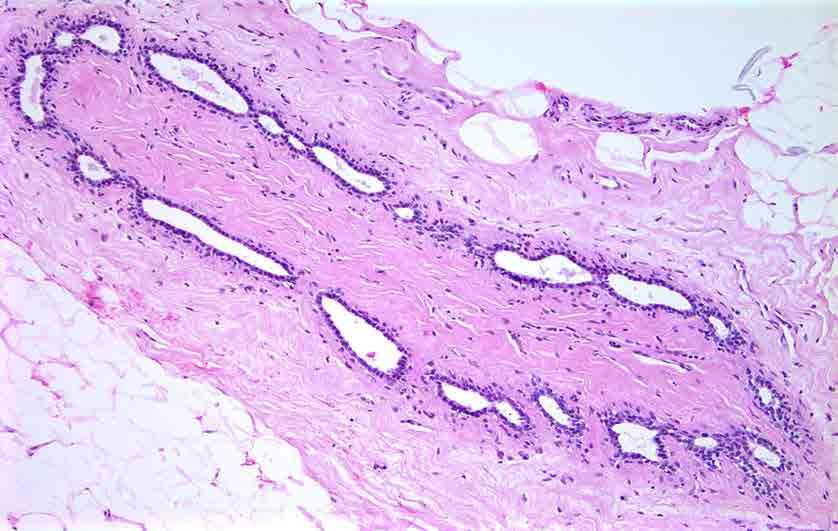
10
11
12
13
14
15 Proposed Pathogenesis Inspissation of secretions, leakage Inflammation Fibrosis Duct dilatation


16 Alternatively, are inflammatory lesion and duct ectasia separate entities? Differences in age, smoking hx
17 Ectatic Ducts = Mammary Duct Ectasia Ectasia of extralobular ducts common 30-40% of women >50 years Clinically evident mammary duct ectasia uncommon

18 Ectatic ducts, not duct ectasia!
19 Topic for Discussion Inflammatory lesions of the breast Mammary duct ectasia Lymphocytic mastopathy Granulomatous lesions Squamous metaplasia of lactiferous ducts (SMOLD) IgG4-related mastitis Reaction to mammary implants Mimics of inflammatory/reactive lesions
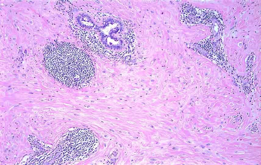
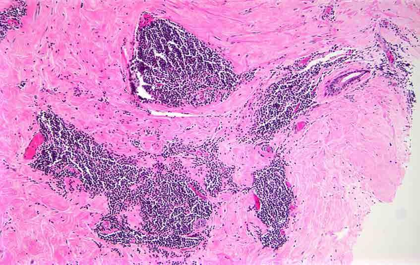
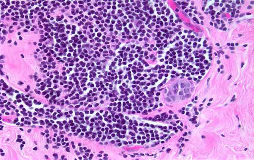
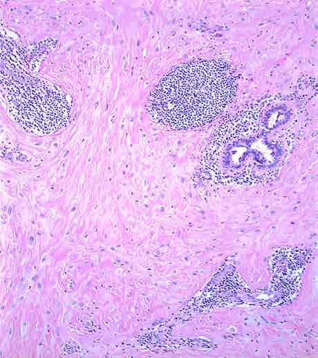
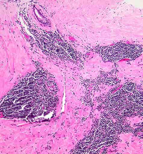
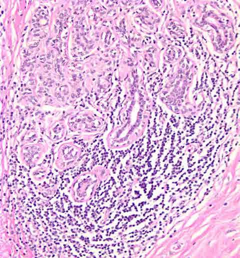
20 Lymphocytic Mastopathy Primarily young to middle-aged women Most commonly associated with type I diabetes (diabetic mastopathy) Similar histologic changes seen in association with other autoimmune diseases, type II diabetes, and in men May present as a palpable mass or mammographic abnormality May be multiple and bilateral Recurrences in up to one third
21 Histologic Features of Lymphocytic Mastopathy Keloidal fibrosis Periductal, perilobular and perivascular lymphocytic infiltrates (primarily B cells) Epithelioid myofibroblasts
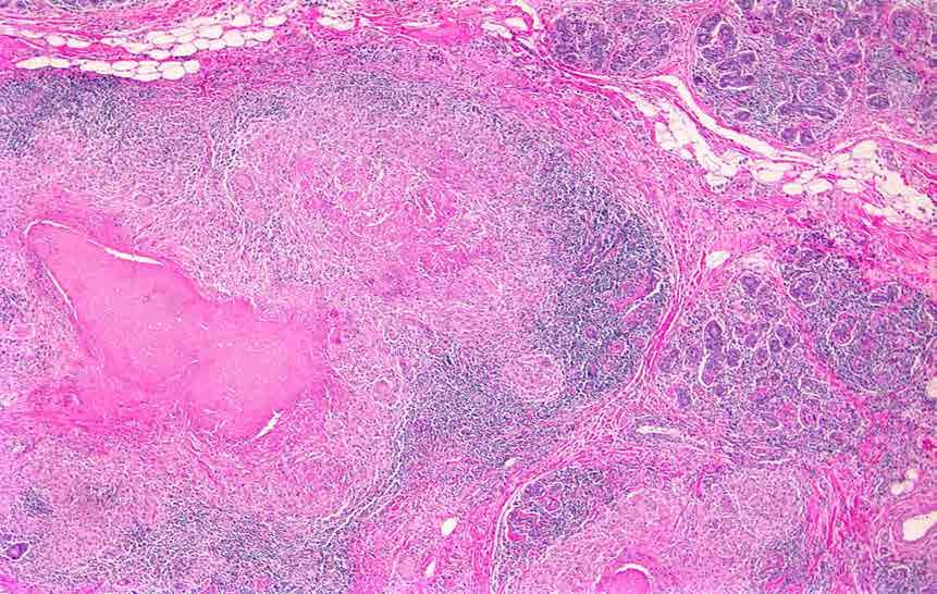
22
23
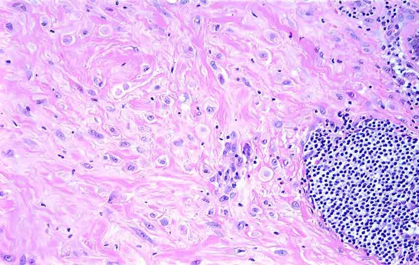
24
25 Lymphocytic Mastopathy Pathogenesis?Autoimmune reaction?resistance of collagen to degradation due to glycosylation and increased cross-linking
26 Topic for Discussion Inflammatory lesions of the breast Mammary duct ectasia Lymphocytic mastopathy Granulomatous lesions Squamous metaplasia of lactiferous ducts (SMOLD) IgG4-related mastitis Reaction to mammary implants Mimics of inflammatory/reactive lesions
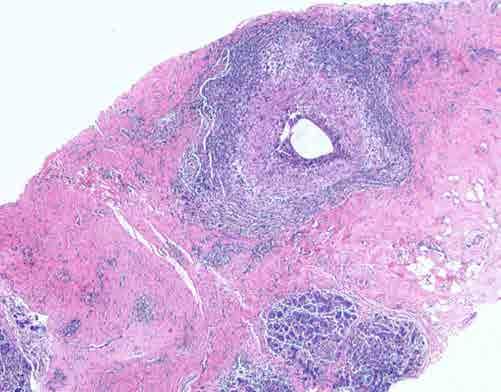
27 Granulomatous Lesions Most often result from same causes as granulomatous lesions in other organs (infection, sarcoid, reaction to foreign materials, reaction to carcinoma) Some due to lesions unique to the breast (duct ectasia, idiopathic granulomatous mastitis, cystic neutrophilic granulomatous mastitis)
28 Histologic Features of Granulomatous Lesions Granulomatous Lesion Mycobacterial, fungal, parasitic infections Sarcoid Mammary duct ectasia Reaction to foreign materials Features Granulomas resemble those seen in comparable infections in other sites; may be necrotizing or non-necrotizing Non-necrotizing granulomas in inter- and intra-lobular stroma Periductal granulomas; may have xanthogranulomatous features Foreign body-type granulomas; foreign body giant cells; foreign material Reaction to carcinoma Non-necrotizing granulomas
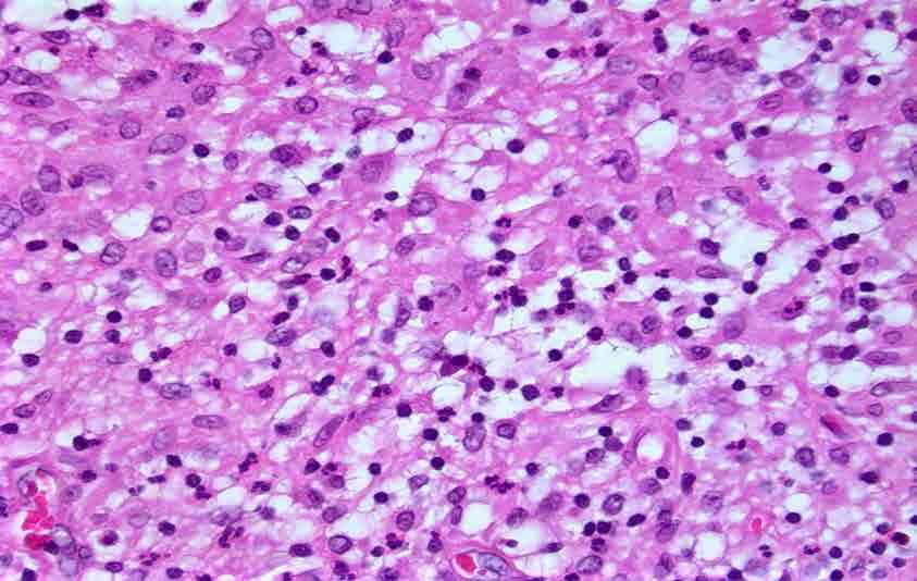
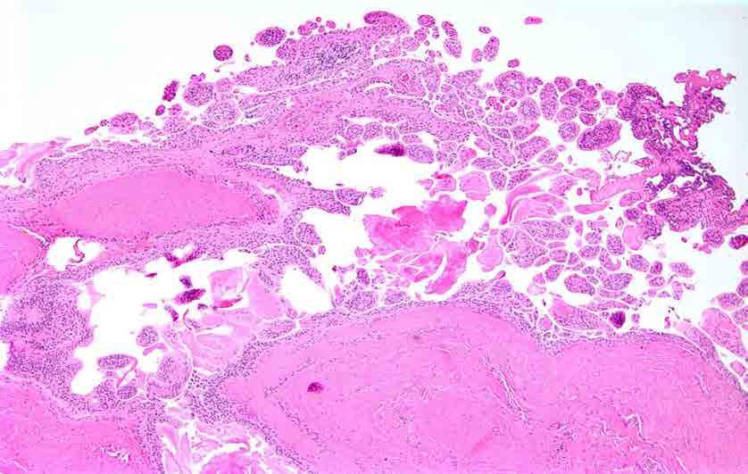
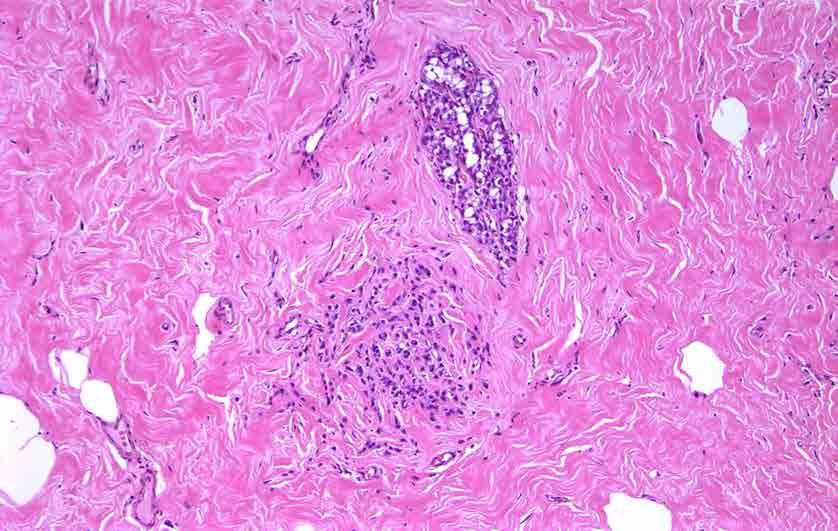
29 Tuberculous Mastitis
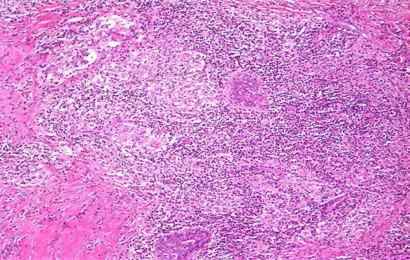
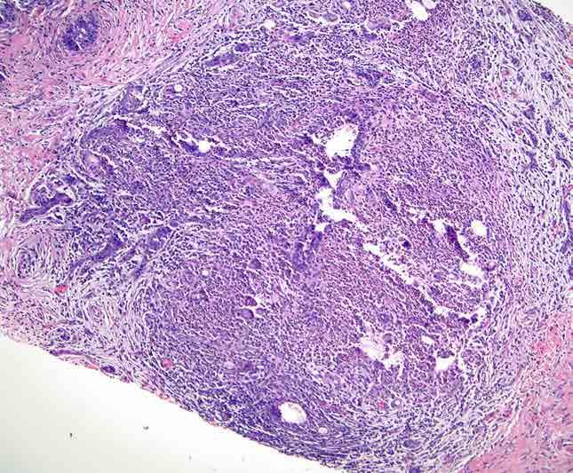
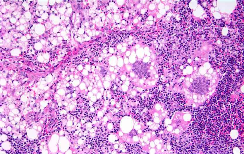
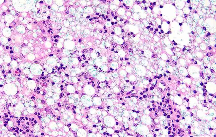
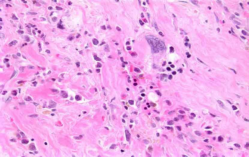
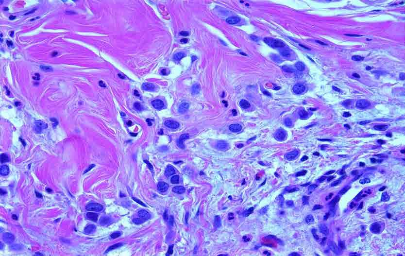
30 Tuberculous Mastitis

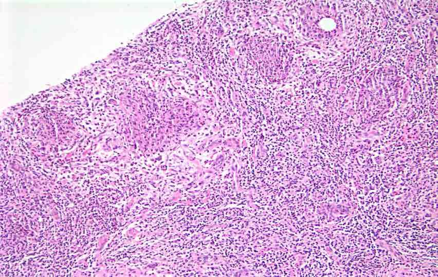
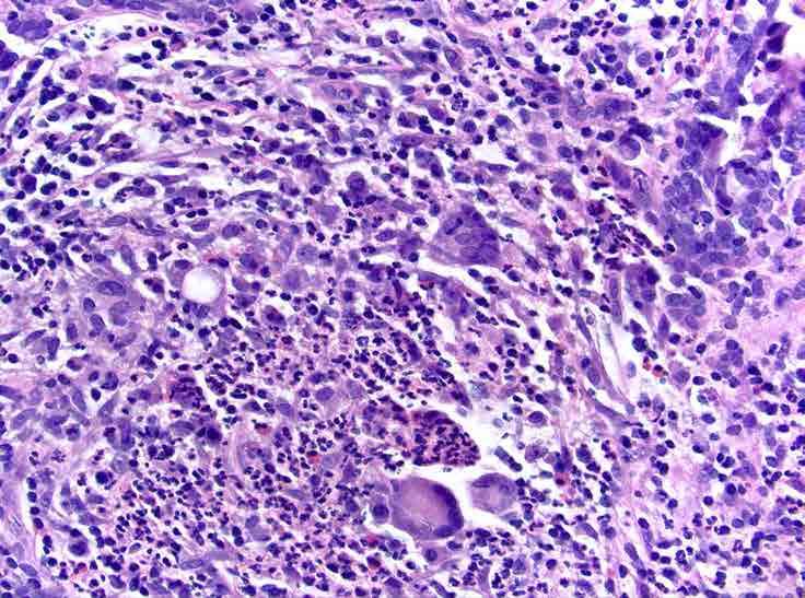
31 Sarcoid involving the breast

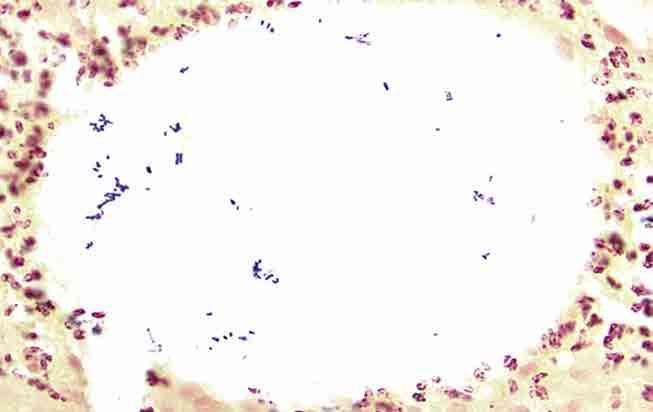
32 Sarcoid involving the breast
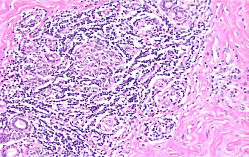
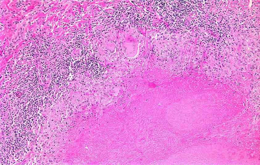
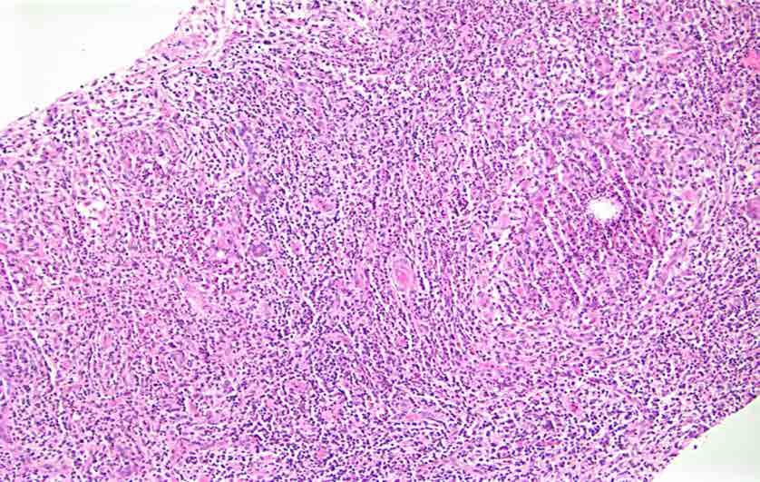
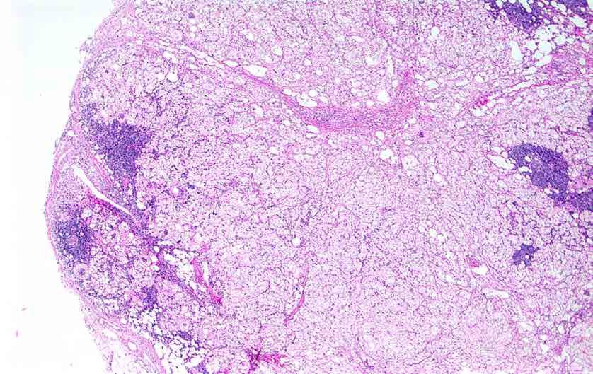
33 Idiopathic Granulomatous Mastitis (Granulomatous Lobular Mastitis) Usually presents as a mass in young parous women, often related to recent pregnancy May clinically simulate carcinoma Histologic features Lobulocentric granulomas, often with neutrophils, microabscesses
34
35
36 Idiopathic Granulomatous Mastitis (Granulomatous Lobular Mastitis) Usually presents as a mass in young parous women, often related to recent pregnancy May clinically simulate carcinoma Histologic features Lobulocentric granulomas, often with neutrophils, microabscesses?corticosteroid responsive (but first rule out infection)
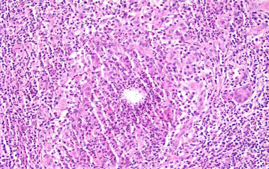
37 Cystic Neutrophilic Granulomatous Mastitis (CNGM) Recently recognized entity Presents as mastitis in parous or lactating women Patients often febrile with leukocytosis Nipple inversion or retraction common; fistulas can occur
38 Cystic Neutrophilic Granulomatous Mastitis (CNGM) Lobulocentric granulomas, often with neutrophils or areas of microabscess formation Empty (cystic) spaces of dissolved lipid surrounded by neutrophils distinguish CNGM from IGM Giant cells may also be present
39 Cystic Neutrophilic Granulomatous Mastitis (CNGM) Faint rod-like structures may be identified within cystic spaces On Gram stain these bacteria are more easily identified as gram positive rods consistent with Corynebaceria C. kroppenstedtii are most commonly implicated Treatment not yet established
40
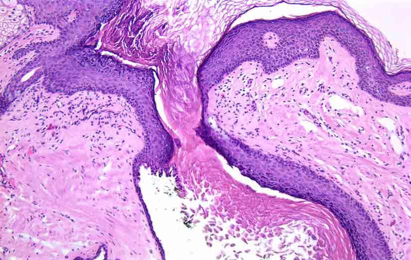
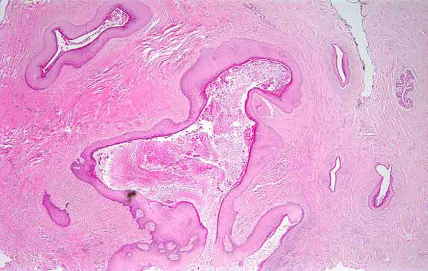
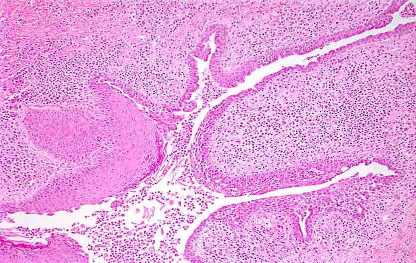
41
42
43
44

45
46 Idiopathic Granulomatous Mastitis vs Cystic Neutrophilic Granulomatous Mastitis Overlap in clinical features Overlap in histologic features Many cases previously diagnosed as idiopathic granulomatous mastitis probably represent examples of cystic neutrophilic granulomatous mastitis in which cysts/bacteria were not identified Johnstone, Pathol, 2017
47 Topic for Discussion Inflammatory lesions of the breast Mammary duct ectasia Lymphocytic mastopathy Granulomatous lesions Squamous metaplasia of lactiferous ducts (SMOLD) IgG4-related mastitis Reaction to mammary implants Mimics of inflammatory/reactive lesions
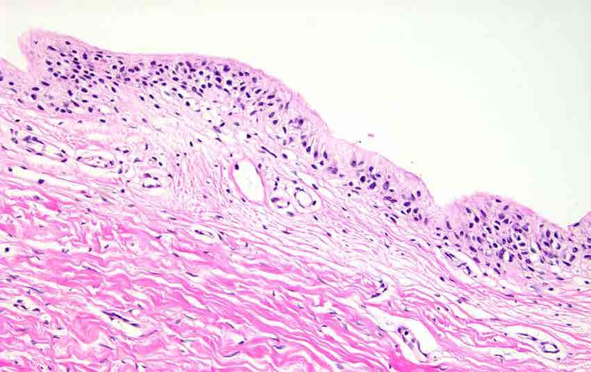
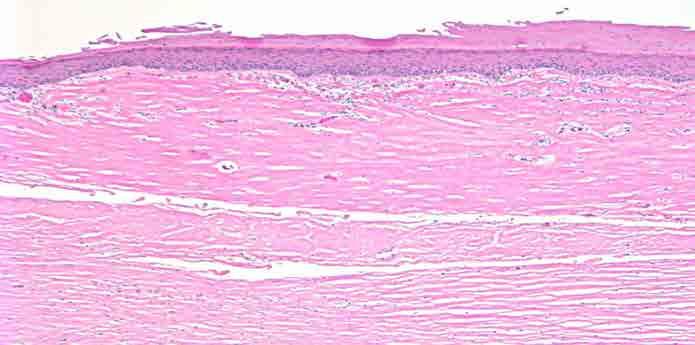
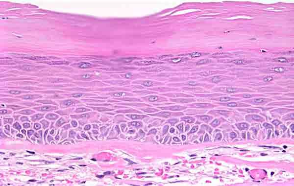
48 Squamous Metaplasia of Lactiferous Ducts (SMOLD) Squamous epithelium normally extends into nipple duct orifices for 1-2mm
49
50 Squamous Metaplasia of Lactiferous Ducts (SMOLD) Squamous epithelium normally extends into nipple duct orifices for 1-2mm If epithelium extends more deeply, keratin may accumulate, fill and obstruct duct (similar to epidermal inclusion cyst) Duct rupture results in extrusion of keratin and inflammatory reaction
51 Squamous Metaplasia of Lactiferous Ducts (SMOLD) May occur at any age Highly associated with smoking Presents as red, painful mass near nipple; usually diagnosed clinically as abscess Antibiotics and I and D generally ineffective Also known as recurrent subareolar abscess, Zuska s disease
52
53

54
55 Squamous Metaplasia of Lactiferous Ducts (SMOLD) Treatment requires complete excision of effected duct(s) Inadequate excision may result in recurrences and/or fistula formation
56 Squamous Metaplasia of Lactiferous Ducts (SMOLD) Initial surgical specimen often I and D of presumptive abscess Specimens may be small and show non-specific findings Search carefully for keratin and for ducts with squamous metaplasia Subsequent excisions usually contain more diagnostic features
57 Topic for Discussion Inflammatory lesions of the breast Mammary duct ectasia Lymphocytic mastopathy Granulomatous lesions Squamous metaplasia of lactiferous ducts (SMOLD) IgG4-related mastitis Reaction to mammary implants Mimics of inflammatory/reactive lesions

58 Part of a growing family of IgG4-related diseases characterized by mass-forming lesions due to: Dense lymphoplasmacytic infiltrates with lymphoid follicles and prominent component of IgG4+ plasma cells Stromal sclerosis with atrophy of lobules Elevated serum IgG4 Other sites may be involved Favorable clinical outcome AJSP, 2009

59 Mod Pathol 2012 Three major histologic features Dense lymphoplasmacytic infiltrate Fibrosis, at least focally in a storiform pattern Obliterative phlebitis Other histologic features: Phlebitis without obliteration of lumens Increased eosinophils Tissue IgG4 counts and IgG4:IgG ratios are secondary in importance (counts vary by organ:10-200/hpf; ratio >40%)

60 Cheuk, AJSP, 2009
61 Topic for Discussion Inflammatory lesions of the breast Mammary duct ectasia Lymphocytic mastopathy Granulomatous lesions Squamous metaplasia of lactiferous ducts (SMOLD) IgG4-related mastitis Reaction to mammary implants Mimics of inflammatory/reactive lesions
62 Reactions to Mammary Implants Fibrous capsule Reaction to silicone gel leakage Capsule, breast, axillary lymph nodes Synovial-like metaplasia Breast implant-associated anaplastic large cell lymphoma?mesenchymal tumors?carcinomas
63
64

65
66
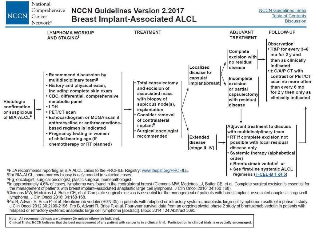
67
68
69
70 Squamous Metaplasia of Implant Capsule
71 Silicone lymphadenitis
72 Silicone lymphadenitis
73 Silicone lymphadenitis
74 Breast Implant-Associated Anaplastic Large Cell Lymphoma Presentation Late onset seroma, capsular contraction, mass Median time to development ~10 yrs (range 1-39 yrs) Median age 51 yrs (range yrs) Significant association with textured implants Clemens, Plast Recon Surg, 2017 de Boer, JAMA Oncol, 2018
75 Breast Implant-Associated Anaplastic Large Cell Lymphoma RR of BIA-ALCL in patients with textured implants is 67x higher than in general population But absolute risk is extremely low 1 in 35,000 at age 50 1 in 12,00 at age 70 1 in 7,000 at age 75 Clemens, Plast Recon Surg, 2017 de Boer, JAMA Oncol, 2018
76 Breast Implant-Associated Anaplastic Large Cell Lymphoma Large, pleomorphic cells in seroma fluid or implant capsule T-cell or null lineage CD30+ Majority ALK-negative Absence of genetic abnormalities at ALK (2q23)

77 Arada, 2014 CD30
78 Breast Implant-Associated Anaplastic Large Cell Lymphoma Differential Diagnosis Inflammation Recurrent carcinoma, particularly invasive lobular carcinoma
79 Breast Implant-Associated Anaplastic Large Cell Lymphoma Treatment and course If confined to capsule, treated conservatively» Removal of implant Most have indolent clinical course (98% 5 year survival)» Patients presenting with mass appear to have worse outcome (?chemotherapy) Must exclude systemic ALCL (may account for some cases with more aggressive clinical course) ~20% have lymph node involvement; more commonly seen when BIA-ALCL extends beyond capsule; can be a mimic for Hodgkin lymphoma; OS reduced to ~75% in patients with LNI
80 Breast Implant-Associated Anaplastic Large Cell Lymphoma Possible etiologies Chronic T-cell stimulation either due to:» Micromotion/friction of textured implant surface» Response to bacterial biofilm Ralstonia sp. implicated; non-fermenting Gram negative bacillus found in soil and water Similarities with H. pylori Interestingly, macrotextured implants were developed in the 1980 s to improve integration of implant with host tissues; widespread use began in 1990s and first report of BIA-ALCL was in 1996, with many more being reported in the 2000 s
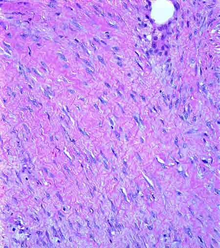
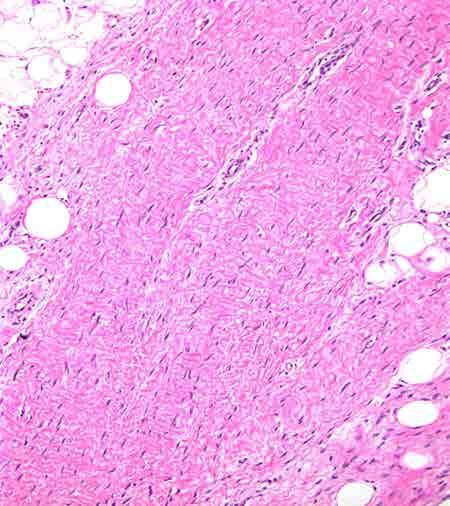
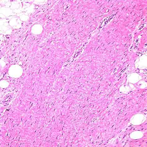
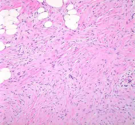
81 Kim, Plast Reconstr Surg, 2015 Nava, Plast Reconstr Surg, experts from multiple disciplines agreed on the following: Late seromas >1 year after implant should always be evaluated Fluid sent for culture, cytology, flow cytometry and cell block evaluation by hematopathologist Surgical removal of implant and capsule Clinical f/u every 6 months for 5 yrs with US for 2 yrs Absolute risk of BIA-ALCL is very low and BIA-ALCL is biologically indolent, but is a clinically important complication of breast reconstruction/augmentation surgery
82
83
84 BIA-ALCL listed as a new provisional entity distinguished from other ALK- ALCL Non-invasive disease with excellent outcome Some would argue should be considered a lymphoproliferative disorder rather than lymphoma Swerdlow, Blood, 2016
85 Implant-Associated Mesenchymal Tumors Balzer, cases reported to date Seen with both saline and silicone implants 2/3 fibromatoses, 1/3 sarcomas Cause and effect relationship between implants and mesenchymal lesions not established Some patients who developed sarcomas had received XRT for breast cancer
86 Implant-Associated Carcinomas Case reports (including squamous cell carcinoma) No relationship between breast augmentation and increased breast cancer risk in two epidemiologic studies
87 Topic for Discussion Inflammatory lesions of the breast Mammary duct ectasia Lymphocytic mastopathy Granulomatous lesions Squamous metaplasia of lactiferous ducts (SMOLD) IgG4-related mastitis Reaction to mammary implants Mimics of inflammatory/reactive lesions
88 Fibromatosis Mimics of Inflammatory and Reactive Lesions Mimic Fibromatosis-like metaplastic carcinoma Spindle cell (metaplastic) carcinoma Scar Scar Inflammatory/Reactive Lesion Nodular fasciitis Hematopoietic/Lymphoid lesions Invasive lobular carcinoma Histiocytoid carcinoma Inflammation Inflammation Fat necrosis
89 Fibromatosis Mimics of Inflammatory and Reactive Lesions Mimic Fibromatosis-like metaplastic carcinoma Spindle cell (metaplastic) carcinoma Scar Scar Inflammatory/Reactive Lesion Nodular fasciitis Hematopoietic/Lymphoid lesions Invasive lobular carcinoma Histiocytoid carcinoma Inflammation Inflammation Fat necrosis

90 Fibromatosis Scar
91 Fibromatosis Scar
92 Fibromatosis Mimics of Inflammatory and Reactive Lesions Mimic Fibromatosis-like metaplastic carcinoma Spindle cell (metaplastic) carcinoma Scar Scar Inflammatory/Reactive Lesion Nodular fasciitis Hematopoietic/Lymphoid lesions Invasive lobular carcinoma Histiocytoid carcinoma Inflammation Inflammation Fat necrosis
93 Fibromatosis-like Metaplastic Ca Scar
94 Fibromatosis Mimics of Inflammatory and Reactive Lesions Mimic Fibromatosis-like metaplastic carcinoma Spindle cell (metaplastic) carcinoma Scar Scar Inflammatory/Reactive Lesion Nodular fasciitis Hematopoietic/Lymphoid lesions Invasive lobular carcinoma Histiocytoid carcinoma Inflammation Inflammation Fat necrosis


95 Spindle Cell Ca Nodular Fasciitis
96 Fibromatosis Mimics of Inflammatory and Reactive Lesions Mimic Fibromatosis-like metaplastic carcinoma Spindle cell (metaplastic) carcinoma Scar Scar Inflammatory/Reactive Lesion Nodular fasciitis Hematopoietic/Lymphoid lesions Invasive lobular carcinoma Histiocytoid carcinoma Inflammation Inflammation Fat necrosis
97
98 CLL
99 Lymphocytic mastopathy CLL

100 Lymphocytic mastopathy CLL

101
102 Extramedullary hematopoiesis
103 Fibromatosis Mimics of Inflammatory and Reactive Lesions Mimic Fibromatosis-like metaplastic carcinoma Spindle cell (metaplastic) carcinoma Scar Scar Inflammatory/Reactive Lesion Nodular fasciitis Hematopoietic/Lymphoid lesions Invasive lobular carcinoma Histiocytoid carcinoma Inflammation Inflammation Fat necrosis
104
105
106 Fibromatosis Mimics of Inflammatory and Reactive Lesions Mimic Fibromatosis-like metaplastic carcinoma Spindle cell (metaplastic) carcinoma Scar Scar Inflammatory/Reactive Lesion Nodular fasciitis Hematopoietic/Lymphoid lesions Invasive lobular carcinoma Histiocytoid carcinoma Inflammation Inflammation Fat necrosis
107
108
109

110 Cytokeratin AE1/AE3/Cam 5.2

111 Histiocytoid ca Fat necrosis
112 Take Home Message Although relatively uncommon, pathologists should be familiar with the spectrum of the important inflammatory and reactive lesions of the breast, and of the lesions that may mimic them
Diseases of the breast (1 of 2)
") Diseases of the breast (1 of 2) Introduction A histology introduction Normal ducts and lobules of the breast are lined by two layers of cells a layer of luminal cells overlying a second layer of myoepithelial
Diseases of the breast (1 of 2) Introduction A histology introduction Normal ducts and lobules of the breast are lined by two layers of cells a layer of luminal cells overlying a second layer of myoepithelial
LYMPHATIC DRAINAGE AXILLARY (MOSTLY) INTERNAL MAMMARY SUPRACLAVICULAR
 INTERNAL MAMMARY SUPRACLAVICULAR") BREAST LYMPHATIC DRAINAGE AXILLARY (MOSTLY) INTERNAL MAMMARY SUPRACLAVICULAR HISTOLOGY LOBE: (10 in whole breast) LOBULE: (many per lobe) ACINUS/I, aka ALVEOLUS/I: (many per lobule) DUCT(S): INTRA- or
BREAST LYMPHATIC DRAINAGE AXILLARY (MOSTLY) INTERNAL MAMMARY SUPRACLAVICULAR HISTOLOGY LOBE: (10 in whole breast) LOBULE: (many per lobe) ACINUS/I, aka ALVEOLUS/I: (many per lobule) DUCT(S): INTRA- or
Lesion Imaging Characteristics Mass, Favoring Benign Circumscribed Margins Intramammary Lymph Node
 Lesion Imaging Characteristics Mass, Favoring Benign Circumscribed Margins Intramammary Lymph Node Oil Cyst Mass, Intermediate Concern Microlobulated Margins Obscured Margins Mass, Favoring Malignant Indistinct
Benign Mimics of Malignancy in Breast Pathology
 Arthur Purdy Stout Society of Surgical Pathologists Companion Meeting Benign Mimics of Malignancy in Breast Pathology Stuart J. Schnitt, M.D. Beth Israel Deaconess Medical Center and Harvard Medical School,
Imaging in breast cancer. Mammography and Ultrasound Donya Farrokh.MD Radiologist Mashhad University of Medical Since
 Imaging in breast cancer Mammography and Ultrasound Donya Farrokh.MD Radiologist Mashhad University of Medical Since A mammogram report is a key component of the breast cancer diagnostic process. A mammogram
Benign, Reactive and Inflammatory Lesions of the Breast
 Benign, Reactive and Inflammatory Lesions of the Breast Marilin Rosa, MD Associate Member Section Head of Breast Pathology Department of Anatomic Pathology Program Director, Breast Pathology Fellowship
BREAST PATHOLOGY. Fibrocystic Changes
 BREAST PATHOLOGY Lesions of the breast are very common, and they present as palpable, sometimes painful, nodules or masses. Most of these lesions are benign. Breast cancer is the 2 nd most common cause
SIGNIFICANT OTHERS. Miscellaneous Benign Breast Conditions
 SIGNIFICANT OTHERS Miscellaneous Benign Breast Conditions Epworth HealthCare 1 FAT NECROSIS TRAUMATIC Cell rupture Seat-Belt injury Blunt trauma Iatrogenic injury Surgery, Flaps, Radiotherapy Pathology
INDEX. in this web service Cambridge University Press
 abscess. See also subareolar abscess acute mastitis, 44 lactational/puerperal mastitis, 55 mammary tuberculosis, 42 tuberculous, 43 adeno gastric, 198, 200 invasive, 157 lung, 197, 200 prostatic, 199 200
University Journal of Pre and Para Clinical Sciences
 ISSN 2455 2879 Volume 2 Issue 1 2016 Metaplastic carcinoma breast a rare case report Abstract : Metaplastic carcinoma of the breast is a rare malignancy with two distinct cell lines described as a breast
1 NORMAL HISTOLOGY AND METAPLASIAS
 1 NORMAL HISTOLOGY AND METAPLASIAS, MD Anatomy and Histology 1 Metaplasias 2 ANATOMY AND HISTOLOGY The female breast is composed of a branching duct system, which begins at the nipple with the major lactiferous
Case year old Chinese female. Radiological echo-distortion in the right breast at o clock. Core biopsy of the o clock lesion.
 Case 3 64 year old Chinese female. Radiological echo-distortion in the right breast at 10-12 o clock. Core biopsy of the 11-12 o clock lesion. Division of Pathology Courtesty of Dr Lester Leong ill-defined,
ASPS Recommended Insurance Coverage Criteria for Third- Party Payers
 ASPS Recommended Insurance Coverage Criteria for Third- Party Payers Breast Implant Associated Anaplastic Large Cell Lymphoma BACKGROUND Anaplastic Large Cell Lymphoma (ALCL) is a rare type of cancer of
Granulomatous mastitis: Radio-pathologic correlation and management
 Granulomatous mastitis: Radio-pathologic correlation and management Poster No.: C-1418 Congress: ECR 2014 Type: Educational Exhibit Authors: S. E. Song, B. K. Seo, K. R. Cho, O. H. Woo, Y.-S. Kim ; 1 1
Abid Irshad, MD Director Breast Imaging. Medical University of South Carolina Charleston
 Abid Irshad, MD Director Breast Imaging Medical University of South Carolina Charleston Cases Financial disclosure: I or my family have no financial interest related to the material discussed in this presentation
Ductal Carcinoma in Situ. Laura C. Collins, M.D. Department of Pathology Beth Israel Deaconess Medical Center and Harvard Medical School Boston, MA
 Ductal Carcinoma in Situ Laura C. Collins, M.D. Department of Pathology Beth Israel Deaconess Medical Center and Harvard Medical School Boston, MA Definition of DCIS WHO 2012 A neoplastic proliferation
BREAST PATHOLOGY MCQS
 BREAST PATHOLOGY MCQS 1) :The most important factor in breast enlargement during pregnancy is A. stromal edema B. secretion of chorionic gonadotropin C. glandular hyperplasia D. proliferation of stroma
Breast implant-associated anaplastic large cell lymphoma. (BIA-ALCL): a collaborative effort for diagnosis and treatment
: a collaborative effort for diagnosis and treatment") Case Report Page 1 of 5 Breast implant-associated anaplastic large cell lymphoma (BIA-ALCL): a collaborative effort for diagnosis and treatment Kerry E. Fine, Matthew Wi, Della C. Bennett Department of
Case Report Page 1 of 5 Breast implant-associated anaplastic large cell lymphoma (BIA-ALCL): a collaborative effort for diagnosis and treatment Kerry E. Fine, Matthew Wi, Della C. Bennett Department of
Case year female. Routine Pap smear
 Case 1 57 year female Routine Pap smear Diagnosis? 1. Atypical glandular cells of unknown significance (AGUS) 2. Endocervical AIS 3. Endocervical adenocarcinoma 4. Endometrial adenocarcinoma 5. Adenocarcinoma
TOTALS 30. Preliminary analysis of NNDEQA 004 (May 2013 TSL workshops) 1) 70 year old female with focal irregularity in the right breast.
 1) 70 year old female with focal irregularity in the right breast.") -- Preliminary analysis of NNDEQA 00 (May 0 TSL workshops) ) 70 year old female with focal irregularity in the right breast. Fat necrosis (with organising thrombus/foreign body granuloma/surgical site
-- Preliminary analysis of NNDEQA 00 (May 0 TSL workshops) ) 70 year old female with focal irregularity in the right breast. Fat necrosis (with organising thrombus/foreign body granuloma/surgical site
Evening specialty conference: Liver
 Evening specialty conference: Liver Joseph Misdraji, M.D. Disclosure of Relevant Financial Relationships Disclosure of Relevant Financial Relationships USCAP requires that all planners (Education Committee)
Breast Pathology. Breast Development
 Breast Pathology Lecturer: Hanina Hibshoosh, M.D. Reading: Kumar, Cotran, Robbins, Basic Pathology, 6th Edition, pages 623-635 Breast Development 5th week - thickening of the epidermis - milk line 5th
COMMON BENIGN DISORDERS AND DISEASES OF THE BREAST
 COMMON BENIGN DISORDERS AND DISEASES OF THE BREAST Aberrations of Normal Development and Involution (ANDI). The basic principles underlying the aberrations of normal development and involution (ANDI) classification
DISORDERS OF THE BREAST Dated. FIBROADENOSIS Other common names: mastitis, fibrocystic disease, cystic mammary dysplasia.
 DISORDERS OF THE BREAST Dated BENIGN BREAST DISORDERS (Essential Surg 2 nd Ed, pp 540) FIBROADENOSIS Other common names: mastitis, fibrocystic disease, cystic mammary dysplasia. Fibroadenosis is the distortion
PRINCIPLES OF BREAST SURGERY & COMPLICATIONS
 PRINCIPLES OF BREAST SURGERY & COMPLICATIONS Adam Cichowitz The Royal Melbourne Hospital ANATOMY Lies in subcutaneous tissue Base: midline to midaxillary line, 2nd to 6th rib Overlies pec major, serratus
Breast pathology. 2nd Department of Pathology Semmelweis University
 Breast pathology 2nd Department of Pathology Semmelweis University Breast pathology - Summary - Benign lesions - Acute mastitis - Plasma cell mastitis / duct ectasia - Fat necrosis - Fibrocystic change/
Mousa. Israa Ayed. Abdullah AlZibdeh. 0 P a g e
 1 Mousa Israa Ayed Abdullah AlZibdeh 0 P a g e Breast pathology The basic histological units of the breast are called lobules, which are composed of glandular epithelial cells (luminal cells) resting on
Table of Contents: Foreword Preface Acknowledgementsi Dedication
 Table of Contents: Foreword Preface Acknowledgementsi Dedication Chapter 1 Problems of concept and nomenclature of benign disorders of the breast The source of the problem History The present and the future
Breast Infections. Epworth Benign Breast Disease Symposium Miss Melanie Walker MBBS(Hons) FRACS Epworth Breast Service
 FRACS Epworth Breast Service") Breast Infections Epworth Miss Melanie Walker MBBS(Hons) FRACS Epworth Breast Service Parenchymal Breast Infection Infections associated with breast feeding Central or subareolar abscesses +/- mammary
Breast Infections Epworth Miss Melanie Walker MBBS(Hons) FRACS Epworth Breast Service Parenchymal Breast Infection Infections associated with breast feeding Central or subareolar abscesses +/- mammary
PAAF vs Core Biopsy en Lesiones Mamarias Case #1
 5/19/2014 PAAF vs Core Biopsy en Lesiones Mamarias Case #1 Fine Needle Aspiration Cytology of Breast: Correlation with Needle Core Biopsy 64-year-old woman Mass in breast Syed Hoda, MD CD31 Post-Radiation
CPC 4 Breast Cancer. Rochelle Harwood, a 35 year old sales assistant, presents to her GP because she has noticed a painless lump in her left breast.
 CPC 4 Breast Cancer Rochelle Harwood, a 35 year old sales assistant, presents to her GP because she has noticed a painless lump in her left breast. 1. What are the most likely diagnoses of this lump? Fibroadenoma
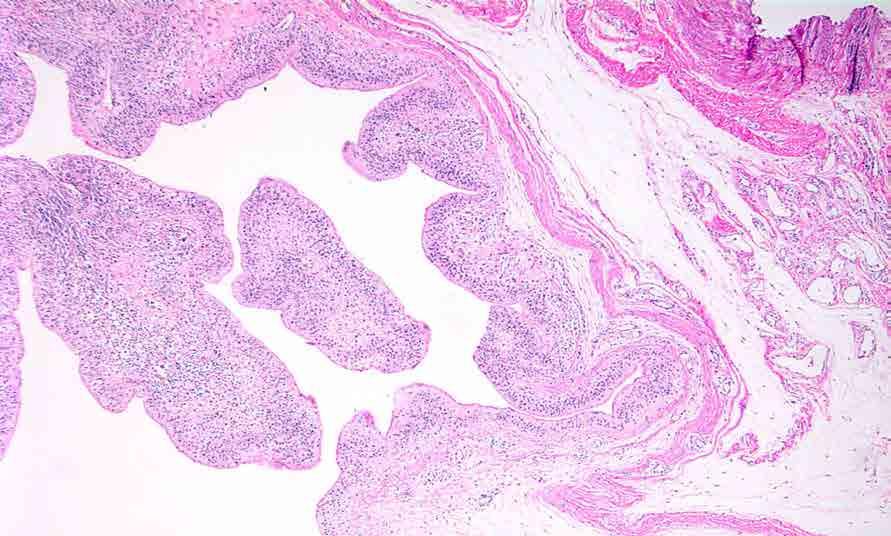
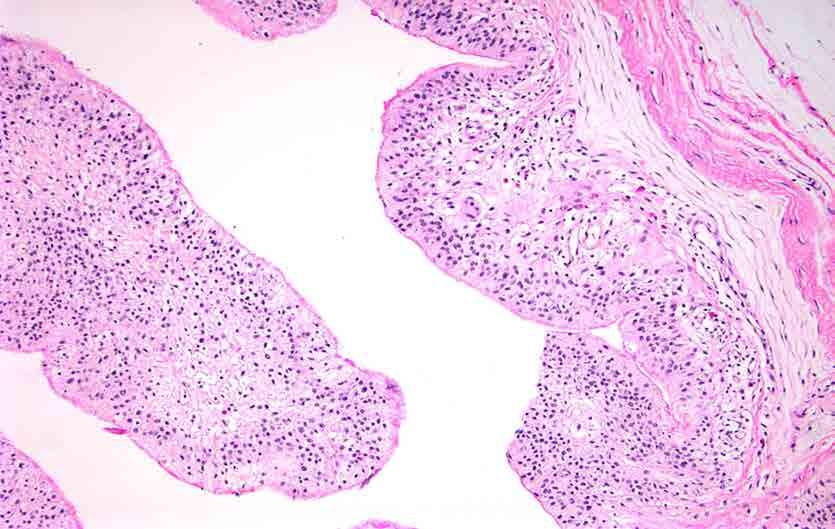
Papillary Lesions of the Breast A Practical Approach to Diagnosis. (Arch Pathol Lab Med. 2016;140: ; doi: /arpa.
 Papillary Lesions of the Breast A Practical Approach to Diagnosis (Arch Pathol Lab Med. 2016;140:1052 1059; doi: 10.5858/arpa.2016-0219-RA) Papillary lesions of the breast Span the spectrum of benign,
Columnar Cell Lesions
 Columnar Cell Lesions Laura C. Collins, M.D. Department of Pathology Beth Israel Deaconess Medical Center and Harvard Medical School Boston, MA Question? Columnar cell lesions are: a) Annoying lesions
Spindle Cell Lesions Of The Breast. Emad Rakha Professor of Breast Pathology and Consultant Pathologist
 Spindle Cell Lesions Of The Breast Emad Rakha Professor of Breast Pathology and Consultant Pathologist * SCLs comprise a wide spectrum of diseases, ranging from reactive processes to aggressive malignant
Triple Negative Breast Cancer
 Triple Negative Breast Cancer Prof. Dr. Pornchai O-charoenrat Division of Head-Neck & Breast Surgery Department of Surgery Faculty of Medicine Siriraj Hospital Breast Cancer Classification Traditional
Pathology & Presentation of Benign Breast Disease Zdenek Dubrava - February 2006
 Pathology & Presentation of Benign Breast Disease Zdenek Dubrava - February 2006 Presentation Lump Pain Nipple Discharge Breast Shape/Size Richard J Santen, Robert Mansel. The New England Journal of Medicine.
PATHOLOGY OF THE BREAST
 UROGENITAL SYSTEM MBBS 2 nd Yr. Lecture Dr. U.S. Khoo October 3, 2002-9:45 AM Underground Lecture Theatre 1 New Clinical Building, QMH PATHOLOGY OF THE BREAST Learning Objectives 1. To understand the normal
Salivary Glands 3/7/2017
 Salivary Glands 3/7/2017 Goals and objectives Focus on the entities unique to H&N Common board type facts Information for your future practice Salivary Glands Salivary Glands Major gland. Paratid. Submandibular.
Anatomy of the biliary tract
 Harvard-MIT Division of Health Sciences and Technology HST.121: Gastroenterology, Fall 2005 Instructors: Dr. Jonathan Glickman Anatomy of the biliary tract Figure removed due to copyright reasons. Biliary
Contents. Basic Ultrasound Principles and Terminology. Ultrasound Nodule Characteristics
 Contents Basic Ultrasound Principles and Terminology Basic Ultrasound Principles... 1 Ultrasound System... 2 Linear Transducer for Superficial Images and Ultrasound-Guided FNA... 3 Scanning Planes... 4
Invasive Papillary Breast Carcinoma
 410 This is an Open Access article licensed under the terms of the Creative Commons Attribution- NonCommercial-NoDerivs 3.0 License (www.karger.com/oa-license), applicable to the online version of the
Among the benign intraepithelial melanocytic proliferations, Inflamed Conjunctival Nevi. Histopathological Criteria. Resident Short Reviews
 Resident Short Reviews Inflamed conjunctival nevi (ICN) may suggest malignancy because of their rapid growth and atypical histology. The objective of this study was to characterize the diagnostic features
CMS Limitations Guide Mammography & Bone Mass Measurement
 Starting October 1, 2015, CMS will update their existing medical necessity limitations on tests and procedures to correspond to ICD-10 codes. This limitations guide provides you with the latest changes.
Final analysis of NNDEQA 004 (May 2013 TSL workshops) 1) 70 year old female with focal irregularity in the right breast.
 1) 70 year old female with focal irregularity in the right breast.") Final analysis of NNDEQA 00 (May 0 TSL workshops) ) 70 year old female with focal irregularity in the right breast. CONSUL TANTS Fat necrosis (with organising thrombus/foreign body granuloma/surgical site
Final analysis of NNDEQA 00 (May 0 TSL workshops) ) 70 year old female with focal irregularity in the right breast. CONSUL TANTS Fat necrosis (with organising thrombus/foreign body granuloma/surgical site
Diseases of the breast (2 of 2) Breast cancer
 Breast cancer") Diseases of the breast (2 of 2) Breast cancer Epidemiology & etiology The most common type of cancer & the 2 nd most common cause of cancer death in women 1 of 8 women in USA Affects 7% of women Peak at
Diseases of the breast (2 of 2) Breast cancer Epidemiology & etiology The most common type of cancer & the 2 nd most common cause of cancer death in women 1 of 8 women in USA Affects 7% of women Peak at
Treatment options for the precancerous Atypical Breast lesions. Prof. YOUNG-JIN SUH The Catholic University of Korea
 Treatment options for the precancerous Atypical Breast lesions Prof. YOUNG-JIN SUH The Catholic University of Korea Not so benign lesions? Imaging abnormalities(10% recall) lead to diagnostic evaluation,
Papillary Lesions of the Breast
 Papillary Lesions of the Breast Laura C. Collins, M.D. Associate Professor of Pathology Associate Director, Division of Anatomic Pathology Beth Israel Deaconess Medical Center and Harvard Medical School
Basement membrane in lobule.
 Bahram Memar, MD Basement membrane in lobule. Normal lobule-luteal phase Normal lobule-follicular phase Lactating breast Greater than 95% are adenocarcinomas in situ carcinomas and invasive carcinomas.
CHRONIC PANCREATITIS OR DUCTAL ADENOCARCINOMA? N. Volkan Adsay, \ MD
 CHRONIC PANCREATITIS OR DUCTAL ADENOCARCINOMA? N. Volkan Adsay, \ MD Case for discussion 67 y/o male Back pain and weight loss CT: 4.5 cm ill-defined, solid lesion in the head FNA/Core bx: Inconclusive
CURRICULUM FOR THE BREAST PATHOLOGY ROTATION UNIVERSITY OF FLORIDA DEPARTMENT OF PATHOLOGY
 CURRICULUM FOR THE BREAST PATHOLOGY ROTATION UNIVERSITY OF FLORIDA DEPARTMENT OF PATHOLOGY JULY, 2003 The following is a conceptual curriculum and set of guidelines for Pathology Residents on the Breast
Jordan University Faculty Of Medicine. Breast. Dr. Ahmed Salman. Assistant professor of anatomy & embryology
 Jordan University Faculty Of Medicine Breast Dr. Ahmed Salman Assistant professor of anatomy & embryology The breasts are specialized accessory glands of the skin that secretes milk. They are situated
Primary Cutaneous CD30-Positive T-cell Lymphoproliferative Disorders
 Primary Cutaneous CD30-Positive T-cell Lymphoproliferative Disorders Definition A spectrum of related conditions originating from transformed or activated CD30-positive T-lymphocytes May coexist in individual
(1/5) PP7 - Spinal Epidural Anaplastic Large Cell Lymphoma associated with breast implants
 PP7 - Spinal Epidural Anaplastic Large Cell Lymphoma associated with breast implants") (1/5) PP7 - Spinal Epidural Anaplastic Large Cell Lymphoma associated with breast implants Athanasiou A 1, Iliadis A 2, Kostopoulos I 2, Tsona A 3, Spiliotopoulos A 1 1 1 st Department of Neurosurgery,
(1/5) PP7 - Spinal Epidural Anaplastic Large Cell Lymphoma associated with breast implants Athanasiou A 1, Iliadis A 2, Kostopoulos I 2, Tsona A 3, Spiliotopoulos A 1 1 1 st Department of Neurosurgery,
Disclosure of Relevant Financial Relationships. Breast Pathology Evening Specialty Conference Case #4. Clinical Case: Pathologic Features
 Breast Pathology Evening Specialty Conference Case #4 K.P. Siziopikou, MD, PhD Professor of Pathology Director of Breast Pathology and Breast Pathology Fellowship Program Northwestern University Feinberg
CLINICAL SIGNIFICANCE OF BENIGN EPITHELIAL CHANGES
 Papillomas. Papillomas are composed of multiple branching fibrovascular cores, each having a connective tissue axis lined by luminal and myoepithelial cells ( Fig. 23-11 ). Growth occurs within a dilated
57th Annual HSCP Spring Symposium 4/16/2016
 An Unusual Malignant Spindle Cell Lesion to Involve the Breast Erinn Downs-Kelly, D.O. Associate Professor of Pathology University of Utah & ARUP Laboratories No disclosures Case 39 y/o female with no
04/10/2018. Intraductal Papillary Neoplasms Of Breast INTRADUCTAL PAPILLOMA
 Intraductal Papillary Neoplasms Of Breast Savitri Krishnamurthy MD Professor of Pathology Deputy Division Head The University of Texas MD Anderson Cancer Center 25 th Annual Seminar in Pathology Pittsburgh,
INTRA-OPERATIVE CYTOLOGY AND FROZEN SECTIONS OF BREAST LESIONS: A COMPARISON FROM A SAUDI TEACHING HOSPITAL
 Bahrain Medical Bulletin, Volume 18, Number 1, March 1996 INTRA-OPERATIVE CYTOLOGY AND FROZEN SECTIONS OF BREAST LESIONS: A COMPARISON FROM A SAUDI TEACHING HOSPITAL Ammar C.Al-Rikabi, MD,MRCPath,FIAC*
Papillary Lesions of the Breast
 Papillary Lesions of the Breast Texas Society of Pathologists 2013 Laura C. Collins, M.D. Associate Professor of Pathology Associate Director, Division of Anatomic Pathology Beth Israel Deaconess Medical
Question 1 A. ER-, PR-, HER+ B. ER+, PR+, HER2- C. ER-, PR+, HER2- D. ER-, PR-, HER2- E. ER-, PR+, HER2+
 Triple Negative Breast Cancer Laura C. Collins, M.D. Department of Pathology Beth Israel Deaconess Medical Center and Harvard Medical School, Boston, MA Question 1 The tumor depicted on the next slide
Immune checkpoint inhibitors, lymphocytes, and breast cancer. Megan L. Troxell, MD/PhD Stanford Pathology
 Immune checkpoint inhibitors, lymphocytes, and breast cancer Megan L. Troxell, MD/PhD Stanford Pathology Objectives Recognize the potential prognostic & predictive importance of tumor infiltrating lymphocytes
SESSION 1: GENERAL (BASIC) PATHOLOGY CONCEPTS Thursday, October 16, :30am - 11:30am FACULTY COPY
 PATHOLOGY CONCEPTS Thursday, October 16, :30am - 11:30am FACULTY COPY") SESSION 1: GENERAL (BASIC) PATHOLOGY CONCEPTS Thursday, October 16, 2008 9:30am - 11:30am FACULTY COPY GOAL: Describe the basic morphologic (structural) changes which occur in various pathologic conditions.
SESSION 1: GENERAL (BASIC) PATHOLOGY CONCEPTS Thursday, October 16, 2008 9:30am - 11:30am FACULTY COPY GOAL: Describe the basic morphologic (structural) changes which occur in various pathologic conditions.
Disclaimer no conflict of interest
 Disclaimer no conflict of interest Benign Breast Disease Alison Hayes FRACS Content Clinical assessment of the breast Triple assessment Focal nodularity Breast pain Cysts Infection Nipple discharge Gynaecomastia
COPE Library Sample
 Breast Anatomy LOBULE LOBE ACINI (MILK PRODUCING UNITS) NIPPLE AREOLA COMPLEX ENLARGEMENT OF DUCT AND LOBE LOBULE SUPRACLAVICULAR NODES INFRACLAVICULAR NODES DUCT DUCT ACINI (MILK PRODUCING UNITS) 8420
3/24/2017 DENDRITIC CELL NEOPLASMS: HISTOLOGY, IMMUNOHISTOCHEMISTRY, AND MOLECULAR GENETICS. Disclosure of Relevant Financial Relationships
 DENDRITIC CELL NEOPLASMS: HISTOLOGY, IMMUNOHISTOCHEMISTRY, AND MOLECULAR GENETICS Jason L. Hornick, M.D., Ph.D. Director of Surgical Pathology and Immunohistochemistry Brigham and Women s Hospital Professor
Diabetic Mastopathy in Long Standing Type I Diabetic Mellitus; A Case Report
 CASE REPORT J Surg Ultrasound 2017;4:25-29 JSU Journal of Surgical Ultrasound Diabetic Mastopathy in Long Standing Type I Diabetic Mellitus; A Case Report Ra Mi Kim, Hunkyoung Lee 1, Heeboong Park 2 Kangnam
ACCME/Disclosures ALK FUSION-POSITIVE MESENCHYMAL TUMORS. Tumor types with ALK rearrangements. Anaplastic Lymphoma Kinase. Jason L.
 Companion Meeting of the International Society of Bone and Soft Tissue Pathology The Evolving Concept of Mesenchymal Tumors ALK FUSION-POSITIVE MESENCHYMAL TUMORS Jason L. Hornick, MD, PhD March 13, 2016
-1- Pathology Department (code: 0605) Final Exam for Third year students Date: Time allowed: 2 hours. Paper II (75 marks).
 Final Exam for Third year students Date: Time allowed: 2 hours. Paper II (75 marks).") -1- BENHA UNIVERSITY FACULTY OF MEDICINE Pathology Department (code: 0605) Final Exam for Third year students Date: 28-5-2011 Time allowed: 2 hours. Paper II (75 marks). Please note that this question
-1- BENHA UNIVERSITY FACULTY OF MEDICINE Pathology Department (code: 0605) Final Exam for Third year students Date: 28-5-2011 Time allowed: 2 hours. Paper II (75 marks). Please note that this question
Columnar Cell Lesions. Columnar Cell Lesions and Flat Epithelial Atypia
 Columnar Cell Lesions and Stuart J. Schnitt, M.D. Beth Israel Deaconess Medical Center and Harvard Medical School Boston, MA, USA Columnar Cell Lesions Lesions characterized by columnar epithelial cells
Note: The cause of testicular neoplasms remains unknown
 - In the 15- to 34-year-old age group, they are the most common tumors of men. - Tumors of the testis are a heterogeneous group of neoplasms that include: I. Germ cell tumors : 95%; all are malignant.
Armed Forces Institute of Pathology.
 Armed Forces Institute of Pathology www.radpath.com Armed Forces Institute of Pathology Breast Disease www.radpath.org Armed Forces Institute of Pathology Interpretation of Breast MRI Leonard M. Glassman
Classification System
 Classification System A graduate of the Breast Oncology training program should be able to care for all aspects of disease and/or provide comprehensive management. When referring to a discipline of training
CELL AND TISSUE INJURY COURSE-II PATHOLOGY LABORATORY
 CELL AND TISSUE INJURY COURSE-II PATHOLOGY LABORATORY PATHOLOGY of INFECTIOUS DISEASES MICROSCOPY Rengin Ahıskalı Macroscopy samples are shown in the macroscopy presentations of the first two courses.
Case Scenario 1 History and Physical 3/15/13 Imaging Pathology
 Case Scenario 1 History and Physical 3/15/13 The patient is an 84 year old white female who presented with an abnormal mammogram. The patient has a five year history of refractory anemia with ringed sideroblasts
Contents. vii. Preface... Acknowledgments... v xiii
 Contents Preface... Acknowledgments... v xiii SECTION I 1. Introduction... 3 Knowledge-Based Diagnosis... 4 Systematic Examination of the Lymph Node... 7 Cell Type Identification... 9 Cell Size and Cellularity...
Review of the AP Part II Practical Examination. Dr David Clift Co Chief Examiner
 Review of the AP Part II Practical Examination Dr David Clift Co Chief Examiner General Remarks The part II practical examination involved 15 cases which were presented with sufficient clinical data to
Dermatopathology. Dr. Rafael Botella Estrada. Hospital La Fe de Valencia
 Dermatopathology Dr. Rafael Botella Estrada. Hospital La Fe de Valencia DERMATOPATHOLOGY CASE CHALLENGE: RECOGNIZING MIMIS AND MASQUERADERS Rosalie Elenitsas. University of Pennsylvania Spectrum Lupus
3/27/2017. Disclosure of Relevant Financial Relationships. Papilloma???
 Management of Papillary Lesions Diagnosed at Rad Path Concordant Core Biopsy (CNB) Disclosure of Relevant Financial Relationships USCAP requires that all planners (Education Committee) in a position to
Papillary Lesions of the breast
 Papillary Lesions of the breast Emad Rakha Professor of Breast Pathology The University of Nottingham Papillary lesions of the breast are a heterogeneous group of disease, which are characterised by neoplastic
Histopathology: granulomatous inflammation, including tuberculosis
 Histopathology: granulomatous inflammation, including tuberculosis These presentations are to help you identify basic histopathological features. They do not contain the additional factual information
University of Washington Radiology Review Course: Strange and Specific Diagnoses. Case #1
 University of Washington Radiology Review Course: Strange and Specific Diagnoses Katherine E. Dee, MD Seattle Breast Center Via Radiology 2014 Case #1 37 year old presents with bilateral palpable lumps.
Columnar Cell Lesions and Flat Epithelial Atypia
 Columnar Cell Lesions and Flat Epithelial Atypia Laura C. Collins, M.D. Department of Pathology Beth Israel Deaconess Medical Center and Harvard Medical School, Boston, MA Terminology for Columnar Cell
Lách
 Lách Lách Lách Lách Splenogonadal fusion. Splenic tissue is attached to testicular tissue. Pseudocyst (false or secondary cyst). A, Outer aspect. Pseudocyst (false or secondary cyst). B, Inner surface.
Case of the month March Dre Sonia Ziadi Dre Ana Barrigón Benítez Institut universitaire de pathologie, Lausanne
 Case of the month March 2018 Dre Sonia Ziadi Dre Ana Barrigón Benítez Institut universitaire de pathologie, Lausanne Clinical history 78 years-old female, past medical history for right breast carcinoma
OUTLINE FIBROADENOMA FIBROADENOMA. FIBROEPITHELIAL LESIONS OF THE BREAST UCSF Current Issues in Anatomic Pathology 2015 FIBROADENOMA PHYLLODES TUMOR
 OUTLINE FIBROADENOMA FIBROEPITHELIAL LESIONS OF THE BREAST UCSF Current Issues in Anatomic Pathology 2015 Gregor Krings, MD PhD Assistant Professor PHYLLODES TUMOR DIFFERENTIAL DIAGNOSIS CELLULAR FIBROEPITHELIAL
Promise of a beautiful day
 Promise of a beautiful day Ductal carcinoma in Situ Lobular Carcinoma in Situ Natural History Manosmed Tartous Oct 2009 Gérard ABADJIAN MD Pathology Department Hôtel-Dieu de France. Associate Professor
Overview of Pathology Evaluation of Breast Lesions and Quality Assurance
 Overview of Pathology Evaluation of Breast Lesions and Quality Assurance 2 Michael O. Idowu, Jaime A. Singh, and Margaret M. Grimes Masses/Densities/Distortions: General Considerations Radiologic evaluation
Breast Evaluation & Management Guidelines
 Breast Evaluation & Management Guidelines Pamela L. Kurtzhals, M.D. F.A.C.S. Head, Dept. of General Surgery Scripps Clinic, La Jolla Objective Review screening & diagnostic guidelines Focused patient complaints
Case study 1. Rie Horii, M.D., Ph.D. Division of Pathology Cancer Institute Hospital, Japanese Foundation for Cancer Research
 NCCN/JCCNB Seminar in Japan April 15, 2012 Case study 1 Rie Horii, M.D., Ph.D. Division of Pathology Cancer Institute Hospital, Japanese Foundation for Cancer Research Present illness: A 50y.o.premenopausal
Atypical Palisaded Myofibroblastoma of Lymph Node: Report of a rare case.
 ISPUB.COM The Internet Journal of Pathology Volume 10 Number 1 Atypical Palisaded Myofibroblastoma of Lymph Node: Report of a rare case. V Kinnera, R Nandyala, M Yootla, K Mandyam Citation V Kinnera, R
Imaging the Symptomatic Patient. Avice M.O Connell MD,FACR,FSBI Professor of Imaging Sciences Director, Women s Imaging University of Rochester
 Imaging the Symptomatic Patient Avice M.O Connell MD,FACR,FSBI Professor of Imaging Sciences Director, Women s Imaging University of Rochester The four most common symptoms Mass Pain Discharge Infection
Merih Guray, Aysegul A. Sahin. University of Texas M. D. Anderson Cancer Center, Houston, Texas, USA
 This material is protected by U.S. Copyright law. Unauthorized reproduction is prohibited. For reprints contact: Reprints@AlphaMedPress.com Breast Cancer Benign Breast Diseases: Classification, Diagnosis,
Differential Diagnosis of Oral Masses. Palatal Lesions
 Differential Diagnosis of Oral Masses Palatal Lesions Palatal Masses Periapical Abscess Torus Palatinus Mucocele Lymphoid Hyperplasia Adenomatous Hyperplasia Benign Salivary Neoplasms Malignant Salivary
Breast Cancer Screening and Surgery. April 26, 2018 Ashley B. Simpson, DO
 Breast Cancer Screening and Surgery April 26, 2018 Ashley B. Simpson, DO Objectives Breast cancer screening Common breast complaints Surgical management of breast cancer Breast Screening Question 1 At
Timby/Smith: Introductory Medical-Surgical Nursing, 9/e
 Timby/Smith: Introductory Medical-Surgical Nursing, 9/e Chapter 60: Caring for Clients With Breast Disorders Slide 1 Infectious and Inflammatory Breast Disorders: Mastitis Pathophysiology and Etiology
Papillary Lesions of the Breast: WHO Update
 Papillary Lesions of the Breast: WHO Update Stuart J. Schnitt, M.D. Department of Pathology Beth Israel Deaconess Medical Center and Harvard Medical School Boston, MA, USA Papillary Lesions of the Breast
Consumer summary Autologous fat transfer for breast augmentation (The report of the Review Group has been adapted for consumers by E.
 ASERNIP S Australian Safety and Efficacy Register of New Interventional Procedures Surgical Consumer summary Autologous fat transfer for breast augmentation (The report of the Review Group has been adapted
Low-Grade Periductal Stromal of Breast: a case report
 Low-Grade Periductal Stromal of Breast: a case report Rosanna Nenna 1 Cosimo Damiano Inchingolo 1 Domenico Palmieri 2 Annalisa De Lucia 1 Giusy Elicio 1 Pina Miscioscia 1 ( 1 ) U.O.C. di Anatomia Patologica,
Objectives. Salivary Gland FNA: The Milan System. Role of Salivary Gland FNA 04/26/2018
 Salivary Gland FNA: The Milan System Dr. Jennifer Brainard Section Head Cytopathology Cleveland Clinic Objectives Introduce the Milan System for reporting salivary gland cytopathology Define cytologic
Primary Tuberculosis of Breast in 22 Year Old Female A Rare Case Report
 IOSR Journal of Dental and Medical Sciences (IOSR-JDMS) e-issn: 2279-0853, p-issn: 2279-0861.Volume 13, Issue 12 Ver. II (Dec. 2014), PP 70-74 Primary Tuberculosis of Breast in 22 Year Old Female A Rare