Spontaneous Neoplasms and Survival in Wistar Han Rats: Compilation of Control Group Data. March, 2003
|
|
|
- Adela Bishop
- 6 years ago
- Views:
Transcription

1 Spontaneous Neoplasms and Survival in Wistar Han Rats: Compilation of Control Group Data March, 2003 Information Prepared by Mary L.A. Giknis Ph.D Charles B. Clifford D.V.M, Ph.D
2 TABLE OF CONTENTS INTRODUCTION...1 PURPOSE...1 COMMON STUDY PARAMETERS... 1 DATA SETS PRESENTED... 2 SUMMARY TABLE CALCULATIONS FOR NEOPLASTIC LESIONS...2 SYNONYMS FOR NEOPLASTIC LESIONS...3 ACKNOWLEDGMENTS...4 REFERENCES...4 TABLE 1: SUMMARY OF INDIVIDUAL STUDY INFORMATION AND SURVIVAL/MALES WEEKS...5 TABLE 2: SUMMARY OF INDIVIDUAL STUDY INFORMATION AND SURVIVAL/FEMALES WEEKS... 6 GRAPH 1: MALE SURVIVAL WEEKS...7 GRAPH 2: FEMALE SURVIVAL WEEKS...8 TABLE 3: NEOPLASMS/MALES WEEKS... 9 TABLE 4: NEOPLASMS/FEMALES WEEKS TABLE 5: INCIDENCE OF NEOPLASMS BY STUDY FOR SELECTED ORGANS/MALES...18 TABLE 6: INCIDENCE OF NEOPLASMS BY STUDY FOR SELECTED ORGANS/FEMALES... 21
3 INTRODUCTION: In the course of data analysis from a carcinogenicity study, statistical tests will occasionally indicate that the incidence of a particular neoplasm is significantly greater in a treated group than in the concurrent control. Since statistical differences can occur as a matter of chance alone, using a positive statistic difference as the sole or definitive evaluation tool could produce a false positive result (1, 2). Alternatively, a slight increase in the incidence of a rare neoplasm would be unlikely to achieve statistical significance by the tests typically employed in toxicology studies. In this type of situation, the use of historical control data could justify the biological significance of even a slight increase in the incidence of an uncommon neoplasm (2). The histopathology and survival data presented in this publication were gathered from ten control groups of animals from toxicology studies of approximately 104 weeks duration. All studies were conducted in accordance with Good Laboratory Practice regulations of the US Food and Drug Administration or the Environmental Protection Agency and/or the Standard Operating Procedures of the participating laboratory. All studies were performed in the United States or Europe by contract laboratories or industrial toxicology facilities. All studies were conducted in support of in-house research or marketing permits. The data presented were provided to us by the individual laboratories. PURPOSE: The purpose of this compilation is to offer the study director, reviewing toxicologist and/or study pathologist some reported incidences of neoplasms and survival data in Wistar Han rats maintained as control animals until approximately weeks of age. This document was prepared for informational purposes only. Diagnoses of the various neoplasms in the compilations are intentionally grouped in a manner to provide the user with a range of reported incidences of similar types of lesions. This compilation is not intended in any way to propose a system of standardized nomenclature nor does it separately include each and every reported variant of each lesion. For these reasons, care should be taken in using these data that are not intended as a substitute for historical data collected within an institution. COMMON STUDY PARAMETERS: The studies included in this publication were initiated prior to 1999 at four different industrial or contract testing facilities in the United States or Europe. All studies used Wistar Han rats. This outbred stock of rat was rederived by GlaxoWellcome from the Han Wistar stock supplied by BRL. VAF/Plus animals were transferred to Charles River UK in 1996 and then transferred to Charles River Laboratories in the U.S. in 1997 and rederived into an isolator maintained Foundation Colony. The rats in the reported studies were from control groups of dietary, gavage, or inhalation dosing studies and were approximately 4-8 weeks of age at study initiation. Rats included in this publication were housed, 2-5 rats/cage, in stainless steel wire mesh cages with free access to water. The animal rooms were generally maintained at average temperatures of 72 +/- 5 degrees Fahrenheit with an average relative humidity of 30-70%. A 12hr/12hr light/dark cycle was employed in all studies. Since these studies were conducted in different facilities, there was some variation in environmental conditions. However, the overall environmental conditions were not considered by those performing the studies to have had any effect on the quality or integrity of the studies. Rats were allowed free access to tap water and one of the following commercial diets; Purina PMI Certified Rodent Chow 5002 or SDS Rat and Mouse No.1 with or without nitrite supplementation. 1
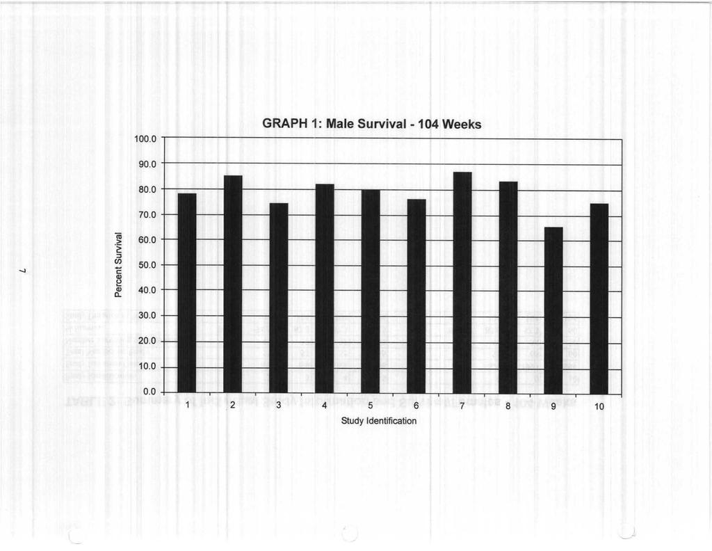
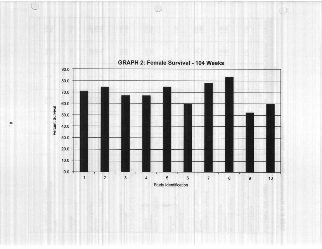
4 DATA SETS PRESENTED: Survival data are presented by study as the actual number surviving to terminal sacrifice and as percent survival at terminal sacrifice (Tables 1 and 2). The survival data are also presented in graphic form (Graphs 1 & 2) The overall incidences of all neoplastic lesions observed in any organ are reported and are summarized in Tables 3 and 4. These data also include neoplastic lesions from rats that died or were found moribund and killed prior to terminal sacrifice, but not from rats that were killed for an interim sacrifice. Due to the apparent diversity in terminology and the variability among studies in the incidence of particular lesions, the individual study incidences of lesions in selected organs/systems are also presented (Tables 5 and 6). These organs/systems include liver; kidney; pituitary; thyroid; adrenal; pancreas; ovary; uterus; cervix; vagina; mammary gland; testes; skin/subcutis; thymus; lymph nodes; and hemolymphoreticular system. SUMMARY TABLE CALCULATIONS FOR NEOPLASTIC LESIONS: The following is a description of how each of the parameters in the tables was calculated. Number of Studies (# Studies) This is the number of studies in which a particular tissue/organ was examined. In this presentation, the number of studies is 10 for males and 10 for females. Total Number of Organs (Total # Organs) This number represents the sum of the total number of tissues or organs examined in all control groups from all studies combined. Widespread tumors that showed involvement of multiple organs were listed on the basis of total number of animals examined. Occasionally a tumor would be noticed in a tissue not designated for histological examination by the study protocol. In these instances, the tumor incidence was based on the total number of animals examined as any such tumor or lesion would have been noticed on gross examination of the animal. Autolysis of tissues did not routinely exclude tissues from diagnosis. Some laboratories presented data separately for different regions within an organ (i.e., duodenum, jejunum and ileum) while most presented data by the organ (i.e., small intestine). When data were presented separately by organ region, they were grouped under the organ and calculations were based on the number of organs examined. Total Number of Lesions (# Lesions) This represents the total number of occurrences of this lesion in the specified organ in all studies examined. Percent of Total These values represent the percent incidence of a particular lesion/diagnosis in the total number (all studies combined) of a particular organ examined. These values were calculated by dividing the total number of lesions by the total number of organs/animals examined and multiplying by 100 to express the values as a percent. Values are expressed to the second decimal place. Some caution is indicated in using this number, since not all pathologists or institutions will include all diagnoses in their lexicon. Number of Studies Using This Diagnosis This is the number of studies in which a particular diagnosis was reported. This number may be useful in interpreting the overall incidence (percent of total) of a particular diagnosis, see above. 2
5 Minimum and Maximum Percent Found (Minimum and Maximum % Found) The range reported is the lowest and highest percent incidence for each lesion from the studies where the diagnosis was made. Therefore, if a study did not include a particular diagnosis, it was excluded from these calculations. The minimum and maximum percent found values should be considered in conjunction with the Number of Studies Using the Diagnosis. The individual study percentages, Minimum % Found and Maximum % Found, were calculated by dividing the number of times each diagnosis was made by the total number of organs examined in each study and then multiplying the resultant value by 100 to express it as a percent. Values are expressed to the second decimal place. SYNONYMS FOR NEOPLASTIC LESIONS: Synonymous terms or diagnoses were frequently encountered in different studies, and were combined under a single, often broad diagnosis, which was considered to be the primary diagnosis, shown below in CAPITAL LETTERS. Although some effort was made to use currently acceptable terms, it is beyond the scope of this publication to propose a system of preferred diagnoses. A current trend in toxicologic pathology is to simplify tumor classification (i.e., "lumping" as opposed to "splitting") and the categories of neoplasms used in this publication are considered to be consistent with that trend. The synonyms which were included in the various diagnoses are presented in the synonym list which follows. Where possible, terminology is consistent with the classification system proposed by the Society of Toxicologic Pathologists. Stomach: NONGLANDULAR MUCOSAISQUAMOUS CELL PAPILLOMA = papilloma; non-glandular mucosa papilloma; squamous cell papilloma NONGLANDULAR MUCOSA, CARCINOMA = squamous cell carcinoma Liver: BILE DUCT ADENOMA = cholangioma Uterus: ENDOMETRIAL STROMAL POLYP = polyp ENDOMETRIUM, ADENOCARCINOMA = adenocarcinoma; endometrium, carcinoma ENDOMETRIAL STROMAL SARCOMA = sarcoma Skin: BASAL CELL CARCINOMA = malignant basal cell tumor Adrenal: CORTEX, CARCINOMA= cortex, adenocarcinoma PHEOCHROMOCYTOMA, BENIGN= medulla neoplasm, benign PHEOCHROMOCYTOMA, MALIGNANT= medulla neoplasm, malignant Pituitary: ADENOMA = adenoma anterior lobe; adenoma pars distalis CARCINOMA = carcinoma pars distalis; adenocarcinoma; adenocarcinoma pars distalis Thyroid: FOLLICULAR CELL CARCINOMA = follicular cell adenocarcinoma Body: WHOLE BODY/MULTIPLE ORGAN = primary site undetermined 3
6 ACKNOWLEDGEMENTS: Our thanks to Joe Frank, Patricia Turck, Daniel Potenta and all of the contributing laboratories without whose help this publication would not have been possible. REFERENCES: 1. Haseman, J.K., Winbush, J.S., and O'Donnell, M.W. (1986) Use of control groups to estimate false positive rates in laboratory animal carcinogenicity studies. Fundam. Appl. Toxicol. 7: Maronpot, R.R., Montgomery, C.A. Jr., Boorman, G.A., and McConnell, E.E. (1986). National toxicology program nomenclature for hepatoproliferative lesions of rats. Toxicol. Pathol. 14:
7 TABLE 1: Summary of Individual Study Information and Survival/Males -104 Weeks Study Identification Study Initiation Date n/a n/a n/a n/a n/a n/a n/a n/a Total Number on Study Number Surviving to Termination % Survival Study Duration in Weeks
8 TABLE 2: Summary of Individual Study Information and Survival/Females -104 Weeks Study Identification Study Initiation Date n/a n/a n/a n/a n/a n/a n/a n/a Total Number on Study Number Surviving to Termination % Survival Study Duration in Weeks
9
10
11 TABLE 3: Neoplasms/Males -104 Weeks #STUDIES #ORGANS PERCENT USING THIS MINIMUM MAXIMUM LOCATION AND TUMOR #STUDIES #LESIONS OF DIAGNOSIS % FOUND % FOUND ORAL CAVITY Squamous Cell Carcinoma (Tongue) SALIVARY GLAND Adenoma Adenocarcinoma ABDOMINAL CAVITY Lipoma Mesothelioma, Malignant STOMACH Nonglandular Mucosa/Squamous Cell Papilloma SMALL INTESTINE Carcinoma Sarcoma LARGE INTESTINE/CECUM/ANUS Leiomyoma Leiomyosarcoma LIVER Hepatocellular Adenoma Sarcoma Bile Duct Sarcoma NASAL CAVITY Adenoma LUNG Bronchioloalveolar Carcinoma Squamous Cell Carcinoma
12 TABLE 3: Neoplasms/Males (cont'd.) #STUDIES #ORGANS PERCENT USING THIS MINIMUM MAXIMUM LOCATION AND TUMOR #STUDIES #LESIONS OF DIAGNOSIS % FOUND % FOUND KIDNEY Hemangiosarcoma Lipoma Liposarcoma Tubular Cell Adenoma Tubular Cell Carcinoma TESTIS Interstitial Cell Adenoma Mesothelioma, Malignant Seminoma SEMINAL VESICLE Adenocarcinoma PROSTATE Adenoma _ Adenocarcinoma EPIDIDYMIS Mesothelioma, Malignant SKIN/SUBCUTIS Basal Cell Tumor, Benign Basal Cell Carcinoma Chrondrosarcoma Fibroma Fibrosarcoma Hemangioma Hemangiosarcoma Keratoacanthoma Lipoma Myxoma Pilomatrixoma Sebaceous Cell Adenoma Sebaceous Cell Carcinoma Schwannoma, Malignant Squamous Cell Papilloma
13 TABLE 3: Neoplasms/Males (cont'd.) #STUDIES #ORGANS PERCENT USING THIS MINIMUM MAXIMUM LOCATION AND TUMOR #STUDIES #LESIONS OF DIAGNOSIS % FOUND % FOUND MAMMARY GLAND Fibroadenoma ADRENAL Cortex Adenoma Cortex Carcinoma Pheochromocytoma, Benign Pheochromocytoma, Malignant PANCREAS Adenoma Islet Cell, Adenoma Islet Cell, Adenocarcinoma Acinar Islet Cell, Adenoma PITUITARY Adenoma Carcinoma THYROID C-Cell Adenoma C-Cell Carcinoma Follicular Cell Adenoma Follicular Cell Carcinoma PARATHYROID Adenoma BRAIN Granular Cell Meningioma, Benign Granular Cell Meningioma, Malignant _ Astrocytoma, Malignant Oligodendrocytic Glioma MUSCLE Chondroma Hemangiosarcoma
14 TABLE 3: Neoplasms/Males (cont'd.) #STUDIES #ORGANS PERCENT USING THIS MINIMUM MAXIMUM LOCATION AND TUMOR #STUDIES #LESIONS OF DIAGNOSIS % FOUND % FOUND BONE Osteosarcoma HEART Endocardial Schwannoma, Malignant SPLEEN Hemangioma THYMUS Adenoma Carcinoma Thymoma, Benign Thymoma, Mixed LYMPH NODES (ALL) Hemangioma Hemangiosarcoma WHOLE BODY/MULTIPLE ORGAN Mesothelioma, Malignant Lymphoma Histiocytic Sarcoma EYE Schwannoma ADIPOSE TISSUE Anaplastic Carcinoma FOOT/LEG Hemangiosarcoma HARDERIAN GLAND Carcinoma
15 TABLE 3: Neoplasms/Males (cont'd.) #STUDIES #ORGANS PERCENT USING THIS MINIMUM MAXIMUM LOCATION AND TUMOR #STUDIES #LESIONS OF DIAGNOSIS % FOUND % FOUND HEMOLYMPHORETICULAR SYSTEM Granulocytic Leukemia, Malignant Hemangiosarcoma Histiocytic Sarcoma Lymphocytic Lymphoma, Malignant Lymphosarcoma Mononuclear Cell Leukemia Pleomorphic Lymphoma, Malignant TAIL Squamous Cell Papilloma THORACIC CAVITY Schwannoma, Malignant ZYMBAL'S GLAND Carcinoma
16 TABLE 4: Neoplasms/Females -104 Weeks #STUDIES #ORGANS PERCENT USING THIS MINIMUM MAXIMUM LOCATION AND TUMOR #STUDIES #LESIONS OF DIAGNOSIS % FOUND % FOUND ORAL CAVITY Squamous Cell Carcinoma Tongue - Papilloma Tongue - Granular Cell Tumor SALIVARY GLAND Adenocarcinoma ABDOMINAL CAVITY Mesothelioma, Malignant MESENTERY Lipoma STOMACH Nonglandular Mucosa/Squamous Cell Papilloma SMALL INTESTINE Fibroma Leiomyoma Leiomyosarcoma Adenocarcinoma LIVER Hepatocellular Adenoma Hepatocellular Adenocarcinoma Bile Duct Adenoma OVARY Mixed Sex Cord Stromal Tumor, Benign Granulosa/Thecal Cell Tumor Tubulo-Stromal Adenoma Papillary Adenoma Thecoma
17 TABLE 4: Neoplasms/Females (cont'd.) #STUDIES #ORGANS PERCENT USING THIS MINIMUM MAXIMUM LOCATION AND TUMOR #STUDIES #LESIONS OF DIAGNOSIS % FOUND % FOUND UTERUS Adenoma Adenocarcinoma Deciduoma Endometrial Polyp Hemangioma Hemangiosarcoma Endometrial Stromal Polyp Endometrial Stromal Sarcoma Squamous Cell Carcinoma CERVIX _ Polyp, Benign Granular Cell Tumor, Benign Schwannoma, Benign Stromal Cell Sarcoma Adenocarcinoma VAGINA Leiomyosarcoma Stromal Polyp Granular Cell Tumor Squamous Cell Carcinoma CLITORAL GLAND Carcinoma Schwannoma, Benign SKIN/SUBCUTIS Basal Cell Tumor, Benign Fibroma Fibrosarcoma Keratoacanthoma Lipoma Myxoma Sebaceous Cell Adenoma Squamous Cell Carcinoma Papilloma
18 TABLE 4: Neoplasms/Females (cont'd.) #STUDIES #ORGANS PERCENT USING THIS MINIMUM MAXIMUM LOCATION AND TUMOR #STUDIES #LESIONS OF DIAGNOSIS % FOUND % FOUND MAMMARY GLAND Adenoma Adenocarcinoma Fibroadenoma ADRENAL Cortex Adenoma Cortex Adenocarcinoma Pheochromocytoma, Benign Pheochromocytoma, Malignant PANCREAS Islet Cell Adenoma Islet Cell Adenocarcinoma Acinar-Islet Cell Adenoma Ductular Adenocarcinoma PITUITARY Adenoma Adenocarcinoma THYROID C-Cell Adenoma C-Cell Carcinoma Follicular Cell Adenoma Follicular Cell Carcinoma PARATHYROID Adenoma BRAIN Granular Cell Meningioma, Benign SPINAL CORD Granular Cell Tumor MUSCLE Granular Cell Tumor, Benign Hemangiosarcoma _ 1.82 _
19 TABLE 4: Neoplasms/Females (cont'd.) #STUDIES #ORGANS PERCENT USING THIS MINIMUM MAXIMUM LOCATION AND TUMOR #STUDIES #LESIONS OF DIAGNOSIS % FOUND % FOUND HEART Liposarcoma, Mediastinum SPLEEN Hemangioma Histiocytic Sarcoma THYMUS Thymoma, Benign Thymoma, Malignant LYMPH NODES Hemangioma Hemangiosarcoma WHOLE BODY/MULTIPLE ORGAN Mesothelioma EAR Granular Cell Tumor, Benign BONE MARROW Hemangioma HEMOLYMPHORETICULAR SYSTEM Malignant Lymphocytic Lymphoma Histiocytic Sarcoma ZYMBAL GLAND Carcinoma
20 TABLE 5: Incidence of Neoplasms by Study for Selected Organs/Males Study Identification LIVER Hepatocellular Adenoma Sarcoma 1 Bile Duct Sarcoma 1 KIDNEY Hemangiosarcoma 1 Lipoma 1 Liposarcoma 1 Tubular Cell Adenoma 1 1 Tubular Cell Carcinoma 1 1 TESTIS Interstitial Cell Adenoma Mesothelioma, Malignant 2 Seminoma 1 SKIN/SUBCUTIS 55 55, Basal Cell Tumor, Benign Basal Cell Carcinoma 2 Chrondrosarcoma 1 Fibroma Fibrosarcoma 2 Hemangioma 1 Hemangiosarcoma 1 Keratoacanthoma Lipoma Myxoma 1 1 Pilomatrixoma 2
21 TABLE 5: Incidence of Neoplasms by Study for Selected Organs/Males (cont'd) Study Identification Sebaceous Cell Adenoma 2 Sebaceous Cell Carcinoma 1 Schwannoma, Malignant Squamous Cell Papilloma ADRENAL Cortex Adenoma Cortex Carcinoma Pheochromocytoma, Benign Pheochromocytoma, Malignant 1 1 PANCREAS Adenoma 3 Islet Cell, Adenoma Islet Cell, Adenocarcinoma 1 1 Acinar Islet Cell, Adenoma PITUITARY Adenoma Carcinoma 1 2 THYROID C-Cell Adenoma C-Cell Carcinoma Follicular Cell Adenoma Follicular Cell Carcinoma
22 TABLE 5: Incidence of Neoplasms by Study for Selected Organs/Males (cont'd Study Identification THYMUS Adenoma 1 Carcinoma 1 Thymoma, Benign Thymoma, Mixed 1 LYMPH NODES (ALL) Hemangioma Hemangiosarcoma HEMOLYMPHORETICULAR SYSTEM Granulocytic Leukemia, Malignant 1 1 Hemangiosarcoma 3 Histiocytic Sarcoma 1 2 Lymphocytic Lymphoma, Malignant Lymphosarcoma 3 Mononuclear cell Leukemia 1 Pleomorphic Lymphoma, Malignant 1 1
23 TABLE 6: Incidence of Neoplasms by Study for Selected Organs/Females Study Identification LIVER Hepatocellular Adenoma 1 1 Hepatocellular Adenocarcinoma 1 Bile Duct Adenoma 1 OVARY Mixed Sex Cord Stromal Tumor, Benign 1 1 Granulosa/Thecal Cell Tumor 1 1 Tubulo-Stromal Adenoma Papillary Adenoma 1 Thecoma 1 UTERUS Adenoma Adenocarcinoma Deciduoma 1 Endometrial Polyp Hemangioma 1 1 Hemangiosarcoma 2 1 Endometrial Stromal Polyp Endometrial Stromal Sarcoma Squamous Cell Carcinoma 1 CERVIX Polyp, Benign 1 1 Granular Cell Tumor, Benign Schwannoma, Benign 1 Stromal Cell Sarcoma 1 Adenocarcinoma 1
24 TABLE 6: Incidence of Neoplasms by Study for Selected Organs/Females (cont'd) Study Identification VAGINA Leiomyosarcoma 1 Stromal Polyp 1 Granular Cell Tumor 6 Squamous Cell Carcinoma 1 SKIN/SUBCUTIS Basal Cell Tumor, Benign Fibroma Fibrosarcoma 1 Keratoacanthoma 2 Lipoma 1 Myxoma 1 Sebaceous Cell Adenoma 1 1 Squamous Cell Carcinoma 1 1 Papilloma 1 MAMMARY GLAND Adenoma Adenocarcinoma Fibroadenoma ADRENAL Cortex Adenoma Cortex Adenocarcinoma 1 Pheochromocytoma, Benign Pheochromocytoma, Malignant
25 TABLE 6: Incidence of Neoplasms by Study for Selected Organs/Females (cont'd) Study Identification PANCREAS Islet Cell Adenoma Islet Cell Adenocarcinoma 1 Acinar-Islet Cell Adenoma 1 Ductular Adenocarcinoma 1 PITUITARY Adenoma Adenocarcinoma THYROID C-Cell Adenoma C-Cell Carcinoma 1 1 Follicular Cell Adenoma Follicular Cell Carcinoma 1 2 THYMUS Thymoma, Benign Thymoma, Malignant 1 1 HEMOLYMPHORETICULAR SYSTEM Malignant Lymphocytic Lymphoma Histiocytic Sarcoma
Spontaneous Neoplastic Lesions in the Crl:CD-1 (ICR)BR Mouse. March, 2000
BR Mouse. March, 2000") Spontaneous Neoplastic Lesions in the Crl:CD-1 (ICR)BR Mouse March, 2000 Information Prepared by Mary L. A. Giknis, Ph.D. Charles B. Clifford, D.V.M., Ph.D. CHARLES RIVER LABORATORIES TABLE OF CONTENTS
Spontaneous Neoplastic Lesions in the Crl:CD-1 (ICR)BR Mouse March, 2000 Information Prepared by Mary L. A. Giknis, Ph.D. Charles B. Clifford, D.V.M., Ph.D. CHARLES RIVER LABORATORIES TABLE OF CONTENTS
Spontaneous Neoplastic Lesions in the CrI:CD-1(ICR) Mouse in Control Groups from 18 Month to 2 year Studies. March, 2005
 Mouse in Control Groups from 18 Month to 2 year Studies. March, 2005") Spontaneous Neoplastic Lesions in the CrI:CD-1(ICR) Mouse in Control Groups from 18 Month to 2 year Studies March, 2005 Information Prepared by Mary L.A. Giknis Ph.D Charles B. Clifford D.V.M, Ph.D 1063
Spontaneous Neoplastic Lesions in the CrI:CD-1(ICR) Mouse in Control Groups from 18 Month to 2 year Studies March, 2005 Information Prepared by Mary L.A. Giknis Ph.D Charles B. Clifford D.V.M, Ph.D 1063
Spontaneous Neoplastic Lesions in the CrI:CD-I (ICR)BR Mouse. March, 2000
BR Mouse. March, 2000") Spontaneous Neoplastic Lesions in the CrI:CD-I (ICR)BR Mouse March, 000 Information Prepared by Mary L. A. Giknis, Ph.D. Charles B. Clifford, D.V.M., Ph. D. CHARLES RIVER LABORATORIES I Dewayne Johnson
Spontaneous Neoplastic Lesions in the CrI:CD-I (ICR)BR Mouse March, 000 Information Prepared by Mary L. A. Giknis, Ph.D. Charles B. Clifford, D.V.M., Ph. D. CHARLES RIVER LABORATORIES I Dewayne Johnson
FORUM Spontaneous Neoplasms in Control Wistar Rats: A Comparison of Reviews
 TOHCOLOGICAL SCIENCES, - (99) ARTICLE NO. TX99 FORUM Spontaneous Neoplasms in Control Wistar Rats: A Comparison of Reviews James Poteracki and Kathleen M. Walsh Department of Pathology and Experimental
THE EFFECT OF DIFFERENT TUMOR GROUPINGS ON FINDINGS OF ANTICARCINOGENIC RESPONSES IN LONG-TERM RODENT BIOASSAYS. Harvard University
 THE EFFECT OF DIFFERENT TUMOR GROUPINGS ON FINDINGS OF ANTICARCINOGENIC RESPONSES IN LONG-TERM RODENT BIOASSAYS George M. Gray #0, Henry Huang +, Igor Linkov *, Michael Polkanov +, and Richard Wilson +
Summary of Inhalation Carcinogenicity Study. of Isopropyl Acetate. in F344 Rats
 Summary of Inhalation Carcinogenicity Study of Isopropyl Acetate in F344 Rats March 2009 Japan Bioassay Research Center Japan Industrial Safety and Health Association PREFACE The tests were contracted
Spontaneous Neoplastic Lesions in Aged Sprague-Dawley Rats
 Exp. Anim. 50(2), 99 103, 2001 Spontaneous Neoplastic Lesions in Aged Sprague-Dawley Rats Motokuni NAKAZAWA, Takeharu TAWARATANI, Hiroshi UCHIMOTO, Akitaka KAWAMINAMI, Makoto UEDA, Akiko UEDA, Yasuhiko
SHN-1 Human Digestive Panel Test results
 SHN-1 Human Digestive Panel Test results HN-30 tongue HN-24 salivary gland HN-12 larynx HN-28 esophagus HN-29 stomach HN-20 pancreas HN-13 liver HN-14 gall bladder HN-27-1 duodenum HN-27-2 ileum HN-27-3
For more information about how to cite these materials visit
 Author(s): Gerald Abrams, M.D., 2009 License: Unless otherwise noted, this material is made available under the terms of the Creative Commons Attribution Non-commercial Share Alike 3.0 License: http://creativecommons.org/licenses/by-nc-sa/3.0/
The stability of historical control data for common neoplasms in laboratory rats and the implications for carcinogenic risk assessment
 Regulatory Toxicology and Pharmacology xxx (2004) xxx xxx Regulatory Toxicology and Pharmacology www.elsevier.com/locate/yrtph The stability of historical control data for common neoplasms in laboratory
Table of Contents. Preface xi. Acknowledgments xiii. Part I Overview of the Diagnostic Process 1. 1 Overview of Grading and Staging 3
 Table of Contents Preface xi Acknowledgments xiii Part I Overview of the Diagnostic Process 1 1 Overview of Grading and Staging 3 Identification of the process 3 Identification of tumor types 5 Grading
Summary of Feed Carcinogenicity Study. of Diphenylamine. in F344 Rats
 Summary of Feed Carcinogenicity Study of Diphenylamine in F344 Rats August 2011 Japan Bioassay Research Center Japan Industrial Safety and Health Association PREFACE The tests were contracted and supported
of 3-Aminophenol in B6D2F1 Mice
 Summary of Drinking Water Carcinogenicity Study of 3-Aminophenol in B6D2F1 Mice July 2012 Japan Bioassay Research Center Japan Industrial Safety and Health Association PREFACE The tests were contracted
Glyphosate Cancer Risks and Failures of the Pesticide Regulatory Process
 Glyphosate Cancer Risks and Failures of the Pesticide Regulatory Process Christopher J. Portier, Ph.D. 11 October, 2017 Brussels, Belgium Disclosures The opinions expressed here and the analyses done to
incidence rate x 100,000/year
 Tier R=rare C=common Cancer Entity European crude and age adjusted incidence by cancer, years of diagnosis 2000 and 2007 Analisys based on 83 population-based cancer registries * applying the European
Tumour Structure and Nomenclature. Paul Edwards. Department of Pathology and Cancer Research UK Cambridge Institute, University of Cambridge
 Tumour Structure and Nomenclature Paul Edwards Department of Pathology and Cancer Research UK Cambridge Institute, University of Cambridge Malignant Metastasis Core idea of cancer Normal Cell Slightly
MT09 - Normal Human Tissue Microarray, FDA
 Reveal Biosciences offers Histochemical Staining, Immunohistochemistry (IHC), In Situ Hybridization (ISH), Whole Slide Imaging, and Quantitative Image Analysis on any TMA MT09 - Normal Human Tissue Microarray,
IMPC phenotyping SOPs in JMC
 IMPC phenotyping SOPs in JMC Tissue Embedding and Block Banking IMPC_BLK_001 Purpose Collect and fix a standard list of tissues from the complete necropsy (see IMPC Gross Pathology & Tissue Collection
The Effect of Different Tumor Groupings on Findings of Anticarcinogenic Responses in Long-Term Rodent Bioassays
 Regulatory Toxicology and Pharmacology 36, 139 148 (2002) doi:10.1006/rtph.2001.1524 The Effect of Different Tumor Groupings on Findings of Anticarcinogenic Responses in Long-Term Rodent Bioassays George
CODING TUMOUR MORPHOLOGY. Otto Visser
 CODING TUMOUR MORPHOLOGY Otto Visser INTRODUCTION The morphology describes the tissue of the tumour closest to normal tissue Well differentiated tumours are closest to normal Undifferentiated tumours show
Cancer Association of South Africa (CANSA)
") Cancer Association of South Africa (CANSA) Fact Sheet on ICD-10 Coding of Neoplasms Introduction The International Statistical Classification of Diseases and Related Health Problems, 10 th Revision (ICD-10)
Cancer Association of South Africa (CANSA) Fact Sheet on ICD-10 Coding of Neoplasms Introduction The International Statistical Classification of Diseases and Related Health Problems, 10 th Revision (ICD-10)
WLH Tumor Frequencies between cohort enrollment and 31-Dec Below the Women Lifestyle and Health tumor frequencies are tabulated according to:
 DESCRIPTION Below the Women Lifestyle and Health tumor frequencies are tabulated according to: Benign =171 (Cervix uteri) treated as not recorded =191 (non-melanoma skin cancer) treated as not recorded
Learning Outcomes: The following list provides the learning objectives that will be covered in the lectures, and tutorials of each week:
 Course Code Course Title ECTS Credits MED-309 Pathology II 6 School Semester Prerequisites Medical School Spring (Semester 6) MED-304 Pathology I Type of Course Field Language of Instruction Required Medicine
Neoplasia literally means "new growth.
 NEOPLASIA Neoplasia literally means "new growth. A neoplasm, defined as "an abnormal mass of tissue the growth of which exceeds and is uncoordinated with that of the normal tissues and persists in the
END-SEMESTER EXAM 2018 ANATOMY, HISTOLOGY AND EMBRYOLOGY FACULTY OF MEDICINE, 2 ND SEMESTER
 University of Szeged, Faculty of Medicine Department of Anatomy, Histology and Embryology Chairman: Prof. Antal Nógrádi MD, PhD, DSc Kossuth L. sgt. 40., H-6724 Szeged, Hungary Tel.: +36-62-545-665 P.
E. Staging a WHO (World Health Organization) b TNM (Tumor Node Metastasis)
 b TNM (Tumor Node Metastasis)") I KNOWLEDGE LIST ONCOLOGY Knowledge of neoplasms in animals (dog, cat, pocket pets, horses, cows) A. Clinical signs, History, Physical Exam (PE) B. Work up 1. Diagnostic Imaging a Bone Scan b Thyroid Scan
I KNOWLEDGE LIST ONCOLOGY Knowledge of neoplasms in animals (dog, cat, pocket pets, horses, cows) A. Clinical signs, History, Physical Exam (PE) B. Work up 1. Diagnostic Imaging a Bone Scan b Thyroid Scan
Supplementary Figure 1. Genotyping strategies for Mcm3 +/+, Mcm3 +/Lox and Mcm3 +/- mice and luciferase activity in Mcm3 +/Lox mice. A.
 Supplementary Figure 1. Genotyping strategies for Mcm3 +/+, Mcm3 +/Lox and Mcm3 +/- mice and luciferase activity in Mcm3 +/Lox mice. A. Upper part, three-primer PCR strategy at the Mcm3 locus yielding
WLH Tumor Frequencies between cohort enrollment and 31-Dec Below the Women Lifestyle and Health tumor frequencies are tabulated according to:
 WLH Tumor Frequencies between cohort enrollment and 31-Dec 2012 DESCRIPTION Below the Women Lifestyle and Health tumor frequencies are tabulated according to: Benign =171 (Cervix uteri) treated as not
Abdulrahman Alhanbali. Bahaa Najjar. Maha shomaf
 14 Abdulrahman Alhanbali Bahaa Najjar Maha shomaf 1 Neoplasia In this lecture we will talk about neoplasia, its features and the nomenclature of different types of tumors. Neoplasia (neo: new and plasia:
Descriptive Histology
 Atlas of Descriptive Histology Michael H. Ross University of Florida College of Medicine Gainesville, Florida Wojciech Pawlina Mayo Medical School College of Medicine, Mayo Clinic Rochester, Minnesota
Neoplasia part I. Dr. Mohsen Dashti. Clinical Medicine & Pathology nd Lecture
 Neoplasia part I By Dr. Mohsen Dashti Clinical Medicine & Pathology 316 2 nd Lecture Lecture outline Review of structure & function. Basic definitions. Classification of neoplasms. Morphologic features.
NEOPLASIA-I CANCER. Nam Deuk Kim, Ph.D.
 NEOPLASIA-I CANCER Nam Deuk Kim, Ph.D. 1 2 Tumor in the hieroglyphics of the Edwin Smith papyrus (1,600 B.C., Breasted s translation 1930) 3 War on Cancer (National Cancer Act, 1971) 4 Cancer Acts in Korea
INTRODUCTION TO ANIMALS
 AP BIOLOGY ANIMALS ACTIVITY #1 NAME DATE HOUR INTRODUCTION TO ANIMALS LEVELS OF ORGANIZATION Animals Activity #1 page 1 HOMEOSTASIS: DEFINITION IMPORTANCE MECHANISMS FOR MAINTAINING HOMEOSTASIS: Animals
CONTINUING EDUCATION IN TOXICOLOGIC PATHOLOGY REPRODUCTIVE SYSTEM
 CONTINUING EDUCATION IN TOXICOLOGIC PATHOLOGY REPRODUCTIVE SYSTEM ORGANIZED BY SOCIETY FOR TOXICOLOGIC PATHOLOGY IN INDIA (STPI) OCTOBER 29-31, 2010 The Atria Hotel, # 1, Palace Road, Bangalore - 560 001
The European Commission s science and knowledge service. Joint Research Centre
 The European Commission s science and knowledge service Joint Research Centre Coding Primary Site and Tumour Morphology JRC-ENCR training course Copenhagen, 25 September 2018 Nadya Dimitrova Outline What
A neoplasm is defined as "an abnormal tissue proliferation, which exceeds that of adjacent normal tissue. This proliferation continues even after
 NEOPLASIA Neoplasia is a very important topic in pathology because neoplasms are both common and serious diseases. A neoplasm literally means a new growth, and this term is used interchangeably with a
FACULTY MEMBERSHIP APPLICATION Tulane Cancer Center
 FACULTY MEMBERSHIP APPLICATION Tulane Cancer Center 1430 Tulane Ave., Box SL-68, New Orleans, LA 70112-2699 J. Bennett Johnston Building, Mezzanine (Floor 1A), Suite A102 (504) 988-6060, fax (504) 988-6077,
CODING PRIMARY SITE. Nadya Dimitrova
 CODING PRIMARY SITE Nadya Dimitrova OUTLINE What is coding and why do we need it? ICD-10 and ICD-O ICD-O-3 Topography coding rules ICD-O-3 online WHAT IS CODING AND WHY DO WE NEED IT? Coding: to assign
Case 3:16-md VC Document Filed 10/28/17 Page 1 of 38 EXHIBIT 96
 Case 3:16-md-02741-VC Document 655-6 Filed 10/28/17 Page 1 of 38 EXHIBIT 96 Case 3:16-md-02741-VC Document 655-6 Filed 10/28/17 Page 2 of 38 UNITED STATES DISTRICT COURT NORTHERN DISTRICT OF CALIFORNIA
Diagnostic Cytology of Cancer Cases
 Diagnostic Cytology of Cancer Cases Somporn Techangamsuwan Companion Animal Cancer Research Unit (CAC-RU) Department of Pathology, Faculty of Veterinary Science, Chulalongkorn University 1 Tumor or Non-tumor
Conflict of Interest Statement
 Specific Aspects and Approaches for Regulatory Evaluation of Pharmaceuticals in Two-Year Rodent Carcinogenicity Studies James A. Popp Stratoxon LLC Morgantown, PA Tel: 610.286.7592 popp@stratoxon.com Conflict
155.2 Malignant neoplasm of liver not specified as primary or secondary. C22.9 Malignant neoplasm of liver, not specified as primary or secondary
 ICD-9 TO ICD-10 Reference ICD-9 150.9 Malignant neoplasm of esophagus unspecified site C15.9 Malignant neoplasm of esophagus, unspecified 151.9 Malignant neoplasm of stomach unspecified site C16.9 Malignant
ONCOLOGY. Csaba Bödör. Department of Pathology and Experimental Cancer Research november 19., ÁOK, III.
 ONCOLOGY Csaba Bödör Department of Pathology and Experimental Cancer Research 2018. november 19., ÁOK, III. bodor.csaba1@med.semmelweis-univ.hu ONCOLOGY Characteristics of Benign and Malignant Neoplasms
Appendix 4: WHO Classification of Tumours of the pancreas 17
 S3.01 The WHO histological tumour type must be recorded. CS3.01a The histological type of the tumour should be recorded based on the current WHO classification 17 (refer to Appendices 4-7). Appendix 4:
Contents. Basic Ultrasound Principles and Terminology. Ultrasound Nodule Characteristics
 Contents Basic Ultrasound Principles and Terminology Basic Ultrasound Principles... 1 Ultrasound System... 2 Linear Transducer for Superficial Images and Ultrasound-Guided FNA... 3 Scanning Planes... 4
Format Of ICD-O Terms In Numerical List Each topographic and morphologic term appears only once The first listed term in Bold Type is the Preferred Te
 Florida Cancer Data System International Classification of Diseases for Oncology ICD-O-3 1 Basic Concepts Primary Site/Topography Histology/Morphology Behavior Grade/Immunophenotype 2 ICD-O 3 Structure/Format
3. Studies of Cancer in Experimental Animals
 NEUTRONS 377 3. Studies of Cancer in Experimental Animals Neutrons have been studied in order to compare their carcinogenicity with that of low-let radiations such as X-radiation and γ-radiation, not only
Cancer Fundamentals. Julie Randolph-Habecker, Ph.D. Director, Experimental Histopathology Shared Resource
 Cancer Fundamentals Julie Randolph-Habecker, Ph.D. Director, Experimental Histopathology Shared Resource Cancer Overview Leading cause of death in US 1.2 million diagnosed each year More common after age
Summary of Inhalation Carcinogenicity Study. of Tetrachloroethylene. in BDF 1 Mice
 Summary of Inhalation Carcinogenicity Study of Tetrachloroethylene in BDF 1 Mice March 1993 Japan Bioassay Laboratory Japan Industrial Safety and Health Association PREFACE The tests were contracted and
Threshold-Based Risk Assessment is the Same for Cancer and Non-cancer Endpoints for Non-DNA Reactive Carcinogens
 Threshold-Based Risk Assessment is the Same for Cancer and Non-cancer Endpoints for Non-DNA Reactive Carcinogens Samuel M. Cohen, MD, PhD Department of Pathology & Microbiology University of Nebraska Medical
Aspartame induces lymphomas and leukaemias in rats Aspartame, a leukaemogenic compound
 Aspartame induces lymphomas and leukaemias in rats Aspartame, a leukaemogenic compound 1 European Journal of Oncology, Vol. 10, No. 2, pp. 00-00, 2005 IN PRESS Morando Soffritti, Fiorella Belpoggi, Davide
8. POST- MORTEM PROTOCOL (John Trupkiewicz)
") 8. POST- MORTEM PROTOCOL (John Trupkiewicz) 8.1. GENERAL COMMENTS Those individuals who may be charged with performing a necropsy examination should formulate a plan of action in the case of the sudden,
8. POST- MORTEM PROTOCOL (John Trupkiewicz) 8.1. GENERAL COMMENTS Those individuals who may be charged with performing a necropsy examination should formulate a plan of action in the case of the sudden,
Test Bank for Robbins and Cotran Pathologic Basis of Disease 9th Edition by Kumar
 Link full download: http://testbankair.com/download/test-bank-for-robbins-cotran-pathologic-basis-of-disease-9th-edition-bykumar-abbas-and-aster Test Bank for Robbins and Cotran Pathologic Basis of Disease
HOSPITAL-BASED CANCER REGISTRY ANNUAL REPORT 2011
 HOSPITAL-BASED CANCER REGISTRY ANNUAL REPORT 2011 SONGKLANAGARIND HOSPITAL FACULTY OF MEDICINE PRINCE OF SONGKLA UNIVERSITY HATYAI SONGKHLA THAILAND EDITOR PARADEE PRECHAWITTAYAKUL, B.Sc. June, 2013 Songklanagarind
The Utility of the Guppy (Poecilia reticulata) and Medaka (Oryzias latipes) in Evaluation of Chemicals for Carcinogenicity
 and Medaka (Oryzias latipes) in Evaluation of Chemicals for Carcinogenicity") TOXICOLOGICAL SCIENCES 92(1), 143 156 (2006) doi:10.1093/toxsci/kfj181 Advance Access publication March 31, 2006 The Utility of the Guppy (Poecilia reticulata) and Medaka (Oryzias latipes) in Evaluation
TOXICOLOGICAL SCIENCES 92(1), 143 156 (2006) doi:10.1093/toxsci/kfj181 Advance Access publication March 31, 2006 The Utility of the Guppy (Poecilia reticulata) and Medaka (Oryzias latipes) in Evaluation
Epithelial tumors. Dr. F.F. Khuzin, PhD Dr. M.O. Mavlikeev
 Epithelial tumors Dr. F.F. Khuzin, PhD Dr. M.O. Mavlikeev Epithelial tumors Tumors from the epithelium are the most frequent among tumors. There are 2 group features of these tumors: The presence in most
A301 VI- Various cancer tissues with corresponding normal tissues
 A301 VI- Various cancer tissues with (formalin fixed) For research use only Specifications: No. of cases: 28 Tissue type: Various cancer tissues with No. of spots: 2 spots from each cancer case (56 spots)
7/4/2018. Key Objectives. A and P 2401 Lecture 2 TWO MECHANISMS USED TO MAINTAIN HOMEOSTASIS. Negative Feedback Examples. Review of Homeostasis
 Key Objectives Review of Homeostasis Negative Feedback Mechanisms Positive Feedback Mechanisms Body Systems and Function A and P 2401 Lecture 2 HOMEOSTASIS TWO MECHANISMS USED TO MAINTAIN HOMEOSTASIS The
Epithelia will be discussed according to the following scheme: Type Number of layers Shape Line drawing. Squamous Cuboidal Columnar
 Epithelia Epithelia will be discussed according to the following scheme: Type Number of layers Shape Line drawing Simple Squamous Cuboidal Columnar Covering and Lining epithelium Pseudostratified Stratified
Test Bank for Robbins and Cotran Pathologic Basis of Disease 9th Edition by Kumar
 Link full download:https://getbooksolutions.com/download/test-bank-for-robbinsand-cotran-pathologic-basis-of-disease-9th-edition-by-kumar Test Bank for Robbins and Cotran Pathologic Basis of Disease 9th
Pancreas. Atrophy, acinar cell. Pathogenesis: Diagnostic key features:
 Pancreas Atrophy, acinar cell Pathogenesis: Decrease in number and/or size of acinar cells may be due to spontaneous or experimentally induced degenerative changes, apoptosis, or a sequel of chronic inflammation.
Cancer in Estonia 2014
 Cancer in Estonia 2014 Estonian Cancer Registry (ECR) is a population-based registry that collects data on all cancer cases in Estonia. More information about ECR is available at the webpage of National
BIO 137 Human Anatomy & Physiology I. Laboratory Manual. Laboratory #1: Measurements, Body Organization and Anatomical Systems
 BIO 137 Human Anatomy & Physiology I Laboratory Manual Laboratory #1: Measurements, Body Organization and Anatomical Systems Lab Exercise 1 Measurements Body Organization Body Systems What you need to
XX. Tumours of the nasal cavity *
 XX. Tumours of the nasal cavity * H. STONZI 1 & B. HAUSER2 Tumours of the nasal cavity are rare in domestic animals, most cases occurring in the dog. Epithelial tumours are the most common type in carnivores
Proceedings of the 36th World Small Animal Veterinary Congress WSAVA
 www.ivis.org Proceedings of the 36th World Small Animal Veterinary Congress WSAVA Oct. 14-17, 2011 Jeju, Korea Next Congress: Reprinted in IVIS with the permission of WSAVA http://www.ivis.org 14(Fri)
Evaluation of Ramazzini Institute Aspartame Studies and EFSA s Assessment
 Evaluation of Ramazzini Institute Aspartame Studies and EFSA s Assessment Lisa Y. Lefferts, MSPH Senior Scientist Center for Science in the Public Interest Who is CSPI and What is Our Agenda? CSPI is a
ICD-O Morphology code. R=Rare Tier Tumour ICD-O Topography code C30.0, C31
 R=Rare Tier Tumour ICD-O Topography code ICD-O Morphology code EPITHELIAL TUMOURS OF NASAL CAVITY AND SINUSES R 2 Squamous cell carcinoma with variants of nasal cavity and sinuses C30.0, C3 C30.0, C3 8000,
A103(9)- Normal tissues, more than single spots
- Normal tissues, more than single spots") A103(9)- Normal tissues, more than single spots (formalin fixed) For research use only Specifications: No. of cases: 45 Tissue type: Normal tissues, more than single spots No. of spots: 2 spots from each
A103(9)- Normal tissues, more than single spots (formalin fixed) For research use only Specifications: No. of cases: 45 Tissue type: Normal tissues, more than single spots No. of spots: 2 spots from each
ANNUAL CANCER REGISTRY REPORT-2005
 ANNUAL CANCER REGISTRY REPORT-25 CANCER STATISTICS Distribution of neoplasms Of a total of 3,115 new neoplasms diagnosed or treated at the Hospital from January 25 to December, 25, 1,473 were seen in males
REPORTABLE CASES MISSISSIPPI For cases diagnosed 10/1/2015 and after
 REPORTABLE CASES MISSISSIPPI For cases diagnosed 10/1/2015 and after The following lists are intended to assist you, as the reporter, in identifying the reportable neoplasms for your facility. Any reportable
Icd 10 code metastatic adenocarcinoma endometrial
 Icd 10 code metastatic adenocarcinoma endometrial 1-10-2017 Free, official coding info for 2018 ICD-10-CM D07.0 - includes detailed rules, notes, synonyms, ICD-9-CM conversion,. 2018 ICD-10-CM Diagnosis
Truman Medical Center-Hospital Hill Cancer Registry 2014 Statistical Summary Incidence
 Truman Medical Center-Hospital Hill Cancer Registry 2014 Statistical Summary Incidence In 2014, there were 452 new cancer cases diagnosed and or treated at Truman Medical Center- Hospital Hill and an additional
Case # nd Annual SEVPAC May 17, Kathy-Anne Clarke
 Case # 10 42 nd Annual SEVPAC May 17, 2014 Kathy-Anne Clarke Google images Babu Babu is 10 year old spayed female French Bulldog Chronic weight loss over 4 months Febrile and lethargic at the referring
Other Sites. Table 2 Continued. MPH Rules 11/8/07. NAACCR Webinar Series 1
 MPH s 11/8/07 Other s 1 Table 2 Continued Use this two-page table to select combination histology codes. Compare the terms in the diagnosis to the terms in Columns 1 and 2. If the terms match, code the
2010 Update. NAACCR Webinar Series 1 4/1/2010. Agenda. Access to 2010 Information. CSv2. Collecting Cancer Data: Soft Tissue Sarcoma
 NAACCR 2009 2010 Webinar Series Collecting Cancer Data: Soft Tissue Sarcoma, Neuroendocrine Tumors (NET) and Gastrointestinal Stromal Tumors (GIST) Agenda Updates Soft Tissue Sarcoma Overview CSv2 MP/H
A Time- and Resource-Efficient Method for Annually Auditing All Reporting Hospitals in Your State: the Inpatient & Outpatient Hospital Discharge Files
 A Time- and Resource-Efficient Method for Annually Auditing All Reporting Hospitals in Your State: the Inpatient & Outpatient Hospital Discharge Files By Dr. Martin A. Whiteside Director, Office of Cancer
List of Available TMAs in the PRN
 TMA RPCI_BrainCa01 RPCI_BrCa03 RPCI_BrCa04 RPCI_BrCa05 RPCI_BrCa0 RPCI_BrCa07 RPCI_BrCa08 RPCI_BrCa15 RPCI_BrCa1 RPCI_BrCa17 RPCI_BrCa18 RPCI_BrCa19 RPCI_BrCa20 RPCI_BrCa21 RPCI_BrCa24 RPCI_BrCa25 RPCI_BrCa2
Gastrointestinal Malignancies. Dr Rodney ITAKI Pathology Division, SMHS, UPNG Anatomical Pathology Discipline
 Gastrointestinal Malignancies Dr Rodney ITAKI Pathology Division, SMHS, UPNG Anatomical Pathology Discipline Esophagus normal anatomy Hollow tube 23-25cm long in adults Extends from pharynx to level of
2011 to 2015 New Cancer Incidence Truman Medical Center - Hospital Hill
 Number of New Cancers Truman Medical Center Hospital Hill Cancer Registry 2015 Statistical Summary Incidence In 2015, Truman Medical Center diagnosed and/or treated 406 new cancer cases. Four patients
Biology Anatomy and Physiology I. Learn and Understand. What is Biology? bios = life -ology = study of
 Biology 2331 Anatomy and Physiology I "If you want something you've never had, then you've got to do something you've never done." Learn and Understand A new language At this stage, science drives the
Radiotherapy in small animals. How is it realized? How does it work? For which patient is it indicated?
 Radiotherapy in small animals How is it realized? How does it work? For which patient is it indicated? Radiotherapy Our linear accelerator can generate photons or electrons. The electron beam is used to
FINALIZED SEER SINQ QUESTIONS
 0076 Source 1: WHO Class CNS Tumors pgs: 33 MP/H Rules/Histology--Brain and CNS: What is the histology code for a tumor originating in the cerebellum and extending into the fourth ventricle described as
Supplementary Online Content
 Supplementary Online Content Chacón MR, Enrico DH, Burton J, Waisberg FD, Videla VM. Incidence of placebo adverse events in randomized clinical trials of targeted and immunotherapy cancer drugs in the
Case year female. Routine Pap smear
 Case 1 57 year female Routine Pap smear Diagnosis? 1. Atypical glandular cells of unknown significance (AGUS) 2. Endocervical AIS 3. Endocervical adenocarcinoma 4. Endometrial adenocarcinoma 5. Adenocarcinoma
Multi-normal human tissues, FDA, 96 samples, 35 organs/sites from three individuals (1.5mm)
") ab178228 Multi-normal human tissues, FDA, 96 samples, 35 organs/sites from three individuals (1.5mm) Instructions for Use Designed for antibody cross reactivity analysis and IHC or ISH based protein or
ab178228 Multi-normal human tissues, FDA, 96 samples, 35 organs/sites from three individuals (1.5mm) Instructions for Use Designed for antibody cross reactivity analysis and IHC or ISH based protein or
Neoplasia 2018 Lecture 1. Dr Heyam Awad MD, FRCPath
 Neoplasia 2018 Lecture 1 Dr Heyam Awad MD, FRCPath Dear All Welcome to this part of your course ( introduction to pathology) where we will study neoplasia in detail. Please note that each lecture builds
Supplementary Information
 Supplementary Information Supplementary Figure 1. Western blotting with ERβ antibodies Full blots corresponding to Fig. 2, along with replicated experiments at different time points, different batches,
Carcinogens and Carcinogenesis
 Special topics in tumor biochemistry Carcinogens and Carcinogenesis Speaker: Prof. Jiunn-Jye Chuu E-Mail: jjchuu@mail.stust.edu.tw What Is Cancer? Cancer is the unregulated multiplication of specific cells
3. Studies of Cancer in Experimental Animals
 FORMALDEHYDE 171 3.1 Inhalation 3.1.1 Mouse 3. Studies of Cancer in Experimental Animals Groups of 4260 C3H mice [sex and age unspecified] were exposed to concentrations of 0, 50, 100 or 200 mg/m 3 formaldehyde
MVST BOD & NST PART IB Thurs. 2 nd & Fri. 3 rd March 2017 Pathology Practical Class 23
 MVST BOD & NST PART IB Thurs. 2 nd & Fri. 3 rd March 2017 Pathology Practical Class 23 Neoplasia I Neoplasia I: Benign and malignant neoplasms in glandular epithelium and mesenchyme 1.0. Aims 1. To understand
S Y M P O S I U M P R O G R A M
 Dear colleagues, dear friends We would like to cordially welcome you to the 10 th International Arkadi M. Rywlin International Pathology Slide Seminar in Anatomic Pathology to be held in Split, Croatia.
Human cell line list Production of Exosome Standards - Cell Lysates
 Human cell line list 2016 - Production of Exosome Standards - Cell Lysates 380 peripheral blood leukemia, pre-b cell 1301 blood leukemia, acute lymphoblastic, T cell 5637 bladder carcinoma 8305C thyroid
TUMOR,NEOPLASM. Pathology Department, Zhejiang University School of Medicine,
 TUMOR,NEOPLASM Pathology Department, Zhejiang University School of Medicine, 马丽琴,maliqin198@zju.edu.cn The points in this chapter What is a neoplasm (conception) Morphology of neoplasm Macroscopy of Neoplasm
SCOPE OF PRACTICE PGY-5
 Recognize normal cytomorphology of cells derived from the respiratory, gastrointestinal, and genitourinary tracts, and body fluid (Cerebrospinal fluid, pleural and peritoneal fluid) Recognize normal cytomorphology
21 Endocrine organs and cells
 21 Endocrine organs and cells The endocrine system consists of discrete organs, portions of organs and distributed cells that secrete hormones into surrounding tissues or structures. Objectives You should
Cellular Pathology Of Glandular Lesions And Uncommon Neoplasms Of The Cervix By Glenn McCluggage;John Tidy;John Smith READ ONLINE
 Cellular Pathology Of Glandular Lesions And Uncommon Neoplasms Of The Cervix By Glenn McCluggage;John Tidy;John Smith READ ONLINE If looking for a book by Glenn McCluggage;John Tidy;John Smith Cellular
MEDICAL POLICY Gene Expression Profiling for Cancers of Unknown Primary Site
 POLICY: PG0364 ORIGINAL EFFECTIVE: 04/22/16 LAST REVIEW: 07/26/18 MEDICAL POLICY Gene Expression Profiling for Cancers of Unknown Primary Site GUIDELINES This policy does not certify benefits or authorization
SUPPLEMENTARY INFORMATION
 Supplementary Table 1 trials currently open for patients with pheochromocytoma and/or paraganglima (from trials.gov) www.clinicaltrials.gov; Active PPGL trials Status Of Sunitinib In Patients With Recurrent
Group B: Organ systems (digestive, respiratory, urinary, genital system, heart, glands and skin) green
 green") Group B: Organ systems (digestive, respiratory, urinary, genital system, heart, glands and skin) green Digestive system 1. Teeth Main points: external and internal structure of a tooth, fixation of a tooth
Group B: Organ systems (digestive, respiratory, urinary, genital system, heart, glands and skin) green Digestive system 1. Teeth Main points: external and internal structure of a tooth, fixation of a tooth
Classification of spontaneous brain tumors in rats
 Classification of spontaneous brain tumors in rats Central Nervous System Neoplasms in the Rat (Solleveld HA, et al., 1991) 1. Tumors of Neuroepithelial Tissue A. Astrocytic and oligodendroglial tumors