Abid Irshad, MD Director Breast Imaging. Medical University of South Carolina Charleston
|
|
|
- Rosalyn Curtis
- 6 years ago
- Views:
Transcription

1 Abid Irshad, MD Director Breast Imaging Medical University of South Carolina Charleston

2 Cases
3 Financial disclosure: I or my family have no financial interest related to the material discussed in this presentation
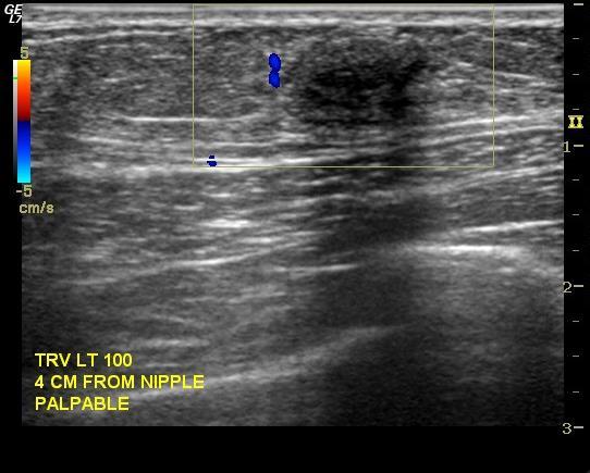
4 Case

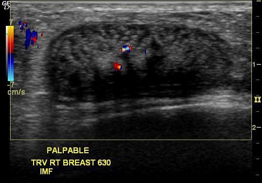
5 30 yo F with h/o recent breast reduction presents with new right breast thickening and swelling.
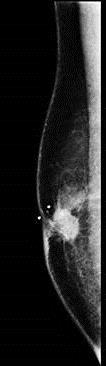
6 What is the differential diagnosis? Seroma? Abscess? Hematoma? Large cyst? Necrotic cancer? Implant?


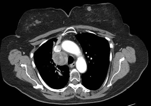
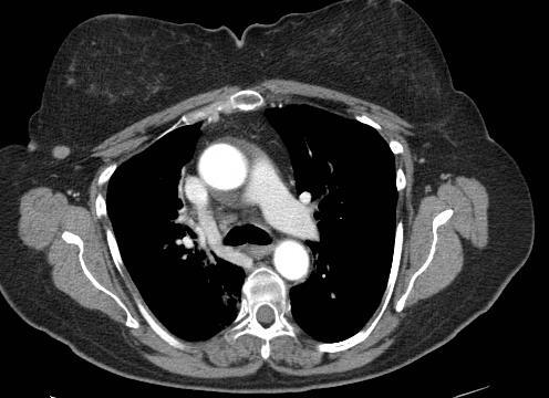
7 Fluid aspiration performed 300 cc of clear fluid aspirated

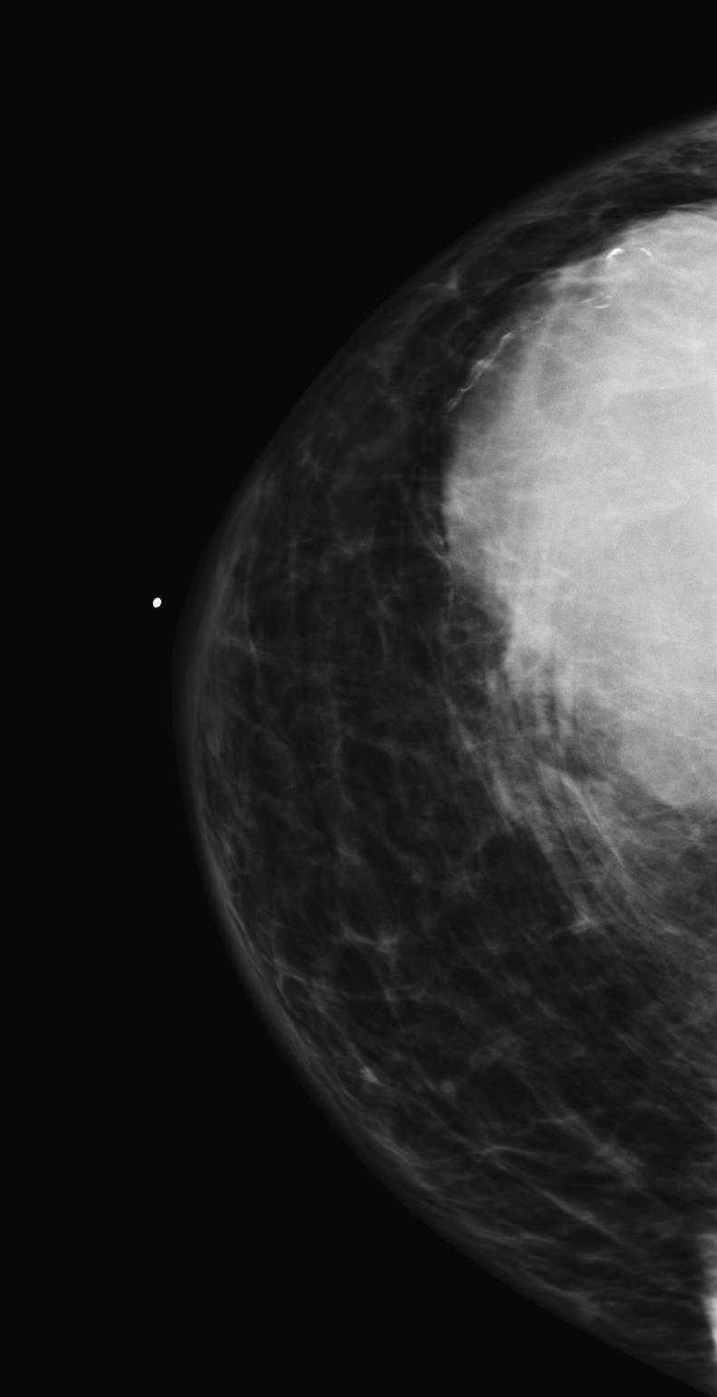
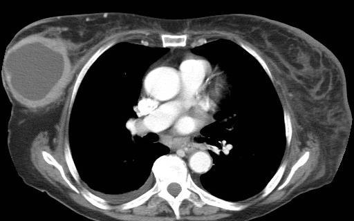
8 Recent plain chest film
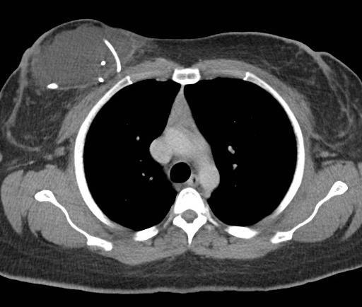
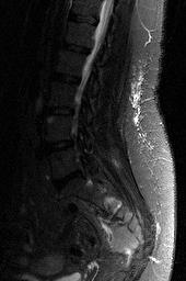
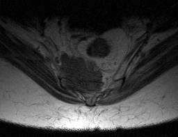
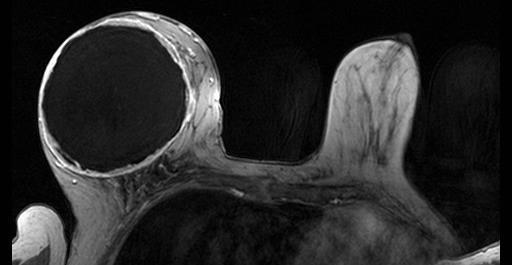
9
10 Diagnosis: CSF collection within the breast The VP shunt pulled up during the breast reduction surgery


11 Old plain films (Prior to breast reduction)
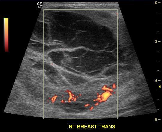
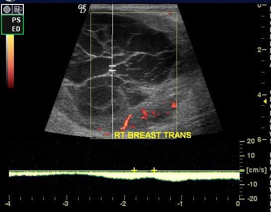
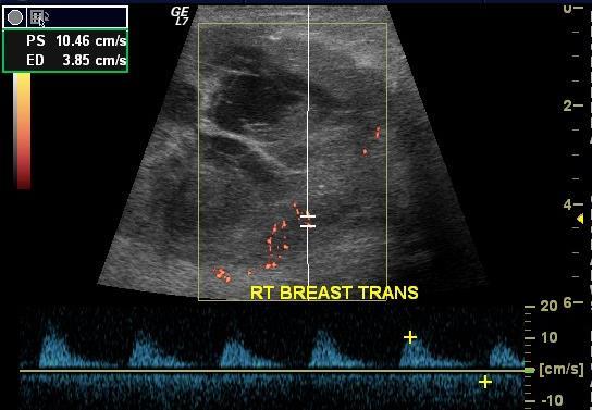
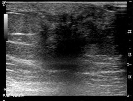
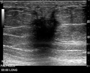
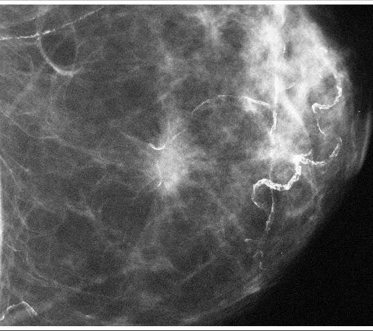
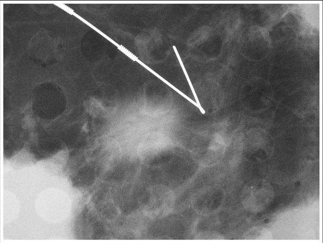
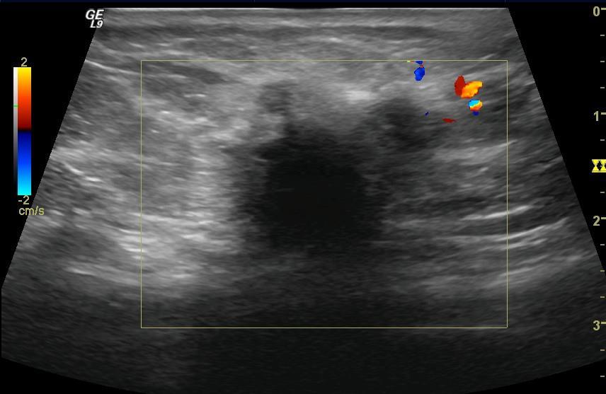
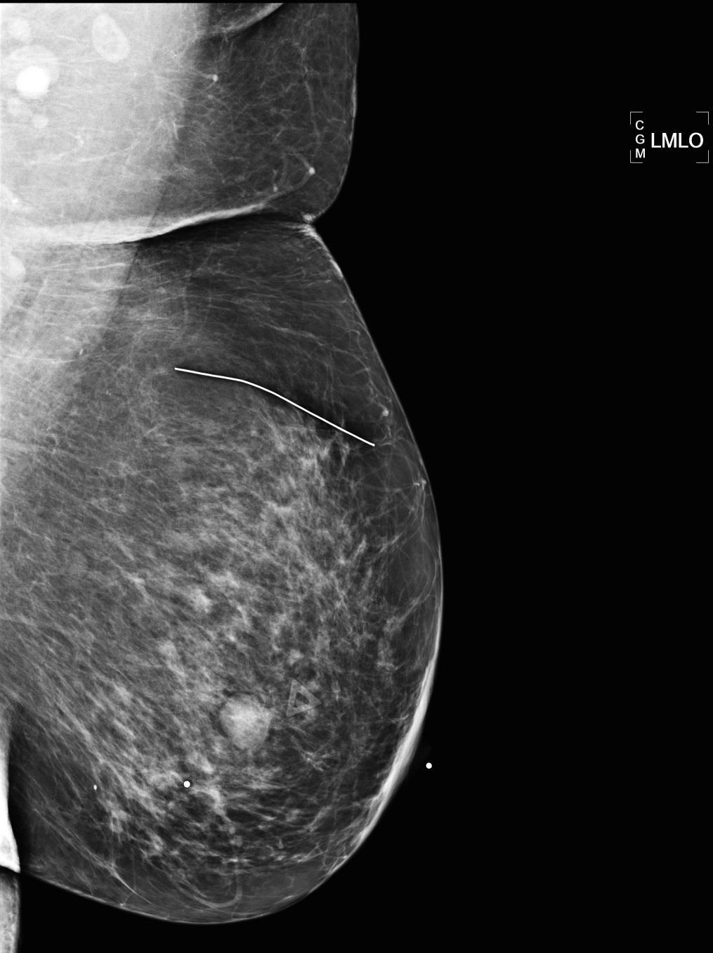
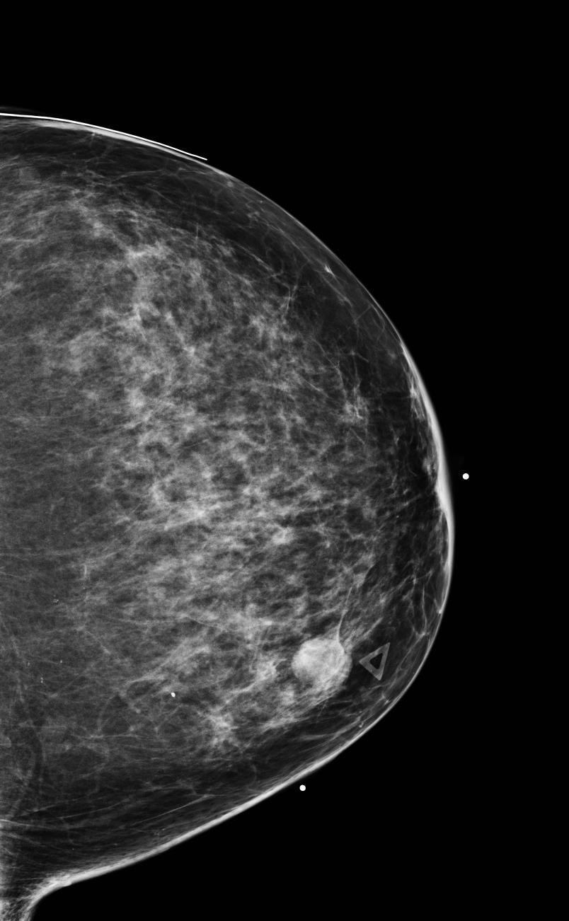
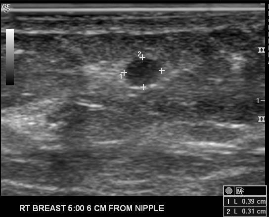
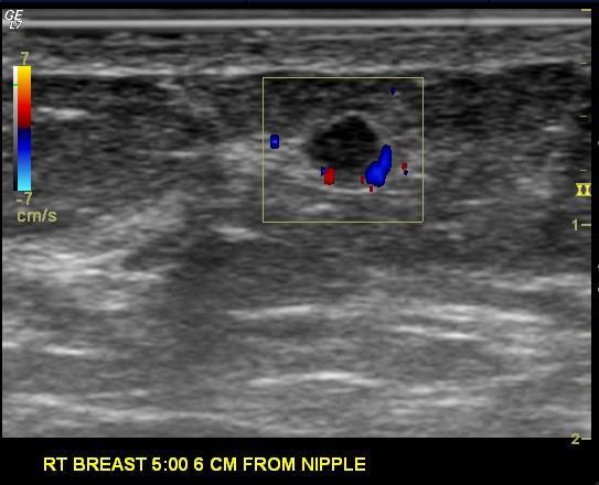
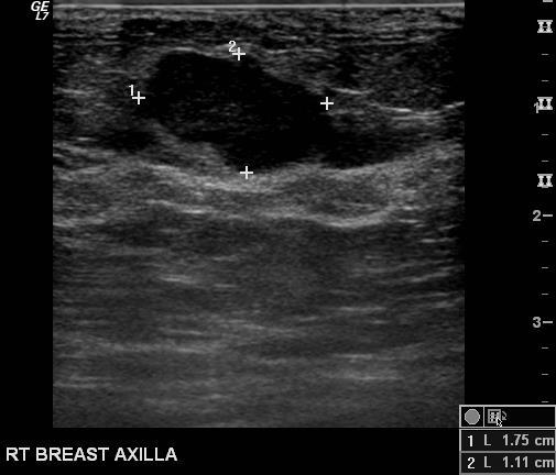
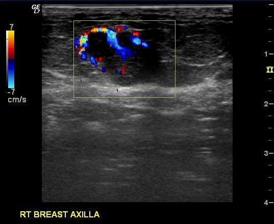
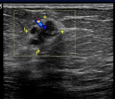
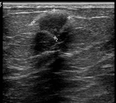
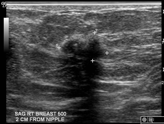
12 Case


13 81 year old female presents with right nipple inversion Old Old
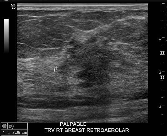
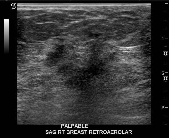
14 US retro-areolar US

15 Core needle biopsy Invasive Lobular Cancer


16 MRI Invasive lobular carcinoma


17 Patient elected mastectomy with implant reconstruction 2015 Replaced with double lumen implant 2013

18 Recently presented with hardness and warmth of the right breast.

19 US Differential diagnosis Recurrent ILC Abscess Granulation tissue Hematoma Implant leak Lymphoma

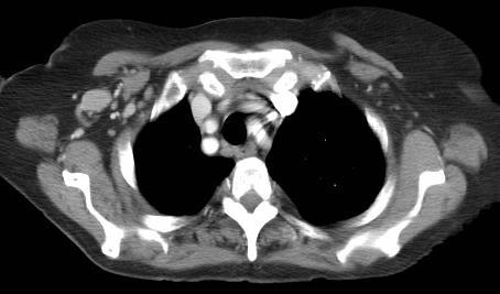
20 Core needle biopsy


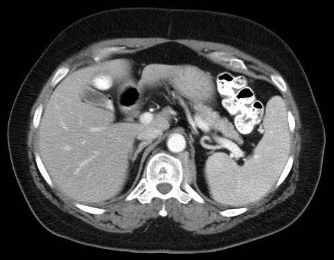
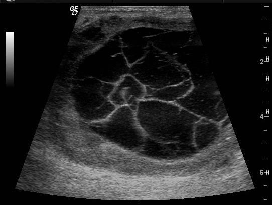
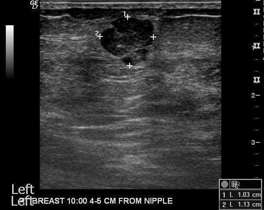
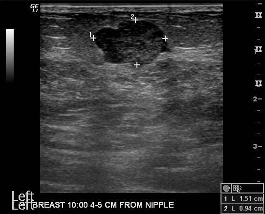
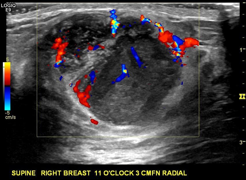
21 Why is the outer surface of mass very smooth?
22 Diagnosis: Spindle cell neoplasm / Metaplastic carcinoma
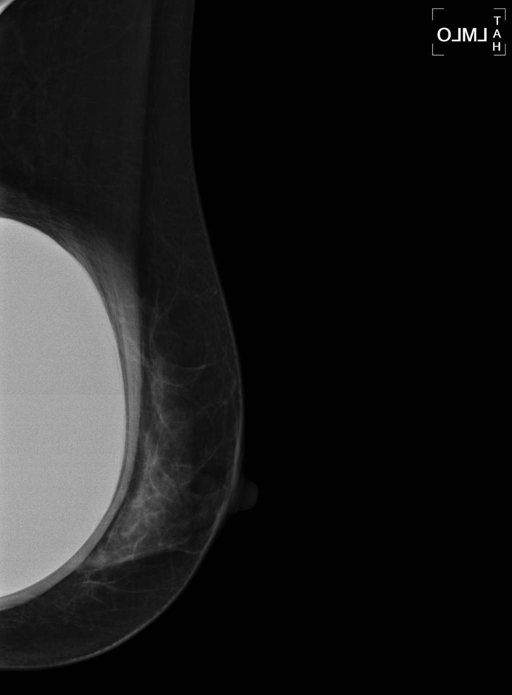
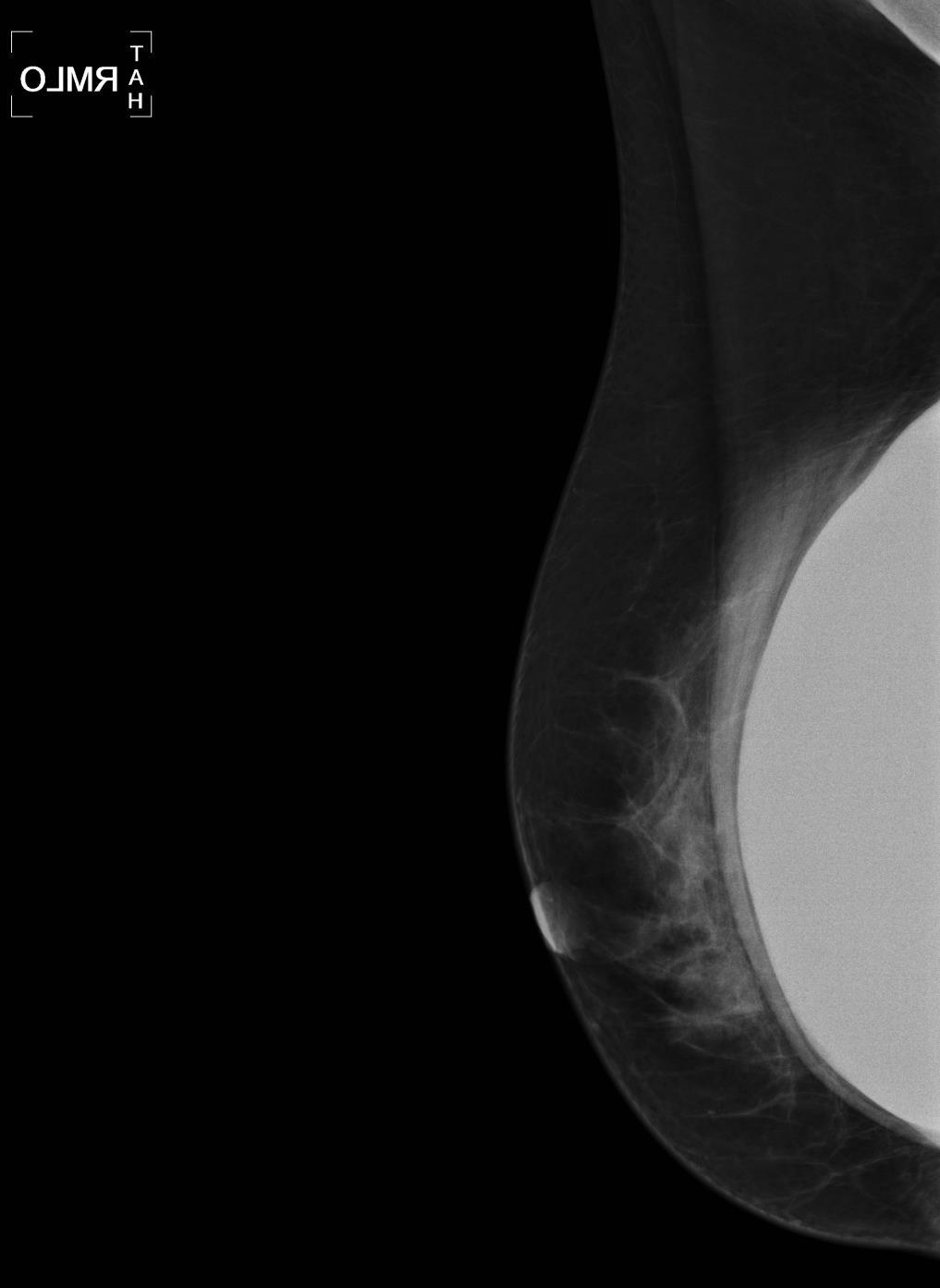
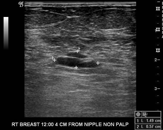
23 Wargotz and Norris proposed Classification System Metaplastic carcinoma subdivided into: Spindle cell type Squamous cell type Carcinosarcoma Matrix producing type Metaplastic carcinoma with osteoclastic giant cells
24 Metaplastic Carcinoma of the Breast Uncommon form of breast cancer (<5%) Heterogeneous group of neoplasms with glandular and non glandular components Mixed epithelial and mesenchymal differentiation Often sporadic development
25 Spindle cell neoplasm Often presents palpable mass Women often greater than 50 years old Larger tumor size Complete surgical excision necessary Variable axillary lymph node involvement reported ranging from 8% to 40% High hematogenous metastatic rather than lymphatic spread


26 Metaplastic Carcinoma- Spindle Cell Type Higher magnification: Elongated spindle cells Squamous cell nest (arrow) portrays differentiation Positive cytokeratin stain marker for epithelial differentiation (sarcoma not reactive to cytokeratin stain)
27 Case
28 History 51 year old female presents with breast masses seen on CT scan
29
30 Mammogram shows two masses with indistinct margins

31 Ultrasound shows two hypoechoic masses which are non-parallel and have indistinct margins
32 Differential Diagnosis: Multifocal cancer Mucinous carcinoma High grade IDC Medullary carcinoma Fibroadenomas Phyllodes tumor Hematomas Metastasis
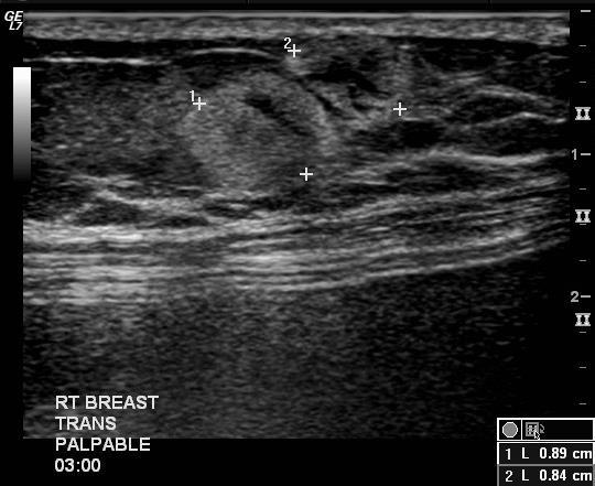
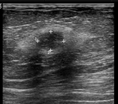
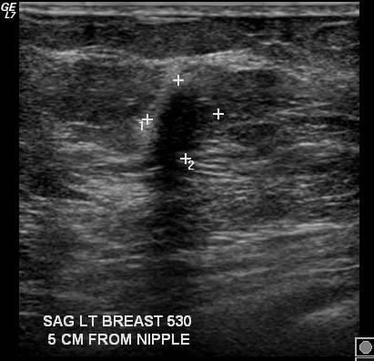
33
34
35 Core needle biopsy done Diagnosis: Plasmacytoma / Myeloma
36 Plasmacytoma Breast plasmacytoma is extremely rare (<100 cases since 1928) Majority 66% were unilateral Most (77%) have occurred in patients with multiple myeloma Imaging features similar to primary breast carcinoma Very radiosensitive, with success rates of 79% 90% 10-year survival rate of 50% 100%


37 Plasmacytoma The needle core biopsy specimen consist of sheets of mature and immature plasma cells

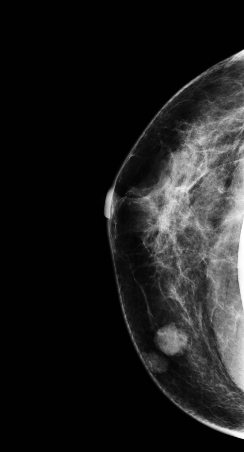
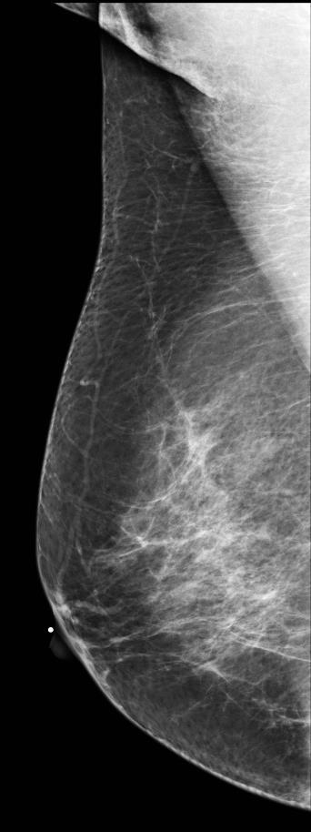
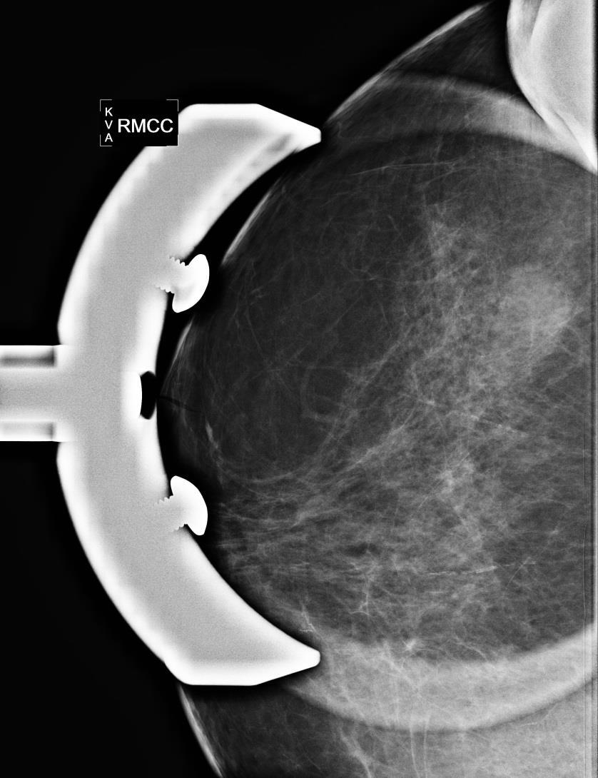
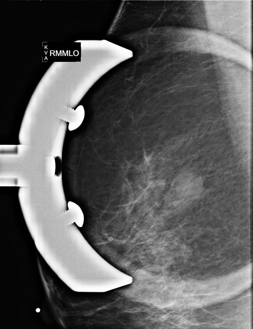
38 Plasmacytoma Tumor cells are immunoreactive for plasma cell marker CD 138
39 Case

40 53 year old female presented with palpable lump
 Benign: Lactating adenoma? PASH? Fat necrosis? Hematoma? Phyllodes?")
41 Ultrasound D/D IDC? ILC? Less common types (mucinous, medullary etc.) Benign: Lactating adenoma? PASH? Fat necrosis? Hematoma? Phyllodes?
42 Core needle biopsy performed Diagnosis: Neuroendocrine tumor (Small cell / Oat cell cancer)
43 Neuroendocrine tumor / Small Cell Carcinoma Very Rare tumors which vary from less aggressive lesions, such as carcinoid to very aggressive such as oat cell cancer Important to rule out breast metastasis from a primary elsewhere in the body, as this is more common than primary breast lesion


44 Small Cell Carcinoma Tumor cells are small with scant cytoplasm, finely granular chromatin and high mitotic rate Necrosis is present Infiltrating tumor cells show organoid pattern of growth
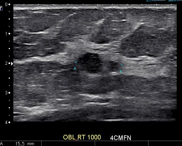
45 Case
46 History 61 y o female with mass in the right breast seen on CT
47
48

49 Right CC Left CC

50 Right CC Right MLO
51

52
53 Differential diagnosis 1. Abscesses 2. Metastasis 3. Fat necrosis 4. Hematomas 5. Lymphoma 6. Multifocal primary breast cancer


54 Core needle biopsy performed Diffuse Large B-cell lymphoma
55
56 Breast Lymphoma 1-2% of all lymphomas occur in the breast Secondary More common than primary lesions (S>P) Most of primary lymphomas are large B-cell type Usually present as non-calcified circumscribed masses or indistinctly marginated spherical or ovoid masses On US, usually show hypoechoic masses with posterior enhancement but may have mixed echogenicity Managed by systemic chemotherapy +/- local excision +/- radiation


57 Large B-Cell Lymphoma The tumor cells are immunoreactive for B-cell marker CD 20 The needle core biopsy specimen consists of diffuse infiltrate of monotonous population of cells with large vesicular nuclei and scant cytoplasm
58 Case
59 History 60 year old male with palpable mass right upper breast for 6 months

60 Right CC Left CC

61 R MLO L MLO
 3. Fat necrosis 4. Hemangioma 5.")
62 Differential diagnosis 1. Breast carcinoma 2. PASH (pseudoangiomatous stromal hyperplasia) 3. Fat necrosis 4. Hemangioma 5. Hematoma 6. Fibroadenoma 7. Lactating adenoma
63 Diagnosis Benign Cavernous hemangioma
64 Cavernous hemangioma Variable appearance on mammogram, often an oval or lobulated mass with circumscribed or microlobulated margins Calcification secondary to phlebolith formation Often located superficially, may have bluish skin Variable echogenicity and margination on US Management in the literature varies from surgical excision to imaging surveillance

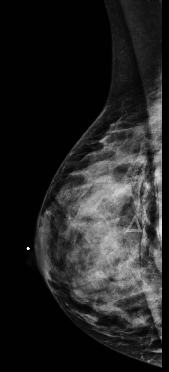
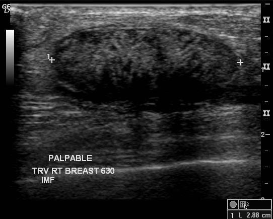
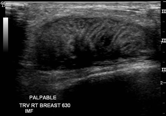
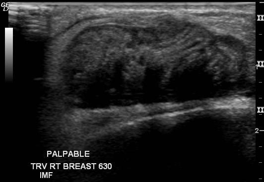
65 Tubular cavernous channels formed within the mass
66 Case
67 History 44 y o white female with palpable lump H/O previous benign excision

68 Right MLO Right CC
69
70
 5.")
71 Differential diagnosis 1. Fibroadenoma 2. Breast carcinoma 3. Granuloma 4. Nodular PASH (pseudo-angiomatous stromal hyperplasia) 5. Epidermal inclusion cyst Benign or malignant?
72 Diagnosis: Suture granuloma Pathology on CNB: Necrotic debris with foreign material on core needle biopsy with organizing granulomatous inflammation on excision
73 Suture granuloma Uncommon, foreign body reaction to suture material Granulomatous inflammation Macrophages and multinucleated giant cells are typical
74 Case
75 History 69 y o white female found to have a large breast mass

76 R CC R MLO

77
78

79 T 2 Post contrast
80 Differential diagnosis Benign or malignant? 1. Breast abscess 2. Mucinous or colloid cancer 3. Papillary cancer 4. Large organizing hematoma 5. Invasive ductal cancer 6. Phyllodes tumor
81
 22 of 22 nodes positive with adenocarcinoma cells in pleural and ascites")
82 Diagnosis: Invasive ductal carcinoma grade III/III ER, PR negative, HER 2 Neu unamplified (triple negative) 22 of 22 nodes positive with adenocarcinoma cells in pleural and ascites fluid
83 Case
84 History 59 y o AA male with palpable right breast mass

85 Right MLO Left MLO
86 Right CC Left CC
87
88 Differential diagnosis 1. Invasive ductal cancer 2. Dense stromal fibrosis 3. Complex sclerosing lesion or radial scar 4. Granular cell tumor 5. Desmoid tumor
89 Diagnosis: Granular cell tumor
90 Granular cell tumor of the breast More common in AA females Relative incidence of about 1-6 per1000 breast cancers Mostly benign but rare malignant reports May be locally invasive, wide surgical excision is recommended Mammographically, non-calcified, many times spiculated masses Great mimickers of breast carcinoma On US, hypoechoic masses with posterior shadowing On MR, may show strong homogeneous or rim enhancement


91 Granular Cell Tumor Cells contain abundant granular eosinophilic cytoplasm and small nuclei Infiltrating nests and cords of large polygonal cells, separated by prominent fibrous septae

92 A different Patient
93
94 Case

95 History 64 yo female with new palpable lump left breast
96

97
98 DDX for new solid circumscribed mass: Malignant Benign Breast carcinoma Invasive ductal Medullary Mucinous Papillary Malignant phyllodes Adenoma Papilloma PASH Lymphoma Benign phyllodes
99 RCC Patient complained of pain on the other side
100 DDx for multiple circumscribed masses Cysts Fibroadenomas Metastasis Lymphoma Multiple primary breast CA s PASH Granulomas
101
102
103

 Remote history of")
104 Biopsied right axillary tail and left breast lesions Diagnosis: Metastatic melanoma (poorly differentiated) Remote history of melanoma leg in 2003
105 Metastatic Disease to the Breast If multiple solid lesions present, consider metastasis Frequently round and fairly well circumscribed; single or multiple, vary in size, non-ductal pattern Melanoma is the most common cancer to metastasize to breast; followed by lung and lymphoma. Others-Ovarian, Cervical and Renal Cell Carcinoma
106
107 Case
108 History 47 year old female with new masses in the left breast
109
110 Differential diagnosis Cysts Fibroadenomas Metastatic lesions Lymphoma Granulomas Fat necrosis Multi-centric cancer PASH

111 US of the left breast
112 3 most suspicious masses were biopsied Diagnosis: PASH Pseudo angiomatous stromal hyperplasia
113 Few years later
114 Few years later
115 Pseudoangiomatous Stromal Hyperplasia (PASH) Uncommon benign lesion likely related to hormones, as it is only seen in premenopausal women Benign overgrowth of fibrous connective tissue stroma that separates into lobules and ducts that resemble vascular spaces. Must distinguish from angiosarcoma On mammography, lesions usually well defined with smooth border and no calcifications, but partially circumscribed borders and even spiculated margins have been reported On ultrasound, usually well defined hypoechoic or mixed echogenicity, solid mass with variable posterior sound properties

116 Pseudoangiomatous Stromal Hyperplasia (PASH) Complex, anastomosing, and empty slit-like spaces in dense collagenous stroma Empty spaces are lined by monomorphic spindle cells resembling endothelial cells
117 Case
118 History 15 year old girl presents with palpable lump in the right upper outer quadrant
 3.")

119 Differential diagnosis 1. Breast Abscess 2. Breast carcinoma (IDC, papillary etc.) 3. Lactating adenoma 4. Phyllodes tumor 5. Hematoma 6. Acute fat necrosis 7. Galactocele
120
121 Core needle biopsy performed Diagnosis: Cystosarcoma Phyllodes
122 Cystosarcoma Phyllodes (Malignant Phyllodes) Malignant high grade phyllodes tumor (sarcomatous features) Usually presents as a rapidly enlarging mass Represents <1% of breast neoplasms Related to fibroadenoma group May recur locally if not completely excised Up to 10% may metastasize, usually hematogeneously to lung or bone


123 Phylloides Tumor-Malignant Marked cytologic atypia and abundant mitosis (>10/HPF) Malignant stroma showing fibrosarcomatous appearance

124 Thank You
125


126 67 yo F on screening mammogram
127
128

129 Adenoid cystic carcinoma

130 33 yo F with palpable lump
131
132 Mass-like Radial Scar (RSL)
133 Adenomyoepithelioma Rare breast tumor which arises from myoepithelial and epithelial cells that are present in normal breast. Most are benign and characterized by biphasic proliferation of epithelial and myoepithelial elements Non specific imaging features Prone to locally recur and may recur several years after surgical removal

134 Adenomyoepithelioma Ultrasound reveals a hypoechoic, solid mass with slightly lobulated borders
135 Adenomatous Lesions Tubular adenomas Pure adenomas without significant fibrous stroma Variant of pericanalicular fibroadenoma, but usually do not calcify Seen in young females Similar imaging features to fibroadenoma, but may mimic malignancy Lactating adenomas Uncommon tumor seen in pregnant or lactating females Variant of fibroadenoma and tubular adenomas and cannot be distinguished by imaging alone Tend to be well circumscribed, hypoechoic masses with posterior enhancement, but occasionally have irregular margins or heterogeneous appearance Many regress spontaneously


136 Tubular Adenoma Lactating Adenoma Ultrasound shows a lobulated oval hypoechoic mass which is indistinguishable from a fibroadenoma Ultrasound shows a well circumscribed, slightly lobulated hypoechoic mass, which is indistinguishable from a fibroadenoma

137 Tubular Adenoma
138 Case


139 17 year old female presents with right breast swelling
Lesion Imaging Characteristics Mass, Favoring Benign Circumscribed Margins Intramammary Lymph Node
 Lesion Imaging Characteristics Mass, Favoring Benign Circumscribed Margins Intramammary Lymph Node Oil Cyst Mass, Intermediate Concern Microlobulated Margins Obscured Margins Mass, Favoring Malignant Indistinct
University of Washington Radiology Review Course: Strange and Specific Diagnoses. Case #1
 University of Washington Radiology Review Course: Strange and Specific Diagnoses Katherine E. Dee, MD Seattle Breast Center Via Radiology 2014 Case #1 37 year old presents with bilateral palpable lumps.
Diseases of the breast (1 of 2)
") Diseases of the breast (1 of 2) Introduction A histology introduction Normal ducts and lobules of the breast are lined by two layers of cells a layer of luminal cells overlying a second layer of myoepithelial
Diseases of the breast (1 of 2) Introduction A histology introduction Normal ducts and lobules of the breast are lined by two layers of cells a layer of luminal cells overlying a second layer of myoepithelial
SIGNIFICANT OTHERS. Miscellaneous Benign Breast Conditions
 SIGNIFICANT OTHERS Miscellaneous Benign Breast Conditions Epworth HealthCare 1 FAT NECROSIS TRAUMATIC Cell rupture Seat-Belt injury Blunt trauma Iatrogenic injury Surgery, Flaps, Radiotherapy Pathology
Imaging in breast cancer. Mammography and Ultrasound Donya Farrokh.MD Radiologist Mashhad University of Medical Since
 Imaging in breast cancer Mammography and Ultrasound Donya Farrokh.MD Radiologist Mashhad University of Medical Since A mammogram report is a key component of the breast cancer diagnostic process. A mammogram
BREAST PATHOLOGY. Fibrocystic Changes
 BREAST PATHOLOGY Lesions of the breast are very common, and they present as palpable, sometimes painful, nodules or masses. Most of these lesions are benign. Breast cancer is the 2 nd most common cause
Ultrasound of the Breast BASICS FOR THE ORDERING CLINICIAN
 Ultrasound of the Breast BASICS FOR THE ORDERING CLINICIAN Breast Ultrasound Anatomy Skin Breast Parenchyma Pectoralis Fascia Pectoralis Breast Ultrasound Anatomy Indications for Breast Ultrasound Palpable
Benign, Reactive and Inflammatory Lesions of the Breast
 Benign, Reactive and Inflammatory Lesions of the Breast Marilin Rosa, MD Associate Member Section Head of Breast Pathology Department of Anatomic Pathology Program Director, Breast Pathology Fellowship
Index. C Calcifications fat necrosis 1, 61 fat necrosis 4, 69 nipple/peri-areolar involvement 1, 165
 A ADH. See Atypical ductal hyperplasia (ADH) American College of Radiology (ACR), BI-RADS background parenchymal enhancement, 8, 9, 81, 82 fibroglandular tissue guidelines, 6 American Joint Committee on
Breast pathology. 2nd Department of Pathology Semmelweis University
 Breast pathology 2nd Department of Pathology Semmelweis University Breast pathology - Summary - Benign lesions - Acute mastitis - Plasma cell mastitis / duct ectasia - Fat necrosis - Fibrocystic change/
Case study 1. Rie Horii, M.D., Ph.D. Division of Pathology Cancer Institute Hospital, Japanese Foundation for Cancer Research
 NCCN/JCCNB Seminar in Japan April 15, 2012 Case study 1 Rie Horii, M.D., Ph.D. Division of Pathology Cancer Institute Hospital, Japanese Foundation for Cancer Research Present illness: A 50y.o.premenopausal
04/10/2018. Intraductal Papillary Neoplasms Of Breast INTRADUCTAL PAPILLOMA
 Intraductal Papillary Neoplasms Of Breast Savitri Krishnamurthy MD Professor of Pathology Deputy Division Head The University of Texas MD Anderson Cancer Center 25 th Annual Seminar in Pathology Pittsburgh,
Salivary Glands 3/7/2017
 Salivary Glands 3/7/2017 Goals and objectives Focus on the entities unique to H&N Common board type facts Information for your future practice Salivary Glands Salivary Glands Major gland. Paratid. Submandibular.
Mousa. Israa Ayed. Abdullah AlZibdeh. 0 P a g e
 1 Mousa Israa Ayed Abdullah AlZibdeh 0 P a g e Breast pathology The basic histological units of the breast are called lobules, which are composed of glandular epithelial cells (luminal cells) resting on
Contents. Basic Ultrasound Principles and Terminology. Ultrasound Nodule Characteristics
 Contents Basic Ultrasound Principles and Terminology Basic Ultrasound Principles... 1 Ultrasound System... 2 Linear Transducer for Superficial Images and Ultrasound-Guided FNA... 3 Scanning Planes... 4
PAAF vs Core Biopsy en Lesiones Mamarias Case #1
 5/19/2014 PAAF vs Core Biopsy en Lesiones Mamarias Case #1 Fine Needle Aspiration Cytology of Breast: Correlation with Needle Core Biopsy 64-year-old woman Mass in breast Syed Hoda, MD CD31 Post-Radiation
Maram Abdaljaleel, MD Dermatopathologist and Neuropathologist University of Jordan, School of Medicine
 Maram Abdaljaleel, MD Dermatopathologist and Neuropathologist University of Jordan, School of Medicine The most common non-skin malignancy of women 2 nd most common cause of cancer deaths in women, following
Criteria of Malignancy. Evaluation Score
 30 5 Diagnostic Criteria Criteria of Malignancy Table 5.2 lists criteria in contrast-enhancing MR mammography that strongly indicate the presence of malignancy or are unspecific. Unifactorial evaluation
University Journal of Pre and Para Clinical Sciences
 ISSN 2455 2879 Volume 2 Issue 1 2016 Metaplastic carcinoma breast a rare case report Abstract : Metaplastic carcinoma of the breast is a rare malignancy with two distinct cell lines described as a breast
Basement membrane in lobule.
 Bahram Memar, MD Basement membrane in lobule. Normal lobule-luteal phase Normal lobule-follicular phase Lactating breast Greater than 95% are adenocarcinomas in situ carcinomas and invasive carcinomas.
RADIOLOGIC EVALUATION OF BREAST CANCER
 RADIOLOGIC EVALUATION OF BREAST CANCER Orsolya Farkas, Gabriella Bodrogi and Gábor Szalai Department of Radiology, Pécs University Orsifarkas@yahoo.com Complex evaluation of the breast Patient history
INDEX. in this web service Cambridge University Press
 abscess. See also subareolar abscess acute mastitis, 44 lactational/puerperal mastitis, 55 mammary tuberculosis, 42 tuberculous, 43 adeno gastric, 198, 200 invasive, 157 lung, 197, 200 prostatic, 199 200
Armed Forces Institute of Pathology.
 Armed Forces Institute of Pathology www.radpath.com Armed Forces Institute of Pathology Breast Disease www.radpath.org Armed Forces Institute of Pathology Interpretation of Breast MRI Leonard M. Glassman
Ultrasonography. Methods. Brief Description. Indications. Device-related Prerequisites. Technical Requirements. Evaluation Criteria
 1 Ultrasonography Brief Description Imaging modality using sound waves Tissue-specific wave reflection. Indications Evaluation of palpable breast nodules Evaluation of clinically occult mammographic findings
LYMPHATIC DRAINAGE AXILLARY (MOSTLY) INTERNAL MAMMARY SUPRACLAVICULAR
 INTERNAL MAMMARY SUPRACLAVICULAR") BREAST LYMPHATIC DRAINAGE AXILLARY (MOSTLY) INTERNAL MAMMARY SUPRACLAVICULAR HISTOLOGY LOBE: (10 in whole breast) LOBULE: (many per lobe) ACINUS/I, aka ALVEOLUS/I: (many per lobule) DUCT(S): INTRA- or
BREAST LYMPHATIC DRAINAGE AXILLARY (MOSTLY) INTERNAL MAMMARY SUPRACLAVICULAR HISTOLOGY LOBE: (10 in whole breast) LOBULE: (many per lobe) ACINUS/I, aka ALVEOLUS/I: (many per lobule) DUCT(S): INTRA- or
BREAST PATHOLOGY MCQS
 BREAST PATHOLOGY MCQS 1) :The most important factor in breast enlargement during pregnancy is A. stromal edema B. secretion of chorionic gonadotropin C. glandular hyperplasia D. proliferation of stroma
57th Annual HSCP Spring Symposium 4/16/2016
 An Unusual Malignant Spindle Cell Lesion to Involve the Breast Erinn Downs-Kelly, D.O. Associate Professor of Pathology University of Utah & ARUP Laboratories No disclosures Case 39 y/o female with no
Leonard M. Glassman MD
 BI-RADS The New BI-RADS Leonard M. Glassman MD FACR Former Chief of Breast Imaging American Institute for Radiologic Pathology Washington Radiology Associates, PC Breast Imaging Reporting and Data System
Spindle Cell Lesions Of The Breast. Emad Rakha Professor of Breast Pathology and Consultant Pathologist
 Spindle Cell Lesions Of The Breast Emad Rakha Professor of Breast Pathology and Consultant Pathologist * SCLs comprise a wide spectrum of diseases, ranging from reactive processes to aggressive malignant
Imaging the Symptomatic Patient. Avice M.O Connell MD,FACR,FSBI Professor of Imaging Sciences Director, Women s Imaging University of Rochester
 Imaging the Symptomatic Patient Avice M.O Connell MD,FACR,FSBI Professor of Imaging Sciences Director, Women s Imaging University of Rochester The four most common symptoms Mass Pain Discharge Infection
Differential Diagnosis of Oral Masses. Palatal Lesions
 Differential Diagnosis of Oral Masses Palatal Lesions Palatal Masses Periapical Abscess Torus Palatinus Mucocele Lymphoid Hyperplasia Adenomatous Hyperplasia Benign Salivary Neoplasms Malignant Salivary
DISORDERS OF THE BREAST Dated. FIBROADENOSIS Other common names: mastitis, fibrocystic disease, cystic mammary dysplasia.
 DISORDERS OF THE BREAST Dated BENIGN BREAST DISORDERS (Essential Surg 2 nd Ed, pp 540) FIBROADENOSIS Other common names: mastitis, fibrocystic disease, cystic mammary dysplasia. Fibroadenosis is the distortion
Pleomorphic adenoma of breast - a case report and distinction with metaplastic carcinoma D Gupta, S Agrawal, N Trivedi, A Tewari
 of breast - a case report and distinction with metaplastic carcinoma D Gupta, S Agrawal, N Trivedi, A Tewari Introduction, also known as mixed tumour, is a benign tumour which typically presents as a painless,
Amammography report is a key component of the breast
 Review Article Writing a Mammography Report Amammography report is a key component of the breast cancer diagnostic process. Although mammographic findings were not clearly differentiated between benign
Salivary Gland Cytology
 Salivary Gland Cytology Diagnostic challenges and potential pitfalls Tarik M. Elsheikh, MD Professor and Medical Director Anatomic Pathology Cleveland Clinic FNA Salivary Gland Lesions Indications Distinguish
Benign Mimics of Malignancy in Breast Pathology
 Arthur Purdy Stout Society of Surgical Pathologists Companion Meeting Benign Mimics of Malignancy in Breast Pathology Stuart J. Schnitt, M.D. Beth Israel Deaconess Medical Center and Harvard Medical School,
Treatment options for the precancerous Atypical Breast lesions. Prof. YOUNG-JIN SUH The Catholic University of Korea
 Treatment options for the precancerous Atypical Breast lesions Prof. YOUNG-JIN SUH The Catholic University of Korea Not so benign lesions? Imaging abnormalities(10% recall) lead to diagnostic evaluation,
Mody. AIS vs. Invasive Adenocarcinoma of the Cervix
 Common Problems in Gynecologic Pathology Michael T. Deavers, M.D. Houston Methodist Hospital, Houston, Texas Common Problems in Gynecologic Pathology Adenocarcinoma in-situ (AIS) of the Cervix vs. Invasive
Case Scenario 1 History and Physical 3/15/13 Imaging Pathology
 Case Scenario 1 History and Physical 3/15/13 The patient is an 84 year old white female who presented with an abnormal mammogram. The patient has a five year history of refractory anemia with ringed sideroblasts
Imaging of giant breast masses with pathological correlation
 P i c t o r i a l E s s a y Singapore Med J 2004 Vol 45(3) : 132 Imaging of giant breast masses with pathological correlation M Muttarak, B Chaiwun ABSTRACT Ultrasonography (US) and mammography are the
Diagnostically Challenging Cases in Gynecologic Pathology
 Diagnostically Challenging Cases in Gynecologic Pathology Eric C. Huang, M.D., Ph.D. Department of Pathology and Laboratory Medicine University of California, Davis Medical Center Case 1 Presentation 38
Alena Levit MD Avice O Connell MD University of Rochester, Rochester, NY
 Alena Levit MD Avice O Connell MD University of Rochester, Rochester, NY Purpose Review imaging spectrum of both common benign and malignant breast lesions Describe and demonstrate CT features with mammogram,
Breast Pathology. Breast Development
 Breast Pathology Lecturer: Hanina Hibshoosh, M.D. Reading: Kumar, Cotran, Robbins, Basic Pathology, 6th Edition, pages 623-635 Breast Development 5th week - thickening of the epidermis - milk line 5th
Mammographic imaging of nonpalpable breast lesions. Malai Muttarak, MD Department of Radiology Chiang Mai University Chiang Mai, Thailand
 Mammographic imaging of nonpalpable breast lesions Malai Muttarak, MD Department of Radiology Chiang Mai University Chiang Mai, Thailand Introduction Contents Mammographic signs of nonpalpable breast cancer
CPC 4 Breast Cancer. Rochelle Harwood, a 35 year old sales assistant, presents to her GP because she has noticed a painless lump in her left breast.
 CPC 4 Breast Cancer Rochelle Harwood, a 35 year old sales assistant, presents to her GP because she has noticed a painless lump in her left breast. 1. What are the most likely diagnoses of this lump? Fibroadenoma
The radiologic workup of a palpable breast mass
 Imaging in Practice CME CREDIT EDUCTIONL OJECTIVE: The reader will consider which breast masses require further workup and which imaging study is most appropriate Lauren Stein, MD Imaging Institute, Cleveland
Case year female. Routine Pap smear
 Case 1 57 year female Routine Pap smear Diagnosis? 1. Atypical glandular cells of unknown significance (AGUS) 2. Endocervical AIS 3. Endocervical adenocarcinoma 4. Endometrial adenocarcinoma 5. Adenocarcinoma
RSNA, /radiol Appendix E1. Methods
 RSNA, 2016 10.1148/radiol.2016151097 Appendix E1 Methods US and Near-infrared Data Acquisition Four optical wavelengths (740 nm, 780 nm, 808 nm, and 830 nm) were used to sequentially deliver the light
FIBROEPITHELIAL LESIONS
 DEFINITIONS FIBROEPITHELIAL LESIONS Suzanne Moore FIBROADENOMA- A discrete benign tumour showing evidence of connective tissue and epithelial proliferation- WHO Fibrous stromal element of these tumours
CLINICAL SIGNIFICANCE OF BENIGN EPITHELIAL CHANGES
 Papillomas. Papillomas are composed of multiple branching fibrovascular cores, each having a connective tissue axis lined by luminal and myoepithelial cells ( Fig. 23-11 ). Growth occurs within a dilated
ORIGINAL ARTICLE EVALUATION OF BREAST LESIONS USING X-RAY MAMMOGRAM WITH HISTOPATHOLOGICAL CORRELATION
 Available online at www.journalijmrr.com INTERNATIONAL JOURNAL OF MODERN RESEARCH AND REVIEWS IJMRR ISSN: 2347-8314 Int. J. Modn. Res. Revs. Volume 3, Issue 10, pp 807-814, October, 2015 ORIGINAL ARTICLE
Giant Ulcerative Lactating Nodule of Ectopic Breast Mimicking Malignancy.
 57 Giant Ulcerative Lactating Nodule of Ectopic Breast Mimicking Malignancy. I. Roy 1 FRCS(C), E Othieno 2 MMed (Path), R Ssentongo 3 FCS (ECSA) 1 Department of Pathology, St. Raphael of St. Francis Hospital,
Gross appearance of nodular hyperplasia in material obtained from suprapubic prostatectomy. Note the multinodular appearance and the admixture of
 Tiền liệt tuyến Tiền liệt tuyến Gross appearance of nodular hyperplasia in material obtained from suprapubic prostatectomy. Note the multinodular appearance and the admixture of solid and microcystic areas.
Papillary Lesions of the Breast A Practical Approach to Diagnosis. (Arch Pathol Lab Med. 2016;140: ; doi: /arpa.
 Papillary Lesions of the Breast A Practical Approach to Diagnosis (Arch Pathol Lab Med. 2016;140:1052 1059; doi: 10.5858/arpa.2016-0219-RA) Papillary lesions of the breast Span the spectrum of benign,
Journal of Medical Imaging and Radiation Oncology
 Journal of Medical Imaging and Radiation Oncology 60 (2016) 506 513 MEDICAL IMAGING PICTORIAL ESSAY Malignant hyperechoic breast lesions at ultrasound: A pictorial essay Stephen Tiang, 1 Cecily Metcalf,
Classification System
 Classification System A graduate of the Breast Oncology training program should be able to care for all aspects of disease and/or provide comprehensive management. When referring to a discipline of training
Papillary lesions of the breast - Imaging findings and diagnostic challenges
 Papillary lesions of the breast - Imaging findings and diagnostic challenges Poster No.: R-0146 Congress: RANZCR-AOCR 2012 Type: Educational Exhibit Authors: P. Jagmohan, F. J. Pool Keywords: Breast, Mammography,
ACRIN 6666 IM Additional Evaluation: Additional Views/Targeted US
 Additional Evaluation: Additional Views/Targeted US For revised or corrected form check box and fax to 215-717-0936. Instructions: The form is completed based on recommendations (from ID form) for additional
Normal endometrium: A, proliferative. B, secretory.
 Normal endometrium: A, proliferative. B, secretory. Nội mạc tử cung Nội mạc tử cung Cyclic changes in endometrium.. Approximate relationship of useful microscopic changes. Arias-Stella reaction in endometrial
Pitfalls and Limitations of Breast MRI. Susan Orel Roth, MD Professor of Radiology University of Pennsylvania
 Pitfalls and Limitations of Breast MRI Susan Orel Roth, MD Professor of Radiology University of Pennsylvania Objectives Review the etiologies of false negative breast MRI examinations Discuss the limitations
Case year old Chinese female. Radiological echo-distortion in the right breast at o clock. Core biopsy of the o clock lesion.
 Case 3 64 year old Chinese female. Radiological echo-distortion in the right breast at 10-12 o clock. Core biopsy of the 11-12 o clock lesion. Division of Pathology Courtesty of Dr Lester Leong ill-defined,
MEDICAL IMAGING AND BREAST DISEASE HOW CAN WE HELP YOU?
 MEDICAL IMAGING AND BREAST DISEASE HOW CAN WE HELP YOU? Barbara M. Preston, M.D. SCREENING MAMMOGRAPHY AVERAGE RISK PATIENTS KAISER RECOMMENDATION: ALL WOMEN (INCLUDING TRANSGENDER FEMALES) Every 1-21
Lung Cytology: Lessons Learned from Errors in Practice
 Lung Cytology: Lessons Learned from Errors in Practice Stephen S. Raab, M.D. Department of Laboratory Medicine Eastern Health and Memorial University of Newfoundland, St. John s, NL and University of Washington,
Intracystic papillary carcinoma of the breast
 Intracystic papillary carcinoma of the breast Poster No.: C-1932 Congress: ECR 2011 Type: Educational Exhibit Authors: V. Dimarelos, F. TZIKOS, N. Kotziamani, G. Rodokalakis, 1 2 3 1 1 1 2 T. MALKOTSI
CURRICULUM FOR THE BREAST PATHOLOGY ROTATION UNIVERSITY OF FLORIDA DEPARTMENT OF PATHOLOGY
 CURRICULUM FOR THE BREAST PATHOLOGY ROTATION UNIVERSITY OF FLORIDA DEPARTMENT OF PATHOLOGY JULY, 2003 The following is a conceptual curriculum and set of guidelines for Pathology Residents on the Breast
Breast Imaging: Multidisciplinary Approach. Madelene Lewis, MD Assistant Professor Associate Program Director Medical University of South Carolina
 Breast Imaging: Multidisciplinary Approach Madelene Lewis, MD Assistant Professor Associate Program Director Medical University of South Carolina No Disclosures Objectives Discuss a multidisciplinary breast
Enterprise Interest None
 Enterprise Interest None B3 lesions of the breast What are they at surgery? Case 4 Edi Brogi MD PhD Attending Pathologist - Director of Breast Pathology Memorial Sloan Kettering Cancer Center New York
performed to help sway the clinician in what the appropriate diagnosis is, which can substantially alter the treatment of management.
 Hello, I am Maura Polansky at the University of Texas MD Anderson Cancer Center. I am a Physician Assistant in the Department of Gastrointestinal Medical Oncology and the Program Director for Physician
40th European Congress of Cytology Liverpool, UK, 2-5 th October 2016
 40th European Congress of Cytology Liverpool, UK, 2-5 th October 2016 EUS FNA of abdominal organs: An approach to reporting and triage for ancillary testing Date and time: Sunday 2 nd October 2016 15.00-16.30
Respiratory Tract Cytology
 Respiratory Tract Cytology 40 th European Congress of Cytology Liverpool, UK Momin T. Siddiqui M.D. Professor of Pathology and Laboratory Medicine Director of Cytopathology Emory University Hospital, Atlanta,
Triple Negative Breast Cancer
 Triple Negative Breast Cancer Prof. Dr. Pornchai O-charoenrat Division of Head-Neck & Breast Surgery Department of Surgery Faculty of Medicine Siriraj Hospital Breast Cancer Classification Traditional
Breast Cancer. Most common cancer among women in the US. 2nd leading cause of death in women. Mortality rates though have declined
 Breast Cancer Most common cancer among women in the US 2nd leading cause of death in women Mortality rates though have declined 1 in 8 women will develop breast cancer Breast Cancer Breast cancer increases
A GP S APPROACH TO BREAST LUMPS AND SYMPTOMS DR KK CHEUNG GPGC WORKSHOP
 A GP S APPROACH TO BREAST LUMPS AND SYMPTOMS DR KK CHEUNG GPGC WORKSHOP 18.08.18 HAVE A SYSTEM HISTORY EXAMINATION INVESTIGATION FOLLOW UP BREAST SYMPTOMS HISTORY DON T FORGET SKIN CHANGES AND NIPPLE CHANGES
Triple-negative breast cancer: which typical features can we identify on conventional and MRI imaging?
 Triple-negative breast cancer: which typical features can we identify on conventional and MRI imaging? Poster No.: C-1862 Congress: ECR 2013 Type: Educational Exhibit Authors: V. Bertani 1, A. Gualano
IBCM 2, April 2009, Sarajevo, Bosnia and Herzegovina
 Preoperative diagnosis and treatment planning in breast cancer The pathologist s perspective L. Mazzucchelli Istituto Cantonale di Patologia Locarno, Switzerland IBCM 2, 23-25 April 2009, Sarajevo, Bosnia
Carcinoma mammario: le istologie non frequenti. Valentina Guarneri Università di Padova IOV-IRCCS
 Carcinoma mammario: le istologie non frequenti Valentina Guarneri Università di Padova IOV-IRCCS Histological diversity of breast adenocarcinomas Different histological types are defined according to specific
PATHOLOGY OF THE BREAST
 UROGENITAL SYSTEM MBBS 2 nd Yr. Lecture Dr. U.S. Khoo October 3, 2002-9:45 AM Underground Lecture Theatre 1 New Clinical Building, QMH PATHOLOGY OF THE BREAST Learning Objectives 1. To understand the normal
BREAST IMAGING and NEW IMAGING MODALITIES- A Surgeons view
 BREAST IMAGING and NEW IMAGING MODALITIES- A Surgeons view DR CHANTEL THORNTON SPECIALIST BREAST CANCER SURGEON BMSc (hons) MBBS (hons) FRACS Epworth Hospital, Richmond- Agora Centre for Women s Health
Lesions Mimicking Adenoid Cystic Carcinoma. Diagnostic Problems in Salivary Gland Pathology An Update 5/29/2009
 Diagnostic Problems in Salivary Gland Pathology An Update Lesions Mimicking Adenoid Cystic Carcinoma Stacey E. Mills, M.D. W.S. Royster Professor of Pathology Director of Surgical and Cytopathology University
Papillary Lesions of the breast
 Papillary Lesions of the breast Emad Rakha Professor of Breast Pathology The University of Nottingham Papillary lesions of the breast are a heterogeneous group of disease, which are characterised by neoplastic
objectives Pitfalls and Pearls in PET/CT imaging Kevin Robinson, DO Assistant Professor Department of Radiology Michigan State University
 objectives Pitfalls and Pearls in PET/CT imaging Kevin Robinson, DO Assistant Professor Department of Radiology Michigan State University To determine the regions of physiologic activity To understand
FNA OF SALIVARY GLANDS: A PRACTICAL APPROACH
 FNA OF SALIVARY GLANDS: A PRACTICAL APPROACH FNA of Salivary Glands: Challenges Wide range of neoplastic and non-neoplastic lesions Cytological overlap between the different benign and malignant tumors
Breast Cancer Diagnosis, Treatment and Follow-up
 Breast Cancer Diagnosis, Treatment and Follow-up What is breast cancer? Each of the body s organs, including the breast, is made up of many types of cells. Normally, healthy cells grow and divide to produce
Protocol for the Examination of Biopsy Specimens From Patients With Invasive Carcinoma of the Breast
 Protocol for the Examination of Specimens From Patients With Invasive Carcinoma of the Breast Version: BreastInvasive 1.0.0.0 Protocol Posting Date: February 2019 Accreditation Requirements The use of
NAME/ AGE/57 SEX/Female AREA/Australia Visit 1: 5/8/2011. Right Breast Cancer Infiltrating Ductal Carcinoma, no specific type, Grade 1 to 2.
 Breast Cancer-CAM Rx NAME/ AGE/57 SEX/Female AREA/Australia Visit 1: 5/8/2011 Case History Discussion Right Breast Cancer Infiltrating Ductal Carcinoma, no specific type, Grade 1 to 2. Patient visited
Case Report Rare Presentation of Pseudoangiomatous Stromal Hyperplasia: A Case Report
 IBIMA Publishing International Journal of Case Reports in Medicine http://www.ibimapublishing.com/journals/ijcrm/ijcrm.html Vol. 2013 (2013), Article ID 331549, 5 pages DOI: 10.5171/2013.331549 Case Report
Gynecomastia and Its Mimics: Not All Male Breast Lesions are Benign
 Gynecomastia and Its Mimics: Not All Male Breast Lesions are Benign Poster No.: C-0139 Congress: ECR 2014 Type: Educational Exhibit Authors: S. A. Choudhery, P. Gupta, S. Foshee, F. Garcia-Morales, G.
A neoplasm is defined as "an abnormal tissue proliferation, which exceeds that of adjacent normal tissue. This proliferation continues even after
 NEOPLASIA Neoplasia is a very important topic in pathology because neoplasms are both common and serious diseases. A neoplasm literally means a new growth, and this term is used interchangeably with a
Normal thyroid tissue
 Thyroid Pathology Overview Normal thyroid tissue Normal thyroid tissue with follicles filled with colloid. Thyroid cells form follicles, spheres of epithelial cells (always single layered in health, usually
Unusual Breast Cancers: What do we know?
 Unusual Breast Cancers: What do we know? Poster No.: C-1267 Congress: ECR 2015 Type: Educational Exhibit Authors: D. A. Puentes Bejarano, C. Oliva Fonte, C. Sibaja Castro, J. Pereda Rodríguez, J. Gonzalez
High risk lesions of the breast : Review of the current diagnostic and management strategies
 High risk lesions of the breast : Review of the current diagnostic and management strategies Poster No.: C-1204 Congress: ECR 2016 Type: Educational Exhibit Authors: P. Jagmohan, F. J. Pool, P. G. Pillay,
Synchronous squamous cell carcinoma of the breast. and invasive lobular carcinoma
 Sentani K et al. 1 Letter to the editor Synchronous squamous cell carcinoma of the breast and invasive lobular carcinoma Kazuhiro Sentani, 1 Takashi Tashiro, 2 Naohide Oue, 1 Wataru Yasui 1 1 Department
Neoplasia literally means "new growth.
 NEOPLASIA Neoplasia literally means "new growth. A neoplasm, defined as "an abnormal mass of tissue the growth of which exceeds and is uncoordinated with that of the normal tissues and persists in the
Breast Evaluation & Management Guidelines
 Breast Evaluation & Management Guidelines Pamela L. Kurtzhals, M.D. F.A.C.S. Head, Dept. of General Surgery Scripps Clinic, La Jolla Objective Review screening & diagnostic guidelines Focused patient complaints
Contrast-enhanced Breast MRI RSSA 2013
 Contrast-enhanced Breast MRI RSSA 2013 Prof. dr. Maurice van den Bosch University Medical Center Utrecht, the Netherlands Index 1) Breast cancer 2) Why MRI of the breast 3) Technique 4) Interpretation
Breast Cancer. Saima Saeed MD
 Breast Cancer Saima Saeed MD Breast Cancer Most common cancer among women in the US 2nd leading cause of death in women 1 in 8 women will develop breast cancer Incidence/mortality rates have declined Breast
Note: The cause of testicular neoplasms remains unknown
 - In the 15- to 34-year-old age group, they are the most common tumors of men. - Tumors of the testis are a heterogeneous group of neoplasms that include: I. Germ cell tumors : 95%; all are malignant.
-1- Pathology Department (code: 0605) Final Exam for Third year students Date: Time allowed: 2 hours. Paper II (75 marks).
 Final Exam for Third year students Date: Time allowed: 2 hours. Paper II (75 marks).") -1- BENHA UNIVERSITY FACULTY OF MEDICINE Pathology Department (code: 0605) Final Exam for Third year students Date: 28-5-2011 Time allowed: 2 hours. Paper II (75 marks). Please note that this question
-1- BENHA UNIVERSITY FACULTY OF MEDICINE Pathology Department (code: 0605) Final Exam for Third year students Date: 28-5-2011 Time allowed: 2 hours. Paper II (75 marks). Please note that this question
Presentation material is for education purposes only. All rights reserved URMC Radiology Page 1 of 98
 Presentation material is for education purposes only. All rights reserved. 2011 URMC Radiology Page 1 of 98 Radiology / Pathology Conference February 2011 Brooke Koltz, Cytopathology Resident Presentation
Outline 11/2/2017. Pancreatic EUS-FNA general aspects. Cytomorphologic features of solid neoplasms/lesions of the pancreas
 ENDOSCOPIC ULTRASOUND GUIDED-FINE NEEDLE ASPIRATION CYTOLOGY OF PANCREAS Khalid Amin M.D. Assistant Professor Department of Laboratory Medicine and Pathology University of Minnesota Outline Pancreatic