Imaging of common diseases of hepatobiliary and GI system
|
|
|
- Mark O’Connor’
- 6 years ago
- Views:
Transcription
1 Imaging of common diseases of hepatobiliary and GI system Natthaporn Tanpowpong, M.D. Diagnostic radiology Faculty of Medicine, Chulalongkorn University

2 Normal plain radiograph
3
4
5
6
7
8
9

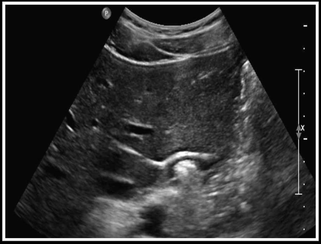
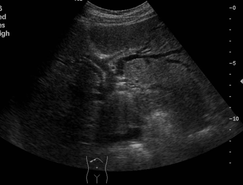
10 A = Common bile duct B = Right hepatic artery C = Portal vein D = IVC A B C D


11 LL CBD ปกต SV SMA IVC S

12 Fundus Neck Body

13 ภาพ transverse scan ท mid SMA Pancreas upper abdomen Splenic vein
14
15
16
17
18
19


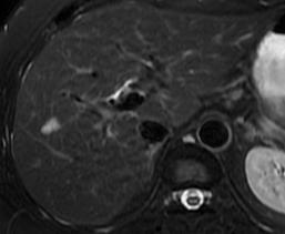
20 T1W in phase T1W out of phase T2W with FS Heavy T2W with FS
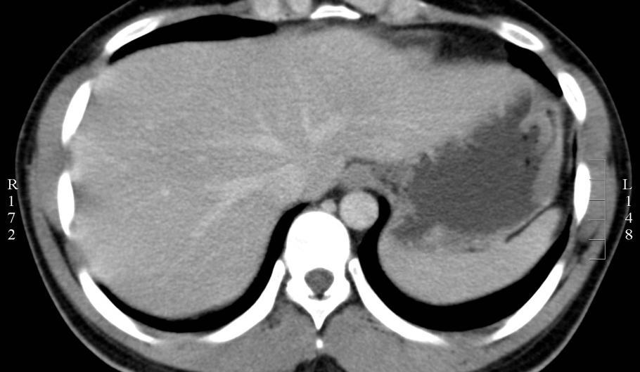
21


22 A-phase PV-phase Equilibrium phase Delayed 3 minutes

23
24 Symptoms Signs Differential diagnosis Investigations Laboratory: blood, urine, stool Imaging
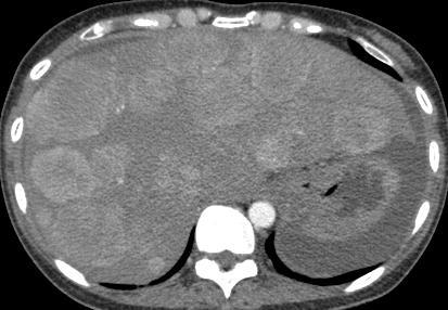
25 Common liver disease Liver parenchymal disease Fatty liver Hepatitis Cirrhosis Focal liver lesions Cyst Hemangioma Abscess Hepatocellular carcinoma Metastasis


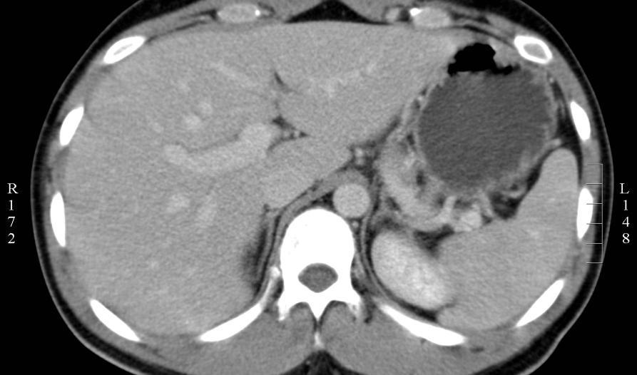
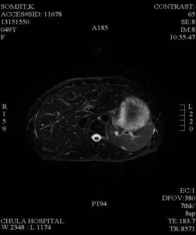
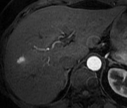
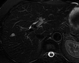
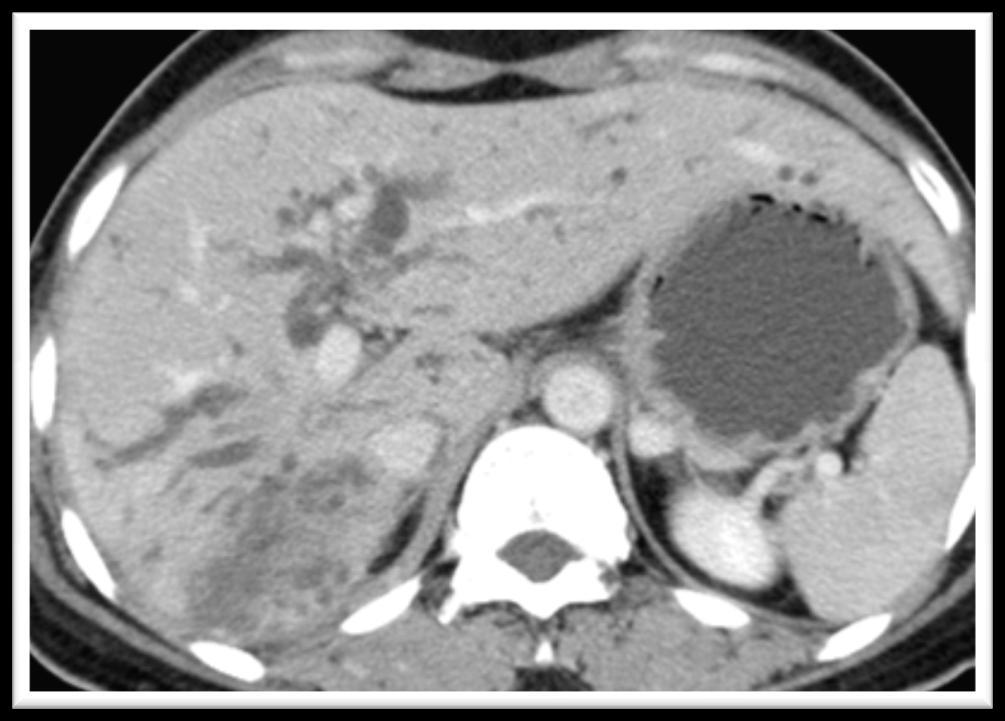
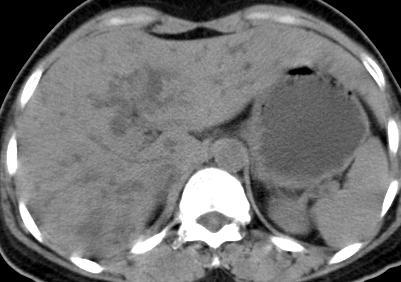
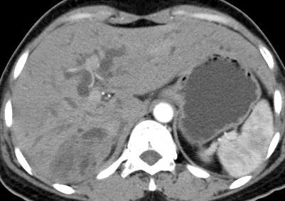
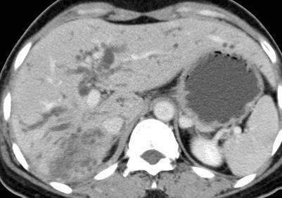
26 Fatty liver Many causes obesity, DM, malnutrition or exposure to ethanol or other toxins Imaging findings vary depending on amount of fat US diffuse increased hepatic parenchymal echogenicity CT diffuse decreased attenuation


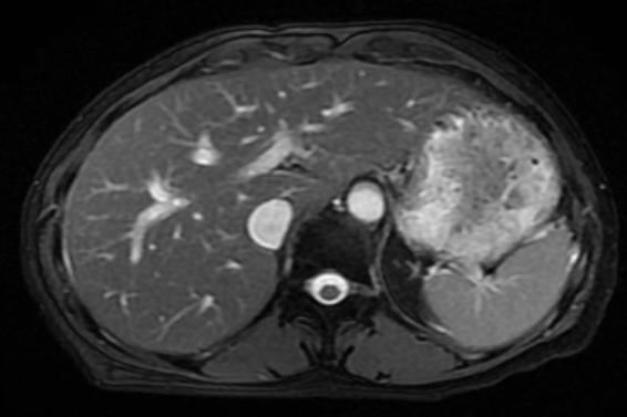
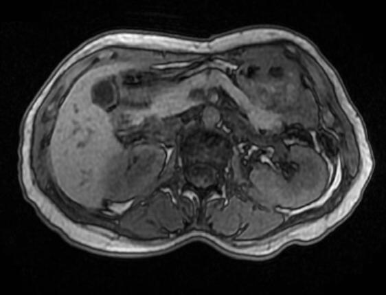
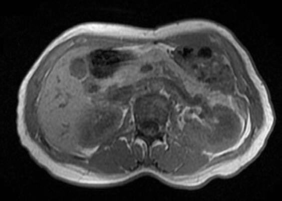
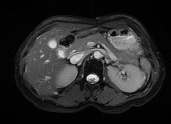
27 HEPATITIS Most cases normal US Diffusely decreased hepatic parenchymal echogenicity, with accentuated brightness of the portal triads and periportal cuffing Hepatomegaly and thickening of gallbladder wall
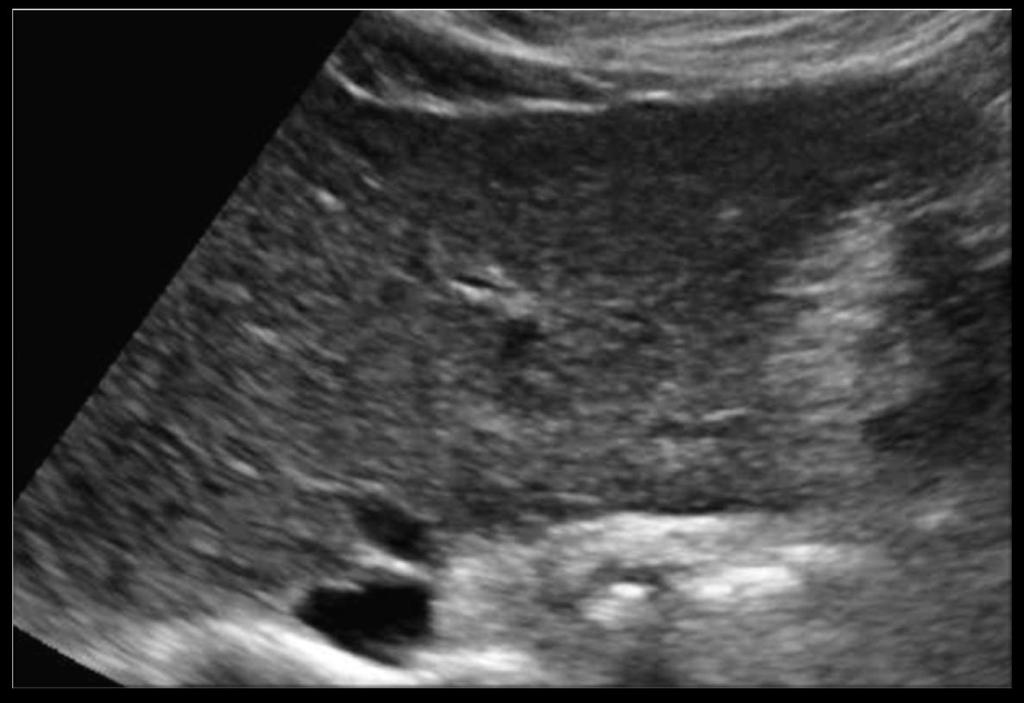
28 Liver cirrhosis ocauses Viral hepatitis: B and C Alcohol Toxin NASH Autoimmune
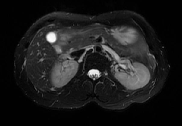
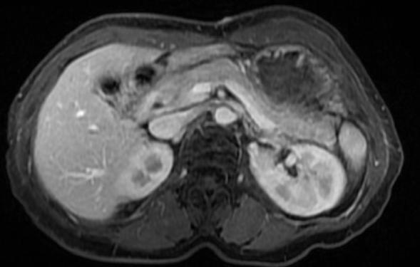
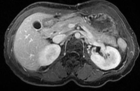
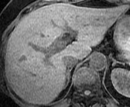
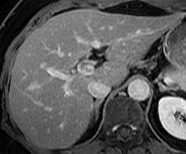
29 Liver cirrhosis Morphologic change Hypertrophy of lateral segment of left hepatic lobe and caudate lobe Atrophy of medial segment of left hepatic lobe and right hepatic lobe Nodular hepatic surface Regenerative nodules Fibrosis Expanded gallbladder fossa sign Dilatation of right inferior phrenic artery Gastrointestinal wall thickening Portal hypertension
30 Liver cirrhosis Portal hypertension o Portosystemic collaterals: increased size and number of retroperitoneal vessels in splenic hilum gastrohepatic ligament paraesophageal region splenorenal shunt canalization of paraumbilical vein o Splenomegaly o Ascites
31


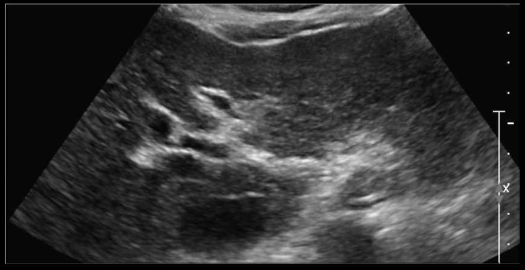
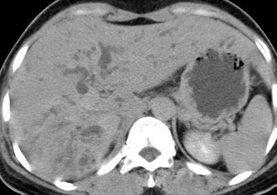
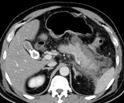
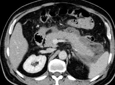
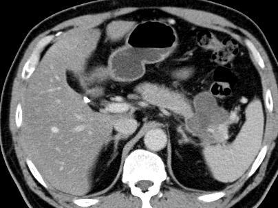
32 Liver cirrhosis with portal hypertension


33 Cyst o Greater prevalence in older patients o Typically asymptomatic o US anechoic lesion with posterior enhancement o CT o Water density (<20 HU) o Not enhance o Well defined border without perceptible walls o No mural nodularity or wall calcification o MR non-enhanced low SI on T1W, high SI on T2W and heavy T2W
34 Hemangioma o M/C benign liver tumor, prevalence up to 20% o Usually incidental finding o Occur in all age groups but more common in adults, particularly women o Solitary or multiple lesions


35 Hemangioma Typical sonographic appearance well defined, homogeneous hyperechoic May appear hypoechoic within background of a fatty infiltrated liver
36 Hemangioma CT findings Well defined hypodensity mass MRI findings T1W low SI T2W and heavy T2W high SI, giant hemangioma central area of either bright, dark or mixed SI and a network of multiple fibrous septae of low SI Typical enhancement pattern peripheral nodular enhancement with centripetal fill-in on later phases
37
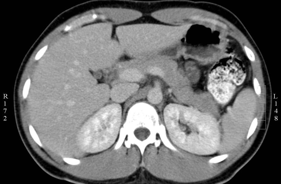
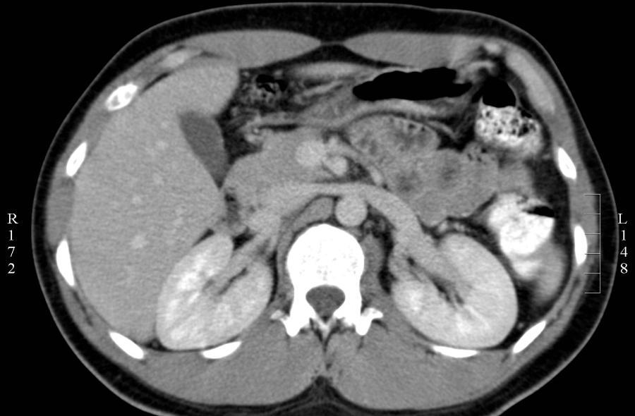
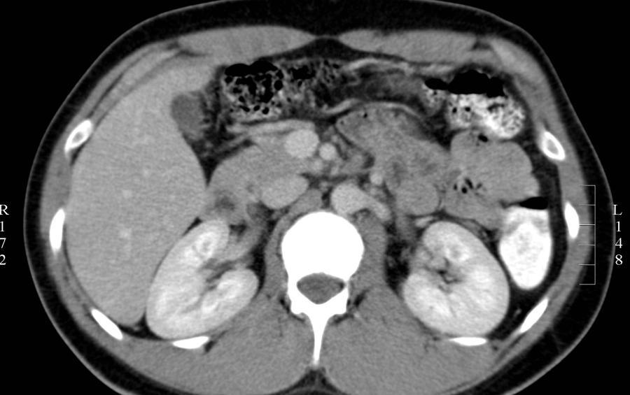
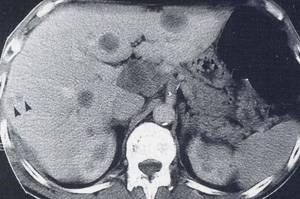
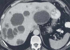
38

39 Abscess o Causes Pyogenic hepatic abscess (M/C) o Clostridium species o Gram-negative bacteria: E.coli, Bacteroides species o Coalescent, grouped appearance Amebic abscess: Entamoeba histolytica Fungal abscess: Candida albicans o Single or multiple o Can be small or large o Hepatomegaly o Pleural effusion o Gas within abscess (esp. Klebsiella)

40 Liver abscess US findings Hypoechoic lesion with well defined mildly echoic rim Posterior acoustic enhancement Low-level echoes/ fluiddebris level Intensely echogenic reflections with reverberations


41 Liver abscess CT findings Centrally hypodense lesion Peripheral enhancing rim Perilesional hyperemia Air bubbles may be present
42 Hepatocellular carcinoma M/C primary malignant hepatic neoplasm Predominant causal factors cirrhosis from alcoholism, viral hepatitis and toxin exposure

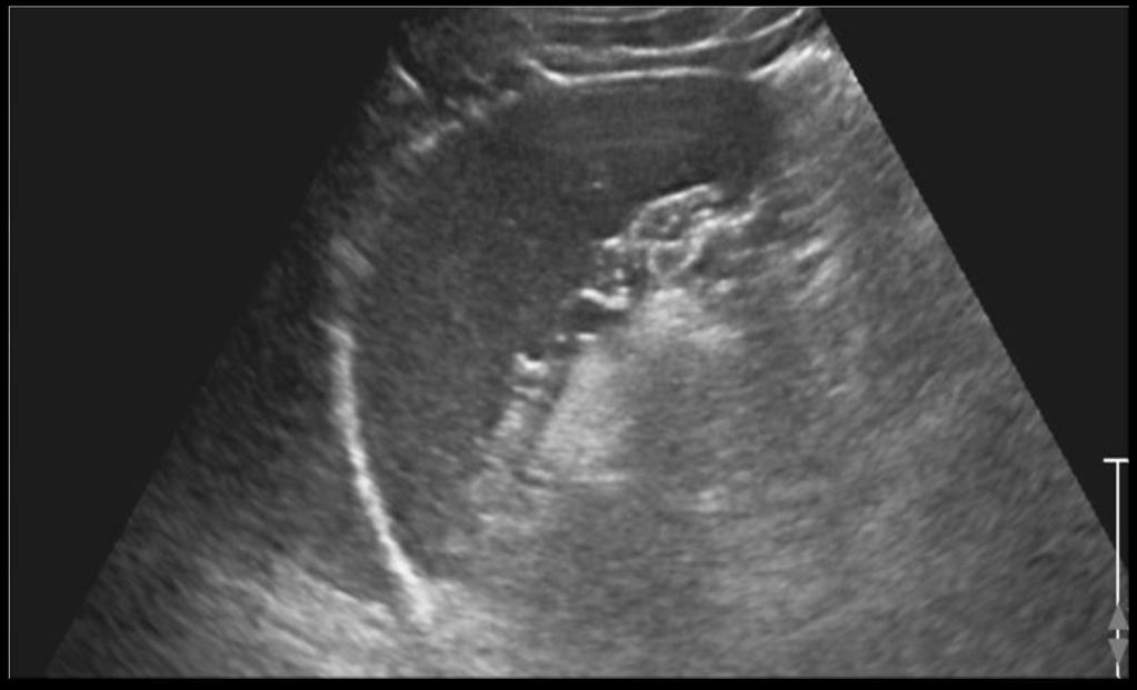
43 Hepatocellular carcinoma Ultrasound o Lower sensitivity and specificity than CT or MR in diagnosing HCC o Variable echogenicity: hypoechoic, hyperechoic and mixed echogenicity o Thin hypoechoic band of capsule


44 Hepatocellular carcinoma CT findings Hypodensity mass ± necrosis, fat, calcification Early enhancing mass with rapid washout on the late phase Late enhancement of capsule Tumor thrombus detection of early enhancement of thrombus during arterial phase
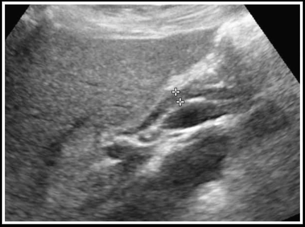
45 Hepatic Metastasis o Most common malignant focal liver lesions in the non-cirrhotic liver o Features of metastasis is vary, may be expansive, infiltrative, surface spreading or miliary, depending on origin of primary tumor


46 Hepatic Metastasis Ultrasound Diagnostic sensitivity over 90% in detection of metastasis Sonographic patterns Hyperechoic Bull s eye or target pattern Hypoechoic Cystic Calcified Infiltrative Multiple lesions
47 METASTASES Hypervascular metastasis Hypovascular metastasis Cystic metastasis
48 Common diseases of biliary system Gallstone Cholecystitis Cholangitis Cholangiocarcinoma

49 Ultrasound Gallstone Imaging tool of choice, accuracy 96% Highly reflective echo, mobile and associated with posterior acoustic shadowing
50 Acute cholecystitis o Ultrasound First imaging modality of choice Most specific findings Gallstone, esp. impacted stone in cystic duct or gallbladder neck Positive sonographic Murphy sign Thickening of gallbladder wall (>3 mm) Distention of gallbladder lumen (diameter > 4 cm) Pericholecystic fluid collection Hyperemic gallbladder wall on Doppler US
51 Acute Cholecystitis
52 Cholangitis Sonography is advocated as the first imaging modality to determine the cause and level of obstruction and to exclude other diseases Sonographic findings Dilatation of the biliary tree Choledocholithiasis and possibly sludge Bile duct wall thickening Hepatic abscess
53 Cholangitis with CBD stone
54 Cholangiocarcinoma
55
56 Common diseases of pancreas Pancreatitis Adenocarcinoma
57 Acute pancreatitis Acute inflammatory process of pancreas with variable involvement of other regional tissue or remote organ systems Etiology Alcohol, stone, metabolic, infection, trauma, drug Clinical severe mid epigastric pain radiating to back Increased serum amylase and lipase Diagnosis based on clinical and laboratory findings
58 Acute pancreatitis Role of ultrasound Detection of gallstone or bile duct stone Survey complication as peripancreatic fluid Follow up complication Guide intervention CT is primary modality of choice to identified necrotic parenchyma and extraparenchymal involvement

59 Acute pancreatitis Sonographic findings o Negative in mild form o Focal pancreatitis focal isoechoic or hypoechoic enlargement of pancreas o Diffuse pancreatitis increasingly hypoechogenic relative to normal liver and increased size o Focal hemorrhage focal echogenic mass


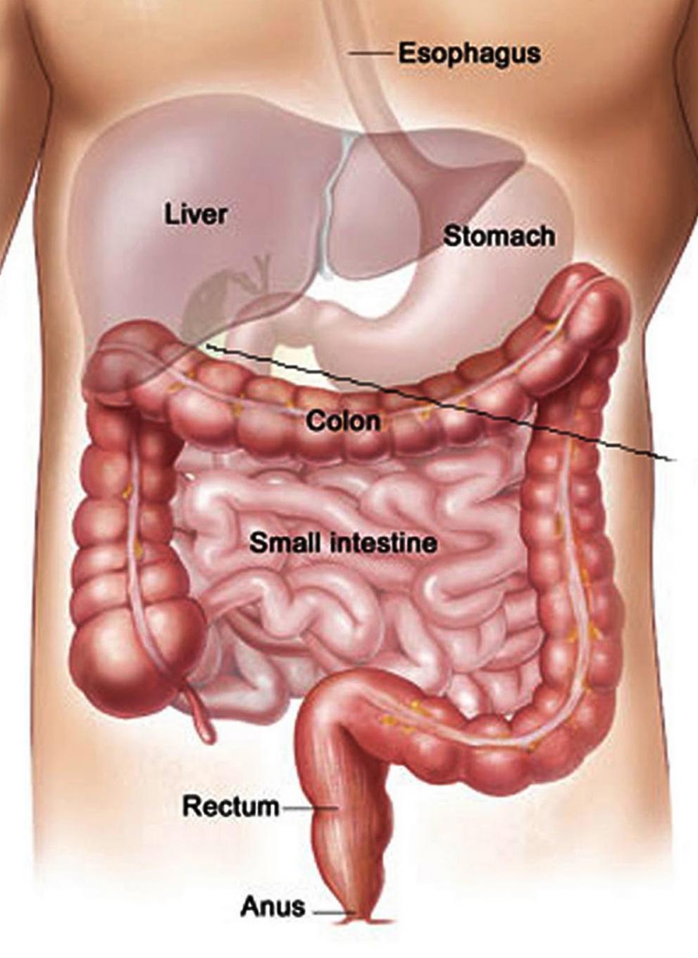
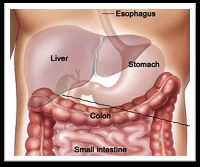
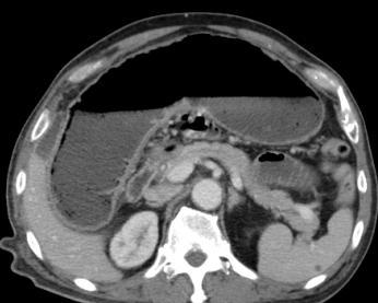
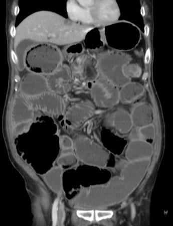
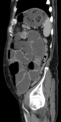
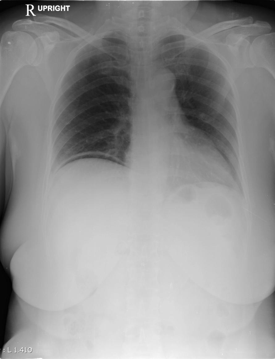
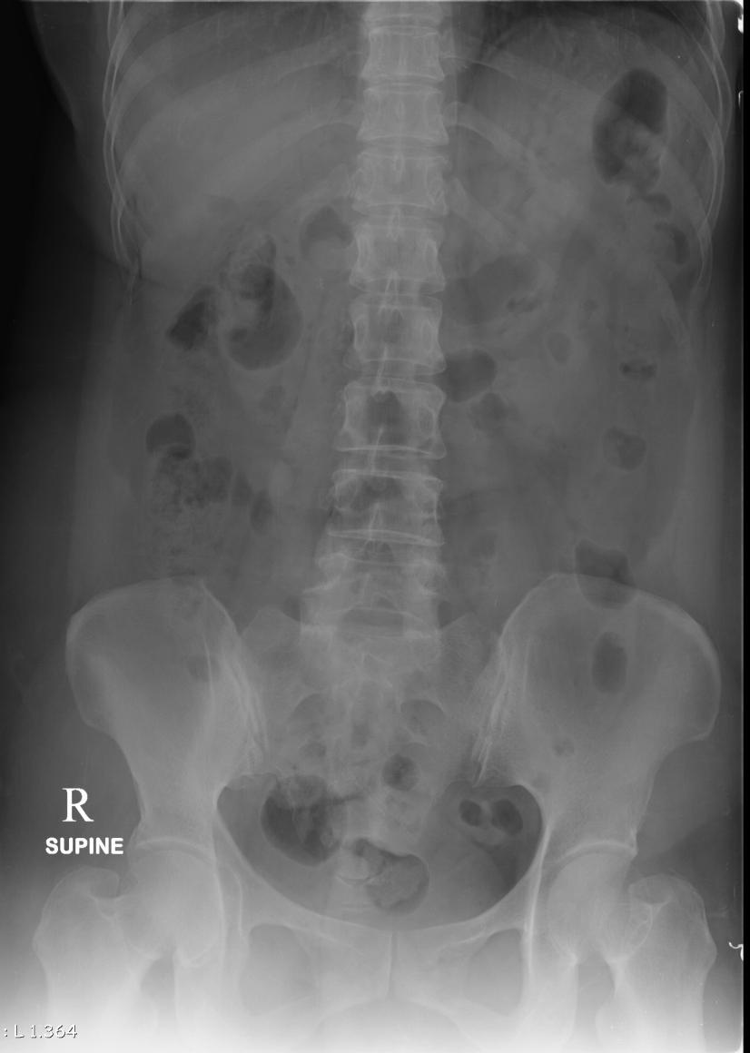
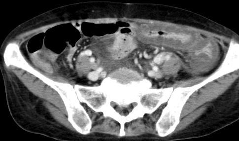
60 Acute pancreatitis CT findings Focal or diffuse pancreatic enlargement Heterogeneous enhancement with nonenhancing necrotic areas Rim enhancement of fluid collection, abscess and pseudocyst Infiltration of peripancreatic fat Gallstone Pseudoaneurysm Pleural effusion and atelectasis of basal lungs

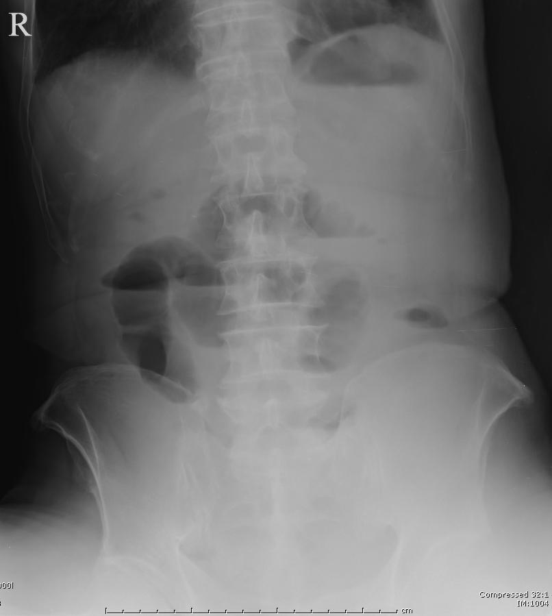
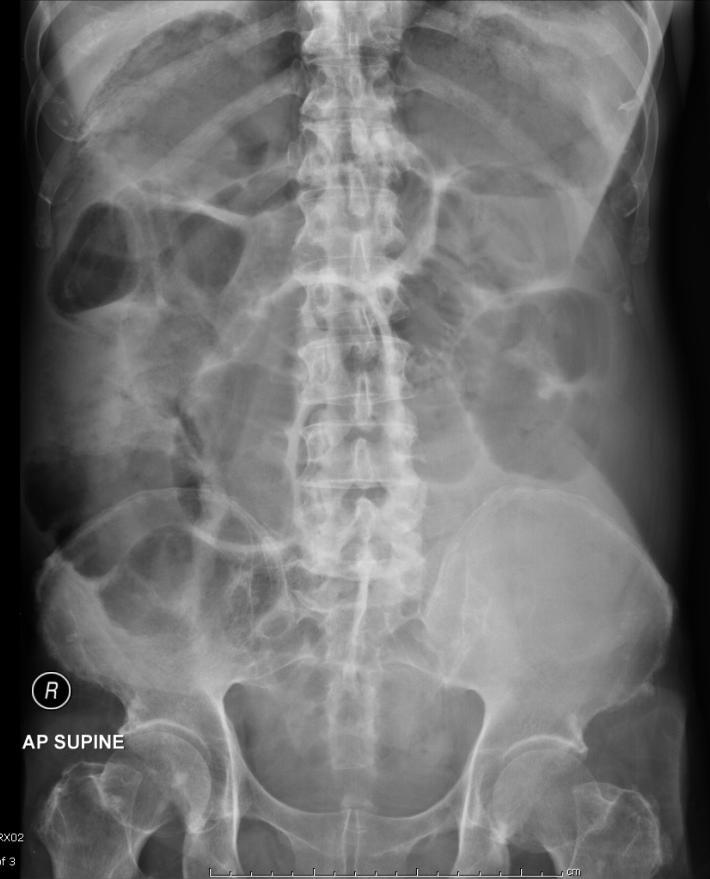
61 Day 4 Day 25 1 y
62 Chronic pancreatitis An inflammatory disease process that leads to progressive and irreversible structural damage of pancreas permanent dysfunction of both endocrine and exocrine pancreatic function Etiology Alcohol abuse Genetic factors Hypertriglyceridemia Hypercalcemia Autoimmunity Pancreatic duct obstruction

63 Chronic pancreatitis Radiography Calcifications projecting over epigastrium can be seen in 30-70% Specific but poorly sensitive

64 Chronic pancreatitis Ultrasound findings Pancreatic calcifications (40%), may be focal or diffuse Pancreatic enlargement or atrophy Ductal dilatation Pseudocyst
65 Pancreatic adenocarcinoma Malignancy arises from ductal epithelium of exocrine pancreas Presentation varies with location Head obstructive jaundice, pain and weight loss Body & tail weight loss and massive hepatic metastasis Mean age at onset 55 years Location Head 60% Body 20% CT scan is recommended for staging, pre- and post therapeutic evaluation, evaluating complication


66 Pancreatic Adenocarcinoma CT findings Heterogeneous poorly enhancing mass Parenchymal atrophy and ductal dilatation upstream from tumor Lesion in head may cause CBD obstruction
67 Common diseases of GI system Bowel obstruction Bowel perforation Infection/inflammation Cancer

68 Small bowel obstruction

69 Large bowel obstruction

70 Large bowel obstruction from colonic cancer
71
72 Colitis
73 Esophageal carcinoma


74 Gastric cancer malignancy arising from gastric mucosa Polypoid or circumferential mass with no peristalsis through lesion Morphology polypoid, ulcerated, infiltrative lesions CT Negative contrast agent Polypoid mass ± ulceration Focal wall thickening with mucosal irregularity or focal infiltration of wall Gas filled ulcer crater within mass Wall thickening with loss of normal rugal fold pattern Enhancing thickened wall CA cardia irregular soft tissue thickening; lobulated mass

")

75 CA colon BE Sessile/ pedunculated lesion Semiannular lesion Annular (apple core) lesion CT Asymmetric mural thickening ± irregular surface
Normal Sonographic Anatomy
 hapter 2:The Liver DUNSTAN ABRAHAM Normal Sonographic Anatomy Homogeneous, echogenic texture (Figure 2-1) Measures approximately 15 cm in length and 10 12.5 cm anterior to posterior; measurement taken
Radiology of hepatobiliary diseases
 GI cycle - Lecture 14 436 Teams Radiology of hepatobiliary diseases Objectives 1. To Interpret plan x-ray radiograph of abdomen with common pathologies. 2. To know the common pathologies presentation.
Abdominal ultrasound:
 Abdominal ultrasound: Non-traumatic acute abdomen Wittanee Na-ChiangMai, MD Department of Radiology ChiangMai University 26/04/2017 Contents Technique of examination Normal anatomy Emergency conditions
Imaging of liver and pancreas
 Imaging of liver and pancreas.. Disease of the liver Focal liver disease Diffusion liver disease Focal liver disease Benign Cyst Abscess Hemangioma FNH Hepatic adenoma HCC Malignant Fibrolamellar carcinoma
IT 의료융합 1 차임상세미나 복부질환초음파 이재영
 IT 의료융합 1 차임상세미나 2013-4-3 복부질환초음파 이재영 나는오늘누구를위하여 종을울리나? 전통적의료 의사 공학설계자 의사 최첨단진단장비들 USG, CT, MRI 환자 환자 현대의료 사용자중심의사고 US in the Abdomen Detection DDx Look Behavior Response by external stimuli Guiding Tool
HEPATO-BILIARY IMAGING
 HEPATO-BILIARY IMAGING BY MAMDOUH MAHFOUZ MD PROF.OF RADIOLOGY CAIRO UNIVERSITY mamdouh.m5@gmail.com www.ssregypt.com CT ABDOMEN Indications Patient preparation Patient position Scanogram Fasting 4-6 hours
Case Study: #3: Gallbladder Carcinoma?
 Case Study: #3: Gallbladder Carcinoma? By: Megan Wyatt K. SON Wyatt 225 2B1 RDMS, RVT Patient: Male 85 YOA Caucasian Indication: Elevated Alkaline Phosphatase History Annual physical showed elevated alkaline
Anatomy Jessica Ferguson Ashley Dobos May 31, 2006 LIVER
 Anatomy Jessica Ferguson Ashley Dobos May 31, 2006 LIVER 1) Other Names: Reidel s Lobe normal anatomic variant; projection of the right lobe that can extend as far as the iliac crest (Tempkin, p.54, Anatomy).
US in non-traumatic acute abdomen. Lalita, M.D. Radiologist Department of radiology Faculty of Medicine ChiangMai university
 US in non-traumatic acute abdomen Lalita, M.D. Radiologist Department of radiology Faculty of Medicine ChiangMai university Sagittal Orientation Transverse (Axial) Orientation Coronal Orientation Intercostal
Guidelines, Policies and Statements D5 Statement on Abdominal Scanning
 Guidelines, Policies and Statements D5 Statement on Abdominal Scanning Disclaimer and Copyright The ASUM Standards of Practice Board have made every effort to ensure that this Guideline/Policy/Statement
Biliary Tree Ultrasound - In a nutshell. Pamela Parker Lead Sonographer
 Biliary Tree Ultrasound - In a nutshell Pamela Parker Lead Sonographer Aims Review what we know about the biliary system Common pathologies Pitfalls Reporting tips The Nutshell Background Biliary examinations
CT 101 :Pancreas and Spleen
 CT 101 :Pancreas and Spleen Shikha Khullar,, MD, MPH Division of Radiology University of South Alabama The Pancreas Normal Pancreas 3 Phase Pancreatic CT Non contrast Arterial phase : 30-35 35 second
Pediatric Hepatobiliary, Pancreatic & Splenic US
 Pediatric Hepatobiliary, Pancreatic & Splenic US Susan J. Back, MD Department of Radiology, The Children s Hospital of Philadelphia No Disclosures Objectives Normal Abnormal: cases and US advances Objectives
Abdominal Ultrasound. Diane Hallinen, MD. Bloodroot
 Abdominal Ultrasound Diane Hallinen, MD Bloodroot Abdominal Ultrasound Vasculature Hepatobiliary Spleen Kidney Bladder Bowel Where to put the probe? Vasculature We are going to talk about Celiac Trunk
Hepatobiliary Ultrasound Rimon Bengiamin, MD, RDMS Assistant Clinical Professor Director of Emergency Ultrasound UCSF Fresno. Objectives. Why?
 Hepatobiliary Ultrasound Rimon Bengiamin, MD, RDMS Assistant Clinical Professor Director of Emergency Ultrasound UCSF Fresno Objectives Discuss the goals of point-of-care biliary ultrasound Review the
Biliary Tree Ultrasound - In a nutshell. Pamela Parker Lead Sonographer
 Biliary Tree Ultrasound - In a nutshell Pamela Parker Lead Sonographer Aims Review what we know about the biliary system Common pathologies Pitfalls Reporting tips The Nutshell Background Biliary examinations
Interesting Cases from Liver Tumor Board. Jeffrey C. Weinreb, M.D.,FACR Yale University School of Medicine
 Interesting Cases from Liver Tumor Board Jeffrey C. Weinreb, M.D.,FACR Yale University School of Medicine jeffrey.weinreb@yale.edu Common Liver Diseases Hemangioma Cyst FNH Focal Fat/Sparing THID Non-Cirrhotic
The Focal Hepatic Lesion: Radiologic Assessment
 The Focal Hepatic Lesion: Radiologic Assessment Kevin Kuo, Harvard Medical School Year III Our Patient: PS 67 y/o female w/ long history of alcohol use Drinking since age 18, up to one bottle of wine/day
Case Discussion Splenic Abscess
 Case Discussion Splenic Abscess Personal Data Gender: male Birth Date: 1928/Mar/06th Allergy: Mefenamic Smoking: 0.5 PPD for 55 years Alcohol: negative (?) 4 Months Ago Abdominal pain: epigastric area
Biliary Ultrasonography Kathleen O Brien MD MPH RDMS Kaiser Permanente South Sacramento
 Biliary Ultrasonography Kathleen O Brien MD MPH RDMS Kaiser Permanente South Sacramento https://www.google.com/search?sa=g&hl=en&q=public+disclosure&tbm=isch&tbs=simg:caqsigeahwelekju2aqaaawlelcmpwgaygpgcamskpib_1qnza7ai
Alice Fung, MD Oregon Health and Science University
 Alice Fung, MD Oregon Health and Science University Disclosure Comments The speaker Alice Fung, MD Has relevant financial relationships to disclose. Received honorarium from (Guerbet). This individual
IMAGING OF LIVER, BILIARY TREE, PANCREAS
 IMAGING OF LIVER, BILIARY TREE, PANCREAS Department of Radiology West China Hospital, Sichuan University Yao Jin Learning Points The methodology for imaging the LBP (liver, biliary tree, and pancreas )
Liver, Pancreas and Biliary System. Wirana Angthong, M.D.
 Liver, Pancreas and Biliary System Wirana Angthong, M.D. Objectives Outline Anatomy Imaging Techniques Common Diseases Outline Anatomy Imaging Techniques Common Diseases Liver anatomy Morphological anatomy:
4/9/2018 OBJECTIVES PANCREAOTO BILIARY ULTRASOUND: BEYOND CHOLECYSTITIS
 PANCREAOTO BILIARY ULTRASOUND: BEYOND CHOLECYSTITIS Jean Yves Sewah Kaiser Permanente West Los Angeles 1 OBJECTIVES Discuss the role of ultrasound in the evaluation of the gallbladder, biliary tree and
GASTROINTESTINAL IMAGING STUDY GUIDE
 GASTROINTESTINAL IMAGING STUDY GUIDE Pharynx Diverticula Foreign bodies Trauma o Motility Disorders Esophagus Diverticula Trauma Esophagitis Barrett esophagus Rings, webs, and strictures Varices Benign
Malignant Focal Liver Lesions
 Malignant Focal Liver Lesions Other Than HCC Pablo R. Ros, MD, MPH, PhD Departments of Radiology and Pathology University Hospitals Cleveland Medical Center Case Western Reserve University Pablo.Ros@UHhospitals.org
Gallbladder & Pancreas Ultrasonography
 복부초음파 : 담낭과췌장 Gallbladder & Pancreas Ultrasonography 김정훈 Department of Radiology 1 Interaction of sound with matter (1) 반사 (Reflection) (2) 굴절 (Refraction) (3) 흡수 (Absorption) (4) 산란 (Scattering) 음향저항
My Patient Has Abdominal Pain PoCUS of the Biliary Tract and the Urinary Tract
 My Patient Has Abdominal Pain PoCUS of the Biliary Tract and the Urinary Tract Objectives PoCUS for Biliary Disease PoCUS for Renal Colic PoCUS for Urinary Retention Biliary Disease A patient presents
Role of imaging in RCC. Ultrasonography. Solid lesion. Cystic RCC. Solid RCC 31/08/60. From Diagnosis to Treatment: the Radiologist Perspective
 Role of imaging in RCC From Diagnosis to Treatment: the Radiologist Perspective Diagnosis Staging Follow up Imaging modalities Limitations and pitfalls Duangkamon Prapruttam, MD Department of Therapeutic
Elastography in the. technically difficult patient. EPIQ ultrasound system. Ultrasound
 Ultrasound Elastography in the technically difficult patient EPIQ ultrasound system Chairman Department of Diagnostic Radiology Allegheny General Hospital Pittsburgh, PA, USA You can offer more information
Anatomy of the biliary tract
 Harvard-MIT Division of Health Sciences and Technology HST.121: Gastroenterology, Fall 2005 Instructors: Dr. Jonathan Glickman Anatomy of the biliary tract Figure removed due to copyright reasons. Biliary
US LI-RADS v2017 CORE
 US LI-RADS v2017 CORE Screening or surveillance US in patient at high risk for HCC US category US-1 US-2 US-3 Negative Subthreshold Positive Category Concept Definition US-1 Negative US-2 Subthreshold
X-Ray Corner. Imaging Approach to Cystic Liver Lesions. Pantongrag-Brown L. Solitary cystic liver lesions. Hepatic simple cyst (Figure 1)
") THAI J 136 Imaging Approach to Cystic Liver Lesions GASTROENTEROL 2013 X-Ray Corner Imaging Approach to Cystic Liver Lesions Pantongrag-Brown L Cystic liver lesions are common findings in daily practice
THAI J 136 Imaging Approach to Cystic Liver Lesions GASTROENTEROL 2013 X-Ray Corner Imaging Approach to Cystic Liver Lesions Pantongrag-Brown L Cystic liver lesions are common findings in daily practice
Contrast enhanced ultrasound (CEUS) in gallbladder and bile duct pathology: technique, interpretation and clinical applications
 in gallbladder and bile duct pathology: technique, interpretation and clinical applications") Contrast enhanced ultrasound (CEUS) in gallbladder and bile duct pathology: technique, interpretation and clinical applications Poster No.: C-2099 Congress: ECR 2011 Type: Scientific Exhibit Authors: E.
Contrast enhanced ultrasound (CEUS) in gallbladder and bile duct pathology: technique, interpretation and clinical applications Poster No.: C-2099 Congress: ECR 2011 Type: Scientific Exhibit Authors: E.
Policies, Standards, and Guidelines. Guidelines for Abdominal Ultrasound Examination
 Policies, Standards, and Guidelines Guidelines for Abdominal Ultrasound Examination Approved by Council Feb 2018 Disclaimer and Copyright The ASUM Standards of Practice Board have made every effort to
Appendix 5. EFSUMB Newsletter. Gastroenterological Ultrasound
 EFSUMB Newsletter 87 Examinations should encompass the full range of pathological conditions listed below A log book listing the types of examinations undertaken should be kept Training should usually
Abdomen and Retroperitoneum Ultrasound Protocols
 Abdomen and Retroperitoneum Ultrasound Protocols Reviewed By: Anna Ellermeier, MD Last Reviewed: March 2018 Contact: (866) 761-4200, Option 1 **NOTE for all examinations: 1. If documenting possible flow
Newcastle HPB MDM updated radiology imaging protocol recommendations. Author Dr John Scott. Consultant Radiologist Freeman Hospital
 Newcastle HPB MDM updated radiology imaging protocol recommendations Author Dr John Scott. Consultant Radiologist Freeman Hospital This document is intended as a guide to aid radiologists and clinicians
Case 1. Intro to Gallbladder & Pancreas Pathology. Case 1 DIAGNOSIS??? Acute Cholecystitis. Acute Cholecystitis. Helen Remotti M.D.
 Cholecystitis acute chronic Gallbladder tumors Adenomyoma (benign) Adenocarcinoma Pancreatitis acute chronic Pancreatic tumors Intro to Gallbladder & Pancreas Pathology Helen Remotti M.D. Case 1 70 year
Evaluation of Liver Mass Lesions. American College of Gastroenterology 2013 Regional Postgraduate Course
 Evaluation of Liver Mass Lesions American College of Gastroenterology 2013 Regional Postgraduate Course Lewis R. Roberts, MB ChB, PhD Division of Gastroenterology and Hepatology Mayo Clinic College of
CTA/MRA of Pediatric Hepatic Masses Radiology-Pathology Correlation
 Acta Radiológica Portuguesa, Vol.XVIII, nº70, pág. 41-50, Abr.-Jun., 2006 CTA/MRA of Pediatric Hepatic Masses Radiology-Pathology Correlation Marilyn J. Siegel Mallinckrodt Institute of Radiology, Washington
Hepatic Imaging: What Every Practitioner Should Know
 Hepatic Imaging: What Every Practitioner Should Know Shuchi K. Rodgers, MD Section Chief, Abdominal Imaging Director of Ultrasound Department of Radiology Einstein Medical Center rodgerss@einstein.edu
Sonography of Gall Bladder
 Sonography of Gall Bladder Vikram Dogra,MD Professor of Radiology, Urology and BME Director of Ultrasound Associate Chair of Education and Research University of Rochester, NY Objectives Describe the Congenital
Dr Claire Smith, Consultant Radiologist St James University Hospital Leeds
 Dr Claire Smith, Consultant Radiologist St James University Hospital Leeds Imaging in jaundice and 2ww pathway Image protocol Staging Limitations Pancreatic cancer 1.2.4 Refer people using a suspected
Radiological Reasoning: Incidentally Discovered Liver Mass
 AJR Integrative Imaging LIFELONG LEARNING FOR RADIOLOGY This Radiological Reasoning article is available for SAM credit and CME credits when completed with the additional educational material provided
Multiple Primary Quiz
 Multiple Primary Quiz Case 1 A 72 year old man was found to have a 12 mm solid lesion in the pancreatic tail by computed tomography carried out during a routine follow up study of this patient with adult
Contents. Basic Ultrasound Principles and Terminology. Ultrasound Nodule Characteristics
 Contents Basic Ultrasound Principles and Terminology Basic Ultrasound Principles... 1 Ultrasound System... 2 Linear Transducer for Superficial Images and Ultrasound-Guided FNA... 3 Scanning Planes... 4
Multi modality Imaging in Acute Pancreatitis. Marsha Lynch, HMS III Gillian Lieberman, MD BIDMC Core Clerkship in Radiology March 2009
 Multi modality Imaging in Acute Pancreatitis Marsha Lynch, HMS III Gillian Lieberman, MD BIDMC Core Clerkship in Radiology March 2009 Our Patient R: Introduction 52M with 10d history of nausea, vomiting
Disorders of the Liver and Pancreas
 Disorders of the Liver and Pancreas Liver Lobule Hexagonal plates Sinusoids Triads Bile duct branch Arteriole Venuole Blood flows from periphery to Central vein Space of Dissé Lobular Microanatomy Hepatocytes
Financial Disclosure
 Benign Liver Masses Adil Abdalla, MBBS Creighton University-CHI Health August 25, 2018 Financial Disclosure Nothing to disclose Financial Disclosure 1 Objectives To assess patients with benign liver tumors
CT EVALUATION OF GASTRIC LESIONS:
 CT EVALUATION OF GASTRIC LESIONS: Pictural essay Hasni Bouraoui I, Kahloun A, Jemni H, Elouni F, Moulahi H, Daadoucha A, Ben Ali A, Sriha B, Tlili Graies K Departments of Radiology, Gastro enterology,
Disorders of the Liver, Gallbladder and Pancreas
 Disorders of the Liver, Gallbladder and Pancreas Objectives: Disorders of the liver Disorders of the gall bladder Disorders of the pancreas Part 1: Disorders of the Liver 1 Jaundice: is a manifestation
Leonard M. Glassman MD
 BI-RADS The New BI-RADS Leonard M. Glassman MD FACR Former Chief of Breast Imaging American Institute for Radiologic Pathology Washington Radiology Associates, PC Breast Imaging Reporting and Data System
OF HEMATOMA INTRACRANIAL HEMORRHAGE
 SCOPE TUTORIAL RADIOLOGY CT BRIAN BREASTS US MRCP Natrada Rawdhetubhai, M.D. Radiology Department, Lerdsin General Hospital CT OF HEMATOMA INTRACRANIAL HEMORRHAGE Intra-axial Extra-axial Intraventricular
Common and unusual CT and MRI manifestations of pancreatic adenocarcinoma: a pictorial review
 Review Article Common and unusual CT and MRI manifestations of pancreatic adenocarcinoma: a pictorial review Min-Jie Yang, Su Li, Yong-Guang Liu, Na Jiao, Jing-Shan Gong Department of Radiology, Shenzhen
Role of Imaging Methods in Diagnosis of Acute Pancreatitis. Válek V. Radiologická klinika, FN Brno a LF MU v Brně
 Role of Imaging Methods in Diagnosis of Acute Pancreatitis Válek V. Radiologická klinika, FN Brno a LF MU v Brně New Classification: Acute Pancreatitis 2007 revision of Atlanta classification and definitions
Gastrointestinal Tract. Anatomy of GI Tract. Anatomy of GI Tract. (Effective February 2007) (1%-5%)
 (1%-5%)") Gastrointestinal Tract (Effective February 2007) (1%-5%) Anatomy of GI Tract Esophagus bulls-eye or target EG junction seen on sagittal scan posterior to left lobe of liver and anterior to aorta Anatomy
Gastrointestinal Tract (Effective February 2007) (1%-5%) Anatomy of GI Tract Esophagus bulls-eye or target EG junction seen on sagittal scan posterior to left lobe of liver and anterior to aorta Anatomy
Use of Ultrasound in NAFLD
 Institute for Liver and Digestive Health Use of Ultrasound in NAFLD Dr. Davide Roccarina Specialist in General Medicine Specialist Doctor in Clinical Ultrasound and non-invasive liver assessment Hepatology
Renal masses - the role of diagnostic imaging
 Renal masses - the role of diagnostic imaging Poster No.: C-2471 Congress: ECR 2015 Type: Educational Exhibit Authors: V. Rai#; Bjelovar/HR Keywords: Cysts, Cancer, Structured reporting, Ultrasound, MR,
Chief Complain. Liver lesion found in routine health check 41 days ago
 Chief Complain Liver lesion found in routine health check 41 days ago Present Illness On 2005-7-26 at 台北署立醫院 he underwent a health check for the first time. Abdominal US showed suspicious of a 6*5 cm hepatoma,
Sex: 女 Age: 51 Occupation: 無 Admission date:92/07/22
 Sex: 女 Age: 51 Occupation: 無 Admission date:92/07/22 Chief complaint Unknown fever for one month Hand tremor and left huge renal tumor was noted Present illness Suffered from fever for one month, hand
Imaging in gastric cancer
 Imaging in gastric cancer Gastric cancer remains a deadly disease because of late diagnosis. Adenocarcinoma represents 90% of malignant tumors. Diagnosis is based on endoscopic examination with biopsies.
Portal Venous Thrombosis: Tumor VS Bland Thrombus
 June 2015 Portal Venous Thrombosis: Tumor VS Bland Thrombus SERGIO ALFARO, HARVARD MEDICAL SCHOOL YEAR III GILLIAN LIEBERMAN, MD Overview 2 Index Patient History Portal Venous Thrombosis (PVT) Imaging
Diffuse Gallbladder Wall Thickening: Differential Diagnosis
 van reda Vriesman et al. Diffuse Gallbladder Wall Thickening Hepatobiliary Imaging Pictorial Essay driaan C. van reda Vriesman 1 Marc R. Engelbrecht 2 Robin H. M. Smithuis 1 Julien. C. M. Puylaert 3 van
Objectives. Hepatobiliary Ultrasound: Anatomy, Technique, Pathology. RUQ: Normal Anatomy. Emergency Ultrasound: Gallbladder Location
 Hepatobiliary Ultrasound: Anatomy, Technique, Pathology Laleh Gharahbaghian, MD FAAEM Associate Director, EM Ultrasound Co-Director, EM Ultrasound Fellowship Stanford University Medical Center Seric Cusick,
DIAGNOSTIC IMAGING: LIVER DISEASE
 Vet Times The website for the veterinary profession https://www.vettimes.co.uk DIAGNOSTIC IMAGING: LIVER DISEASE Author : Abby Caine Categories : Vets Date : February 1, 2010 ABBY CAINE reviews both established
CT and MRI of Hepatic Contour Abnormalities
 Hepatobiliary Imaging Pictorial Essay CT and MRI of Jafi A. Lipson 1, Aliya Qayyum 1, David E. Avrin 2, Antonio Westphalen 1, Benjamin M. Yeh 1, Fergus V. Coakley 1 Fig. 1. 38-year-old woman imaged for
Abdominal Imaging - 9 Topics in 90 min
 Abdominal Imaging 9 topics in 90 min Antonio C. Westphalen, MD PhD Departments of Radiology and Biomedical Imaging, and Urology Liver Biliary tree Gallbladder Pancreas Kidneys Small bowel Colon Abscess?
Job Task Analysis for ARDMS Abdomen Data Collected: June 30, 2011
 Job Task Analysis for ARDMS Abdomen Data Collected: June 30, 2011 Reported: Analysis Summary for: Abdomen Examination Survey Dates 06/13/2011-06/26/2011 Invited Respondents 6,000 Surveys with Demographics
Cholangiocarcinoma: appearances and mimics
 Cholangiocarcinoma: appearances and mimics Poster No.: C-1572 Congress: ECR 2011 Type: Educational Exhibit Authors: C. Cardenas Valencia, J. Fernandez Jara, J. Cubero Carralero, B. Corral Ramos, P. Perez
Personal Profile. Name: 劉 XX Gender: Female Age: 53-y/o Past history. Hepatitis B carrier
 Personal Profile Name: 劉 XX Gender: Female Age: 53-y/o Past history Hepatitis B carrier Chief complaint Fever on and off for 2 days Present illness 94.10.14 Sudden onset of epigastric pain 94.10.15 Fever
GENERAL ABDOMINAL IMAGING PERITONEAL SPACE, PANCREAS, & SPLEEN. VMB 960 March 25, 2013
 GENERAL ABDOMINAL IMAGING PERITONEAL SPACE, PANCREAS, & SPLEEN VMB 960 March 25, 2013 REFERENCE Chapters 35-36 Pages 650-678 Chapter 37 Pages 694-701 Chapter 3 Pages 38-49 OBJECTIVES Radiography and Ultrasound
Hepatocellular carcinoma Cholangiocarcinoma. Jewels of hepatobiliary cancer imaging : what to look for? Imaging characteristics of HCC.
 Outline : Imaging Jewels Jewels of hepatobiliary cancer imaging : what to look for? Hepatocellular carcinoma Cholangiocarcinoma Surachate Siripongsakun, M.D. Chulabhorn Cancer Center Imaging characteristics
Intraductal papillary mucinous neoplasm of the bile ducts: a rare form of premalignant lesion of invasive cholangiocarcinoma
 Intraductal papillary mucinous neoplasm of the bile ducts: a rare form of premalignant lesion of invasive cholangiocarcinoma Authors: R. Revert Espí, Y. Fernandez Nuñez, I. Carbonell, D. P. Gómez valencia,
MALIGNANT HEPATIC NEOPLASMS: USING ULTRASONOGRAPHY AS A MEANS OF DEFINING HEPATIC LESIONS. 1.5 Contact Hours. Presented by: CEU Professor 7
 MALIGNANT HEPATIC NEOPLASMS: USING ULTRASONOGRAPHY AS A MEANS OF DEFINING HEPATIC LESIONS 1.5 Contact Hours Presented by: CEU Professor 7 www.ceuprofessoronline.com Copyright 8 2007 The Magellan Group,
Pediatric Retroperitoneal Masses Radiologic-Pathologic Correlation
 Acta Radiológica Portuguesa, Vol.XVIII, nº 70, pág. 61-70, Abr.-Jun., 2006 Pediatric Retroperitoneal Masses Radiologic-Pathologic Correlation Marilyn J. Siegel Mallinckrodt Institute of Radiology, Washington
State of the Art Imaging for Hepatic Malignancy: My Assignment
 State of the Art Imaging for Hepatic Malignancy: My Assignment CT vs MR vs MRCP Which one to choose for HCC vs Cholangiocarcinoma What special protocols to use for liver tumors Role of PET and Duplex US
of Thyroid Lesions Comet Tail Crystals
 2 Ultrasound Features of Thyroid Lesions There are many different features indicating a certain benign or malignant tumor type, but many of these are overlapping signs. Combining several features is considered
Essentials of Clinical MR, 2 nd edition. 65. Benign Hepatic Masses
 65. Benign Hepatic Masses Pulse sequences acquired for abdominal MRI typically consist of fast acquisition schemes such as single-shot turbo spin echo (i.e. HASTE) and gradient echo schemes such as FLASH
Liver Ultrasound - Beyond the Basics. Pamela Parker Lead Sonographer
 Liver Ultrasound - Beyond the Basics Pamela Parker Lead Sonographer Aims Review what we know about the liver Reasons for imaging Focal lesions Diffuse disease Can we do more? The Liver The Liver The Liver
PATHOLOGY MCQs. The Pancreas
 PATHOLOGY MCQs The Pancreas A patient with cystic fibrosis is characteristically: A. more than 45 years of age B. subject to recurring pulmonary infections C. obese D. subject to spontaneous fractures
Q129. Which of the following is NOT true about lymph node?
 Q129. Which of the following is NOT true about lymph node? (1). Normal lymph node is not seen in the ultrasound image (2). It is general that high frequency probe is used due to normal lymph node is located
ACUTE PANCREATITIS: NEW CLASSIFICATION OF AN OLD FOE. T Barrow, A Nasrullah, S Liong, V Rudralingam, S A Sukumar
 ACUTE PANCREATITIS: NEW CLASSIFICATION OF AN OLD FOE T Barrow, A Nasrullah, S Liong, V Rudralingam, S A Sukumar LEARNING OBJECTIVES q Through a series of cases illustrate the updated Atlanta symposium
Biliary cancers: imaging diagnosis. Study of 30 cases
 Biliary cancers: imaging diagnosis. Study of 30 cases N Hammoune, S Semlali, M Eddarai, T. Amil, M Zentar, S. El Kandri,, M Benameur,, S Chaouir. Radiology Department. Mohamed V Military Hospital. Rabat-
Liver Tumors. Prof. Dr. Ahmed El - Samongy
 Liver Tumors Prof. Dr. Ahmed El - Samongy Objective 1. Identify the most important features of common benign liver tumors 2. Know the risk factors, diagnosis, and management of hepatocellular carcinoma
Incidental Esophageal Findings on Chest CT. Amira Hussien, MD, Elliot Fishman, MD, Bouchra Younes, MD, Ahmed Hatw. Johns Hopkins Medical Institution
 Incidental Esophageal Findings on Chest CT Amira Hussien, MD, Elliot Fishman, MD, ouchra Younes, MD, Ahmed Hatw. Johns Hopkins Medical Institution I have nothing to disclose. DISCLOSURE INTRODUCTION Although
Emergent Right Upper Quadrant Sonography
 Image Presentation Emergent Right Upper Quadrant Sonography Susanna C. Spence, MD, Davis Teichgraeber, MD, Chitra Chandrasekhar, MD Objective. The purpose of this presentation is to review the sonographic
Liver Cancer (Hepatocellular Carcinoma or HCC) Overview
 Overview") Liver Cancer (Hepatocellular Carcinoma or HCC) Overview Recent advances in liver cancer care seek to address the rising incidence of liver cancer, which has steadily increased over the past three decades.
Liver Cancer (Hepatocellular Carcinoma or HCC) Overview Recent advances in liver cancer care seek to address the rising incidence of liver cancer, which has steadily increased over the past three decades.
Evidence based imaging of the pancreas
 Evidence based imaging of the pancreas D.Vanbeckevoort, D.Bielen, K.Op de beeck, R.Vanslembrouck Department of Radiology Chairman Prof. Dr. R.Oyen Non-invasive imaging tests available for the diagnosis
Navigating the Biliary Tract with CT & MR: An Imaging Approach to Bile Duct Obstruction
 Navigating the Biliary Tract with CT & MR: An Imaging Approach to Bile Duct Obstruction Ann S. Fulcher, MD Medical College of Virginia Virginia Commonwealth University Richmond, Virginia Objectives To
Ankur A. Gupta 1, Danny C. Kim, Glenn A. Krinsky, Vivian S. Lee
 Pictorial Essay T and MRI of irrhosis and its Mimics nkur. Gupta 1, Danny. Kim, Glenn. Krinsky, Vivian S. Lee irrhosis is among the leading causes of death in the western world. irrhosis and its associated
Intrahepatic Cholangiocarcinoma (ICC) Detected by Sonography
 Detected by Sonography") 661245JDMXXX10.1177/8756479316661245Journal of Diagnostic Medical SonographyHamer research-article2016 Case Study Intrahepatic Cholangiocarcinoma (ICC) Detected by Sonography Journal of Diagnostic Medical
661245JDMXXX10.1177/8756479316661245Journal of Diagnostic Medical SonographyHamer research-article2016 Case Study Intrahepatic Cholangiocarcinoma (ICC) Detected by Sonography Journal of Diagnostic Medical
X-ray Corner. Imaging of The Pancreas. Pantongrag-Brown L
 X-ray Corner 125 Imaging of The Pancreas Modern imaging modalities commonly used in pancreas include ultrasound (US), CT, and MRI. Pancreas is a retroperitoneal organ which makes it difficult to visualize
Abdominal Sonography Review
 L I F E I S A T E S T... P A S S I T! 1-2-3 Step Ultrasound Education & Test Preparation Step 1 Review text Step 2 Mock examination Step 3 Q&A memory skills flashcard drill Abdominal Sonography Review
Approach to the Patient with Liver Disease
 Approach to the Patient with Liver Disease Diagnosis of liver disease Careful history taking Physical examination Laboratory tests Radiologic examination and imaging studies Liver biopsy Liver diseases
6 th August 2018 Day 1 - Gallbladder & Bile duct Topic
 Venue: Sterling Hospital Auditorium, Sterling Hospitals, Gurukul Road Ahmedabad, Gujarat 6 th August 2018 Day 1 - Gallbladder & Bile duct Registration(8:00am-8:15am) Inauguration(8:15am-8:30am) Welcome
Cystic Fibrosis in Children and Young Adults: Findings on Routine Abdominal Sonography
 bdominal Sonography in Cystic Fibrosis bdominal Imaging Pictorial Essay Downloaded from www.ajronline.org by 37.44.206.10 on 01/08/18 from IP address 37.44.206.10. Copyright RRS. For personal use only;
Jaundice. Agnieszka Dobrowolska- Zachwieja, MD, PhD
 Jaundice Agnieszka Dobrowolska- Zachwieja, MD, PhD Jaundice definition Jaundice, as in the French jaune, refers to the yellow discoloration of the skin. It arises from the abnormal accumulation of bilirubin
Treatment of chronic calcific pancreatitis endoscopy versus surgery
 Treatment of chronic calcific pancreatitis endoscopy versus surgery 35 - year old ladypresented to LPC Mumbai with intermittent abdominal pain. Pain was intermittent, colicky, more in epigastrium and periumbilical
Pancreas Case Scenario #1
 Pancreas Case Scenario #1 An 85 year old white female presented to her primary care physician with increasing abdominal pain. On 8/19 she had a CT scan of the abdomen and pelvis. This showed a 4.6 cm mass