Supplemental Figure 1. Quantification of proliferation in thyroid of WT, Ctns -/- and grafted
|
|
|
- Madison Tucker
- 6 years ago
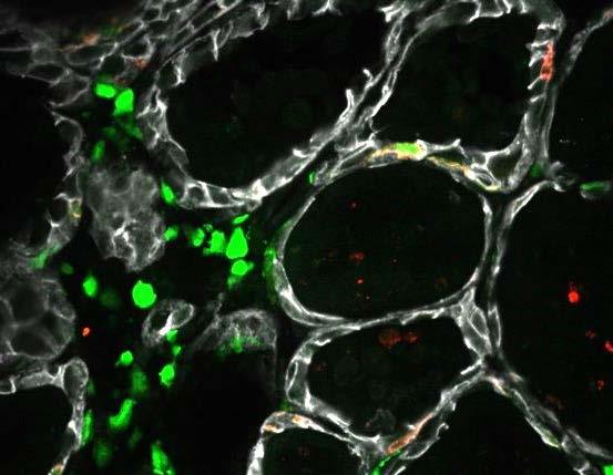
- Views:
Transcription
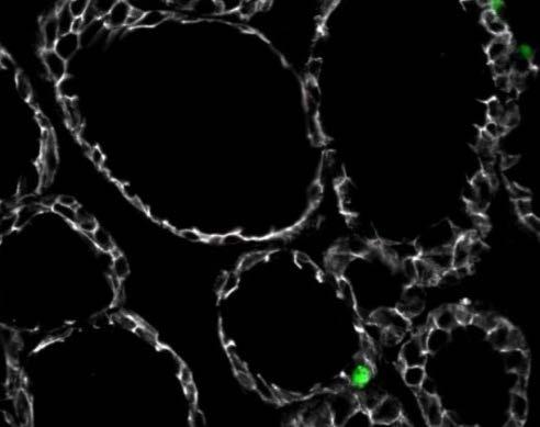
1 Supplemental Figure 1. Quantification of proliferation in thyroid of WT, Ctns -/- and grafted Ctns -/- mice. Cells immunolabeled for the proliferation marker (Ki-67) were counted in sections (n=3 WT, n=4 Ctns -/-, n=4 grafted Ctns -/- mice); 7 representative fields spanning each entire thyroid and corresponding to µm 2 were analyzed. Notice significant decrease of proliferation in thyrocytes of grafted compared to non-grafted Ctns -/- mice.* p<0.05 for comparison of thyrocytes. Supplemental Figure 2. Electron microscopy. Upper panel. Two low-magnification views of grafted Ctns -/- thyroid at 8 months / 6 months post-transplant. For comparison with non-grafted Ctns -/- thyroid, see (Gaide Chevronnay et al. Endocrinology 2015). At (a), three preserved columnar thyrocytes (activated) with well-defined apical junctions (yellow arrowheads). CD, colloid droplet; asterisk, cluster of normal lysosomes. At (b), cluster of thyrocytes with pale chromatin (suggesting proliferation). Double asterisk denotes a huge apical vesicle filled with finely granular material, of comparable structure as colloid droplet identified at (a). At lower left, # indicates a cell body with electron-dense elongated nucleus, infiltrated within basement lamina boundaries (red dotted line) and extending a cone penetrating between thyrocytes (yellow arrow), suggesting an HSC with projecting radial TNT. Lower panel, lysosomes. c,d. Two thyrocytes in non-grafted Ctns -/- mice with crystal-bearing lysosomes of normal size (~400nm sectioned diameter) within preserved cytoplasm. e. Grafted Ctns -/- thyrocyte showing a cluster of normal lysosomes (~500nm sectioned diameter, arrowhead points to single limiting membrane with pale halo wrapping homogenous electron-dense matrix). Note the vicinity with longitudinal sections of TNTs (yellow arrows); filamentous core is best seen at double arrow.
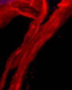
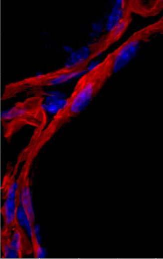
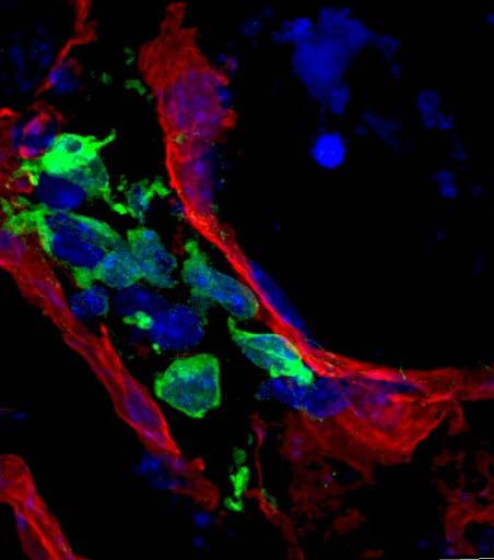
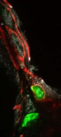
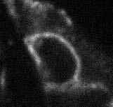
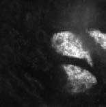
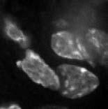
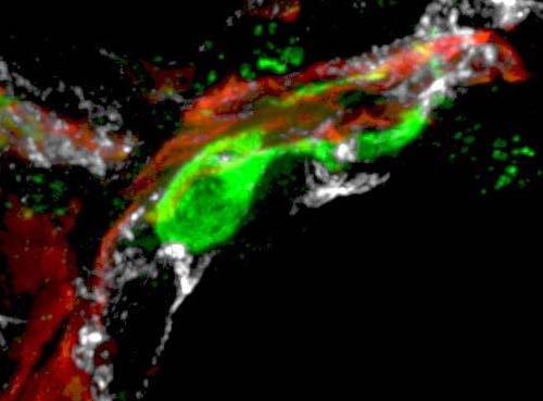
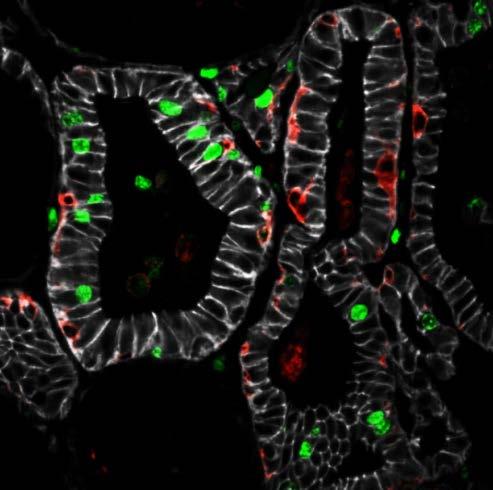
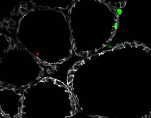
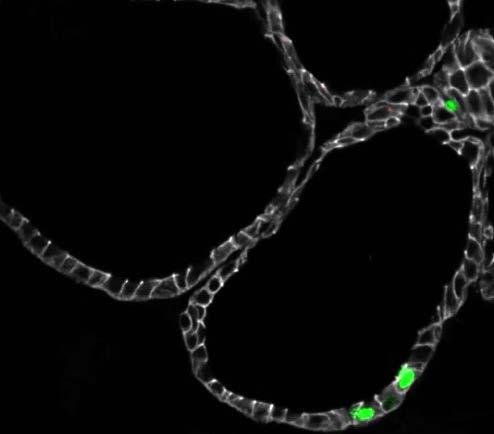
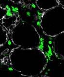
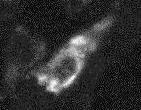
2 Supplemental Figure 3. Inserted WT-eGFP-HSC emits thin and thick cytoplasmic extensions, allowing contacting several thyrocytes. 3-D reconstruction of z-stack series images (0.23 µm increment, 5 µm total thickness) after double immunolabelling for laminin (red, basement lamina) and GFP (green, HSCs). This inserted HSC with dendritic shape emits both thin (white arrows) and thick (yellow arrows) cytoplasmic extensions. Supplemental Figure 4. Fine structure of basal lamina. Grazing views in WT (a,d), Ctns -/- (b,e), and grafted Ctns -/- (c,f). Lower panels are enlargements of areas boxed above. Notice homogenous structure and regular circular orientation of laminin in WT thyroid, contrasting with scalloped contour and enclosures/cavities in Ctns -/-. HSCs grafting largely preserves circularity and homogeneity of follicle basement lamina, except at small discontinuities frequently occupied by HSC projections. Supplemental Figure 5. Follicle inserted GFP + -WT HSCs do not transdifferentiate into thyrocytes. A. GFP + -WT HSCs insert between follicular basement and thyrocytes. 3-D reconstruction of z-stack series images (0.23 µm increment, 5 µm total thickness) obtained after triple immunolabelling for E-cadherin (white, thyrocytes baso-lateral membrane), laminin (red, basement lamina) and GFP (green, HSCs). DsRed signal is not shown here, for clarity. A HSC has passed through the follicle basement lamina and is intertwined within the thyrocyte monolayer (at least two thyrocytes are contacted, one by the cell body at left, and the second by a large extension (yellow arrowhead) B. Follicle-inserted HSC do not form tight junction with thyrocytes. Triple immunostaining for E-cadherin (red), ZO-1 (white, thyrocyte tight junctions) and GFP (green, HSCs). The boxed HSC is inserted into the thyrocyte monolayer and appears circumscribed by E-cadherin but does not engage into apical tight junction, nor reaches the
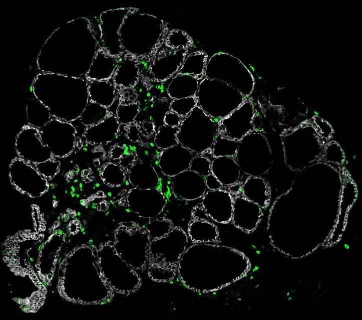
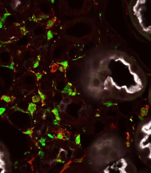
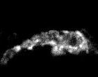
3 lumen. C. Follicle-inserted HSC do not transdifferentiate in thyrocytes. Triple immunolabelling for E-cadherin (white), Nkx2.1 (red, thyroid-specific transcription factor) and GFP (green, HSCs). All nuclei are further stained with DAPI (blue). In contrast to thyrocyte nuclei (white arrowhead), the nucleus of inserted HSCs (red arrowhead) is not labelled for Nkx2.1. Boxed areas at B and C are split below into black/white images for the three indicated channels. Supplemental Figure 6. Endogenous macrophage recruitment in Ctns -/- thyroid correlates with thyrocyte proliferation and structural alterations. Time-course of endogenous macrophage infiltration was analyzed by immunolabelling (F4/80, red) and compared to proliferative repair (Ki-67, green) in thyroid of WT or Ctns -/- mice at 3, 6 and 9 months. Macrophages invade the cystinotic tissue between 6 and 9 months, concomitant with structural tissue alterations (E-cadherin; white) and increased cell proliferation (Ki67; green). As in Ctns -/- kidney (Gaide Chevronnay et al. JASN 2014), notice silent phase for at least 3 months. Starting around 6 months and maximal at ~9 months of age, endogenous, cystinosin-deficient macrophages insert within cystinotic follicles (red arrowheads, best seen in enlarged box), exactly as GFP + -WT HSCs bearing normal cystinosin, but fail to provide protection. Supplemental Figure 7. Pattern of GFP + -WT HSC infiltration in the thyroid of grafted Ctns -/- mice. (a) Immunolabelling of grafted WT-eGFP-HSCs (GFP, green) in one representative mouse out of 5 reveals the pattern of infiltrating HSCs in the entire cystinotic thyroid. Thyroid full view was obtained using MosaiX module of AxioVision Software allowing tilling and stitching of images captured across the thyroid section. Enlargement (b) shows individual HSCs
4 (white arrowheads), inserted within the epithelium (E-cadherin, white); enlargement (c) shows 3 foci of HSC clusters mainly in the interstitium of the center of the gland. Supplemental Figure 8. In contrast to kidney, only few WT-eGFP-HSCs grafted in the thyroid express F4/80 differentiation marker. Multiplex fluorescence labeling for GFP (green, HSC), F4/80 (red, macrophages) and E-cadherin (white, thyrocyte basolateral membrane) or by LT-lectin (white, kidney proximal tubule cells apical membrane). In contrast to endogenous macrophages, few WT-eGFP-HSCs grafted in the thyroid express detectable F4/80 marker. In kidney, both endogenous macrophages and grafted HSCs show strong comparable F4/80 signal. Boxed fields point to cells clearly immunolabelled for F4/80, enlarged in the insets as black/white.
5 Proliferative events * * * thyrocytes, * p <0.05 interstitium, NS HSC 0 WT Ctns -/- grafted Ctns -/- Supplemental Figure 1


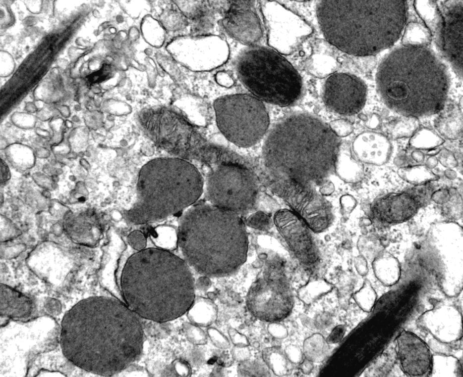
6 a b colloid ** CD * # capillary 2 µm c e 200 nm d 200 nm 500 nm Supplemental Figure 2 2 µm

7 laminin, WT-eGFP-HSCs lumen 5µm Supplemental Figure 3


8 laminin, WT-eGFP-HSCs, Hoechst a. WT b. Ctns -/- c. grafted Ctns -/- 10µm d. WT e. Ctns -/- f. grafted Ctns -/- 5µm Supplemental Figure 4
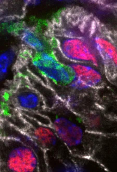
9 A E-cadherin, laminin, WT-eGFP-HSCs a lumen 5µm B E-cadherin, ZO-1, WT-EGFP-HSCs lumen 5µm E-cadherin GFP ZO-1 C E-cadherin, Nkx2.1, WT-eGFP-HSCs, DAPI GFP TTF1 DAPI 5µm Supplemental Figure 5

10 E-cadherin, Ki-67, F4/80 3 months 6 months 9 months WT 20µm Ctns -/- 20µm Supplemental Figure 6

11 E-cadherin, WT-eGFP-HSCs a b c 100µm b c 50µm 50µm Supplemental Figure 7


12 A. Thyroid E-cadherin, F4/80, WT-eGFP-HSCs a. Ctns -/- b. grafted Ctns -/- 20µm 20µm B. Kidney LT-lectin, F4/80, WT-eGFP-HSCs a. Ctns -/- b. grafted Ctns -/- 20µm 20µm Supplemental Figure 8
Supplementary Figure 1 Expression of Crb3 in mouse sciatic nerve: biochemical analysis (a) Schematic of Crb3 isoforms, ERLI and CLPI, indicating the
 Schematic of Crb3 isoforms, ERLI and CLPI, indicating the") Supplementary Figure 1 Expression of Crb3 in mouse sciatic nerve: biochemical analysis (a) Schematic of Crb3 isoforms, ERLI and CLPI, indicating the location of the transmembrane (TM), FRM binding (FB)
Supplementary Figure 1 Expression of Crb3 in mouse sciatic nerve: biochemical analysis (a) Schematic of Crb3 isoforms, ERLI and CLPI, indicating the location of the transmembrane (TM), FRM binding (FB)
Supplementary Figure 1: Signaling centers contain few proliferating cells, express p21, and
 Supplementary Figure 1: Signaling centers contain few proliferating cells, express p21, and exclude YAP from the nucleus. (a) Schematic diagram of an E10.5 mouse embryo. (b,c) Sections at B and C in (a)
Supplementary Materials for
 www.sciencetranslationalmedicine.org/cgi/content/full/4/117/117ra8/dc1 Supplementary Materials for Notch4 Normalization Reduces Blood Vessel Size in Arteriovenous Malformations Patrick A. Murphy, Tyson
Suppl Video: Tumor cells (green) and monocytes (white) are seeded on a confluent endothelial
 and monocytes (white) are seeded on a confluent endothelial") Supplementary Information Häuselmann et al. Monocyte induction of E-selectin-mediated endothelial activation releases VE-cadherin junctions to promote tumor cell extravasation in the metastasis cascade
Supplementary Information Häuselmann et al. Monocyte induction of E-selectin-mediated endothelial activation releases VE-cadherin junctions to promote tumor cell extravasation in the metastasis cascade
A adipose cells. B capillary. C epithelium
 EPITHELIA Objective The objective of this class is to observe how different epithelia vary in terms of cell shape, size and number of cell layers enabling them to be well adapted for functions in different
Loss of RhoA promotes skin tumor formation. Supplementary Figure 1. Loss of RhoA does not impair F-actin organization.
 Supplementary Figure Legends Supplementary Figure 1. Loss of RhoA does not impair F-actin organization. a. Representative IF images of F-actin staining of big and small control (left) and RhoA ko tumors
Supplementary Figure 1: GFAP positive nerves in patients with adenocarcinoma of
 SUPPLEMENTARY FIGURES AND MOVIE LEGENDS Supplementary Figure 1: GFAP positive nerves in patients with adenocarcinoma of the pancreas. (A) Images of nerves stained for GFAP (green), S100 (red) and DAPI
Ahtiainen et al., http :// /cgi /content /full /jcb /DC1
 Supplemental material JCB Ahtiainen et al., http ://www.jcb.org /cgi /content /full /jcb.201512074 /DC1 THE JOURNAL OF CELL BIOLOGY Figure S1. Distinct distribution of different cell cycle phases in the
African Trypanosomes
 African Trypanosomes Giemsa-stained blood smear of African trypanosomes viewed under the 100X objective lens. The block arrows denote trypomastigote forms of the African trypanosomes found within the blood
SUPPLEMENTARY INFORMATION
 SUPPLEMENTARY INFORMATION Advances in pancreatic islet monolayer culture on glass surfaces enable superresolution microscopy and insights into beta cell ciliogenesis and proliferation Edward A. Phelps,
Tissues. tissue = many cells w/ same structure and function. cell shape aids function tissue shape aids function. Histology = study of tissues
 Tissues tissue = many cells w/ same structure and function cell shape aids function tissue shape aids function Histology = study of tissues 4 types of tissues Epithelial coverings contact openings Connective
CD4 and CD8 T cells show a similar accumulation in the tumor stroma.
 Fig S1 CD4 Fibronectin EpCM CD8 CD4 and CD8 T cells show a similar accumulation in the tumor stroma. Fluorescently-labeled CD4 (CMFD, green) and CD8 (Hoechst, yellow) T cells were added to a human lung
F-actin VWF Vinculin. F-actin. Vinculin VWF
 a F-actin VWF Vinculin b F-actin VWF Vinculin Supplementary Fig. 1. WPBs in HUVECs are located along stress fibers and at focal adhesions. (a) Immunofluorescence images of f-actin (cyan), VWF (yellow),
Supplementary Figure 1. Electroporation of a stable form of β-catenin causes masses protruding into the IV ventricle. HH12 chicken embryos were
 Supplementary Figure 1. Electroporation of a stable form of β-catenin causes masses protruding into the IV ventricle. HH12 chicken embryos were electroporated with β- Catenin S33Y in PiggyBac expression
Project report October 2012 March 2013 CRF fellow: Principal Investigator: Project title:
 Project report October 2012 March 2013 CRF fellow: Gennaro Napolitano Principal Investigator: Sergio Daniel Catz Project title: Small molecule regulators of vesicular trafficking to enhance lysosomal exocytosis
Fibrinogen-induced perivascular microglial clustering is required for the. development of axonal damage in neuroinflammation
 SUPPLEMENTARY INFORMATION Fibrinogen-induced perivascular microglial clustering is required for the development of axonal damage in neuroinflammation Dimitrios Davalos, Jae Kyu Ryu, Mario Merlini, Kim
SUPPLEMENTARY INFORMATION
 b 350 300 250 200 150 100 50 0 E0 E10 E50 E0 E10 E50 E0 E10 E50 E0 E10 E50 Number of organoids per well 350 300 250 200 150 100 50 0 R0 R50 R100 R500 1st 2nd 3rd Noggin 100 ng/ml Noggin 10 ng/ml Noggin
sfigure 1: Detection of L-fucose in normal mouse renal cortex using the plant lectin LTL
 sfigure 1: Detection of L-fucose in normal mouse renal cortex using the plant lectin LTL LTL staining Negative control Fluorescence microscopy of normal (CL-11 +/+ ) mouse renal tissue after staining with
Dentin Formation(Dentinogenesis)
") Lecture four Dr. Wajnaa Oral Histology Dentin Formation(Dentinogenesis) Dentinogenesis begins at the cusp tips after the odontoblasts have differentiated and begin collagen production. Dentinogenesis growth
Lecture four Dr. Wajnaa Oral Histology Dentin Formation(Dentinogenesis) Dentinogenesis begins at the cusp tips after the odontoblasts have differentiated and begin collagen production. Dentinogenesis growth
Tissues. tissue = many cells w/ same structure and function. cell shape aids its function tissue shape aids its function
 Tissues tissue = many cells w/ same structure and function cell shape aids its function tissue shape aids its function Histology = study of tissues 4 types of tissues Epithelial coverings contact openings
J. Cell Sci. 129: doi: /jcs : Supplementary information
 Movie 1. AgLDL is contained in small sub-regions of the lysosomal synapse that are acidic. J774 cells were incubated with agldl dual labeled with a ph sensitive and a ph insensitive fluorophore for 1 hr.
Supplementary Figure 1. Nature Neuroscience: doi: /nn.4547
 Supplementary Figure 1 Characterization of the Microfetti mouse model. (a) Gating strategy for 8-color flow analysis of peripheral Ly-6C + monocytes from Microfetti mice 5-7 days after TAM treatment. Living
Histology of the myocardium and blood vessels. Prof. Abdulameer Al-Nuaimi
 Histology of the myocardium and blood vessels Prof. Abdulameer Al-Nuaimi E-mail: a.al-nuaimi@sheffield.ac.uk E-mail: abdulameerh@yahoo.com Histology of blood vessels The walls of arteries and veins are
The Endocrine System Pituitary
 The Endocrine System Pituitary Look at your slide of the human pituitary with your naked eye. You should see a cellular region and a more fibrous region. Then view each region with your microscope under
SUPPLEMENTARY INFORMATION
 doi: 10.1038/nature06994 A phosphatase cascade by which rewarding stimuli control nucleosomal response A. Stipanovich*, E. Valjent*, M. Matamales*, A. Nishi, J.H. Ahn, M. Maroteaux, J. Bertran-Gonzalez,
mm Distance (mm)
") b a Magnet Illumination Coverslips MPs Objective 2575 µm 1875 µm 1575 µm 1075 µm 875 µm 545 µm 20µm 2 3 0.5 0.3mm 1 1000 100 10 1 0.1 1000 100 10 1 0.1 Field Induction (Gauss) 1.5 0 5 10 15 20 Distance
b a Magnet Illumination Coverslips MPs Objective 2575 µm 1875 µm 1575 µm 1075 µm 875 µm 545 µm 20µm 2 3 0.5 0.3mm 1 1000 100 10 1 0.1 1000 100 10 1 0.1 Field Induction (Gauss) 1.5 0 5 10 15 20 Distance
Endogenous TNFα orchestrates the trafficking of neutrophils into and within lymphatic vessels during acute inflammation
 SUPPLEMENTARY INFORMATION Endogenous TNFα orchestrates the trafficking of neutrophils into and within lymphatic vessels during acute inflammation Samantha Arokiasamy 1,2, Christian Zakian 1, Jessica Dilliway
Santulli G. et al. A microrna-based strategy to suppress restenosis while preserving endothelial function
 ONLINE DATA SUPPLEMENTS Santulli G. et al. A microrna-based strategy to suppress restenosis while preserving endothelial function Supplementary Figures Figure S1 Effect of Ad-p27-126TS on the expression
SUPPLEMENTARY INFORMATION
 DOI:.38/ncb3399 a b c d FSP DAPI 5mm mm 5mm 5mm e Correspond to melanoma in-situ Figure a DCT FSP- f MITF mm mm MlanaA melanoma in-situ DCT 5mm FSP- mm mm mm mm mm g melanoma in-situ MITF MlanaA mm mm
WSC , Conference 9, Case 1. Tissue from a nyala.
 WSC 2009-2010, Conference 9, Case 1. Tissue from a nyala. MICROSCOPIC DESCRIPTION: Heart, atrium (1 pt.): Approximately 40% of the atrial myocardium is replaced by areas of fibrous connective tissue (1
a 0,8 Figure S1 8 h 12 h y = 0,036x + 0,2115 y = 0,0366x + 0,206 Labeling index Labeling index ctrl shrna Time (h) Time (h) ctrl shrna S G2 M G1
 Time (h) ctrl shrna S G2 M G1") (GFP+ BrdU+)/GFP+ Labeling index Labeling index Figure S a, b, y =,x +, y =,x +,,,,,,,, Time (h) - - Time (h) c d S G M G h M G S G M G S G h Time of BrdU injection after electroporation (h) M G S G M
(GFP+ BrdU+)/GFP+ Labeling index Labeling index Figure S a, b, y =,x +, y =,x +,,,,,,,, Time (h) - - Time (h) c d S G M G h M G S G M G S G h Time of BrdU injection after electroporation (h) M G S G M
Supplemental Figure 1. Intracranial transduction of a modified ptomo lentiviral vector in the mouse
 Supplemental figure legends Supplemental Figure 1. Intracranial transduction of a modified ptomo lentiviral vector in the mouse hippocampus targets GFAP-positive but not NeuN-positive cells. (A) Stereotaxic
20 2 Stomach Fig. 2.1 An illustration showing different patterns of the myenteric plexus peculiar to the regions in the guinea-pig stomach stained wit
 Stomach 2 The stomach is unique in that ICC have a different distribution in proximal and distal regions of the same organ. ICC-CM and ICC-LM are densely distributed throughout the thick circular and longitudinal
Epithelia will be discussed according to the following scheme: Type Number of layers Shape Line drawing. Squamous Cuboidal Columnar
 Epithelia Epithelia will be discussed according to the following scheme: Type Number of layers Shape Line drawing Simple Squamous Cuboidal Columnar Covering and Lining epithelium Pseudostratified Stratified
Supplementary Figure 1 The ability to regenerate an ear hole is discontinuous with wound healing. Ear-hole closure at D85 for each sex within each
 Supplementary Figure 1 The ability to regenerate an ear hole is discontinuous with wound healing. Ear-hole closure at D85 for each sex within each species observed. Data show a binary response to a 4 mm
Nature Immunology: doi: /ni eee Supplementary Figure 1
 eee Supplementary Figure 1 Hyphae induce NET release, but yeast do not. (a) NET release by human peripheral neutrophils stimulated with a hgc1 yeast-locked C. albicans mutant (yeast) or pre-formed WT C.
McWilliams et al., http :// /cgi /content /full /jcb /DC1
 Supplemental material JCB McWilliams et al., http ://www.jcb.org /cgi /content /full /jcb.201603039 /DC1 THE JOURNAL OF CELL BIOLOGY Figure S1. In vitro characterization of mito-qc. (A and B) Analysis
Unit I Problem 9 Histology: Basic Tissues of The Body
 Unit I Problem 9 Histology: Basic Tissues of The Body - What is the difference between cytology and histology? Cytology: it is the study of the structure and functions of cells and their contents. Histology:
Nature Neuroscience: doi: /nn Supplementary Figure 1. MADM labeling of thalamic clones.
 Supplementary Figure 1 MADM labeling of thalamic clones. (a) Confocal images of an E12 Nestin-CreERT2;Ai9-tdTomato brain treated with TM at E10 and stained for BLBP (green), a radial glial progenitor-specific
PHENOTYPIC DYNAMICS OF MICROGLIAL AND MONOCYTE-DERIVED CELLS IN GLIOBLASTOMA-BEARING MICE.
 SUPPLEMENTARY FIGURES, TABLES AND VIDEOS PHENOTYPIC DYNAMICS OF MICROGLIAL AND MONOCYTE-DERIVED CELLS IN GLIOBLASTOMA-BEARING MICE. Clément Ricard 1,2,3,4, Aurélie Tchoghandjian 2,4, Hervé Luche 5, Pierre
Postn MCM Smad2 fl/fl Postn MCM Smad3 fl/fl Postn MCM Smad2/3 fl/fl. Postn MCM. Tgfbr1/2 fl/fl TAC
 A Smad2 fl/fl Smad3 fl/fl Smad2/3 fl/fl Tgfbr1/2 fl/fl 1. mm B Tcf21 MCM Tcf21 MCM Smad3 fl/fl Tcf21 MCM Smad2/3 fl/fl Tcf21 MCM Tgfbr1/2 fl/fl αmhc MCM C 1. mm 1. mm D Smad2 fl/fl Smad3 fl/fl Smad2/3
Essentials of Anatomy and Physiology, 9e (Marieb) Chapter 3 Cells and Tissues. Short Answer. Figure 3.1
 Chapter 3 Cells and Tissues. Short Answer. Figure 3.1") Essentials of Anatomy and Physiology, 9e (Marieb) Chapter 3 Cells and Tissues Short Answer Figure 3.1 Using Figure 3.1, match the following: 1) The illustration of simple cuboidal epithelium is. Answer:
Essentials of Anatomy and Physiology, 9e (Marieb) Chapter 3 Cells and Tissues Short Answer Figure 3.1 Using Figure 3.1, match the following: 1) The illustration of simple cuboidal epithelium is. Answer:
川北医学院讲稿. Under low power note the testis is enclosed by a strong fibrous. layer of serous epithelium. These fibrous tissue
 川北医学院讲稿 Experiment 5: Male and Female Reproductive System Hello, everybody, class is begin,keep quiet, please. And this is the last experimental class. Today we will learn 5 slices and review all structures
2. Epithelial Tissues Dr. Manal Othman
 Biology-232 GENERAL HISTOLOGY 2. Epithelial Tissues Dr. Manal Othman Anatomy Department CMMS, AGU HISTOLOGY: w Study of the structure and function of tissues and organs at the microscopic levels. w Tissues
The Fine Structure of the Epithelial Cells of the Mouse Prostate* II. Ventral Lobe Epithelium
 Published Online: 1 June, 1960 Supp Info: http://doi.org/10.1083/jcb.7.3.511 Downloaded from jcb.rupress.org on September 28, 2018 The Fine Structure of the Epithelial Cells of the Mouse Prostate* II.
Biology. Dr. Khalida Ibrahim
 Dr. Khalida Ibrahim Biology Histology: Histology: is the study of the tissues of the body. Tissue: group of similar cells combined to perform a common function. The human body is composed of only 4 basic
Basic histology 5/4/2015
 Male reproductive system The male reproductive system is composed of the testes, genital ducts (the adjoining epididymis, and the vas deferens, a accessory sex glands (the seminal vesicles, the prostrate
Extra notes for lab- 1 histology. Slide 1 : cross section in the elastic artery ( aortic arch, ascending aorta, descending aorta )
") Extra notes for lab- 1 histology Slide 1 : cross section in the elastic artery ( aortic arch, ascending aorta, descending aorta ) - twin of ascending aorta is the pulmonary trunk. Ascending aorta represents
Extra notes for lab- 1 histology Slide 1 : cross section in the elastic artery ( aortic arch, ascending aorta, descending aorta ) - twin of ascending aorta is the pulmonary trunk. Ascending aorta represents
HISTOLOGICAL PARAMETERS
 HISTOLOGICAL PARAMETERS ADDITIONAL FILE FOR THE MANUSCRIPT: Peritoneal Negative Pressure Therapy Prevents Multiple Organ Injury in a Chronic Porcine Sepsis and Ischemia/Reperfusion model Brian D. Kubiak
D1120 Connective Tissue and Muscle Laboratory Module. 1) Connective tissue
 Connective tissue") D1120 Connective Tissue and Muscle Laboratory Module 1) Connective tissue Objectives: 1) identify the components (cells, fibres) present in "ordinary" connective tissue 2) differentiate the three types
D1120 Connective Tissue and Muscle Laboratory Module 1) Connective tissue Objectives: 1) identify the components (cells, fibres) present in "ordinary" connective tissue 2) differentiate the three types
Supplementary Figure S1: TIPF reporter validation in the wing disc.
 Supplementary Figure S1: TIPF reporter validation in the wing disc. a,b, Test of put RNAi. a, In wildtype discs the Dpp target gene Sal (red) is expressed in a broad stripe in the centre of the ventral
Tissues. Tissues - Overview. Bio 101 Laboratory 3. Epithelial Tissues and Integument
 Bio 101 Laboratory 3 Epithelial Tissues and Integument 1 Tissues Tissues to be examined under the microscope Epithelial Tissue Integument Connective Tissue **We will be doing muscle and nervous tissues
Macrophages form functional vascular mimicry channels in vivo. SI Figures and Legend
 Macrophages form functional vascular mimicry channels in vivo Authors: *Faith H. Barnett, *Mauricio Rosenfeld, Malcolm Wood, William Kiosses, Yoshihiko Usui, Valentina Marchetti, Edith Aguilar, and Martin
Histopathology: Glomerulonephritis and other renal pathology
 Histopathology: Glomerulonephritis and other renal pathology These presentations are to help you identify basic histopathological features. They do not contain the additional factual information that you
THE sebaceous glands of the rabbit consist of clusters of about ten cells
 79 On the Relationship between Mammary, Sweat, and Sebaceous Glands By D. B. CARLISLE (From the Department of Zoology and Comparative Anatomy, Oxford, and the Plymouth Laboratory of the Marine Biological
Supplementary Information. Tissue-wide immunity against Leishmania. through collective production of nitric oxide
 Supplementary Information Tissue-wide immunity against Leishmania through collective production of nitric oxide Romain Olekhnovitch, Bernhard Ryffel, Andreas J. Müller and Philippe Bousso Supplementary
Nature Neuroscience doi: /nn Supplementary Figure 1. Characterization of viral injections.
 Supplementary Figure 1 Characterization of viral injections. (a) Dorsal view of a mouse brain (dashed white outline) after receiving a large, unilateral thalamic injection (~100 nl); demonstrating that
Expression of acid base transporters in the kidney collecting duct in Slc2a7 -/-
 Supplemental Material Results. Expression of acid base transporters in the kidney collecting duct in Slc2a7 -/- and Slc2a7 -/- mice. The expression of AE1 in the kidney was examined in Slc26a7 KO mice.
CINtec p16 INK4a Staining Atlas
 CINtec p16 INK4a Staining Atlas Rating Rating Positive The rating positive will be assigned if the p16 INK4a -stained slide shows a continuous staining of cells of the basal and parabasal cell layers of
Dr. Heba Kalbouneh. Dr. Heba Kalbouneh. Dr. Heba Kalbouneh
 Dr. Heba Kalbouneh Dr. Heba Kalbouneh Dr. Heba Kalbouneh Basement membrane: What is the basement membrane? - It is a layer of ECM separating the epithelial cells from the underlying connective tissue Basement
Real-time imaging reveals the single steps of brain metastasis fo mation r
 Real-time imaging reveals the single steps of brain metastasis fo mation r Yvonne Kienast, Louisa von Baumgarten, Martin Fuhrmann, Wolfgang E.F. Klinkert, Roland Goldbrunner, Jochen Herms and Frank Winkler
SUPPLEMENTARY INFORMATION
 doi:1.138/nature1554 a TNF-α + in CD4 + cells [%] 1 GF SPF 6 b IL-1 + in CD4 + cells [%] 5 4 3 2 1 Supplementary Figure 1. Effect of microbiota on cytokine profiles of T cells in GALT. Frequencies of TNF-α
Supplementary Figure 1. Genotyping strategies for Mcm3 +/+, Mcm3 +/Lox and Mcm3 +/- mice and luciferase activity in Mcm3 +/Lox mice. A.
 Supplementary Figure 1. Genotyping strategies for Mcm3 +/+, Mcm3 +/Lox and Mcm3 +/- mice and luciferase activity in Mcm3 +/Lox mice. A. Upper part, three-primer PCR strategy at the Mcm3 locus yielding
Supplementary Figure 1. Chimeric analysis of inner ears. (A-H) Chimeric inner ears with fluorescent ES cells and (I,J) Rainbow inner ears.
 Chimeric inner ears with fluorescent ES cells and (I,J) Rainbow inner ears.") Supplementary Figure 1. himeric analysis of inner ears. (A-H) himeric inner ears with fluorescent ES cells and (I,J) Rainbow inner ears. (A,B) omposite images showing three colors in different vestibular
Supplementary Figure 1. himeric analysis of inner ears. (A-H) himeric inner ears with fluorescent ES cells and (I,J) Rainbow inner ears. (A,B) omposite images showing three colors in different vestibular
(a) Schematic diagram of the FS mutation of UVRAG in exon 8 containing the highly instable
 Schematic diagram of the FS mutation of UVRAG in exon 8 containing the highly instable") Supplementary Figure 1. Frameshift (FS) mutation in UVRAG. (a) Schematic diagram of the FS mutation of UVRAG in exon 8 containing the highly instable A 10 DNA repeat, generating a premature stop codon
Supplementary Figure 1. Frameshift (FS) mutation in UVRAG. (a) Schematic diagram of the FS mutation of UVRAG in exon 8 containing the highly instable A 10 DNA repeat, generating a premature stop codon
Histology of Male Reproductive system (1)
") Histology of Male Reproductive system (1) Prof. Dr. Malak A. Al-yawer Learning Objectives At the end of this lecture, the medical student will be able to: State the organization of the testis Define seminiferous
Histology of Male Reproductive system (1) Prof. Dr. Malak A. Al-yawer Learning Objectives At the end of this lecture, the medical student will be able to: State the organization of the testis Define seminiferous
Histology = the study of tissues. Tissue = a complex of cells that have a common function
 { EPITHELIAL TISSUE Histology = the study of tissues Tissue = a complex of cells that have a common function The Four Primary Tissue Types: Epithelium (epithelial tissue) covers body surfaces, lines body
T H E J O U R N A L O F C E L L B I O L O G Y
 T H E J O U R N A L O F C E L L B I O L O G Y Supplemental material Krenn et al., http://www.jcb.org/cgi/content/full/jcb.201110013/dc1 Figure S1. Levels of expressed proteins and demonstration that C-terminal
(a) Significant biological processes (upper panel) and disease biomarkers (lower panel)
 Significant biological processes (upper panel) and disease biomarkers (lower panel)") Supplementary Figure 1. Functional enrichment analyses of secretomic proteins. (a) Significant biological processes (upper panel) and disease biomarkers (lower panel) 2 involved by hrab37-mediated secretory
Supplementary Figure 1. Functional enrichment analyses of secretomic proteins. (a) Significant biological processes (upper panel) and disease biomarkers (lower panel) 2 involved by hrab37-mediated secretory
MULTIPLE CHOICE. Choose the one alternative that best completes the statement or answers the question.
 Exam Name MULTIPLE CHOICE. Choose the one alternative that best completes the statement or answers the question. 1) All of the following are synthesized along various sites of the endoplasmic reticulum
Transport of Sperm. Endocrinology of the Epididymis and Sperm Maturation. Vas Efferentia. John Parrish Department of Animal Sciences
 Endocrinology of the Epididymis and Sperm Maturation John Parrish Department of Animal Sciences References: he Physiology of Reproduction, Knobil and Neill, 2006; Chapter on the Epididymis by Robaire ransport
Dr. Abeer.c.Yousif. Histology -2 nd stage. What is histology?
 What is histology? Histology is the science of microscopic anatomy of cells and tissues, in Greek language Histo= tissue and logos = study and it's tightly bounded to molecular biology, physiology, immunology
Tissues. How do cells form tissues?
 Tissues How do cells form tissues? Using cell junctions Tissues Epithelial tissue Connective tissue Muscle tissue Nervous tissue Epithelial Tissue Closely packed cells in continuous sheets connected by
Non-Invasive Follicular Thyroid Neoplasm with Papillary-like Nuclei (NIFTP)
") Papillary Thyroid Carcinoma: Follicular Variant Encapsulated Type Replaced by: Non-Invasive Follicular Thyroid Neoplasm with Papillary-like Nuclei (NIFTP) Lester D. R. Thompson www.lester-thompson.com
Papillary Thyroid Carcinoma: Follicular Variant Encapsulated Type Replaced by: Non-Invasive Follicular Thyroid Neoplasm with Papillary-like Nuclei (NIFTP) Lester D. R. Thompson www.lester-thompson.com
SUPPLEMENTARY INFORMATION
 SUPPLEMENTARY INFORMATION Supplementary Figure 1. The expression of ephrin-b2 H2BGFP persists in the post-hearingonset organ of Corti and is specifically restricted to supporting cells. Sox2 immunolabeling
T H E J O U R N A L O F C E L L B I O L O G Y
 Supplemental material Brooks and Wallingford, http://www.jcb.org/cgi/content/full/jcb.201204072/dc1 T H E J O U R N A L O F C E L L B I O L O G Y Figure S1. Quantification of ciliary compartments in control
Nature Neuroscience: doi: /nn Supplementary Figure 1. PICALM expression in brain capillary endothelium in human brain and in mouse brain.
 Supplementary Figure 1 PICALM expression in brain capillary endothelium in human brain and in mouse brain. a, Double immunostaining for PICALM (red, left) and lectin positive endothelial profiles (blue,
GFP/Iba1/GFAP. Brain. Liver. Kidney. Lung. Hoechst/Iba1/TLR9!
 Supplementary information a +KA Relative expression d! Tlr9 5!! 5! NSC Neuron Astrocyte Microglia! 5! Tlr7!!!! NSC Neuron Astrocyte! GFP/Sβ/! Iba/Hoechst Microglia e Hoechst/Iba/TLR9! GFP/Iba/GFAP f Brain
MALE REPRODUCTIVE SYSTEM
 MALE REPRODUCTIVE SYSTEM The male reproductive system consists of primary sex organs (testes) and secondary or accessory sex organs. The secondary organs consist of a series of genital ducts (ductules
ROCK/Cdc42-mediated microglial motility and gliapse formation lead to phagocytosis of degenerating dopaminergic neurons in vivo
 Supplementary Information ROCK/Cdc42-mediated microglial motility and gliapse formation lead to phagocytosis of degenerating dopaminergic neurons in vivo Carlos Barcia* 1,2, Carmen M Ros 1,2, Valentina
T H E J O U R N A L O F C E L L B I O L O G Y
 Supplemental material Beck et al., http://www.jcb.org/cgi/content/full/jcb.201011027/dc1 T H E J O U R N A L O F C E L L B I O L O G Y Figure S1. Membrane binding of His-tagged proteins to Ni-liposomes.
Supplementary Figure 1: Validation of labeling specificity of immature OSNs and presynaptic terminals. (A) (B) (C) (D) (E)
 (B) (C) (D) (E)") Supplementary Figure 1: Validation of labeling specificity of immature OSNs and presynaptic terminals. (A) Confocal images of septal olfactory epithelium of an adult Gγ8-sypGFP-tdTom mouse showing colocalization
Supplementary Figure 1: Validation of labeling specificity of immature OSNs and presynaptic terminals. (A) Confocal images of septal olfactory epithelium of an adult Gγ8-sypGFP-tdTom mouse showing colocalization
Supplementary Materials for
 advances.sciencemag.org/cgi/content/full/3/8/e1700521/dc1 Supplementary Materials for Functional vascularized lung grafts for lung bioengineering N. Valerio Dorrello, Brandon A. Guenthart, John D. O Neill,
Quantitative PPARγ expression affects the balance between tolerance and immunity
 Quantitative PPARγ expression affects the balance between tolerance and immunity Ya-Hui Liu 1, Yau-Sheng Tsai 1,2,3, Shih-Chieh Lin 4, Nan-Shih Liao 5, Ming-Shiou Jan 6, Chung-Tiang Liang 7, Shih-Wen Hsu
Lecture Overview. Marieb s Human Anatomy and Physiology. Chapter 4 Tissues: The Living Fabric Epithelial Tissues Lecture 9. Introduction to Tissues
 Marieb s Human Anatomy and Physiology Marieb Hoehn Chapter 4 Tissues: The Living Fabric Epithelial Tissues Lecture 9 Lecture Overview Introduction to Tissues Epithelial Tissues Location General characteristics
RENAL HISTOPATHOLOGY
 RENAL HISTOPATHOLOGY Peter McCue, M.D. Department of Pathology, Anatomy & Cell Biology Sidney Kimmel Medical College There are no conflicts of interest. 1 Goals and Objectives! Goals Provide introduction
Tongue In the buccal cavity of the digestive system
 Tongue In the buccal cavity of the digestive system same layers as those of tubular organs Mucosa, submucosa, and muscularis muscularis = the muscularis externa no muscularis mucosa 1 Tongue ling = tongue
04_polarity. The formation of synaptic vesicles
 Brefeldin prevents assembly of the coats required for budding Nocodazole disrupts microtubules Constitutive: coatomer-coated Selected: clathrin-coated The formation of synaptic vesicles Nerve cells (and
Tissues. Tissues - Overview. Bio211 Laboratory 2. Epithelial and Connective Tissues
 Bio211 Laboratory 2 Epithelial and Connective Tissues 1 Tissues Tissues to be examined under the microscope Epithelial Tissue (p. 79 Lab Manual) [TODAY] Connective Tissue (p. 93 Lab Manual) [TODAY] Muscle/Nervous
Supplementary information - Table (1), Figures (12), and Videos (5)
, Figures (12), and Videos (5)") Supplementary information - Table (1), Figures (12), and Videos (5) A soft, transparent, freely accessible cranial window for chronic imaging and electrophysiology Chaejeong Heo 1, Hyejin Park 1, 2, Yong-Tae
Supplementary information - Table (1), Figures (12), and Videos (5) A soft, transparent, freely accessible cranial window for chronic imaging and electrophysiology Chaejeong Heo 1, Hyejin Park 1, 2, Yong-Tae
Prelab #4 BLOOD; BONE MARROW; RESPIRATORY; INTEGUEMENT Page 1
 Prelab #4 BLOOD; BONE MARROW; RESPIRATORY; INTEGUEMENT Page 1 Blood Slide 101 This a classic slide of blood cells using a Wright stain. Inspect red blood cells and their appearance. Note the approximate
SUPPLEMENTARY INFORMATION
 DOI: 0.038/ncb33 a b c 0 min 6 min 7 min (fixed) DIC -GFP, CenpF 3 µm Nocodazole Single optical plane -GFP, CenpF Max. intensity projection d µm -GFP, CenpF, -GFP CenpF 3-D rendering e f 0 min 4 min 0
Supplementary Information
 Supplementary Information Title Degeneration and impaired regeneration of gray matter oligodendrocytes in amyotrophic lateral sclerosis Authors Shin H. Kang, Ying Li, Masahiro Fukaya, Ileana Lorenzini,
Lecture Overview. Chapter 4 Epithelial Tissues Lecture 9. Introduction to Tissues. Epithelial Tissues. Glandular Epithelium
 Visual Anatomy & Physiology First Edition Martini & Ober Chapter 4 Lecture 9 Lecture Overview Introduction to Tissues Location General characteristics Functions Classification Glandular Epithelium 2 Where
Supplemental Figure 1: Lrig1-Apple expression in small intestine. Lrig1-Apple is observed at the crypt base and in insterstial cells of Cajal, but is
 Supplemental Figure 1: Lrig1-Apple expression in small intestine. Lrig1-Apple is observed at the crypt base and in insterstial cells of Cajal, but is not co-expressed in DCLK1-positive tuft cells. Scale
(A) PCR primers (arrows) designed to distinguish wild type (P1+P2), targeted (P1+P2) and excised (P1+P3)14-
 PCR primers (arrows) designed to distinguish wild type (P1+P2), targeted (P1+P2) and excised (P1+P3)14-") 1 Supplemental Figure Legends Figure S1. Mammary tumors of ErbB2 KI mice with 14-3-3σ ablation have elevated ErbB2 transcript levels and cell proliferation (A) PCR primers (arrows) designed to distinguish
1 Supplemental Figure Legends Figure S1. Mammary tumors of ErbB2 KI mice with 14-3-3σ ablation have elevated ErbB2 transcript levels and cell proliferation (A) PCR primers (arrows) designed to distinguish
SUPPLEMENTARY INFORMATION
 DOI: 10.1038/ncb2988 Supplementary Figure 1 Kif7 L130P encodes a stable protein that does not localize to cilia tips. (a) Immunoblot with KIF7 antibody in cell lysates of wild-type, Kif7 L130P and Kif7
Basic Urinary Tract Anatomy and Histology
 Basic Urinary Tract Anatomy and Histology The two kidneys are located in the retroperitoneum on either side of the vertebral bladder and the contraction of the detrusor muscle. Any mechanical barrier,
Disorders of Cell Growth & Neoplasia. Histopathology Lab
 Disorders of Cell Growth & Neoplasia Histopathology Lab Paul Hanna April 2010 Case #84 Clinical History: 5 yr-old, West Highland White terrier. skin mass from axillary region. has been present for the
Supplementary Figure 1: Hsp60 / IEC mice are embryonically lethal (A) Light microscopic pictures show mouse embryos at developmental stage E12.
 Light microscopic pictures show mouse embryos at developmental stage E12.") Supplementary Figure 1: Hsp60 / IEC mice are embryonically lethal (A) Light microscopic pictures show mouse embryos at developmental stage E12.5 and E13.5 prepared from uteri of dams and subsequently genotyped.
Supplementary Figure 1: Hsp60 / IEC mice are embryonically lethal (A) Light microscopic pictures show mouse embryos at developmental stage E12.5 and E13.5 prepared from uteri of dams and subsequently genotyped.