Dual-Energy CT: The Technological Approaches
|
|
|
- Hugh Smith
- 5 years ago
- Views:
Transcription


1 Dual-Energy CT: The Technological Approaches Dushyant Sahani, M.D Director of CT Associate Professor of Radiology Massachusetts General Hospital Harvard Medical School

2 Disclosure Research Grant Support GE Healthcare BD Consultant, Bracco Diagnostics


3 Single Energy CT Standard FBP Fast Image Data Space Raw data recon Raw Data Space Single Source CT One X-Ray Tube and Detector Assembly X-Ray Tube kvp 80, 100, 120 and 140
 Image noise and")
 Radiation Dose kvp define upper limit for Polychromatic X")
4 Photon energy Challenges with Single Energy CT Tissue characterization Lesion detection (small lesion) Image noise and quality at low kv scanning Complex scanning protocols (multi-phase imaging) Radiation Dose kvp define upper limit for Polychromatic X ray kv 140 kv spectrum

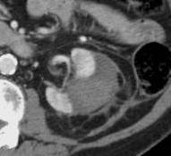
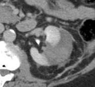

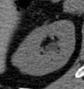

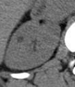
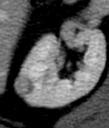
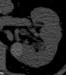

5 Challenges in Lesion Characterization I- I+ I- I+ Mass Enhancement RFA

6 Attenuation on CT Material differentiation on CT is based on X-ray attenuation Attenuation is caused by absorption and scattering of radiation Two main mechanisms Compton scatter and Photon effect Compton effect is energy independent Photo effect is strongly energy-dependent Changing the kv setting results in an alteration of photon energy Johnson et al Eur Rad 2007

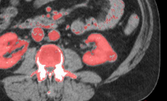
7 DE CT - Background Attenuation is energy-dependent At 80kVp Iodine signal is 2x of 140 kvp CT numbers do not vary with beam energy for soft tissues but do for high z materials By analyzing absorption properties of different material at different x-ray energies, materials can be distinguished (Base material decomposition)


8 80kV 140kV Bone 670 HU Iodine 296 HU Bone 450 HU Iodine 144 HU 80kV 140kV
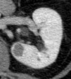
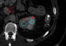
9 Stone composition Element composition Uric Acid Stone Light Elements (H, C, N, O) Non uric acid stone Heavy Elements (P, Ca, S) Attenuation at 80 kvp Lower HU Higher HU Attenuation at 140 kvp Higher HU Lower HU Calcium 710 HU Uric Acid 290 HU Calcium 480 HU Uric Acid 315 HU 80kV 140kV
10 Material Attenuation NIST* Curves cm 2 /g X-Ray Mass Attenuation Coefficients Iodine bone water Photon Energy, kev Note: Bone attenuation curve may be represented by a linear combination of iodine and water. X-ray mass attenuation coefficients *National Institute of Standards and Technology US Dept. of Commerce

11 u(e) (cm2/mg) Material Attenuation Iodine Material Attenuation Coefficient Material Attenuation Coefficient Monochromatic 1 Bone Water (kvp) Photon Energy (kev) 80 kvp 140 kvp 70 KeV 100 KeV


12 Liver Fat quantification- CT Dual Energy CT As the kvp increases, the fat attenuation increases Steatotic liver exhibits greater change in attenuation between 80kVp and 140kVp than normal liver Liver Fat Index = HU at 140 kvp - HU at 80 kvp Liver Fat Index >10HU suggests HS >25%

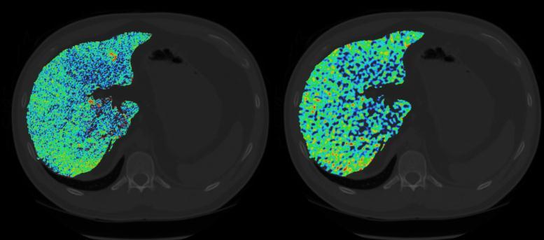
13 DE-CT: Liver Fat Quantification Courtsey: Saad Sirhoi GE


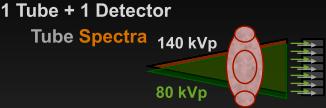
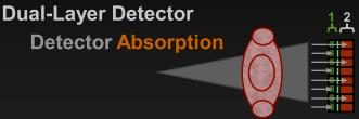
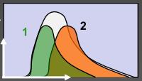
14 # X-rays # X-rays # X-rays Dual-Energy CT-Approaches Siemens Dual-Source Energy GE Fast Switching (Gemstone) Energy Philips Dual-Layer Energy

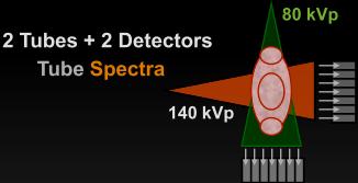
15 Dual Source Dual Energy Scanning Tube A Tube B B A 50 cm FOV 26/33 cm FOV Both tubes can be operated independently or in unison with respect to their kv and ma settings Coursey C A et al. Radiographics 2010;30:
16 Dual Source Single Energy 120 Dual Energy Both tubes at same kvp Both tubes at different kvp Faster image acquisition Higher power (160kW) Improved temporal resolution (83ms) for cardiac imaging Improved image quality In Obese patients 500 lbs (220kg) Tissue material Differentiation

17 DS-DECT Image Processing Algorithm 80kVp 140kVp Weighed120kVp Iodine Map 3 material Decomposition Algorithm Soft tissue, Fat & Iodine Virtual NECT
 Three")


18 Dual Source Dual Energy CT Data from the two acquisitions (80 and 140 kvp) Three material decomposition algorithm Soft tissue, Fat, Iodine Water, Uric acid, calcium Calcium stone Iodine Distribution Image Virtual Un-enhanced Image Stone Composition
19 Iodine (perfusion) Imaging Qualitative Quantitative RM mg/cc RP mc/cc


20 Calcium stone Uric Acid stone Mixed stones, struvite stones and cystine stones show a mixture of red and blue color Stolzman et al Urol Res 2008, Graser et al 2008 Invest Radiol, Graser et al 2009 Eur Radiol, Thomas et al Eur Radiol 2009


21 80kVp 140kVp Limitations Limited field of view of B tube than the standard field of view with the A tube. FOV limitation will not be a problem for the aorta, pancreas, adrenal glands or small bowel Dual energy acquisition are ineffective in obese patients or in patients with very large abdomens (BMI>30).

22 Single Source DE CT Single Source - Dual Energy CT SSDE CT

 1( i) Pl o( wi )")
 Ph i g( ih ).")
 2Ph i g( ih ) 2Pl o( .")

23 GSI Acquisition and Projection Based Material Decomposition GSI Data Acquisition Interleaved High- and Low-kVp Projections Attenuation-to-material density transformation iodine water 2 2 P1 ( i) 1( i) Pl o( wi ) 1( i) Ph i g( ih ) 1P l o( wi ) 1P h i g( ih ) 1P l o( wi ) Ph i g( ih ) P2 ( i) 2( i) Pl o( wi ) 2( i) Ph i g( ih ) 2P l o( wi ) 2Ph i g( ih ) 2Pl o( wi ) Ph i g( ih )... Iodine Projections Water Projections split Low kvp Projections High kvp Projections Image Reconstruc tion Image reconstruction MD Iodine MD Water Monochromatic Generation 140 kvp
 Iodine")

24 cm 2 /g Monochromatic Image Generation Water (mg/ml) Iodine (mg/ml) p ( E) d ( E) d Monochromatic iodine E 70 kev (HU) water kev


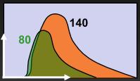
25 Flux Polychromatic vs Monochromatic images 70 kev MONOchromatic Xray beam 70 ke V Polychromatic Xray beam 80 kvp 140 kvp Energ y kvp kev specifies defines the the upper photon limit energy x-ray for a polychromatic monochromatic Xray x-ray beam source 80 kvp 140 kvp kvp ~ KeV


")
26 Single Source DE CT Dual Energy Acquisition 80/140 kvp datasets Material Density images Effective Z Monochromatic images( kev) Water Image Iodine Image
.")
27 SS DECT in differentiating material using spectral attenuation curve. The figure shows spectral attenuation curve of three different materials (contrast in abdominal aorta, fat and soft tissue - corresponding ROI marked). The spectral attenuation curve may be a signature sign of individual material and facilitate differentiation of tissues having similar attenuation values on single energy CT. 27


28 Effective Z (Zeff) MD Water MD Iodine High and low kvp Rapid kv twitching Uric Acid stone = MD Water +, MD Iodine Non-Uric Acid stone = MD Water +, MD Iodine + Effective Z Image Effective z number scatter plot
. Single X-ray tube with 50cm FOV capable of ultrafast kv switching between 80 and 140kV. ma ma on each tube can be controlled ma is fixed for both 80 and 140kV acquisition cycle.")
 Contrast exploitation from Lower kev images Projection space based Monochromatic Images (40-140 kev) Contrast exploitation from Lower")
29 FEATURES DS DECT SS DECT X Ray tube Two Tube mounted at 90 degrees Single X-ray tube & Gemstone detector with fast processing speed Tube potential Two Tube A (140kV, 50cm FOV) & Tube B (80/100kV, only 33cm FOV). Single X-ray tube with 50cm FOV capable of ultrafast kv switching between 80 and 140kV. ma ma on each tube can be controlled ma is fixed for both 80 and 140kV acquisition cycle. Reconstruction technique Post processing of data Image space based Three material decomposition Soft tissue, fat, Iodine Iodine map and virtual non contrast DE data (80/100kV kv) water, uric acid, calcium Stone analysis Monochromatic Images ( kev) Contrast exploitation from Lower kev images Projection space based Monochromatic Images ( kev) Contrast exploitation from Lower kev images DE data (80kV + 140kV) Material density images from two material decomposition Allows separation of different material/tissue Effective Z image Distinguishing tissues based on effective atomic number


30 DECT Spectrum Monochromatic VNC MD Water MD Iodine Iodine map Effective Z Exploitation of contrast Replacement to unenhanced CT Material differentiation and replacement to unenhanced CT Material differentiation, qualitative and quantitative iodine uptake assessment Assessment of iodine uptake both qualitative and quantitatively Material differentiation based on effective atomic number


31 Why Dual Energy CT? Challenges with Single energy CT Advantages of DECT MD Water MD Iodine Tissue composition Uric acid (UA) or non UA stone Better material differentiation enhancing tissue characterization Stone seen on MD water image only suggesting UA stone HU value reliability More consistent HU values on Monochromatic images Beam hardening and metal artifact Reduction of Beam hardening and metal artifact True vs pseudo enhancement HU is 33 True /pseudenhancement More Reliable detection of enhancement with Iodine map Iodine content 0.83mg/cc which is very negligible iodine uptake suggesting benign cyst on Iodine image Protocol complexity Simplification of multiphase protocol TNC VNC DE Arterial Single DE acquisitions giving both phases
32 Scanner Accessibility Cost Hardware Reconstruction time Technologist Training for post processing dual energy data Work Flow Challenges PACS Large data DE soft ware are not integrated on PACS Quantification of material images Radiologist Training Familiarity of different data sets More image series for analysis Standard analysis


33 Summary DECT is an exciting technology that provides information about how substances behave at different energies Enabling improved detection of iodinecontaining substances on low-kvp/kev images Material differentiation The ability to generate virtual unenhanced images These capabilities are promising for improved detection and characterization Evaluation of vascular structures. Work-flow challenges Validation
Role of DE-CT in Oncology
 Role of DE-CT in Oncology Dushyant Sahani, M.D Director of CT Associate Professor of Radiology Massachusetts General Hospital Harvard Medical School Email-dsahani@partners.org Disclosure Research Grant
Liver, Liver Fat, Fibrosis and HCC Screening with DECT
 Liver, Liver Fat, Fibrosis and HCC Screening with DECT Dushyant Sahani, M.D Director of CT Associate Professor of Radiology Massachusetts General Hospital Harvard Medical School Email-dsahani@partners.org
Dual-Energy CT Applications in Radiation Therapy
 THE UNIVERSITY OF WISCONSIN MADISON Dual-Energy CT Applications in Radiation Therapy - Jessica Miller 1 Disclosures Funding provided by Siemens Medical 2 Learning objectives General principles of dual
Dual Energy CT of the Liver
 34th Annual Course October 2011 Washington, DC Dual Energy CT of the Liver Vassilios Raptopoulos, MD Beth Israel Deaconess Medical Center Harvard Medical School Dual Energy CT (DECT) Different materials
8/3/2016. Consultant for / research support from: Astellas Bayer Bracco GE Healthcare Guerbet Medrad Siemens Healthcare. Single Energy.
 U. Joseph Schoepf, MD Prof. (h.c.), FAHA, FSCBT-MR, FNASCI, FSCCT Professor of Radiology, Medicine, and Pediatrics Director, Division of Cardiovascular Imaging Consultant for / research support from: Astellas
Dual Energy CT Aortography: Can We Reduce Iodine Dose??
 Dual Energy CT Aortography: Can We Reduce Iodine Dose?? William P. Shuman MD, FACR FSCBTMR Department of Radiology University of Washington SCBTMR Annual Course Boston, October 10, 2012 Conflict of Interest
True Dual Energy. Dr. Stefan Ulzheimer, Siemens Healthcare GmbH. DEfinitely Siemens
 DEfinitely Siemens True Dual Energy Dr. Stefan Ulzheimer, Siemens Healthcare GmbH International version. Not for distribution in the US. Unrestricted Siemens AG 2015 All rights reserved. The products/features
An Introduction to Dual Energy Computed Tomography
 An Introduction to Dual Energy Computed Tomography Michael Riedel University of Texas Health Science Center at San Antonio Introduction The idea of computed tomography (CT) was first introduced in the
CT Urography. Ureter. Stuart G. Silverman, M.D.
 CT Urography Stuart G. Silverman, M.D. Professor of Radiology Harvard Medical School Director, Abdominal Imaging and Intervention Brigham and Women s Hospital Ureter Boston, MA CT Urography Stuart G. Silverman,
Les Outils Cliniques de Demain en Scanner Cardiaque. Cardiaque Status en ECR 2018 From Diagnosis to Prognosis
 ECR 2018 From Diagnosis to Prognosis ECR 2018 From Diagnosis to Prognosis Thursday, March 1, 2018/08:30-10:00/Room N Les Outils Cliniques de Demain en Scanner Cardiaque Cardiaque Status en 2018 Rodrigo
Dual-Energy 101: Principles, Methods and Dose
 Dual-Energy 101: Principles, Methods and Dose Juan Carlos Ramirez-Giraldo, Ph.D Staff Scien2st, Collabora2ons Manager SE Region ISCT San Francisco, 2017 Siemens Medical Solu2ons USA, Inc., 2017 Page 1
How do the Parameters affect Image Quality and Dose for Abdominal CT? Image Review
 How do the Parameters affect Image Quality and Dose for Abdominal CT? Image Review Mannudeep K. Kalra, MD, DNB Massachusetts General Hospital Harvard Medical School Financial Disclosure This presentation
Typical PET Image. Elevated uptake of FDG (related to metabolism) Lung cancer example: But where exactly is it located?
 Lung cancer example: But where exactly is it located?") Typical PET Image Elevated uptake of FDG (related to metabolism) Lung cancer example: But where exactly is it located? PET/CT Oncology Imaging Anatometabolic fusion images are useful in the management
Typical PET Image Elevated uptake of FDG (related to metabolism) Lung cancer example: But where exactly is it located? PET/CT Oncology Imaging Anatometabolic fusion images are useful in the management
Ultrasound. Computed tomography. Case studies. Utility of IQon Spectral CT in. cardiac imaging
 Ultrasound Computed tomography Case studies Utility of IQon Spectral CT in cardiac imaging Cardiac imaging is a challenging procedure where it is necessary to image a motion-free heart. This requires a
Dual Energy Spectral CT of Focal Liver Lesions in Advanced Cirrhosis: Early Experience
 Dual Energy Spectral CT of Focal Liver Lesions in Advanced Cirrhosis: Early Experience William P. Shuman MD, FACR University of Washington SCBTMR Annual Course Washington DC, October 23-26, 2011 Conflict
To Shield or Not to Shield? Lincoln L. Berland, M.D.
 To Shield or Not to Shield? Lincoln L. Berland, M.D. Disclosures Consultant to: Nuance, Inc. Page 2 Breast Radiation on CT Use of chest CT has increased in women vulnerable to cancer induction by radiation.
Scanner Spectral. Double Energie et Comptage Photonique. Principes et Premiers Résultats
 Scanner Spectral Double Energie et Comptage Photonique Principes et Premiers Résultats Philippe Douek, Loic Boussel, Monica Sigovan, Salim Si-Mohamed Daniel Bar ness Plan 1. Rappel Techniques: Attenuation
Neuro CT What s a Good Head Exam?
 Neuro CT What s a Good Head Exam? Rajiv Gupta, PhD, MD Neuroradiology Massachusetts General Hospital Harvard Medical School Outline What we need to see? Routine Head CT protocols Dose optimization strategies
Gemstone Spectral Imaging quantifies lesion characteristics for a confident diagnosis
 GE Healthcare Gemstone Spectral Imaging quantifies lesion characteristics for a confident diagnosis CT clinical case study lesion characterization Desiree Morgan, MD Vice Chair of Clinical Research Professor
Combined Anatomical and Functional Imaging with Revolution * CT
 GE Healthcare Case studies Combined Anatomical and Functional Imaging with Revolution * CT Jean-Louis Sablayrolles, M.D. Centre Cardiologique du Nord, Saint-Denis, France Case 1 Whole Brain Perfusion and
Dual-Energy Imaging of Bone Marrow Edema on a Dedicated Multi-Source Cone-Beam CT System for the Extremities
 Dual-Energy Imaging of Bone Edema on a Dedicated Multi-Source Cone-Beam CT System for the Extremities W Zbijewski, 1 A Sisniega, 1 JW Stayman, 1 N Packard, 2 J Yorkston, 2 G Thawait, 3 S Demehri, 3 J Fritz,
Emerging Applications in Musculoskeletal CT Imaging
 Emerging pplications in Musculoskeletal CT Imaging y K Murali MD(RD), PDCC, Director of Interventional Radiology, G. Francis DMRD, DN (RD), Consultant Radiologist, and R. Madan, MS, MD, Consultant Radiologist,
Dual Energy CT of Pulmonary Embolism
 Dual Energy CT of Pulmonary Embolism U. Joseph Schoepf, MD, FAHA, FSCBT MR, FSCCT Professor of Radiology, Medicine, and Pediatrics Director of Cardiovascular Imaging Disclosures Consultant for / research
Dual Energy CT: a new tool in evaluation of the urinary tract stones composition in clinical practice - initial study
 Dual Energy CT: a new tool in evaluation of the urinary tract stones composition in clinical practice - initial study Poster No.: C-2279 Congress: ECR 2013 Type: Scientific Exhibit Authors: M. Guzi#ski,
Bone Densitometry Radiation dose: what you need to know
 Bone Densitometry Radiation dose: what you need to know John Damilakis, PhD Associate Professor and Chairman University of Crete, Iraklion, Crete, GREECE Estimation of bone status using X-rays Assessment
Low Dose Era in Cardiac CT
 Low Dose Era in Cardiac CT DIANA E. LITMANOVICH, MD Department of Radiology Beth Israel Deaconess Medical Center Harvard Medical School Disclosures Neither I nor my immediate family members have a financial
Managing Radiation Risk in Pediatric CT Imaging
 Managing Radiation Risk in Pediatric CT Imaging Mahadevappa Mahesh, MS, PhD, FAAPM, FACR, FACMP, FSCCT. Professor of Radiology and Cardiology Johns Hopkins University School of Medicine Chief Physicist
Ultralow Dose Chest CT with MBIR
 Ultralow Dose Chest CT with MBIR Ella A. Kazerooni, M.D. Professor & Director Cardiothoracic Radiology Associate Chair for Clinical Affairs University of Michigan Disclosures Consultant: GE Healthcare
CT Perfusion. U. Joseph Schoepf, MD, FAHA, FSCBT MR, FSCCT Professor of Radiology, Medicine, and Pediatrics Director of Cardiovascular Imaging
 CT Perfusion U. Joseph Schoepf, MD, FAHA, FSCBT MR, FSCCT Professor of Radiology, Medicine, and Pediatrics Director of Cardiovascular Imaging Disclosures Consultant for / research support from Bayer Bracco
Acknowledgments. A Specific Diagnostic Task: Lung Nodule Detection. A Specific Diagnostic Task: Chest CT Protocols. Chest CT Protocols
 Personalization of Pediatric Imaging in Terms of Needed Indication-Based Quality Per Dose Acknowledgments Duke University Medical Center Ehsan Samei, PhD Donald Frush, MD Xiang Li PhD DABR Cleveland Clinic
Spectral CT imaging as a new quantitative tool? Assessment of perfusion defects of pulmonary parenchyma in patients with lung cancer
 Original Article Spectral CT imaging as a new quantitative tool? Assessment of perfusion defects of pulmonary parenchyma in patients with lung cancer Ying-Shi Sun, Xiao-Yan Zhang, Yong Cui, Lei Tang, Xiao-Ting
Dual Energy CT of the Heart: Perfusion and Beyond
 Dual Energy CT of the Heart: Perfusion and Beyond U. Joseph Schoepf, MD, FAHA, FSCBT MR, FSCCT Professor of Radiology, Medicine, and Pediatrics Director of Cardiovascular Imaging Disclosures Consultant
A Snapshot on Nuclear Cardiac Imaging
 Editorial A Snapshot on Nuclear Cardiac Imaging Khalil, M. Department of Physics, Faculty of Science, Helwan University. There is no doubt that nuclear medicine scanning devices are essential tool in the
New Technologies for Cardiac CT. Geoffrey D. Rubin, MD, MBA, FACR, FNASCI Duke University
 1996 New Technologies for Cardiac CT Geoffrey D. Rubin, MD, MBA, FACR, FNASCI Duke University New Technology The Long View Levels of Efficacy Endpoint Examples 1: Technical Imaging resolution 2: Diagnostic
Usefulness of New CT Technologies for Interventional Cardiovascular Procedures
 Usefulness of New CT Technologies for Interventional Cardiovascular Procedures Ronen Rubinshtein, MD FACC FESC Department of Cardiovascular Medicine Lady Davis Carmel Medical Center & Technion Israel Institute
Ask EuroSafe Imaging. Tips & Tricks. Paediatric Imaging Working Group. Shielding in pediatric CT
 Ask EuroSafe Imaging Tips & Tricks Paediatric Imaging Working Group Shielding in pediatric CT Claudio Granata (IRCCS Istituto Giannina Gaslini, IT) Joana Santos (ESTeSC-Coimbra Health School, PT) Elina
Fundamentals, Techniques, Pitfalls, and Limitations of MDCT Interpretation and Measurement
 Fundamentals, Techniques, Pitfalls, and Limitations of MDCT Interpretation and Measurement 3 rd Annual Imaging & Physiology Summit November 20-21, 21, 2009 Seoul, Korea Wm. Guy Weigold, MD, FACC Cardiovascular
X-Ray & CT Physics / Clinical CT
 Computed Tomography-Basic Principles and Good Practice X-Ray & CT Physics / Clinical CT INSTRUCTORS: Dane Franklin, MBA, RT (R) (CT) Office hours will be Tuesdays from 5pm to 6pm CLASSROOM: TIME: REQUIRED
Translating Protocols Across Patient Size: Babies to Bariatric
 Translating Protocols Across Patient Size: Babies to Bariatric Cynthia H. McCollough, PhD, FACR, FAAPM Professor of Radiologic Physics Director, CT Clinical Innovation Center Department of Radiology Mayo
Cardiac CTA Prospective Gating Broad Beam
 Cardiac CTA Prospective Gating Broad Beam ACQUISITION- Broad Beam Gating: Prospective Non Contrast Scan-Calcium Score Patient Position Supine Feet First into Gantry Heart Isocenter Scanogram AP and Lateral
Estimating Iodine Concentration from CT Number Enhancement
 Estimating Iodine Concentration from CT Number Enhancement Rosemary Eaton, Andrew Shah, Jane Shekhdar Medical Physics, Mount Vernon Hospital CT Users Group 4 th October 212, Edinburgh Summary Background
ESTABLISHING DRLs in PEDIATRIC CT. Keith Strauss, MSc, FAAPM, FACR Cincinnati Children s Hospital University of Cincinnati College of Medicine
 ESTABLISHING DRLs in PEDIATRIC CT Keith Strauss, MSc, FAAPM, FACR Cincinnati Children s Hospital University of Cincinnati College of Medicine CT Dose Indices CTDI INTRODUCTION CTDI 100, CTDI w, CTDI vol
Biomarkers and the Future of. John R. Votaw CBIS 5 th Year Anniversary Celebration/Look to the future February 8, 2013
 Biomarkers and the Future of Radiology John R. Votaw CBIS 5 th Year Anniversary Celebration/Look to the future February 8, 2013 Statistics/Radiology Collaboration The utility of Radiologic procedures
Cardiac CT Lowering the Dose Dramatically
 Cardiac CT Lowering the Dose Dramatically U. Joseph Schoepf, MD, FAHA, FSCBT MR, FSCCT Professor of Radiology, Medicine, and Pediatrics Director of Cardiovascular Imaging Disclosures Consultant for / research
Technology Assessment Institute: Summit on CT Dose Cardiac CT - Optimal Use of Evolving Scanner Technologies
 Cardiac CT - Optimal Use of Evolving Scanner Technologies P. Rogalla, M.D. Dept. of Medical Imaging University of Toronto Special thanks to Dr. Lembcke, Dr. Hein Charité, Berlin Disclosures No salaries
Clinical Applica*on of Dual-Source Dual-Energy CT in Children
 Clinical Applica*on of Dual-Source Dual-Energy CT in Children Jing Qi, MD, Eric Eutsler, MD, and Marilyn Siegel, MD Pediatric Radiology, Mallinckrodt Ins*tute of Radiology Washington University School
Evaluation of energy spectrum CT for the measurement of thyroid iodine content
 Shao et al. BMC Medical Imaging (2016) 16:47 DOI 10.1186/s12880-016-0151-y RESEARCH ARTICLE Open Access Evaluation of energy spectrum CT for the measurement of thyroid iodine content Weiguang Shao, Jingang
Giorgio Ascenti Angelo Vanzulli Carlo Catalano Rendon C. Nelson. CT of the. Retroperitoneum. From Conventional to Multi-energy Imaging
 CT of the Giorgio Ascenti Angelo Vanzulli Carlo Catalano Rendon C. Nelson Retroperitoneum From Conventional to Multi-energy Imaging 123 CT of the Retroperitoneum Giorgio Ascenti Angelo Vanzulli Carlo Catalano
SPECIFIC PRINCIPLES FOR DOSE REDUCTION IN HEAD CT IMAGING. Rajiv Gupta, MD, PhD Neuroradiology, Massachusetts General Hospital Harvard Medical School
 SPECIFIC PRINCIPLES FOR DOSE REDUCTION IN HEAD CT IMAGING Rajiv Gupta, MD, PhD Neuroradiology, Massachusetts General Hospital Harvard Medical School OUTLINE 1 st Presentation: Dose optimization strategies
Radiation Dose Reduction: Should You Use a Bismuth Breast Shield?
 Radiation Dose Reduction: Should You Use a Bismuth Breast Shield? Lincoln L. Berland, M.D., F.A.C.R. Michael V. Yester, Ph.D. University of Alabama at Birmingham Breast Radiation on CT Use of chest CT
CT Myocardial Perfusion
 1 CT Myocardial Perfusion Ting-Yim Lee, PhD, FCCPM, FCOMP Aaron So, PhD, FSCCT Gerald Wisenberg, MD, FRCPC Ali Islam, MD, FRCPC Lawson Health Research Institute Robarts research Institute The University
Metal Artefact Reduction in CT
 Metal Artefact Reduction in CT DANIEL MARRINER Metal Artefact Reduction in CT Metal Artefact Clinical Indications for MAR SEMAR and How It Works Technical Considerations Case Studies utilising SEMAR Metal
Radiology Update 2017
 Radiology Update 2017 John K. Phillips, MD Affiliated Assistant Professor of Radiology University of Tennessee Health Sciences Center Chief, Radiology and Nuclear Medicine VA Memphis Disclosures Financial:
Cardiac CT - Coronary Calcium Basics Workshop II (Basic)
") Cardiac CT - Coronary Calcium Basics Workshop II (Basic) J. Jeffrey Carr, MD, MSCE Dept. of Radiology & Public Health Sciences Wake Forest University School of Medicine Winston-Salem, NC USA No significant
Cardiac CT - Coronary Calcium Basics Workshop II (Basic) J. Jeffrey Carr, MD, MSCE Dept. of Radiology & Public Health Sciences Wake Forest University School of Medicine Winston-Salem, NC USA No significant
Metal Artifact Reduction by Dual Energy CT
 Metal Artifact Reduction by Dual Energy CT Poster No.: C-0108 Congress: ECR 2011 Type: Authors: Keywords: DOI: Scientific Paper T. Johnson, F. Bamberg, A. Dierks, H.-C. Becker, M. F. Reiser; Munich/DE
Customizing Contrast Injection for Body MDCT: Algorithmic Approach
 Customizing Contrast Injection for Body MDCT: Algorithmic Approach Lincoln L. Berland, M.D., F.A.C.R. University of Alabama at Birmingham Before Contrast Prep and Hydration Hydration single most important
Case Report Three-Dimensional Dual-Energy Computed Tomography for Enhancing Stone/Stent Contrasting and Stone Visualization in Urolithiasis
 Case Reports in Urology Volume 2013, Article ID 646087, 4 pages http://dx.doi.org/10.1155/2013/646087 Case Report Three-Dimensional Dual-Energy Computed Tomography for Enhancing Stone/Stent Contrasting
Uozu city/jp, Minatoku, Tokyo/JP Bones, Extremities, CT, Surgery, Physics, Artifacts, Image verification /ecr2014/C-0462
 Metal Artifact Reduction Algorithm enablesreduce metal artifacts and improvement of diagnosis in the postoperative Pedicle screws implant for Spinal Fusion: A Phantom Study Poster No.: C-0462 Congress:
CT Myocardial Perfusion: Is there Added Value to Coronary CT?
 CT Myocardial Perfusion: Is there Added Value to Coronary CT? U. Joseph Schoepf, MD, FAHA, FSCBT MR, FSCCT Professor of Radiology, Medicine, and Pediatrics Director of Cardiovascular Imaging Disclosures
Utility of Dual-Energy CT to Evaluate Patients with Hip and Pelvis Pain in the ER Setting
 Utility of Dual-Energy CT to Evaluate Patients with Hip and Pelvis Pain in the ER Setting Johnson, T., Moran, E., Glazebrook, K., Leng, S., Fletcher, J., and McCollough, C. An educational review ER011
Fused monochromatic imaging acquired by single source dual energy CT in hepatocellular carcinoma during arterial phase: an initial experience
 Original Article Fused monochromatic imaging acquired by single source dual energy CT in hepatocellular carcinoma during arterial phase: an initial experience Shun-Yu Gao, Xiao-Peng Zhang, Yong Cui, Ying-Shi
Cone-Beam CT for MSK Extremities
 8/6/0 Diagnostic Image Quality Evaluation of an Extremity Cone-Beam CT Scanner: Pre-Clinical and First Clinical Results Abdullah Muhit Wojciech Zbijewski, J Webster Stayman John Yorkston, Nathan Packard,
Dual-Energy CT Applications in the Abdomen
 Dual-Energy CT Review Dual-Energy CT Review Tobias Heye 1 Rendon C. Nelson Lisa M. Ho Daniele Marin Daniel T. Boll Heye T, Nelson RC, Ho LM, Marin D, Boll DT Keywords: abdomen, dual-energy CT, iodine extraction,
Is Coronary Stent Assessment Improved with Spectral Analysis of Dual Energy CT? 1
 Is Coronary Stent Assessment Improved with Spectral Analysis of Dual Energy CT? 1 Ethan J. Halpern, MD, David J. Halpern, Jeffrey H. Yanof, PhD, Sigal Amin-Spector, PhD, David Fischman, MD, Galit Aviram,
Certainty. lives in layers. Philips IQon Spectral CT. Computed tomography
 Computed tomography Certainty *The Centers for Medicare & Medicaid Services requires United States hospitals that treat Medicare patients to participate in the national Hospital Consumer Assessment of
Alessandro Albonico Philips
 Alessandro Albonico Philips Alessandro.albonico@philips.com Noise (Standard Deviation in HU) Virtually noise-free Characteristic of a true knowledge-based IR 80 70 Standard Recon idose4 Level6 1 mm Slice
CVIA. Dual-Layer Computed Tomography in Cardiovascular Imaging INTRODUCTION
 CVIA REVIEW ARTICLE pissn 2508-707X / eissn 2508-7088 https://doi.org/10.22468/cvia.2018.00066 CVIA 2018;2(2):49-57 Received: March 15, 2018 Revised: April 2, 2018 Accepted: April 9, 2018 Corresponding
HHS Public Access Author manuscript Abdom Imaging. Author manuscript; available in PMC 2017 June 27.
 Motion Artifacts in Kidney Stone Imaging Using Single-Source and Dual-Source Dual-Energy CT Scanners. A Phantom Study El-Sayed H. Ibrahim 1,2,*, Joseph G. Cernigliaro 1, Robert A. Pooley 1, James C. Williams
CT angiography techniques. Boot camp
 CT angiography techniques Boot camp Overview Basic concepts Contrast administration arterial opacification Time scan acquisition during the arterial phase Protocol examples Helical non-gated CTA Pulmonary
Automated CT Protocol Design Advantages and Pitfalls of Algorithm-Based Technique Selection in Pediatrics. Disclosures 7/22/2014. Learning Objectives
 Automated CT Protocol Design Advantages and Pitfalls of Algorithm-Based Technique Selection in Pediatrics Robert MacDougall, M.Sc. Department of Radiology Boston Children s Hospital Disclosures 2 Learning
MEDICAL UNIVERSITY of SOUTH CAROLINA
 U. Joseph Schoepf, MD Prof. (h.c.), FAHA, FSCBT-MR, FNASCI, FSCCT Professor of Radiology, Medicine, and Pediatrics Director, Division of Cardiovascular Imaging MEDICAL UNIVERSITY of SOUTH CAROLINA Disclosures
RADIATION PROTECTION IN DIAGNOSTIC AND INTERVENTIONAL RADIOLOGY. L19: Optimization of Protection in Mammography
 IAEA Training Material on Radiation Protection in Diagnostic and Interventional Radiology RADIATION PROTECTION IN DIAGNOSTIC AND INTERVENTIONAL RADIOLOGY L19: Optimization of Protection in Mammography
CT Quality Control Manual FAQs
 CT Quality Control Manual FAQs General Question: How often will the QC Manual be updated and how will those updates be communicated? Answer: The ACR CT Physics Subcommittee will review any comments, issues
University of Groningen. Quantitative CT myocardial perfusion Pelgrim, Gert
 University of Groningen Quantitative CT myocardial perfusion Pelgrim, Gert IMPORTANT NOTE: You are advised to consult the publisher's version (publisher's PDF) if you wish to cite from it. Please check
Introducing Wollongong Diagnostics Corrimal
 Introducing Wollongong Diagnostics Corrimal 432-434 PRINCES HIGHWAY, CORRIMAL NSW 2518 www.wollongongdiagnostics.com.au WELCOME TO SERVICES MR MSK Neurology Prostate Body Imaging MULTISLICE CT CT angiography
CT Optimisation for Paediatric SPECT/CT Examinations. Sarah Bell
 CT Optimisation for Paediatric SPECT/CT Examinations Sarah Bell Sarah.bell14@nhs.net Outline 1. Introduction 2. Aims and Objectives 3. Methods 4. Results 5. Discussion 6. Conclusions 7. References Introduction
Variation in tube voltage dependence of X-ray CT image contrast attributed to the difference of object materials
 589-8511 377-2 565-0871 1-7 2008 9 19 2008 11 25 Variation in tube voltage dependence of X-ray CT image contrast attributed to the difference of object materials Tatsuo KONISHI, Yoshiyuki ASAI, Masanobu
(Non-EKG Gated) CTA Thoracic Aorta = CTA Chest
 CTA Thoracic Aorta = CTA Chest") (Non-EKG Gated) CTA Thoracic Aorta = CTA Chest Reviewed By: Dan Verdini, MD, Rachael Edwards, MD Last Reviewed: January 2019 Contact: (866) 761-4200, Option 1 In accordance with the ALARA principle, TRA
(Non-EKG Gated) CTA Thoracic Aorta = CTA Chest Reviewed By: Dan Verdini, MD, Rachael Edwards, MD Last Reviewed: January 2019 Contact: (866) 761-4200, Option 1 In accordance with the ALARA principle, TRA
STRUCTURED EDUCATION REQUIREMENTS EFFECTIVE: JANUARY 1, 2016
 Computed Tomography The purpose of structured education is to provide the opportunity for individuals to develop mastery of discipline-specific knowledge that, when coupled with selected clinical experiences,
Pediatric chest HRCT using the idose 4 Hybrid Iterative Reconstruction Algorithm: Which idose level to choose?
 Journal of Physics: Conference Series PAPER OPEN ACCESS Pediatric chest HRCT using the idose 4 Hybrid Iterative Reconstruction Algorithm: Which idose level to choose? To cite this article: M Smarda et
Introduction Pediatric malignancies Changing trends & Radiation burden Radiation exposure from PET/CT Image gently PET & CT modification - PET/CT
 Introduction Pediatric malignancies Changing trends & Radiation burden Radiation exposure from PET/CT Image gently PET & CT modification - PET/CT protocols Tips Leukaemia / lymphoma: ~ 35% acute lymphoblastic
Preparing for Medical Physics Components of the ABR Core Examination
 Preparing for Medical Physics Components of the ABR Core Examination The ABR core examination for radiologists contains material on medical physics. This content is based on the medical physics that is
Toshiba Aquillion 64 CT Scanner. Phantom Center Periphery Center Periphery Center Periphery
 Comparison of radiation dose and imaging performance for the standard Varian x-ray tube and the Richardson Healthcare ALTA750 replacement tube for the Toshiba Aquillion CT scanners. by Robert L. Dixon,
Contrast Enhanced Spectral Mammography (CESM) Updates
 Updates") Contrast Enhanced Spectral Mammography (CESM) Updates Georgeta Mihai, PhD, DABR Medical Physicist, BIDMC, Boston Assistant Professor, Harvard Medical School, Boston Disclosures None Acknowledgments: Da
Contrast Enhanced Spectral Mammography (CESM) Updates Georgeta Mihai, PhD, DABR Medical Physicist, BIDMC, Boston Assistant Professor, Harvard Medical School, Boston Disclosures None Acknowledgments: Da
CT Pancreas 3 Phase CT Abdomen WO W - NC.A.V
 CT Pancreas 3 Phase CT Abdomen WO W - NC.A.V Reviewed By: Rachael Edwards, MD; Anna Ellermeier, MD; Brett Mollard, MD Last Reviewed: January 2019 Contact: (866) 761-4200, Option 1 In accordance with the
Photon Attenuation Correction in Misregistered Cardiac PET/CT
 Photon Attenuation Correction in Misregistered Cardiac PET/CT A. Martinez-Möller 1,2, N. Navab 2, M. Schwaiger 1, S. G. Nekolla 1 1 Nuklearmedizinische Klinik der TU München 2 Computer Assisted Medical
Calculation methods in Hermes Medical Solutions dosimetry software
 Calculation methods in Hermes Medical Solutions dosimetry software Helena McMeekin MSc. Clinical Applications Scientist, Hermes Medical Solutions MRTDosimetry Scientific Workshop The Principals and Clinical
Radiation Dose Optimization in Cardiac CT: A Technical Review. CHENG Wai Kwong. 17 May Contents
 Radiation Dose Optimization in Cardiac CT: A Technical Review CHENG Wai Kwong 17 May 2014 Contents 1. Introduction and background 2. Cardiac CT synchronization techniques 3. 4. Conclusion 1 Introduction
Radiology Rounds A Newsletter for Referring Physicians Massachusetts General Hospital Department of Radiology
 Radiology Rounds A Newsletter for Referring Physicians Massachusetts General Hospital Department of Radiology Minimizing CT Radiation Dose CT examinations improve health care and are an essential part
Paediatric Dose Reduction and Image Quality
 Paediatric Dose Reduction and Image Quality Alan Whiteside The majority of this work was undertaken as part of MSc Thesis of Helen Dixon. Introduction Paediatric CT protocols result in a higher effective
Liver 4 Phase CT Abdomen WO W - NC.A.V.D
 Liver 4 Phase CT Abdomen WO W - NC.A.V.D Reviewed By: Rachael Edwards, MD; Anna Ellermeier, MD; Brett Mollard, MD Last Reviewed: January 2019 Contact: (866) 761-4200, Option 1 In accordance with the ALARA
8/1/2017. Financial Disclosures. Dose Tracking at MGH. How Dose Tracking Affected Protocol Optimization in a Tertiary Quaternary Healthcare Center
 How Dose Tracking Affected Protocol Optimization in a Tertiary Quaternary Healthcare Center Mannudeep K. Kalra, MD Webster Center for Quality and Safety Massachusetts General Hospital Harvard Medical School
Whole Body CT Protocol Update 2018
 Whole Body CT Protocol Update 2018 10 th Nordic Course in Trauma Radiology Gothenburg, Sweden K.SHANMUGANATHAN M.D. Disclosure of Commercial Interest Neither I nor my immediate family members have a financial
Medical Physics 4 I3 Radiation in Medicine
 Name: Date: 1. This question is about radiation dosimetry. Medical Physics 4 I3 Radiation in Medicine Define exposure. A patient is injected with a gamma ray emitter. The radiation from the source creates
EDUCATION ON THE STANDS PROGRAMME
 Monday 12 June EDUCATION ON THE STANDS PROGRAMME 9.30 Carestream RIS, PACS & VNA users 9.30 Phllips Dose reduction and workflow improvement with Philips SkyFlow We will explain the function of digital
GE Healthcare. Rad Rx. White Paper
 GE Healthcare Rad Rx White Paper Introduction This publication is part of a series of white papers aimed at communicating the importance of each component in the image chain of a PET/CT study. From data
Dual-Energy CT for Quantification of Urinary Stone Composition in Mixed Stones: A Phantom Study
 Genitourinary Imaging Original Research Leng et al. Dual-Energy CT of Urinary Stones Genitourinary Imaging Original Research Shuai Leng 1 Alice Huang 1 Juan Montoya Cardona 1 Xinhui Duan 1,2 James C. Williams
The Computed Tomography Examination
 CONTENT SPECIFICATIONS The Computed Tomography Examination The purpose of The American Registry of Radiologic Technologists (ARRT ) Computed Tomography Examination is to assess the knowledge and cognitive