Nuclear medicine. Zámbó Katalin Department of Nuclear Medicine
|
|
|
- Lynn Dickerson
- 5 years ago
- Views:
Transcription
1 Nuclear medicine Zámbó Katalin Department of Nuclear Medicine
2 Imaging tehniques Anatomy Physiology Metabolism Molecular X-ray / CT Nuclear medicine / SPECT / PET MRI MR spectroscopy fmri Ultrasound Hybrid imaging: SPECT/CT, PET/CT, PET/MRI
3 The short history of nuclear medicine - Discovery of radioactivity (Bequerel 1896) - Using of radioactive material as a tracer (György Hevesy 1923) - Development of arteficial radioactivity (Irene Curie és Frederic Joliot Curie 1934) - Gamma-camera (Anger 1951)
4 Radioactivity is the spontaneous desintegration (decay) of the nucleus of a radioactive atom, while the element becomes to an other one.
5 The hydrogen atom
6 Sub-atomic particles
7 Number of protons = elemental identity number Number of protons and neutrons = mass number - Atoms with the same number of protons but differing number of neutrons are called isotopes of that element. - The behaviour of the different radioactive isotopes of an element is the same as the stable form in every conditions.
8 Radioactive isotopes Only certain combinations of protons and neutrons are stable, the other ones are radioactive, which become stable form with the emitting different radioactive radiations.
9 Activity of a radioactive atom is usually given in desintegrations per second or minute, this is the dps or dpm. The unit of the activity - 1 Bq = 1 disintegration/second - 1 kbq = 10 3 disintegration/second - 1 MBq = 10 6 disintegration/second (used in practice) Measurement - counts/second (cps) or counts/minute (cpm)
10 Half-life is defined as the time required for one-half of the atoms in a group of radioactive atoms to decay. - Physical half-life is characteristic for an element, independent on the external conditions. - Biological half-life is depend on the physiological conditions (e.g. increased fluid input). - Effective half-life: 1/T eff = 1/T phys + 1/T biol Energy is emitted during the decay. Units: ev, kev or MeV (1 ev is extremly small!)
 2.")
11 Three kind of the radioactive radiation 1. Corpuscular : alpha -beta, +beta (positron) 2. Electromagnetic: gamma Lead Paper mm mmm PENETRATION
12 Alpha radiation the emission of a helium nucleus (2 protons + 2 neutrons) the ionizating property and biological effectivity is great range in tissue is a few micrometers can not be detected outside! e.g. 226 Radium for therapy (it is a new trend!)
13 Beta radiation - the emission of high-speed electrons - the biological effectivity is smaller than the alpha radiation - the range in tissue is a few millimeters - external detection is impossible, too - the biological damage to tissues is high - very suitable for radiotherapy - e.g. 131 Iodine for thyroid ablation
14 Gamma radiation - really an electromagnetic radiation - phisically similar to X-rays, but it comes from the nucleus of the atom - very penetrated and easily pass trough tissue - it can be detected externally well! - e.g. 99mTechnetium for the diagnosis
15 The most commonly used isotopes Isotope Radiation Half-time Energy 99m-Technetium 6 hours 140 kev 131-iodine 8 days 364 kev ( 180 kev) 123-iodine 13.2 hours 159 kev 111-indium 2.8 days 172 kev 201-thallium 3.1 days 76 kev

16 Equipments I. Gamma-camera - it sees the whole entire area below the detector
17 The layout of gamma-camera DIGITAL PICTURES ARE PRINTED NaI CRYSTAL


18 Equipments II. SPECT Single Photon Emission Computer Tomograph SPECT/CT Multimodality! - the computer program reconstruates the transversal, sagittal and coronal slices of the organ + fusion imaging
19 The principle of the SPECT Patient The detectors whirl around the patient and make pictures from different steps. The reconstruction and/or the reorientation are made by the computer program from this pictures after the imaging. Transversal, sagittal and coronal slices are reconstruated and evaluated.
20 +Beta (positron) radiation too many protons are in the nucleus its life is very short, when it slows down, it combines with a normal electron in a process known annihilation, which destroyes both electron and positron and produces two energetic gamma photons each with 511 kev isotopes with ultrashort half-life (11C, 15O, 13N, 18F) are used for PET examinations the metabolic changes of the tumors, the brain and the heart can be examined e.g. 18Fluor-FDG shows the increased glucose metabolism of the tumors


21 Equipments III. PET: Positron Emission Tomograph PET/CT: multimodality! Multiplex bone metastases by FDG

22 The principle of the PET Computers Gamma rays from the patient Preprocessing Evaluation, printing Ring detectors
23 Radiation exposure - principle of ALARA (as low as reasonable achieveble) both the patients and the staff - correct indication of the examination! - examination of pregnant women is contraindicated - children should be examined carefully
24 Scintigraphies need - gamma radiating isotope is detected by outside - carrier molecule is participating in the examined function of the organs - together is radiofarmaceutical - administered in sterile intravenous NaCl injection - delayed times are different before the examinations - imaging by scintillation detector
25 In vivo radionuclide studies - are based on the function of an organ or an organ system - are very sensitive, but aspecific methods - are easily performed - need no premedication - are not associated with any morbidity and complication, have only minimal risk - are very good for screening studies
26 The types of the examinations Static examinations (scintigraphy): - an optimal time-period after the subject administration is delayed and several photos are made of the organ from different directions Dynamic studies: - a frame-serie is stored in the computer from the time of the isotope injection during an optimal time-period of the examined organ function
27 Static examinations Thyroid with 99mTc-pertechnetate Lung with MAA (big particulums of HSA) Bone with MDP (methyl-diphosphonate) Bone marrow with Nanoalbumon (small particulated colloid) Liver and spleen with Fyton (big particulated colloid) Kidney with DMSA (dimercapto-succinate) Brain with DTPA (diethylen-triaminepentaacetate)
28 Dynamic studies Hepatobiliary scintigraphy Measurment of the hepatobiliary function from the blood through the liver to the bowels Camera-renography Measurment of the renal function from the blood through the kidneys to the bladder Perfusion studies Measurment of the perfusion of the several organs with fast excreted radiopharmaceuticals through the kidneys
29 Nuclear oncology Sentinel lymph node examination by human serum albumin (99mTc-Sentiscint) Neuroendocrine receptor study by 123I- or 131I-MIBG (pheochromocytoma, neuroblastoma) Somatostatin receptor study by 111Inoctreotide or 99mTc-depreotide (carcinoid tumors, small cell lung cancer, medullary thyroid cancer)
30 Sentinel lymph node examination Indications: - mamma cancer - melanoma malignum - vulvar and penis malignancies Method: - peritumoral injections by 4x15 MBq (4x0.2 ml) HSA colloid (99mTc-Sentiscint) - static images from the lymph nodes 1, 3 és 24 hours after the injection - the sentinel lymph nodes are marked on the skin, the operation is on the following day with help of intraoperative gamma-probe

31 Sentinel lymph node scintigraphy in breast cancer in the left side Anterior 1 hour 3 hours 24 hours axilla jugulum Lateral 1 hour 3 hours 24 hours axilla


32 Sentinel lymph node scintigraphy in adenocc cervicis uteri in the left parailiacalis region CT SPECT Fúzió
33 Adrenerg receptor scintigraphy Injected subject: 185 MBq 123-iodine-MIBG (metaiodobenzyl-guanidine) is binding to adrenerg receptors of the tumor-cells Imaging time: 6 és 24 hours after the intravenous injection (SPECT/CT imaging!) Indications: - neuroendocrine tumors - pheochromocytoma - neuroblastoma

34 Phaeochromocytoma in the right adrenal gland

35 Phaeochromocytoma in the right adrenal gland by SPECT/CT CT SPECT SPECT/CT

36 Metastases in retroperitoneal lymph nodes after operation of small intestine NET CT SPECT SPECT/CT

37 Multiplex 123-iodine-MIBG cumulation in malignant pheochromocytoma ANTERIOR POSTERIOR
38 Somatostatin receptor scintigraphy Injected subject: 122 MBq 111-Indium-pentetreotide or 740 MBq 99m-Tc-depreotide (somatostatin analog peptids are binding to the receptors overexpressed on the surface of tumor cells) Imaging time: - 99m-Tc on the same day 2 hours later In 24 and 48 hours after the intravenous injection (SPECT/CT imaging!) Indications: - carcinoid és GEP tumors - small cell lung cancer - medullary thyroid cancer

39 St. p. pancreas head carcinoid operation, metastases? 111In-Octreoscan-study ANTERIOR POSTERIOR

40 Multiplex liver metastases of carcinoid CT SPECT SPECT/CT

41 Multiplex bone metastases of carcinoid CT SPECT SPECT/CT


42 Carcinoid in the left lung? 99mTc-Neospect examination Whole body scan CT SPECT SPECT/CT

43 Liver metastasis of rectal cancer by 18F-FDG


44 Multiplex metastases of pancreas tail cancer by 18F-FDG


45 Multiplex liver metastases of sigmatumor by 18F-FDG Before therapy After therapy
46 Nuclear cardiology Rest myocardial perfusion study Stress/rest myocardial perfusion study Radionuclide ventriculography (RNV), multigated analysis (MUGA)
47 Myocardial perfusion imaging in rest conditions The myocardium is shown by radioactive tracers (99mTc-MIBI, 99mTc-tetrofosmin, 201-Tl-clorid) Reconstruated and reorientated slices are investigated from the left ventricle by SPECT The impairment of the myocardial perfusion is indicated by decreased activity or lack of the activity

48 Coronary anatomy
49 Long axis and short axis slices of the myocardium by SPECT

50 The transversal, sagittal and coronal slices of the myocardium

51 Infero-septal + antero-septal hypoperfusion
52 Stress/rest myocardial perfusion study Physical or pharmacological stress (Dipyridamol, Dobutrex) is applied The isotope is administered on the top of the stress» SPECT-imaging Rest SPECT-imaging is on the same day (Tl), or a day later (Tc-MIBI) Evaluation by two independent nuclear medicine experts + cardiologist

53 The method of the imaging by fixed 90 degree double-haed SPECT (Physical or Dipyridamol stress is used commonly)

54 Normal stress/rest myocardial perfusion study

55 Transient ischaemy in the apex and in the apical part of the antero-septal wall

56 CAD in the infero-septal wall

57 CAD in the basal part of the septum Polar map: short-axis circumferential profiles for quantification of tracer uptake in percentage of the reference zone

58 Severe infero-lateral transient ischaemy

59 Severe infero-lateral transient ischaemy 3D processing

60 Scar in the inferior, infero-septal and infero-lateral wall
61 Viability of the myocardium When fix defect is found in both stress and rest situation (scar or hibernated myocardium) to assess the possibility for succesful revascularization 201Tl-chlorid has a specific redistribution pattern after 3-4 hours in rest, which depends on the wash-out from the myocytes After the reinjection the activity of the myocardium depends on primarily the perfusion by the coronary arteries

62 Viability examination by 201Tl-clorid stress redistr. reinjection


63 PET study Perfusionmetabolic match scar MIBI-FDG mismatch myocardium is viable

64 3D SPECT/CT imaging: stenosis of right coronary artery apical hypoperfusion

65 4. pts Balogh I. Kerecsen G.
66 Radionuclide ventriculography (RNV), multigated analysis (MUGA) The blood-pool of the heart is labelled (99mTc-pyrophosphate-RBC) Gamma-camera-computer-R wave monitor system EF=ED - ES/ED-BG (%) Wall-motion is analysed by parametric pictures
 326-222/1229 Intézetvezető: dr.")


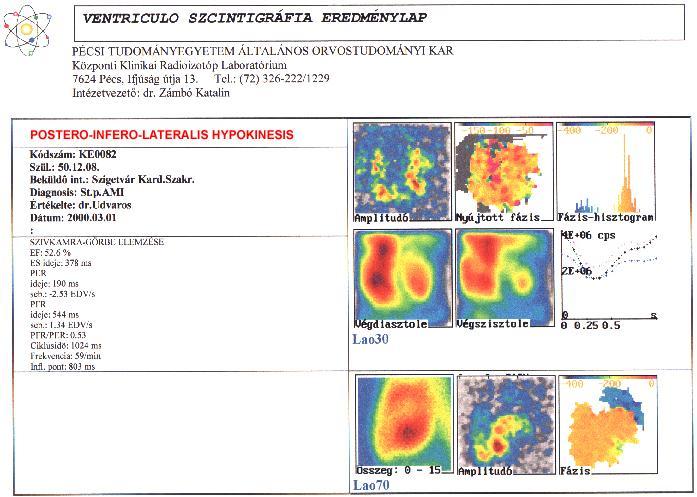
67 VENTRICULO SZCINTIGRÁFIA EREDMÉNYLAP PÉCSI TUDOMÁNYEGYETEM ÁLTALÁNOS ORVOSTUDOMÁNYI KAR Központi Klinikai Radioizotóp Laboratórium 7624 Pécs, Ifjúság útja 13. Tel.: (72) /1229 Intézetvezető: dr. Zámbó Katalin LARGE PARADOX WALL-MOTION IN THE APICAL PART OF THE HEART Kódszám: KE0100 Szül.: Beküldô int.: Komló Bel. Diagnosis: ISZB Értékelte: Dr.Schmidt Dátum: : SZIVKAMRA-GÖRBE ELEMZÉSE EF: 25.2 % ES ideje: 270 ms PER ideje: 137 ms seb.: EDV/s PFR ideje: 392 ms seb.: 1.26 EDV/s PFR/PER: 0.79 Ciklusidö: 592 ms Frekvencia: 101/min Lao30 Lao70 Makro Eq A nyomtatás készült: 2000/03/16
68 An average cycle is generated from more hundred heart cycles
69 The ejection fraction curve


70 VENTRICULO SZCINTIGRÁFIA EREDMÉNYLAP PÉCSI TUDOMÁNYEGYETEM ÁLTALÁNOS ORVOSTUDOMÁNYI KAR Központi Klinikai Radioizotóp Laboratórium 7624 Pécs, Ifjúság útja 13. Tel.: (72) /1229 Intézetvezető: dr. Zámbó Katalin NORMAL FUNCTION OF THE LEFT VENTRICLE Kódszám: KE0351 Szül.: Beküldô int.: Szigetvár Bel. Diagnosis: St.p.inf.myoc. Értékelte: Dr.Schmidt Dátum: : SZIVKAMRA-GÖRBE ELEMZÉSE EF: 64.1 % ES ideje: 398 ms PER ideje: 180 ms seb.: EDV/s PFR ideje: 550 ms seb.: 2.18 EDV/s PFR/PER: 0.87 Ciklusidö: 944 ms Frekvencia: 64/min Infl. pont: 768 ms Lao30 Lao70

71 Parametric pictures Amplitude picture The colours represent the amplitude of the change of the activity of the pixels. Phase picture The colours represent the phase of the change of the activity of the pixels.
72

73 LARGE HYPOKINESIS IN DCM
74 Brain perfusion study The cortex and the basal ganglia are shown by a lypophil radioactive subject (99mTc-HM- PAO hexamethyl-propilenamine-oxyme) Reconstruated and reorientated transversal, sagittal and coronal slices from the brain The impairment of the brain perfusion is indicated by decreased activity or lack of the activity

75 The parts of the brain 1. Seeing cortex 2. Occipital lobe 3. Thalamus 4. Frontal lobe 5. N. caudatus 6. Temporal lobe 7. Chambers

76 Normal brain perfusion

77 CT SPECT SPECT/CT SPECT/CT imaging of brain perfusion CT SPECT SPECT/CT CT SPECT SPECT/CT

78 The changes of the brain perfusion in different diseases

79 Pick disease: atrophy in both frontal lobes
 CT SPECT")
80 Perfusion defect (stroke) in the right occipital region (produced blindness) CT SPECT SPECT/CT

81 Occlusion of left internal carotis artery Before operation After operation

82 Cerebral glioma by 18-FDG-PET

83 Recidiv parasagittal meningeoma after operation (18-FDG)

84 Thank you for your attention!
Nuclear cardiology. Zámbó Katalin Department of Nuclear Medicine
 Nuclear cardiology Zámbó Katalin Department of Nuclear Medicine Imaging techniques Morphology Physiology Metabolism Molecules X-ray / CT MRI NM - SPECT/ PET MR spectroscopy fmri Ultrasound Hybrid imaging:
Itroduction to the Nuclear Medicine: biophysics and basic principles. Zámbó Katalin Department of Nuclear Medicine
 Itroduction to the Nuclear Medicine: biophysics and basic principles Zámbó Katalin Department of Nuclear Medicine NUCLEAR MEDICINE Application of the radioactive isotopes in the diagnostics and in the
Nuclear medicine studies of the digestiv system. Zámbó Katalin Department of Nuclear Medicine
 Nuclear medicine studies of the digestiv system Zámbó Katalin Department of Nuclear Medicine Imaging tehniques Anatomy Physiology Metabolism Molecular Rtg. / CT PET / SPECT MRI MR spectroscopy fmri Ultrasound
Nuclear pulmonology. Katalin Zámbó Department of Nuclear Medicine
 Nuclear pulmonology Katalin Zámbó Department of Nuclear Medicine Imaging techniques Morphology Physiology Metabolism Molecules X-ray / CT MRI NM - SPECT/ PET MR spectroscopy fmri Ultrasound Hybrid imaging:
Nuclear medicine studies of the digestiv system. Zámbó Katalin Department of Nuclear Medicine
 Nuclear medicine studies of the digestiv system Zámbó Katalin Department of Nuclear Medicine Anatomy of the liver Liver scintigraphy The labelled colloid (200 MBq 99mTc-Fyton) is phagocyted by the Kuppfer-cells
Nuclear neurology. Zámbó Katalin Department of Nuclear Medicine
 Nuclear neurology Zámbó Katalin Department of Nuclear Medicine To refresh your memory Brain has a high rate of oxidative metabolism. It has no reserves either of oxygen or of glucose and has a very limited
Nuclear Medicine in Oncology
 Radiopharmaceuticals Nuclear Medicine in Oncology Practice Pharmaceutical Radionuc lide Function Tumor type Diphosphonates Tc-99m Osteoblast Bone tumor & metast. Ga-citrate Ga-67 Fe-analogue Bronchogenous
OTHER NON-CARDIAC USES OF Tc-99m CARDIAC AGENTS Tc-99m Sestamibi for parathyroid imaging, breast tumor imaging, and imaging of other malignant tumors.
 DEFINITION OF CARDIAC RADIOPHARMACEUTICAL: A radioactive drug which, when administered for purpose of diagnosis of heart disease, typically elicits no physiological response from the patient. Even though
Basics of nuclear medicine
 Basics of nuclear medicine Prof. dr. Davor Eterović Prof. dr. Vinko Marković Radioisotopes are used both in diagnostics and in therapy Diagnostics gamma emitters are used since gamma rays can penetrate
Recent initiatives of the FANC. Michel Biernaux Health Protection Service Health and Environment Department
 Recent initiatives of the FANC Michel Biernaux Michel.biernaux@fanc.fgov.be Health Protection Service Health and Environment Department Reminder objectives of the national survey : 1.Review the average
PHYSICS 2: HSC COURSE 2 nd edition (Andriessen et al) CHAPTER 20 Radioactivity as a diagnostic tool (pages 394-5)
 CHAPTER 20 Radioactivity as a diagnostic tool (pages 394-5)") PHYSICS 2: HSC COURSE 2 nd edition (Andriessen et al) CHAPTER 20 Radioactivity as a diagnostic tool (pages 394-5) 1. (a) A radioisotope is an isotope that is unstable and will emit particles from the nucleus
PHYSICS 2: HSC COURSE 2 nd edition (Andriessen et al) CHAPTER 20 Radioactivity as a diagnostic tool (pages 394-5) 1. (a) A radioisotope is an isotope that is unstable and will emit particles from the nucleus
Radiopharmacy. Prof. Dr. Çetin ÖNSEL. CTF Nükleer Tıp Anabilim Dalı
 Prof. Dr. Çetin ÖNSEL CTF Nükleer Tıp Anabilim Dalı What is Nuclear Medicine? Nuclear Medicine is the branch of medicine concerned with the use of radionuclides in the study and the diagnosis of diseases.
NUCLEAR CARDIOLOGY UPDATE
 Nuclear Cardiology David K. Shelton, Jr., MD NUCLEAR CARDIOLOGY UPDATE No Conflicts. No Disclosures. No Smoking. David K. Shelton UCDMC Nuclear Cardiology Nuclear Cardiology Radionuclide Ventriculography
Medical imaging X-ray, CT, MRI, scintigraphy, SPECT, PET Györgyi Műzes
 Medical imaging X-ray, CT, MRI, scintigraphy, SPECT, PET Györgyi Műzes Semmelweis University, 2nd Dept. of Medicine Medical imaging: definition technical process of creating visual representations about
Option D: Medicinal Chemistry
 Option D: Medicinal Chemistry Basics - unstable radioactive nuclei emit radiation in the form of smaller particles alpha, beta, positron, proton, neutron, & gamma are all used in nuclear medicine unstable
Nuclear Medicine: Manuals. Nuclear Medicine. Nuclear imaging. Emission imaging: study types. Bone scintigraphy - technique
 Nuclear Medicine - Unsealed radioactive preparations the tracer mixes with the patients body fluids on a molecular level (e.g. after intravenous injection) - 3 main fields: - In vitro : measuring concentrations
Department of Nuclear Medicine with Positron Emission Tomography
 (PET) Unit [1] Contact information: Registration: +48 41 367 4850 Main office: +48 41 367 4860 Fax: +48 41 367 4887 e-mail: zmnsco@onkol.kielce.pl [2] Head of the Department: Professor Janusz Braziewicz
Nuclear medicine in oncology. 1. Diagnosis 2. Therapy
 Nuclear medicine in oncology 1. Diagnosis 2. Therapy Diagnosis - Conventional methods - Nonspecific radiopharmaceuticals cumulating in tumours - Specific radiopharmaceuticals (receptor- and immunoscintigraphy)
Fundamentals of Nuclear Cardiology. Terrence Ruddy, MD, FRCPC, FACC
 Fundamentals of Nuclear Cardiology Terrence Ruddy, MD, FRCPC, FACC Objectives To understand the Principles of Nuclear Cardiac Imaging Radiotracers Image acquisition and processing Stress protocols To appreciate
Nuclear Medicine and PET. D. J. McMahon rev cewood
 Nuclear Medicine and PET D. J. McMahon 150504 rev cewood 2018-02-15 Key Points Nuclear Medicine and PET: Imaging: Understand how Nuc Med & PET differ from Radiography & CT by the source of radiation. Be
Radiopharmaceutical Activities Administered for Diagnostic and Therapeutic Procedures in Nuclear Medicine in Argentine: Results of a National Survey
 Radiopharmaceutical Activities Administered for Diagnostic and Therapeutic Procedures in Nuclear Medicine in Argentine: Results of a National Survey A. M. Bomben, C. A. Chiliutti Autoridad Regulatoria
weighing risks against benefits ALARA principle appropriate activities (radiopharmaceutical doses)
") weighing risks against benefits ALARA principle appropriate activities (radiopharmaceutical doses) based on EANM references adequate appointment method (patient booking system) Appropriate activities (doses)
weighing risks against benefits ALARA principle appropriate activities (radiopharmaceutical doses) based on EANM references adequate appointment method (patient booking system) Appropriate activities (doses)
CEREBRAL BLOOD FLOW AND METABOLISM
 Supported by: HURO/0901/069/2.3.1 HU-RO-DOCS CEREBRAL BLOOD FLOW AND METABOLISM Part 3 Modern imaging methods SPECT, PET, nmri History of Nuclear Medicine Starts with the invention of the X-ray 1946: radioactive
NUCLEAR MEDICINE Molecular Imaging + Endo-Radiotherapy
 NUCLEAR MEDICINE Molecular Imaging + Endo-Radiotherapy Istvan Szilvási Dept. of Nuclear Medicine Semmelweis University and HDF Medical Centre 2016 DEFINITION OF NUCLEAR MEDICINE Medical applications of
Radionuclides in Medical Imaging. Danielle Wilson
 Radionuclides in Medical Imaging Danielle Wilson Outline Definitions History and development Radionuclide applications & techniques in imaging Conclusion Definition #1 : Radionuclide An unstable nucleus
SPECT-CT: Τι πρέπει να γνωρίζει ο Καρδιολόγος
 SPECT-CT: Τι πρέπει να γνωρίζει ο Καρδιολόγος Δρ Αναστασία Κίτσιου Διευθύντρια, Καρδιολογική Κλινική, Σισμανόγλειο ΓΝΑ Chair, Education Committee, Section on Nuclear Cardiology & Cardiac CT, EACVI, ESC
Physical Bases : Which Isotopes?
 Physical Bases : Which Isotopes? S. Gnesin Institute of Radiation Physics, Lausanne University Hospital, Lausanne, Switzerland 1/53 Theranostic Bruxelles, 2 Octobrer 2017 Theranostic : use of diagnostic
E LT O N A. M O S M A N OUTLINE
 34 NUCLEAR MEDICINE E LT O N A. M O S M A N OUTLINE Principles of nuclear medicine, 388 Historical development, 389 Comparison with other modalities, 390 Physical principles of nuclear medicine, 392 Radiation
General Nuclear Medicine
 General Nuclear Medicine What is General Nuclear Medicine? What are some common uses of the procedure? How should I prepare? What does the equipment look like? How does the procedure work? How is the procedure
Austin Radiological Association Ga-68 NETSPOT (Ga-68 dotatate)
") Austin Radiological Association Ga-68 NETSPOT (Ga-68 dotatate) Overview Ga-68 dotatate binds to somatostatin receptors, with highest affinity for subtype 2 receptors (sstr2). It binds to cells that express
Austin Radiological Association Ga-68 NETSPOT (Ga-68 dotatate) Overview Ga-68 dotatate binds to somatostatin receptors, with highest affinity for subtype 2 receptors (sstr2). It binds to cells that express
Gated blood pool ventriculography: Is there still a role in myocardial viability?
 Gated blood pool ventriculography: Is there still a role in myocardial viability? Oliver C. Alix, MD Adult Clinical and Nuclear Cardiology St. Luke s Medical Centre - Global City Case Presentation A 62-year-old
Contrast Agents and Radiopharmaceuticals 2017
 Contrast Agents and Radiopharmaceuticals 207 Covered: Code Code Description Allow with Code(s) Code Description Max Units A464 Radiopharmaceutical, diagnostic, not otherwise classified n/a Invoice Req'd
EMPHISIS ON PHYSIOLOGY PHYSIOLOGY REQUIRES TIME QUALITATIVE vs. QUANTITATIVE ISOTOPES TO RADIOPHARMACEUTICALS
 1926 Herman Blumgard used solutions of radon gas to measure what he called velocity of the circulation. 1927 Blumberg and Soma Weiss wrote article in (Journal of Clinical Investigation) 1929 Werner Forssmann
KEYWORDS: nuclear medicine; gamma camera; radiopharmaceutical activities.
 Radiopharmaceutical Activities Administered for Diagnostic Procedures in Nuclear Medicine in the First Six Months of the Gamma Camera Use in the Clinical Center of Montenegro - Podgorica Nevenka Antovic
AN INTRODUCTION TO NUCLEAR MEDICINE
 AN INTRODUCTION TO NUCLEAR MEDICINE WITH RESPECT TO THYROID DISORDERS By: B.Shafiei MD Nuclear Physician Taleghani Medical Center Radioactive: An element with Unstable Nucleus (Excess Energy), can achieve
Isotopes in Functional Cancer Imaging
 Seeing and Believing: i Medical Isotopes in Functional Cancer Imaging François Bénard, MD, FRCPC BCCancer Cancer Agency and University of British Columbia Nuclear Medicine 101 A radioactive atom is produced
Medical Use of Radioisotopes
 Medical Use of Radioisotopes Therapy Radioisotopes prove to be useful in the application of brachytherapy, the procedure for using temporary irradiation close to the area of disease (i.e. cancer) 10% Medical
Radioactivity. Alpha particles (α) :
 :") Radioactivity It is the property of an element that causes it to emit radiation Discovered by Becquerel (1896) Radiation comes from the nucleus of the atom There are three types of radiation : alpha particles
Radioactivity It is the property of an element that causes it to emit radiation Discovered by Becquerel (1896) Radiation comes from the nucleus of the atom There are three types of radiation : alpha particles
Typical PET Image. Elevated uptake of FDG (related to metabolism) Lung cancer example: But where exactly is it located?
 Lung cancer example: But where exactly is it located?") Typical PET Image Elevated uptake of FDG (related to metabolism) Lung cancer example: But where exactly is it located? PET/CT Oncology Imaging Anatometabolic fusion images are useful in the management
Typical PET Image Elevated uptake of FDG (related to metabolism) Lung cancer example: But where exactly is it located? PET/CT Oncology Imaging Anatometabolic fusion images are useful in the management
SPECT TRACERS Tl-201, Tc-99m Sestamibi, Tc-99m Tetrofosmin
 SPECT TRACERS Tl-201, Tc-99m Sestamibi, Tc-99m Tetrofosmin Elmer Jasper B. Llanes, M.D. Nuclear Cardiology St. Luke s Medical Center Outline Ideal Physiologic Characteristics of MPI radioactive tracers
Radiologic Assessment of Myocardial Viability
 November 2001 Radiologic Assessment of Myocardial Viability Joshua Moss, Harvard Medical School Year III Patient EF 66yo female with a 3-year history of intermittent chest pain previously relieved by sublingual
1. LV function and remodeling. 2. Contribution of myocardial ischemia due to CAD, and
 1 The clinical syndrome of heart failure in adults is commonly associated with the etiologies of ischemic and non-ischemic dilated cardiomyopathy, hypertrophic cardiomyopathy, hypertensive heart disease,
Clinical indications for positron emission tomography
 Clinical indications for positron emission tomography Oncology applications Brain and spinal cord Parotid Suspected tumour recurrence when anatomical imaging is difficult or equivocal and management will
Myocardial viability testing. What we knew and what is new
 Myocardial viability testing. What we knew and what is new Dr B K S Sastry, MD, DM. CARE Hospitals, Hyderabad What is Viability Viability Dysfunctional myocardium subtended by diseased coronary arteries
Click here for Link to References: CMS Website HOPPS CY 2018 Final Rule. CMS Website HOPPS CY2018 Final Rule Updated November 2017.
 Final Compared to 3Q 2017 Rates Medicare Hospital Outpatient Prospective Payment System HOPPS () Nuclear Cardiology Procedures, Radiopharmaceuticals, and Drugs Click here for Link to References: CMS Website
Description MRI, TMJ C T Head Without Contrast C T Head With Contrast C T Head Without & With Contrast
 s Requiring Prior Authorization for the Advanced Imaging 70336 MRI, TMJ 70450 C T Head Without Contrast 70460 C T Head With Contrast 70470 C T Head Without & With Contrast 70480 C T Orbit Without Contrast
A Snapshot on Nuclear Cardiac Imaging
 Editorial A Snapshot on Nuclear Cardiac Imaging Khalil, M. Department of Physics, Faculty of Science, Helwan University. There is no doubt that nuclear medicine scanning devices are essential tool in the
Anthem Blue Cross and Blue Shield Virginia Advanced Imaging Procedures Requiring Precertification Revised 02/13/2013
 Anthem Blue Cross and Blue Shield Virginia Advanced Imaging Procedures Requiring Precertification Revised 02/13/2013 Modality and CT Head CTA Head: Cerebrovascular MRI Head MRA Head: Cerebrovascular Functional
Cardiovascular nuclear imaging employs non-invasive techniques to assess alterations in coronary artery flow, and ventricular function.
 National Imaging Associates, Inc. Clinical guidelines CARDIOVASCULAR NUCLEAR MEDICINE -MYOCARDIAL PERFUSION IMAGING -MUGA CPT4 Codes: Refer to pages 6-9 LCD ID Number: L33960 J 15 = KY, OH Responsible
Functional aspects of anatomical imaging techniques
 Functional aspects of anatomical imaging techniques Nilendu Purandare Associate Professor & Consultant Radiologist Tata Memorial Centre Functional/metabolic/molecular imaging (radioisotope scanning) PET
Index. Surg Oncol Clin N Am 16 (2007) Note: Page numbers of article titles are in boldface type.
 Note: Page numbers of article titles are in boldface type.") Surg Oncol Clin N Am 16 (2007) 465 469 Index Note: Page numbers of article titles are in boldface type. A Adjuvant therapy, preoperative for gastric cancer, staging and, 339 B Breast cancer, metabolic
Surg Oncol Clin N Am 16 (2007) 465 469 Index Note: Page numbers of article titles are in boldface type. A Adjuvant therapy, preoperative for gastric cancer, staging and, 339 B Breast cancer, metabolic
Tc-99m Sestamibi/Tetrofosmin Stress-Rest Myocardial Perfusion Scintigraphy
 APPROVED BY: Director of Radiology Page 1 of 6 Tc-99m Sestamibi/Tetrofosmin Stress-Rest Myocardial Primary Indications: Evaluation of myocardial perfusion and viability in patients with known or suspected
COMENIUS-Project: SM&CLIL Radiation & Medicine
 Medical imaging refers to the techniques and processes used to create images of the human body (or parts thereof) for clinical purposes. Thanks to modern mathematics and computer technology, medical imaging
RADIOTRACERS FOR MYOCARDIAL PERFUSION IMAGING
 RADIOTRACERS FOR MYOCARDIAL PERFUSION IMAGING RAYMOND TAILLEFER, M.D. FRCP(c), ABNM DIRECTOR, DEPARTMENT OF NUCLEAR MEDICINE HOPITAL ST-JEAN-SUR-RICHELIEU Disclosures to Report: Grant Research Support:
PET IMAGING (POSITRON EMISSION TOMOGRAPY) FACT SHEET
 FACT SHEET") Positron Emission Tomography (PET) When calling Anthem (1-800-533-1120) or using the Point of Care authorization system for a Health Service Review, the following clinical information may be needed to
Positron Emission Tomography (PET) When calling Anthem (1-800-533-1120) or using the Point of Care authorization system for a Health Service Review, the following clinical information may be needed to
Pearls & Pitfalls in nuclear cardiology
 Pearls & Pitfalls in nuclear cardiology Maythinee Chantadisai, MD., NM physician Division of Nuclear Medicine, Department of radiology, KCMH Principle of myocardial perfusion imaging (MPI) Radiotracer
45 Hr PET Registry Review Course
 45 HR PET/CT REGISTRY REVIEW COURSE Course Control Document Timothy K. Marshel, MBA, R.T. (R), (N)(CT)(MR)(NCT)(PET)(CNMT) The PET/CT Training Institute, Inc. SNMMI-TS 028600-028632 45hr CEH s Voice Credits
Cardiovascular nuclear imaging employs non-invasive techniques to assess alterations in coronary artery flow, and ventricular function.
 National Imaging Associates, Inc. Clinical guidelines CARDIOVASCULAR NUCLEAR MEDICINE -MYOCARDIAL PERFUSION IMAGING -MUGA Original Date: October 2015 Page 1 of 9 FOR CMS (MEDICARE) MEMBERS ONLY CPT4 Codes:
NUCLEAR MEDICINE OF HEART AND LUNG
 Clinical decision tree in CAD NUCLEAR MEDICINE OF HEART AND LUNG Abnormal pump function Slides of lectures + electronic books: Stress-rest Stress: defect MPI Rest: better or normal Glucose metab? László
Multiple Gated Acquisition (MUGA) Scanning
 Scanning") Multiple Gated Acquisition (MUGA) Scanning Dmitry Beyder MPA, CNMT Nuclear Medicine, Radiology Barnes-Jewish Hospital / Washington University St. Louis, MO Disclaimers/Relationships Standard of care research
Multiple Gated Acquisition (MUGA) Scanning Dmitry Beyder MPA, CNMT Nuclear Medicine, Radiology Barnes-Jewish Hospital / Washington University St. Louis, MO Disclaimers/Relationships Standard of care research
Austin Radiological Association Nuclear Medicine Procedure BONE MINERAL STUDY (Tc-99m-MDP, Tc-99m-HMDP)
") Austin Radiological Association Nuclear Medicine Procedure BONE MINERAL STUDY (Tc-99m-MDP, Tc-99m-HMDP) Overview The Bone Mineral Study, with either Tc-99m-MDP or Tc-99m-HMDP, depicts the distribution
Austin Radiological Association Nuclear Medicine Procedure BONE MINERAL STUDY (Tc-99m-MDP, Tc-99m-HMDP) Overview The Bone Mineral Study, with either Tc-99m-MDP or Tc-99m-HMDP, depicts the distribution
György HEVESY ( ) 1943 Nobel Laureate in Chemistry for his work on the use of isotopes as tracers in the study of chemical processes
 1943 Nobel Laureate in Chemistry for his work on the use of isotopes as tracers in the study of chemical processes") József Varga Introduction to UCLEAR MEDICIE Department of uclear Medicine University of Debrecen 2011. UCLEAR MEDICIE uclear Medicine: Manuals Required reading: Taylor A., Alazraki., and Schuster D.M.:
József Varga Introduction to UCLEAR MEDICIE Department of uclear Medicine University of Debrecen 2011. UCLEAR MEDICIE uclear Medicine: Manuals Required reading: Taylor A., Alazraki., and Schuster D.M.:
Chapter 1. Introduction
 Chapter 1 Introduction 1.1 PRINCIPLES OF NUCLEAR MEDICINE Nuclear medicine techniques use radioactive tracers and imaging devices, mainly to provide diagnostic information, but in some cases also for therapeutic
Molecular Imaging Guided Therapy: The Perfect Storm. David M Schuster, MD Emory University Department of Radiology Atlanta, GA
 Molecular Imaging Guided Therapy: The Perfect Storm David M Schuster, MD Emory University Department of Radiology Atlanta, GA Talk can be found at radiology.emory.edu Let s start with a case 74 year
Positron Emission Tomography Computed Tomography (PET/CT)
") Positron Emission Tomography Computed Tomography (PET/CT) What is Positron Emission Tomography Computed Tomography (PET/CT) Scanning? What are some common uses of the procedure? How should I prepare for
Positron Emission Tomography Computed Tomography (PET/CT) What is Positron Emission Tomography Computed Tomography (PET/CT) Scanning? What are some common uses of the procedure? How should I prepare for
Cardiac PET. John Buscombe
 Cardiac PET John Buscombe Why PET? Improved resolution-not really required in cardiology Improved sensitivity this may be important-financially as reduced acquisition time Improved attenuation correction-good
Reversible defect of 123 I-15-(p-iodophenyl)-9-(R,S)-methylpentadecanoic acid indicates residual viability within infarct-related area
-9-(R,S)-methylpentadecanoic acid indicates residual viability within infarct-related area") ORIGINAL ARTICLE Annals of Nuclear Medicine Vol. 16, No. 3, 183 187, 2002 Reversible defect of 123 I-15-(p-iodophenyl)-9-(R,S)-methylpentadecanoic acid indicates residual viability within infarct-related
ORIGINAL ARTICLE Annals of Nuclear Medicine Vol. 16, No. 3, 183 187, 2002 Reversible defect of 123 I-15-(p-iodophenyl)-9-(R,S)-methylpentadecanoic acid indicates residual viability within infarct-related
Nuclear Medicine Head and Neck Region. Bán Zsuzsanna, MD University of Pécs, Department of Nuclear Medicine
 Nuclear Medicine Head and Neck Region Bán Zsuzsanna, MD University of Pécs, Department of Nuclear Medicine Thyroid scintigraphy Parathyroid scintigraphy F18-FDG PET examinations in head and neck cancer
MRI and CT of the CNS
 MRI and CT of the CNS Dr.Maha ELBeltagy Assistant Professor of Anatomy Faculty of Medicine The University of Jordan 2018 Computed Tomography CT is used for the detection of intracranial lesions. CT relies
Nuclear Medicine Diagnosis
 Anatomy : MRI Fonction : Nucl Med Nuclear Medicine Diagnosis Functional imaging = distribution of a (radio)- tracer in organs Each tracer is a SPY of a function, usually through a metabolic pathway flow
PET-MRI in malignant bone tumours. Lars Stegger Department of Nuclear Medicine University Hospital Münster, Germany
 PET-MRI in malignant bone tumours Lars Stegger Department of Nuclear Medicine University Hospital Münster, Germany Content From PET to PET/MRI General considerations Bone metastases Primary bone tumours
Nuclear medicine methods in the urogenital system
 Nuclear medicine methods in the urogenital system Anatomy of the kidneys I. Anatomy of the kidneys II. The types of examinations Static examinations (scintigraphy): 1) the radiopharmaceutical is administered
Dr Alfred O Ankrah FCNP
 Dr Alfred O Ankrah FCNP Outline Introduction Brief history of Nuclear Medicine in Ghana Current situation of Nuclear Medicine in Ghana Use of Nuclear medicine in various disciplines Future of Nuclear Medicine
POSITRON EMISSION TOMOGRAPHY (PET)
") Status Active Medical and Behavioral Health Policy Section: Radiology Policy Number: V-27 Effective Date: 08/27/2014 Blue Cross and Blue Shield of Minnesota medical policies do not imply that members should
Status Active Medical and Behavioral Health Policy Section: Radiology Policy Number: V-27 Effective Date: 08/27/2014 Blue Cross and Blue Shield of Minnesota medical policies do not imply that members should
Bone PET/MRI : Diagnostic yield in bone metastases and malignant primitive bone tumors
 Bone PET/MRI : Diagnostic yield in bone metastases and malignant primitive bone tumors Lars Stegger, Benjamin Noto Department of Nuclear Medicine University Hospital Münster, Germany Content From PET to
Cardiac Imaging. Kimberly Delcour, DO, FACC. Mahi Ashwath, MD, FACC, FASE. Director, Cardiac CT. Director, Cardiac MRI
 Cardiac Imaging Kimberly Delcour, DO, FACC Director, Cardiac CT Mahi Ashwath, MD, FACC, FASE Director, Cardiac MRI Cardiac Imaging Discuss the clinical applications of and indications for: Cardiac CT Nuclear
Austin Radiological Association Nuclear Medicine Procedure WHITE BLOOD CELL MIGRATION STUDY (In-111-WBCs, Tc-99m-HMPAO-WBCs)
") Austin Radiological Association Nuclear Medicine Procedure WHITE BLOOD CELL MIGRATION STUDY (In-111-WBCs, Tc-99m-HMPAO-WBCs) Overview Indications The White Blood Cell Migration Study demonstrates the distribution
Austin Radiological Association Nuclear Medicine Procedure WHITE BLOOD CELL MIGRATION STUDY (In-111-WBCs, Tc-99m-HMPAO-WBCs) Overview Indications The White Blood Cell Migration Study demonstrates the distribution
NUCLEAR CARDIOLOGY AND ADVANCED VASCULAR IMAGING. Joel Kahn, MD, FACC
 NUCLEAR CARDIOLOGY AND ADVANCED VASCULAR IMAGING Joel Kahn, MD, FACC Short History Nuclear Cardiology Hermann blumgart-1927-injected radon to measure circulation time Hal Anger-1952-gamma camera-beginning
THE PARATHYROID GLAND THEORY AND NUCLEAR MEDICINE PRACTICE
 THE PARATHYROID GLAND THEORY AND NUCLEAR MEDICINE PRACTICE George N. Sfakianakis MD Professor of Radiology and Pediatrics Director, Division of Nuclear Medicine UM/JMMC Miami FL October 2009 ENDONCRINE
Radionuclide & Radiopharmaceuticals
 Radionuclide & Radiopharmaceuticals 1. Generator & Reactors 2. Cyclotrons & PET tracer 3. Quality control 4. Renal 5. GIT 6. CNS & Psychiatrics 7. Tumor Diagnosis & Treatment 8. Bones & joints 9. Thyroid
Austin Radiological Association Nuclear Medicine Procedure PROSTATE CANCER STUDY (In-111-Capromab Pendetide [ProstaScint ])
![Austin Radiological Association Nuclear Medicine Procedure PROSTATE CANCER STUDY (In-111-Capromab Pendetide [ProstaScint ])](/thumbs/81/82771892.jpg "Austin Radiological Association Nuclear Medicine Procedure PROSTATE CANCER STUDY (In-111-Capromab Pendetide [ProstaScint ])") Austin Radiological Association Nuclear Medicine Procedure PROSTATE CANCER STUDY (In-111-Capromab Pendetide [ProstaScint ]) Overview Indications The Prostate Cancer Study with an indium-111 labeled murine
Austin Radiological Association Nuclear Medicine Procedure PROSTATE CANCER STUDY (In-111-Capromab Pendetide [ProstaScint ]) Overview Indications The Prostate Cancer Study with an indium-111 labeled murine
INDICATIONS AND USAGE
 1. INDICATIONS AND USAGE a) Axumin is indicated for positron emission tomography (PET) in men with suspected prostate cancer recurrence based on elevated blood prostate specific antigen (PSA) levels following
General Nuclear Medicine
 Scan for mobile link. General Nuclear Medicine Nuclear medicine imaging uses small amounts of radioactive materials called radiotracers that are typically injected into the bloodstream, inhaled or swallowed.
Value of Assessment of Viable and Ischemic Myocardium and Techniques Such as MRI, Radionuclide Imaging
 Chapter 2 Imaging for Viable and Ischemic Myocardium Value of Assessment of Viable and Ischemic Myocardium and Techniques Such as MRI, Radionuclide Imaging Catalin Loghin and K. Lance Gould Introduction
Nuclear Cardiology Reimbursement. Todd Lamb, BS, AS, CNMT Clinical Operations Mgr Regions Hospital St. Paul, MN
 Nuclear Cardiology Reimbursement Todd Lamb, BS, AS, CNMT Clinical Operations Mgr Regions Hospital St. Paul, MN Slides are not to be reproduced without the permission of the author Slides are not to be
Prior Authorization for Non-emergency Cardiac Imaging Procedures
 Attention: All Providers Prior Authorization for Non-emergency Cardiac Imaging Procedures The N.C. Medicaid Program is considering implementation of a prior authorization (PA) program for non-emergency
Peptide Receptor Radionuclide Therapy using 177 Lu octreotate
 Peptide Receptor Radionuclide Therapy using 177 Lu octreotate BLR Kam, Erasmus Medical Centre, Rotterdam DJ Kwekkeboom, Erasmus Medical Centre, Rotterdam Legal aspects As 177 Lu-[DOTA 0 -Tyr 3 ]octreotate
Radiopharmaceuticals. Radionuclides in NM. Radionuclides NUCLEAR MEDICINE. Modes of radioactive decays DIAGNOSTIC THERAPY CHEMICAL COMPOUND
 Univerzita Karlova v Praze - 1. Lékařská fakulta Radiation protection NUCLEAR MEDICINE Involving the application of radioactive substances in the diagnosis and treatment of disease. Nuclear medicine study
Last Updated: 2/10/2017 Implementation date: 4/3/2017 Radiology & Cardiology Prior Authorization / Utilization Management Procedure List
 Last Updated: 2/10/2017 Implementation date: 4/3/2017 Radiology & Cardiology Prior Authorization / Utilization Management Procedure List Deal Sheet Group Product Category CPT CPT Description 3D Imaging
Nuclear medicine in endocrinology
 Nuclear medicine in endocrinology Thyroid gland: anatomy, function, inflammation, Nuclear medicine in endocrinology tumor dignitiy Parathyroid gland: localisation Adrenal cortex: function Adrenal medulla:
Radiation physics and radiation protection. University of Szeged Department of Nuclear Medicine
 Radiation physics and radiation protection University of Szeged Department of Nuclear Medicine Radiation doses to the population 1 Radiation doses to the population 2 Sources of radiation 1 Radiation we
HIP RADIOLOGY PROGRAM CODE LISTS
 EFFECTIVE OCTOBER 1, 2012 70336 MAGNETIC RESONANCE IMAGING TMJ 70450 COMPUTED TOMOGRAPHY HEAD/BRAIN WITHOUT 70460 COMPUTED TOMOGRAPHY HEAD/BRAIN WITH 70470 COMPUTED TOMOGRAPHY HEAD/BRAIN WITHOUT AND WITH
The Integral Role of Metabolic and Perfusion Imaging in Assessment of Myocardial Scar: Comparison between 18F-FDG PET and 99Tc-Sestamibi
 Med. J. Cairo Univ., Vol. 82, No. 1, June: 285-289, 2014 www.medicaljournalofcairouniversity.net The Integral Role of Metabolic and Perfusion Imaging in Assessment of Myocardial Scar: Comparison between
Radionuclide detection of sentinel lymph node
 Radionuclide detection of sentinel lymph node Sophia I. Koukouraki Assoc. Professor Department of Nuclear Medicine Medicine School, University of Crete 1 BACKGROUND The prognosis of malignant disease is
Nuclear Medicine in the Diabetic Foot
 26.11.2015, Uniklinik Balgrist Nuclear Medicine in the Diabetic Foot Martin Hüllner Nuklearmedizin und Neuroradiologie, USZ / UZH Outline A. Imaging modalities brief technical overview B. Nuclear medicine
HEALTHFIRST 2011 RADIOLOGY PROGRAM CODE LIST
 HEALTHFIRST 2011 RADIOLOGY PROGRAM CODE LIST Outpatient Radiology utilization call Carecore at 1-877-773-6964 Modality CPT CODE Description CT SCANS 70450 CT HEAD/BRAIN W/O CONTRAST CT SCANS 70460 CT HEAD/BRAIN
Nuclear Medicine: Basics to therapy
 Nuclear Medicine: Basics to therapy RCP Medical careers day Dr Sabina Dizdarevic MD MSc PhD FRCP Dr Deena Neriman MBBS FRCR Ms Charlotte Weston CEO BNMS On behalf of the British Nuclear Medicine Society
The Value of Tc-99m MIBI Washout Rate in Detection of Ischemia compared with standard Myocardial Perfusion Imaging
 Egyptian J. Nucl. Med., Vol. 10, No. 2, Dec 2014 18 Original article, Cardiology The Value of Tc-99m MIBI Washout Rate in Detection of Ischemia compared with standard Myocardial Perfusion Imaging Moustafa,
Case 1: 79 yr-old woman with a lump in upper outer quadrant of left breast.
 Case 1: 79 yr-old woman with a lump in upper outer quadrant of left breast. Giuliano Mariani Regional Center of Nuclear Medicine, University of Pisa Medical School, Pisa (Italy) Relevant history 79-yr
Radioisotopes in Nuclear Medicine
 Radioisotopes in Nuclear Medicine Anthony J. McGoron Florida International University Biomedical Engineering Institute 10555 West Flagler Street Miami, FL, 33199, USA 305-348-1352 Anthony.mcgoron@fiu.edu