Riste Saat ITKH, HUS. MRT-kool Kõrva ja temporaalluu MRT
|
|
|
- Cynthia Harrell
- 6 years ago
- Views:
Transcription
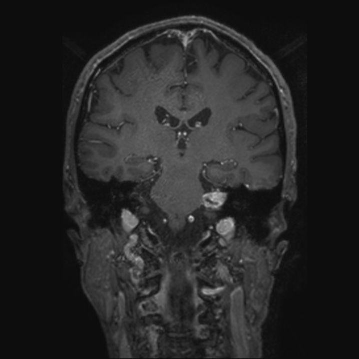
1 Kõrva ja temporaalluu MRT MRT-kool 2012 Riste Saat ITKH, HUS
2 Näidustused Tehnika Anatoomia Patoloogia Pseudolesioonid ja diagnostilised vead
3 Kõrva MRT näidustused n Sensorineuraalne kuulmislangus, tinnitus, vestibulaarsed häired h Temporaaluu ekspansiivsed lesioonid Komplitseerunud välis v lis- ja keskkõrva põletikud
4 MRT tehnika MRT tehnika Vähemalt üks sekvents kogu ajust 2-3mm lõigud temporaalluust Vähemalt üks kõrge resolutsiooniga 3D sekvents kõrvadest Vajadusel Gd Vajadusel DWI Vajadusel MRA

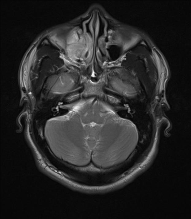
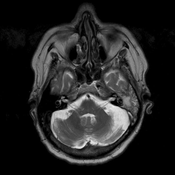
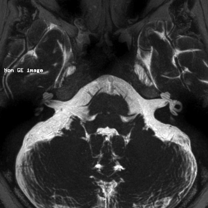
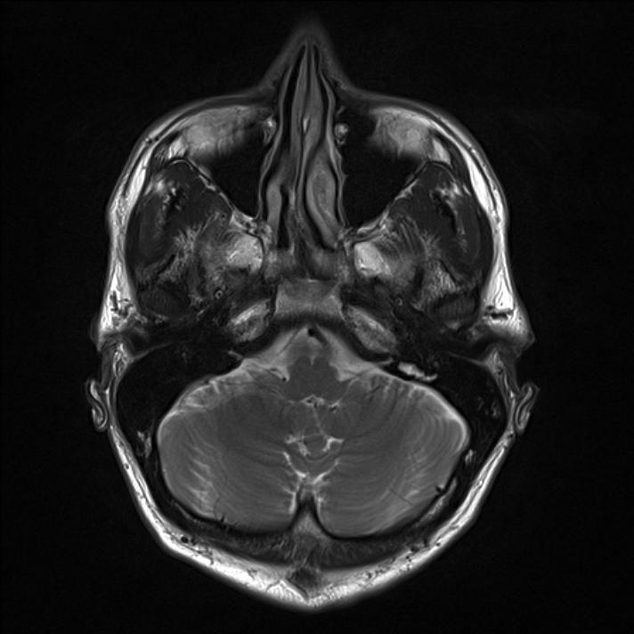
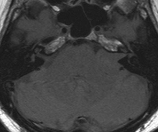
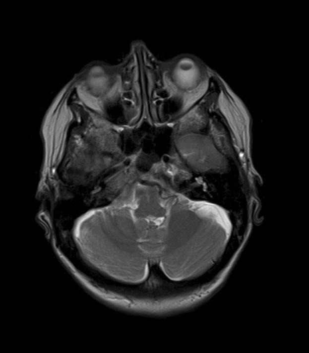
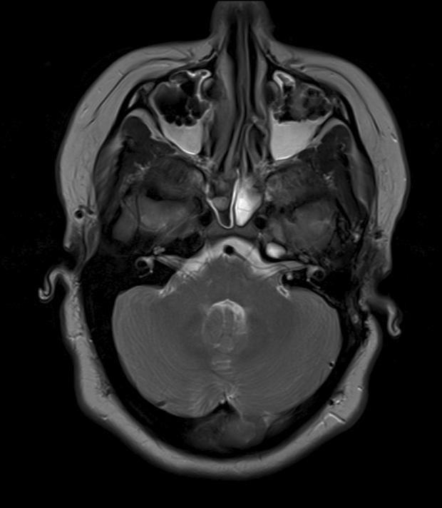
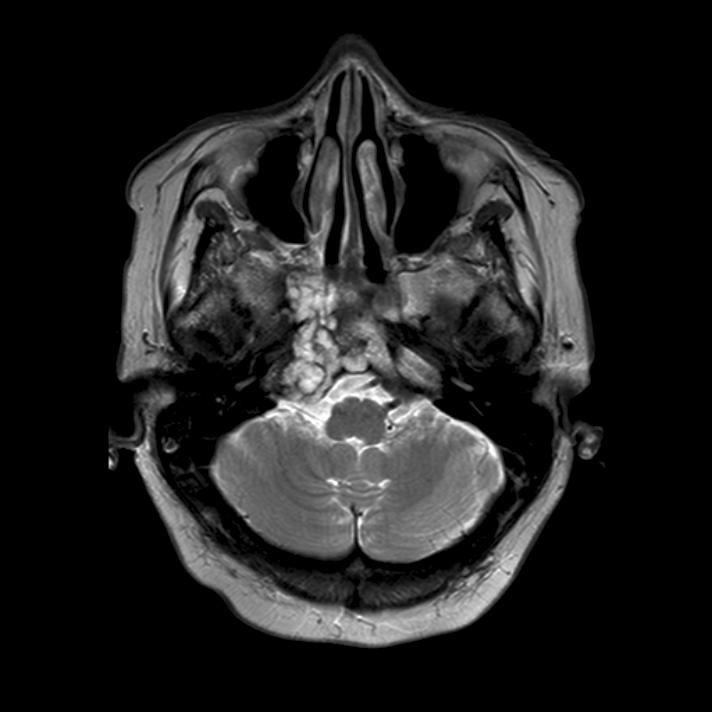
5 Tehnika: T2 ax

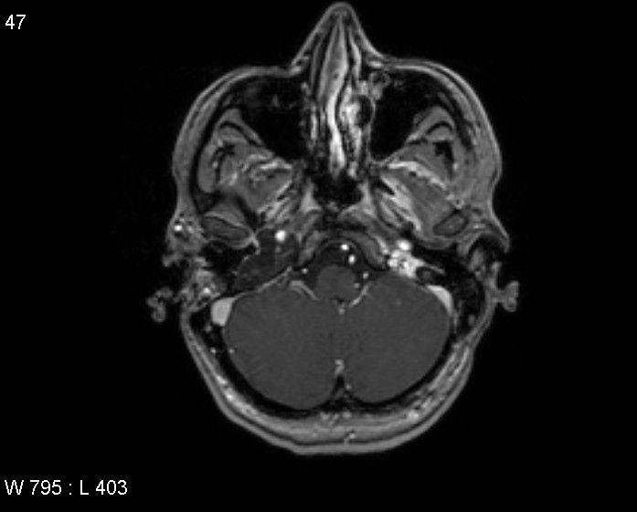
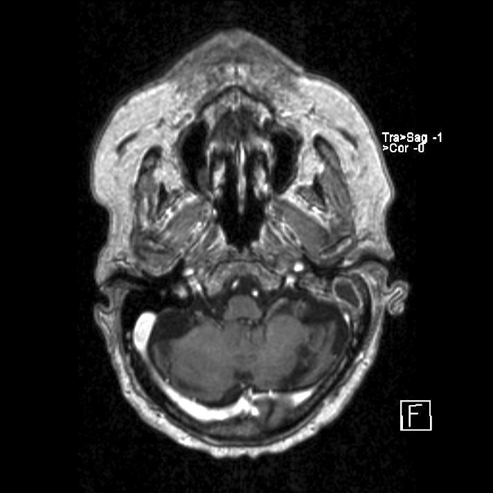
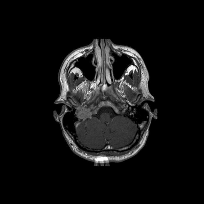
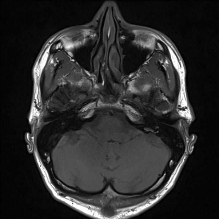
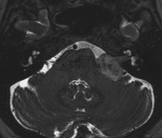
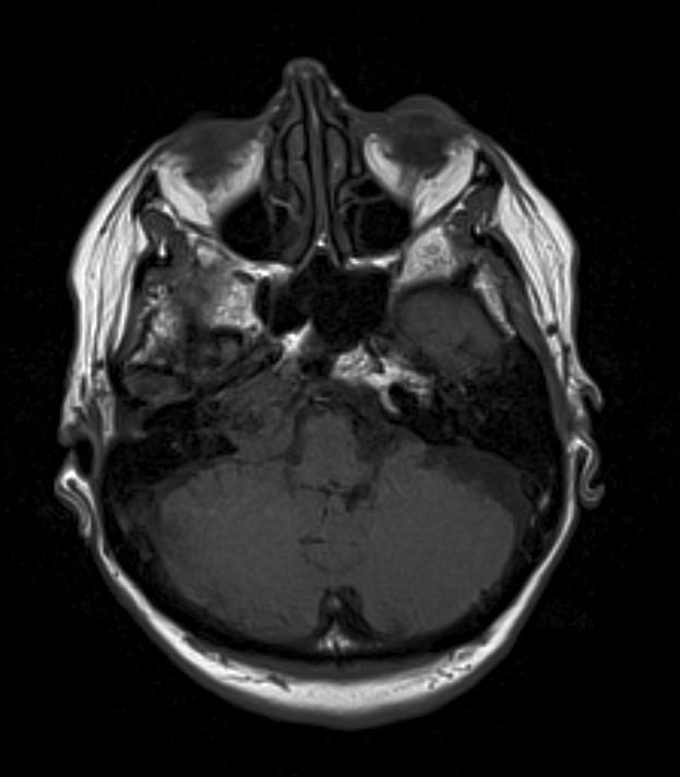
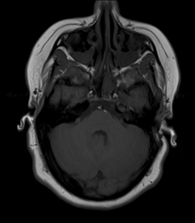
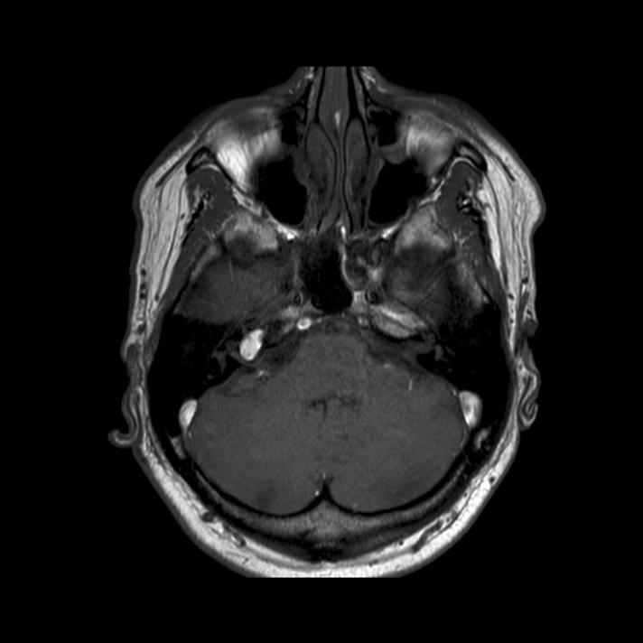
6 Tehnika: T1 ax

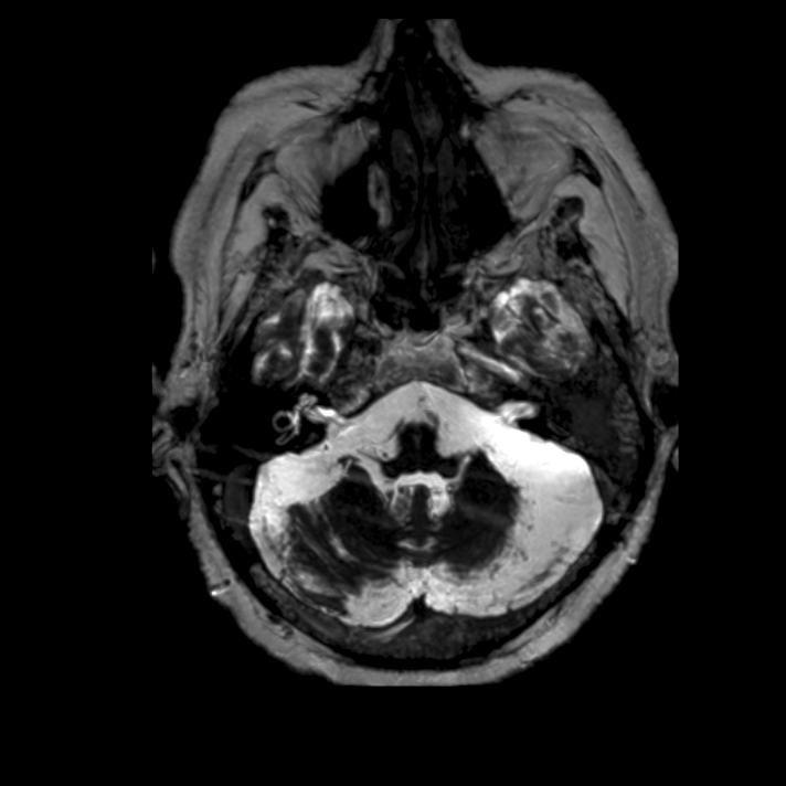
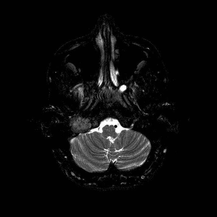
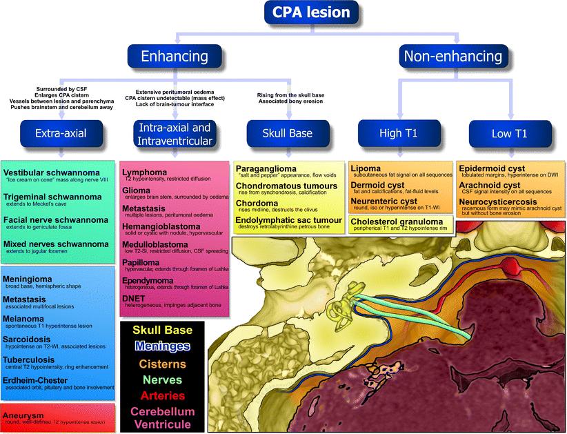
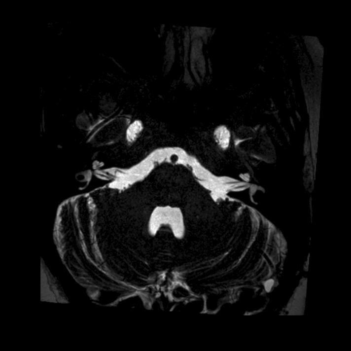
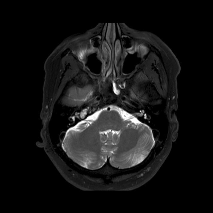
7 HR3D FT T2 ax (CISS/ DRIVE/ FIESTA)


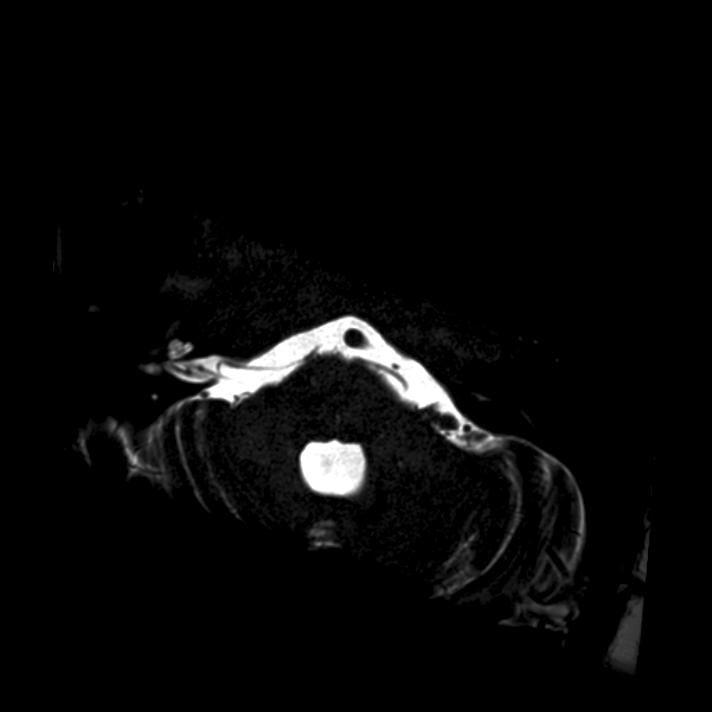
8 HR 3D FT T1 Gd sag (SPGR, GRASS, MPRAGE)
")

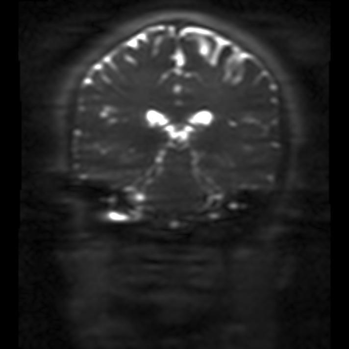
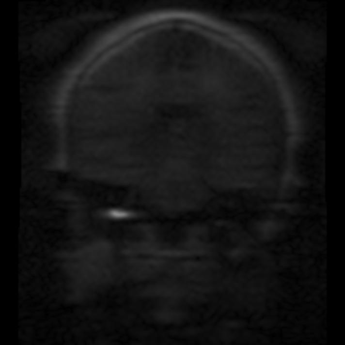
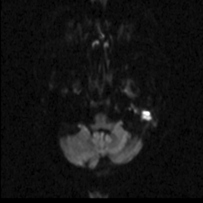
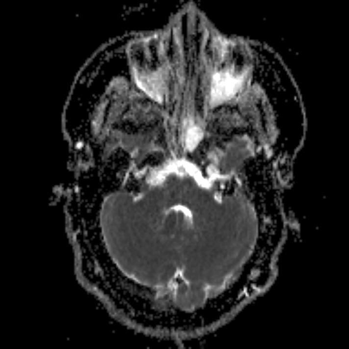
9 SS-TSE DWI (non-epi) cor EPI-DWI ax Non-EPI DWI cor
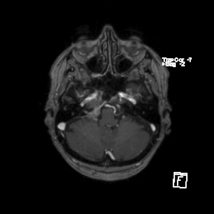
10 TOF MRA
11 Anatoomia
12 Kõrva ja temporaalluu osad Kõrva ja temporaalluu osad T2 T1 Väliskõrv Keskkõrv Sisekõrv ja sisemine kuulmekäik Apex
13 Keskkõrv: anatoomia Keskkõrv: anatoomia
 A = antrum mastoideum A T C SCC")
14 Keskkõrv: anatoomia T = trummiõõs (tympanum) ja kuulmeluukesed C = cochlea MAI = meatus acuticus internus V = vestibulum SCC = poolringkanalid (canales semicirculares) A = antrum mastoideum A T C SCC MAI V
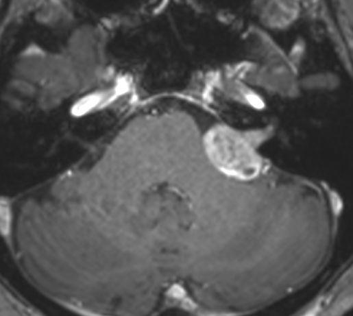
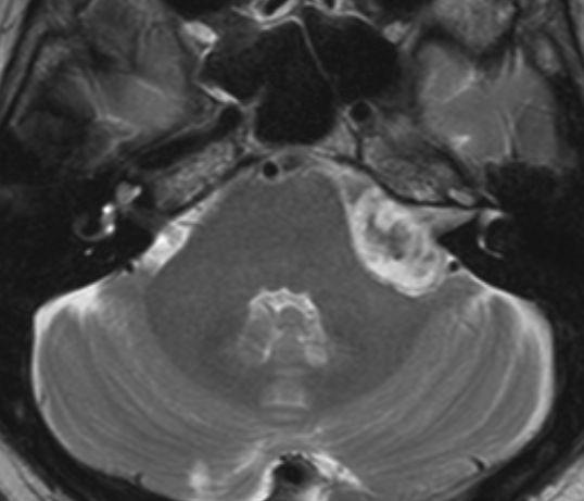
15 Keskkõrv: patoloogia Keskkõrv: patoloogia Kolesteatoom ja epidermoid Põletikud: otiit, mastoidiit ja nende komplikatsioonid Neoplaasiad Paraganglioom Meningioom Schwannoom Adenoom Metastaasid, Lümfoom, L Rhabdomyosarkoom
16 Kolesteatoom (Williams Eur Radiol 2003;13: ) MR imaging using delayed CE SE T1 was able to detect residual or recurrent cholesteatoma as small as 3mm with high specificity: except for cholesteatoma, all other tissues that may be present in the tympanic cavity demonstrate a variable enhancement after contrast injection.
17 Middle Ear Cholesteatoma: Non Echo-planar Diffusion-weighted MR Imaging versus Delayed Gadolinium-enhanced T1-weighted MR Imaging Value in Detection De Foer B. Radiology: Volume 255: Number 3 June 2010 Delayed T1Gd Non-EPI DWI T1Gd+DWI PPV 88.0% 96.0% 96.3% NPV 27% 56.5% 59.6%
18 Retsidiivkolesteatoom
19 Retsidiivkolesteatoom
20 põletikud Otiit? Mastoidiit? Retentsioon?
21 Incidental diagnosis of mastoiditis on MRI. Polat S et al. Eur Arch Otorhinolaryngol Aug;268(8):
22 Mastoidiit, abstsess Mastoidiit, abstsess
23 Mastoidiit, abstsess Mastoidiit, abstsess
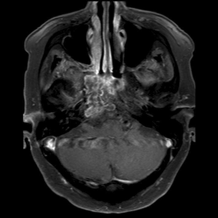
24 Tuumorid: paraganglioom

25 paraganglioom TOF-MRA TOF-MRA: MIP
26 Sisekõrv, MAI, PCN: probleemid Sisekõrv, MAI, PCN: probleemid Kaasasündinud või omandatud kuulmislangus Tinnitus Implantatsioonieelne kaardistamine Vestibulaarsed häired h Varasema juhuleiu täpsustamine t Perifeerne näon n onärvi parees


27 Sisekõrv: anatoomia

28 Harnsberger. Imaging Anatomy: H&N biology.clc.uc.edu
29 DRIVE

30 scala vestibuli scala tympani d. endolymphaticus cochlea vestibulum lat. SCC post. SCC
31 Sisekõrv: patoloogia Sisekõrv: patoloogia Kaasasündinud anomaaliad Labürintiit (Osteodüstroofiad) stroofiad) (Trauma) Neoplaasiad Schwannoom Sacculus endolymphaticuse tuumor

32 DRIVE Sisekõrva patoloogia: kongenitaalsed anomaaliad - LVA

33 Sisekõrva patoloogia: kaasasündinud anomaaliad - Mondini düsplaasia d DRIVE


34 CISS T1Gd 3d-MPR Sisekõrva patoloogia: äge labürintiit


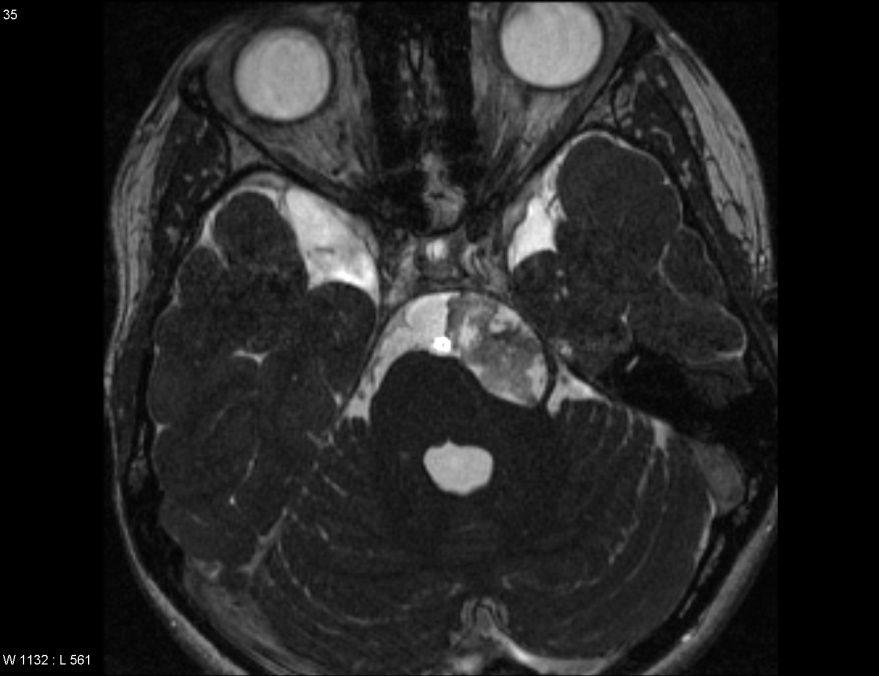
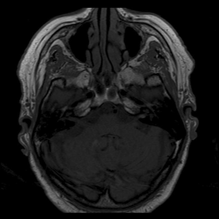
35 Sisekõrva patoloogia: subakuutne fibroseeriv labürintiit CISS


36 CISS Sisekõrva patoloogia: osteodüstroofiad stroofiad - äge otoskleroos T1Gd 3d-MPR


37 Sisekõrva patoloogia: tuumorid - intrakohleaarne schwannoom T1Gd 3d-MPR CISS
38 Sisekõrva patoloogiad: neoplaasiad - sacculus endolymphaticuse tuumor PILDID:
39 Pseudolesioon: kollane luuüdi

40 n. vest. inf. Harnsberger. Imaging Anatomy: H&N n. cochl. n. fac.. n. vest. sup. n. cochlearis n. facialis n. vestibularis inf. n. vestibularis sup. Sisemine kuulmekäik ja PCN


41 VII VIII PCN porus, ülaosa VII n. v. sup. närvid porus, alaosa VIII n. cochl. n. v. inf.
42 MAI ja PCN: patoloogia MAI ja PCN: patoloogia Kuulmisnärvi kaasasündinud düsplaasiad d Aplaasia Hüpoplaasia Põletikulised haigused, neuriidid Neuriit Granulomatoossed põletikud Ekspansioonid Epidermoid Arahnoidaaltsüst st Neoplaasiad Schwannoom Meningioom Lipoom Hemangioom Kartsinomatoos, lümfoom l

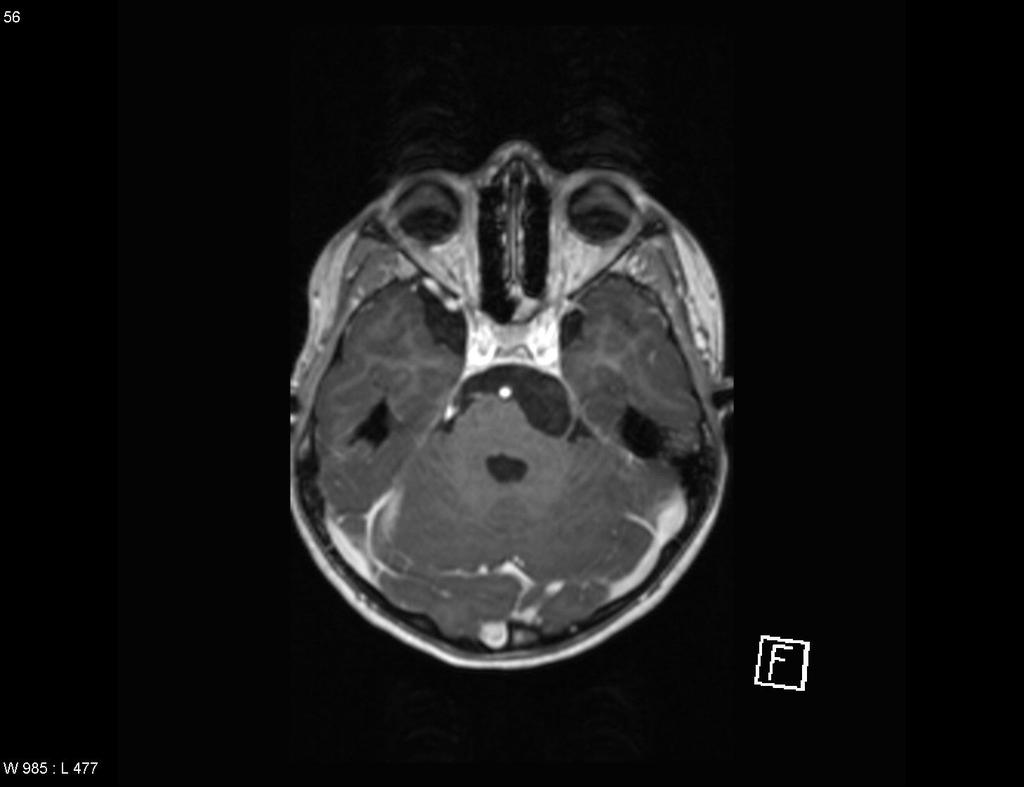
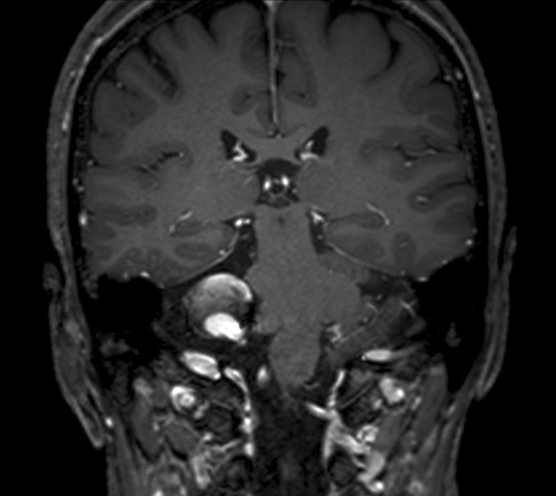
43 T1Gd 3d-MPR schwannoom
44 schwannoom T2 T1Gd DRIVE T1


45 NF II NF II DRIVE T1Gd 3d-MPR bilateraalne!
46 PCA meningioom T1 T2 T1Gd
47 PCA epidermoid T2 FSE CISS T1Gd 3d-MPR

48 PCA epidermoid EPI DWI b=1000 ADC


49 AICA aneurüsm T2 T1 T1Gd

50 lipoom DRIVE T1
51 Bonneville 2007
52 DRIVE hypoplasia n. cochlearis dex *

53 n. facialise neuriit T1Gd 3d-MPR
 T1Gd 3d-MPR")
54 Bilat. n. facialise neuriit (neuroborrelioos) Bilat. n. facialise neuriit (neuroborrelioos) T1Gd 3d-MPR
55 DRIVE neurosarkoidoos T1Gd 3d-MPR
56 Temporaalapex: patoloogia Temporaalapex: patoloogia Pseudoleesioonid Asümmeetriline luuüdi / pneumatisatsioon Vedelikuretentsioon (trapped fluid) Põletikulised haigused temporaalapitsiit Ekspansioonid Kolesteatoom ja epidermoid Kolesteroolgranuloom Neoplaasiad Kondrosarkoom Meningioom Metastaasid, müeloom m L-rakuline histiotsütoos, toos, hiidrakuline tuumor sarkoom
57 Pseudolesioon: trapped fluid Pseudolesioon: trapped fluid T2 flair T1 T1Gd DWI ADC
58 Kolesteroolgranuloom T2fs T1 T1fs nat

59 Temporaalapitsiit T1Gd
60 kondrosarkoom T2 T1fsGd
61 Summa HR 3D sekventsid (T2 ja T1Gd) Keskkõrv: : kõik, mis pole MRT-s s must, on patoloogiline. Tõlgendus! Keskkõrv Sisekõrv ja sisemine kuulmekäik ik: : kas normaalne anatoomia on tuvastatav? Signaal ja kontrasteerumine? DWI (non-epi): retsidiivkolesteatoom ja temporaalluu lesioonide dif.dgn.
62 Kirjandus: Harnsberger et al. Diagnostic Imaging: Head and Neck Som, Curtin. Head and Neck Imaging Swartz. Sensorineural hearing deficit: a systematic approach based on imaging findings. Radiographics 1996 Bonneville et al. Imaging of cerebellopontine angle lesions: an update Part 1: Eur Radiol Oct;17(10): Epub 2007 Jun 12 Part 2: Eur Radiol Nov;17(11): Casselman, Mukherji. Cranial Nerves. Neuroim Clin of NA Anatomy/Temporal-Bone Bone-CT
63 Tänan!
Case Studies in CPA/IAC
 Outline Case Studies in CPA/IAC Atul K Mallik MD PhD Department of Radiology and Imaging Sciences University of Utah Health Sciences Center Salt Lake City, Utah, USA Case based review of cerebellopontine
Dr. T. Venkat Kishan Asst. Prof Department of Radiodiagnosis
 Dr. T. Venkat Kishan Asst. Prof Department of Radiodiagnosis Schwannomas (also called neurinomas or neurilemmomas) constitute the most common primary cranial nerve tumors. They are benign slow-growing
MRI ANATOMY OF THE CRANIAL NERVES. Alexandra Borges Radiology Dpt. Instituto Português de Oncologia de Lisboa
 MRI ANATOMY OF THE CRANIAL NERVES Alexandra Borges Radiology Dpt. Instituto Português de Oncologia de Lisboa SENR 2014 CRANIAL NERVES Olfactory: I Optic: II Oculomotor nerves: III, IV, VI Trigeminal nerve:
Real role of MRI in ORL diagnosis protocol in hearing loss and vertiginous syndrome: A daily challenge for the clinician and the radiologist
 Real role of MRI in ORL diagnosis protocol in hearing loss and vertiginous syndrome: A daily challenge for the clinician and the radiologist Poster No.: C-0848 Congress: ECR 2012 Type: Scientific Exhibit
20 The ear (Vestibulo-acoustic Organs)
") 20 The ear (Vestibulo-acoustic Organs) Median line Sella turcica Tuba auditiva Cavum tympani A. carotis int. Superior border of petrous part Membrana tympani Cochlea N. facialis Meatus acusticus internus
20 The ear (Vestibulo-acoustic Organs) Median line Sella turcica Tuba auditiva Cavum tympani A. carotis int. Superior border of petrous part Membrana tympani Cochlea N. facialis Meatus acusticus internus
NEURO PROTOCOLS MRI NEURO PROTOCOLS (SIEMENS SCANNERS)
") Page 1 NEURO PROTOCOLS Brain Stroke Brain Brain with contrast Brain for seizures Brain for MS Brain for Pineal gland Sella FAST Scan for hydrocephalus MRA/MRV Brain MRA carotids 8 th nerve Cranial nerves
Page 1 NEURO PROTOCOLS Brain Stroke Brain Brain with contrast Brain for seizures Brain for MS Brain for Pineal gland Sella FAST Scan for hydrocephalus MRA/MRV Brain MRA carotids 8 th nerve Cranial nerves
Neuroradiology MR Protocols
 Neuroradiology MR Protocols Brain protocols N 1: Brain MRI without contrast N 2: Pre- and post-contrast brain MRI N 3 is deleted N 4: Brain MRI without or pre-/post-contrast (seizure protocol) N 5: Pre-
Clinical Utility of MRI for Cholesteatoma Recurrence
 Curr Surg Rep (2014) 2:63 DOI 10.1007/s40137-014-0063-0 EAR SURGERY (CJ LIMB, SECTION EDITOR) Clinical Utility of MRI for Cholesteatoma Recurrence Jonathan McJunkin Richard Chole Published online: 24 June
Imaging of Hearing Loss
 Contemporary Imaging of Sensorineural Hearing Loss Imaging of Hearing Loss Discussion Outline (SNHL) Imaging Approaches Anatomic Relationships Lesions: SNHL KL Salzman, MD University of Utah School of
Received 20 June 2006
 American Journal of Otolaryngology Head and Neck Medicine and Surgery 28 (2007) 230 234 www.elsevier.com/locate/amjoto Value of high-resolution computed tomography and magnetic resonance imaging in the
Magnetic Resonance Imaging. Basics of MRI in practice. Generation of MR signal. Generation of MR signal. Spin echo imaging. Generation of MR signal
 Magnetic Resonance Imaging Protons aligned with B0 magnetic filed Longitudinal magnetization - T1 relaxation Transverse magnetization - T2 relaxation Signal measured in the transverse plane Basics of MRI
Pediatric Temporal Bone
 Pediatric Temporal Bone Suresh K. Mukherji, MD, FACR Professor and Chief of Neuroradiology Professor of Radiology, Otolaryngology Head Neck Surgery, Radiation Oncology and Periodontics & Oral Medicine
Major Anatomic Components of the Orbit
 Major Anatomic Components of the Orbit 1. Osseous Framework 2. Globe 3. Optic nerve and sheath 4. Extraocular muscles Bony Orbit Seven Bones Frontal bone Zygomatic bone Maxillary bone Ethmoid bone Sphenoid
New EAONO Cholesteatoma Classification with imaging illustration. Milan Profant, Katarina Sláviková
 New EAONO Cholesteatoma Classification with imaging illustration Milan Profant, Katarina Sláviková EAONO/JOS Joint Consensus Statements on the Definitions, Classification and Staging of Middle Ear Cholesteatoma
UC SF. Safe Surgery Rule #1. Cholesteatoma. It s hard to have a surgical complication when you are not operating
 UC SF Cholesteatoma Chronic Ear Surgery: Staying Out of Trouble! Lawrence R. Lustig, MD Department of Oto-HNS University of California San Francisco Ligaments and folds Spaces NU Epitympanic Cholesteatoma
1. Axial view, left temporal bone. Plane through the upper antrum (A), superior semicircular canal (SSC) and IAC.
, superior semicircular canal (SSC) and IAC.") PA IAC SSC A 1. Axial view, left temporal bone. Plane through the upper antrum (A), superior semicircular canal (SSC) and IAC. IAC VII M I LSC Plane through the IAC, malleus head and incus and the lateral
PA IAC SSC A 1. Axial view, left temporal bone. Plane through the upper antrum (A), superior semicircular canal (SSC) and IAC. IAC VII M I LSC Plane through the IAC, malleus head and incus and the lateral
RADIOLOGY TEACHING CONFERENCE
 RADIOLOGY TEACHING CONFERENCE John Athas, MD Monica Tadros, MD Columbia University, College of Physicians & Surgeons Department of Otolaryngology- Head & Neck Surgery September 27, 2007 CT SCAN IMAGING
HASTE diffusion-weighted MRI for the reliable detection of cholesteatoma
 Diagn Interv Radiol 2012; 18:153 158 Turkish Society of Radiology 2012 HEAD AND NECK IMAGING ORIGINAL ARTICLE HASTE diffusion-weighted MRI for the reliable detection of A. Turan Ilıca, Yusuf Hıdır, Nail
The Temporal Bone Anatomy & Pathology
 Department of Radiology University of California San Diego The Temporal Bone Anatomy & Pathology John R. Hesselink, M.D. Temporal Bone Axial View Temporal Bone Coronal View Longitudinal Fracture The Temporal
Correlation of HRCT mastoid with clinical presentation and operative findings in ear diseases
 International Journal of Otorhinolaryngology and Head and Neck Surgery Chintale SG et al. Int J Otorhinolaryngol Head Neck Surg. 2017 Jul;3(3):656-660 http://www.ijorl.com pissn 2454-5929 eissn 2454-5937
The cochlea: auditory sense. The cochlea: auditory sense
 Inner ear apparatus 1- Vestibule macula and sacculus sensing acceleration of the head and direction of gravity 2- Semicircular canals mainly for sensing direction of rotation of the head 1 3- cochlea in
Detection of Postoperative Residual Cholesteatoma With Non/EchoYPlanar Diffusion-Weighted Magnetic Resonance Imaging
 Otology & Neurotology 29:513Y517 Ó 2008, Otology & Neurotology, Inc. Detection of Postoperative Residual Cholesteatoma With Non/EchoYPlanar Diffusion-Weighted Magnetic Resonance Imaging *Bert De Foer,
Radiologic Evaluation of Petrous Apex Masses. Pavan Kavali, MS-IV Morehouse School of Medicine November 16, 2009
 Radiologic Evaluation of Petrous Apex Masses Pavan Kavali, MS-IV Morehouse School of Medicine November 16, 2009 Roadmap Petrous Apex Anatomy Patient D.S.: Clinical Presentation Differential diagnosis of
Refresher Course EAR TUMOR. Sasikarn Chamchod, MD Chulabhorn Hospital
 Refresher Course EAR TUMOR Sasikarn Chamchod, MD Chulabhorn Hospital Reference: Perez and Brady s Principles and Practice of radiation oncology sixth edition Outlines Anatomy Epidemiology Clinical presentations
SMRT Student Scope Submission
 SMRT Student Scope Submission Title & Author Title: MRI in the Detection and Diagnosis of Acoustic Neuroma Author: Manya Lovesky Andersen, student Massachusetts College of Pharmacy and Health Sciences,
Abnormal direction of internal auditory canal and vestibulocochlear nerve
 Medicine Otorhinolaryngology fields Okayama University Year 2004 Abnormal direction of internal auditory canal and vestibulocochlear nerve Shin Kariya kazunori Nishizaki Hirofumi Akagi Michael M. Paparella
Anatomy of the ear: Lymphatics
 Anatomy of the ear: 1. External ear which consist of auricle and external auditory canal. The auricle has a framework of cartilage except the lobule, the skin is closely adherent to perichonderium at the
Title. Author(s) Takahashi, Haruo. Issue Date Right.
 Takahashi, Haruo. Issue Date Right.") NAOSITE: Nagasaki University's Ac Title Author(s) Citation A case with posterior fossa epiderm symptoms caused by insufficiency of usefulness of free DICOM image view Takasaki, Kenji; Kumagami, Hidetaka
NAOSITE: Nagasaki University's Ac Title Author(s) Citation A case with posterior fossa epiderm symptoms caused by insufficiency of usefulness of free DICOM image view Takasaki, Kenji; Kumagami, Hidetaka
MR Evaluation of Vestibulocochlear Anomalies Associated with Large Endolymphatic Duct and Sac
 AJNR Am J Neuroradiol 20:1435 1441, September 1999 MR Evaluation of Vestibulocochlear Anomalies Associated with Large Endolymphatic Duct and Sac H. Christian Davidson, H. Ric Harnsberger, Marc M. Lemmerling,
Cholesteatoma is a collection of keratinous debris lined by
 ORIGINAL RESEARCH M.H.G. Dremmen P.A.M. Hofman J.R. Hof R.J. Stokroos A.A. Postma The Diagnostic Accuracy of Non-Echo-Planar Diffusion-Weighted Imaging in the Detection of Residual and/or Recurrent Cholesteatoma
15 Marzo 2014 Aspetti radiologici dei disordini vestibolari: approccio multidisciplinare
 15 Marzo 2014 Aspetti radiologici dei disordini vestibolari: approccio multidisciplinare MR Imaging of inner ear endo-perilymphatic spaces at 3T after intratympanic contrast agent administration in Definite
Outline. Neuroradiology. Diffusion Imaging in. Clinical Applications of. Basics of Diffusion Imaging. Basics of Diffusion Imaging
 Clinical Applications of Diffusion Imaging in Neuroradiology No disclosures Stephen F. Kralik Assistant Professor of Radiology Indiana University School of Medicine Department of Radiology and Imaging
1Pulse sequences for non CE MRA
 MRI: Principles and Applications, Friday, 8.30 9.20 am Pulse sequences for non CE MRA S. I. Gonçalves, PhD Radiology Department University Hospital Coimbra Autumn Semester, 2011 1 Magnetic resonance angiography
Carlos Torres MD, FRCPC, Associate Professor of Radiology Department of Radiology, University of Ottawa
 Carlos Torres MD, FRCPC, Associate Professor of Radiology Department of Radiology, University of Ottawa catorres@toh.on.ca None 1. Simplify the complex imaging anatomy of the BP using clear anatomical
Imaging Profile of the Ear in Hearing Loss Patients in Hospital Universiti Sains Malaysia: 5 year Cross Sectional Analysis at a Tertiary
 Imaging Profile of the Ear in Hearing Loss Patients in Hospital Universiti Sains Malaysia: 5 year Cross Sectional Analysis at a Tertiary Otologic Centre Rohaizam bin Japar 1, Dinsuhaimi bin Sidek 1, Suzina
Cranial Nerve VII & VIII
 Cranial Nerve VII & VIII Lecture Objectives Follow up the course of facial nerve from its point of central connections, exit and down to its target areas. Follow up the central connections of the facial
Anatomy of the Ear Region. External ear Middle ear Internal ear
 Ear Lecture Objectives Make a list of structures making the external, middle, and internal ear. Discuss the features of the external auditory meatus and tympanic membrane. Describe the shape, position,
Magnetic Resonance Imaging (MRI) and High Resolution Computed Tomography (HRCT): Can they improve the evaluation of Middle ear cholesteatoma?
 and High Resolution Computed Tomography (HRCT): Can they improve the evaluation of Middle ear cholesteatoma?") Magnetic Resonance Imaging (MRI) and High Resolution Computed Tomography (HRCT): Can they improve the evaluation of Middle ear cholesteatoma? Poster No.: C-1249 Congress: ECR 2013 Type: Educational Exhibit
Magnetic Resonance Imaging (MRI) and High Resolution Computed Tomography (HRCT): Can they improve the evaluation of Middle ear cholesteatoma? Poster No.: C-1249 Congress: ECR 2013 Type: Educational Exhibit
Acute mastoiditis (AM) is a complication of otitis media in. MR Imaging Features of Acute Mastoiditis and Their Clinical Relevance
 is a complication of otitis media in. MR Imaging Features of Acute Mastoiditis and Their Clinical Relevance") ORIGINAL RESEARCH HEAD & NECK MR Imaging Features of Acute Mastoiditis and Their Clinical Relevance R. Saat, A.H. Laulajainen-Hongisto, G. Mahmood, L.J. Lempinen, A.A. Aarnisalo, A.T. Markkola, and J.P.
ORIGINAL RESEARCH HEAD & NECK MR Imaging Features of Acute Mastoiditis and Their Clinical Relevance R. Saat, A.H. Laulajainen-Hongisto, G. Mahmood, L.J. Lempinen, A.A. Aarnisalo, A.T. Markkola, and J.P.
THE MANAGEMENT of COMPLICATED OTITIS MEDIA. IFOS, Lima, 2018
 THE MANAGEMENT of COMPLICATED OTITIS MEDIA IFOS, Lima, 2018 VINCENT C COUSINS ENT-Otoneurology Unit, The Alfred Hospital & Department of Surgery. Monash University MELBOURNE, AUSTRALIA Otologic Complications
AUDITORY APPARATUS. Mr. P Mazengenya. Tel 72204
 AUDITORY APPARATUS Mr. P Mazengenya Tel 72204 Describe the anatomical features of the external ear Describe the tympanic membrane (ear drum) Describe the walls of the middle ear Outline the structures
High-resolution Computed Tomography Study of Temporal Bone Pathologies
 Original Article Print ISSN: 2321-6379 Online ISSN: 2321-595X DOI: 10.17354/ijss/2016/375 High-resolution Computed Tomography Study of Temporal Bone Pathologies Manjit Bagul Senior Resident, Department
Evaluation of acquired cholesteatoma with PROPELLER diffusion imaging
 The Egyptian Journal of Radiology and Nuclear Medicine (2011) 42, 9 17 Egyptian Society of Radiology and Nuclear Medicine The Egyptian Journal of Radiology and Nuclear Medicine www.elsevier.com/locate/ejrnm
Ear. Utricle & saccule in the vestibule Connected to each other and to the endolymphatic sac by a utriculosaccular duct
 Rahaf Jreisat *You don t have to go back to the slides. Ear Inner Ear Membranous Labyrinth It is a reflection of bony labyrinth but inside. Membranous labyrinth = set of membranous tubes containing sensory
DOI: / ORIGINAL ARTICLE. Tomographic evaluation of the contralateral ear in patients with severe chronic otitis media
 Braz J Otorhinolaryngol. 2013;79(4):475-9. DOI: 10.5935/1808-8694.20130085 ORIGINAL ARTICLE.org BJORL Tomographic evaluation of the contralateral ear in patients with severe chronic otitis media Maurício
THE EAR AND HEARING Be sure you have read and understand Chapter 16 before beginning this lab. INTRODUCTION: hair cells outer ear tympanic membrane
 BIOLOGY 211: HUMAN ANATOMY & PHYSIOLOGY ****************************************************************************************************** THE EAR AND HEARING ******************************************************************************************************
Chronic Ear Disease. Daekeun Joo Resident Lecture Series 11/18/09
 Chronic Ear Disease Daekeun Joo Resident Lecture Series 11/18/09 ETD URIs Viral-induced damage to ET lining resulting in decreased mucociliary clearance Viral invasion of ME mucosa results in inflamm Reflux
Imaging: When to get MRI, CT or PET-CT?
 Imaging: When to get MRI, CT or PET-CT? Alina Uzelac, D.O. Assistant Clinical Professor Neuroradiology UCSF Department of Radiology and Biomedical Imaging San Francisco General Hospital Overview CT MRI
Petrous Apex Cephalocele: Report of Two Cases and
 Petrous Apex Cephalocele: Report of Two Cases and Review of the Literature 1 Bo Seong Jeong, M.D., Ghi Jai Lee, M.D., Jae Chan Shim, M.D., Jae Myeong Lee, M.D., Mee Young Nam, M.D., Ho Kyun Kim, M.D. A
Endolymphatic hydrops in patients with vestibular migraine and auditory symptoms
 DOI 10.1007/s00405-013-2751-2 OTOLOGY Endolymphatic hydrops in patients with vestibular migraine and auditory symptoms Robert Gürkov Claudia Kantner Michael Strupp W. Flatz Eike Krause Birgit Ertl-Wagner
Imaging in patients undergoing cochlear implant: CT and MR technique
 Imaging in patients undergoing cochlear implant: CT and MR technique Poster No.: C-0102 Congress: ECR 2012 Type: Educational Exhibit Authors: G. Posillico Keywords: Ear / Nose / Throat, CT, MR, Comparative
ORIGINAL ARTICLE. A New Staging System for Tympano-mastoid Cholesteatoma. Aziz Belal, Mahmoud Reda, Ahmed Mehana, Yousef Belal
 Int. Adv. Otol. 2012; 8:(1) 63-68 ORIGINAL ARTICLE A New Staging System for Tympano-mastoid Cholesteatoma Aziz Belal, Mahmoud Reda, Ahmed Mehana, Yousef Belal Alexandria Ear Hospital Alexandria Egypt (AB,
Chapter 3: Anatomy and physiology of the sensory auditory mechanism
 Chapter 3: Anatomy and physiology of the sensory auditory mechanism Objectives (1) Anatomy of the inner ear Functions of the cochlear and vestibular systems Three compartments within the cochlea and membranes
Cerebellopontine Angle Masses June 2004
 TITLE: Cerebellopontine Angle Masses SOURCE: Grand Rounds Presentation, UTMB, Dept. of Otolaryngology DATE: June 2, 2004 RESIDENT PHYSICIAN: Alan L. Cowan, MD FACULTY ADVISOR: Arun Gadre, MD SERIES EDITORS:
For this lab you will use parts of Exercise #18 in your Wise lab manual. Please be sure to read those sections before coming to lab
 Bio 322 Human Anatomy Objectives for the laboratory exercise The Eye and Ear Required reading before beginning this lab: Saladin, KS: Human Anatomy 5 th ed (2017) Chapter 17 For this lab you will use parts
Parotiidnäärme tuumorid. Liisi Talv
 Parotiidnäärme tuumorid Liisi Talv I patsient Aasta tagasi tekkis väike tuumor vasakul põsel, mis asümptomaatiliselt pidevalt suuremaks kasvas KT alusel sai diagnoosihüpoteesiks pandud pleomorfne adenoom
Modern Imaging & Current Controversies
 Temporal Bone: Modern Imaging & Current Controversies Suresh K. Mukherji, MD, FACR Professor and Chief of Neuroradiology Professor of Radiology, Otolaryngology Head Neck Surgery, Radiation i Oncology,
Dr Melanie Souter. Consultant Otolaryngologist/Otologist Christchurch Public Hospital Christchurch. 12:00-12:15 Ears Made Easy
 Dr Melanie Souter Consultant Otolaryngologist/Otologist Christchurch Public Hospital Specialists @nine Christchurch 12:00-12:15 Ears Made Easy Ears made Easy Dr Melanie Souter Otology / Otolaryngology
Neurovascular elements of the PCF
 Level I: Neurovascular elements of the PCF Trigeminal and Abducent Superior cerebellar artery and vein Dandy s vein Level II: Facial and Cochleovestibular AICA and internal auditory artery, veins Level
Clinician s Guide To Ordering NeuroImaging Studies
 Clinician s Guide To Ordering NeuroImaging Studies MRI CT South Jersey Radiology Associates The purpose of this general guide is to assist you in choosing the appropriate imaging test to best help your
ORIGINAL ARTICLE. Temporal Lobe Injury in Temporal Bone Fractures. imaging (MRI) to evaluate lesions of the temporal
 to evaluate lesions of the temporal") ORIGINAL ARTICLE Temporal Lobe Injury in Temporal Bone Fractures Richard M. Jones, MD; Michael I. Rothman, MD; William C. Gray, MD; Gregg H. Zoarski, MD; Douglas E. Mattox, MD Objective: To determine the
ORIGINAL ARTICLE Temporal Lobe Injury in Temporal Bone Fractures Richard M. Jones, MD; Michael I. Rothman, MD; William C. Gray, MD; Gregg H. Zoarski, MD; Douglas E. Mattox, MD Objective: To determine the
Narrowest segment of the ear canal. Limited microscopic. Wide endoscopic. field of view. field of view
 Endoscopic Transcanal Management of Cholesteatoma M. Tarabichi American Hospital-Dubai The Endoscope in Otology Mostly for documentation. Mostly diagnostic. Exploration of old mastoid cavities Endoscopic
Arastoo Vossough, M.D., Ph.D. Associate Professor of Radiology
 Disorders of the Temporal Bone Arastoo Vossough, M.D., Ph.D. Associate Professor of Radiology 1st Branchial Cleft Cyst Remnant of 1 st branchial apparatus Rare Cystic lesions: I- around pinna II- connecting
Combined TBH / GSH meeting. 11 September 2007 Eric F Post
 Combined TBH / GSH meeting 11 September 2007 Eric F Post Case 32 yo male Presenting: Vertigo x since a.m. Tinnitus 5/7 Hearing loss worsened 4/7 Otorhea (L) x 2/ 52 Case History Ear surgery 1990 @ Umtata
Disclosures. Diffusion and Perfusion Imaging in the Head and Neck. Learning objectives ???
 Disclosures No relevant financial disclosures Diffusion and Perfusion Imaging in the Head and Neck Ashok Srinivasan, MD Associate Professor Director of Neuroradiology University of Michigan Health System
Educational Exhibit Authors: P. Digge, R. K. N. Solanki, S. M. Paruthikunnan, D. S. Shah ; 1
 High-field MRI versus high-resolution CT of temporal bone in inner ear pathologies of children with bilateral profound sensorineural hearing loss: A pictorial essay. Poster No.: C-0867 Congress: ECR 2015
INTRODUCTION: ANATOMY UNDERLYING CLINICAL TESTS OF CRANIAL NERVES
 INTRODUCTION: ANATOMY UNDERLYING CLINICAL TESTS OF CRANIAL NERVES CRANIAL NERVE I - OLFACTORY I - OLFACTORY NERVE - SMELL TEST: SMELL ODORS (note: not ammonia; pain in nasal cavity CN5 DAMAGE: LOSS OF
A&P 1. Ear, Hearing & Equilibrium Lab. Basic Concepts. These notes follow Carl s Talk at the beginning of lab
 A&P 1 Ear, Hearing & Equilibrium Lab Basic Concepts These notes follow Carl s Talk at the beginning of lab In this "Lab Exercise Guide", we will be looking at the basics of hearing and equilibrium. NOTE:
Complications of At t i c o a n t ral Cholesteato ma:
 Complications of t t i c o a n t ral Cholesteato ma: MR Manifestations 1 Jeong Hyun Lee, M.D., Ho Kyu Lee, M.D., Soo Mi Lim, M.D., Ji Hoon Shin, M.D., Choong Gon Choi, M.D., Dae Chul Suh, M.D., Kwang Sun
The Child s Ear. Normal? Abnormal? And what do we do next?
 The Child s Ear Normal? Abnormal? And what do we do next? Anatomy of the Ear: Outer (External) Ear External Ear: Middle Ear: Inner Ear: Inner Ear: Cochlea Inner Ear: Semicircular Canals Why do we care?
Understanding the relevance of neutropenic sepsis as a radiologist and who is at risk.
 What to do next above the diaphragm 08:30 09:00 Investigating suspected chest infection in febrile neutropaenia Dr Anna Sharman, Manchester University NHS Foundation Trust Understanding the relevance of
Intralabyrinthine schwannomas (ILSs) are benign tumors
 are benign tumors") ORIGINAL RESEARCH A. Tieleman J.W. Casselman T. Somers J. Delanote R. Kuhweide J. Ghekiere B. De Foer E.F. Offeciers Imaging of Intralabyrinthine Schwannomas: A Retrospective Study of 52 Cases with Emphasis
ORIGINAL RESEARCH A. Tieleman J.W. Casselman T. Somers J. Delanote R. Kuhweide J. Ghekiere B. De Foer E.F. Offeciers Imaging of Intralabyrinthine Schwannomas: A Retrospective Study of 52 Cases with Emphasis
Imaging of sensorineural hearing loss: a pattern-based approach to diseases of the inner ear and cerebellopontine angle
 Insights Imaging (2012) 3:139 153 DOI 10.1007/s13244-011-0134-z PICTORIAL REVIEW Imaging of sensorineural hearing loss: a pattern-based approach to diseases of the inner ear and cerebellopontine angle
Congenital Middle Ear Cholesteatoma in Children; Retrospective Review of 35 Cases
 J Korean Med Sci 2009; 24: 126-31 ISSN 1011-8934 DOI: 10.3346/jkms.2009.24.1.126 Copyright The Korean Academy of Medical Sciences Congenital Middle Ear Cholesteatoma in Children; Retrospective Review of
Kingdom of Bahrain Arabian Gulf University College of Medicine and Medical Sciences Year 6 ENT SMC Otitis Media (Dr.
 Kingdom of Bahrain Arabian Gulf University College of Medicine and Medical Sciences Year 6 ENT SMC Otitis Media (Dr. Jalal Almarzooq) - Anatomy of the ear: The ear is divided into 3 parts: External ear.
6/23/2009. Inversion Recovery (IR) Techniques and Applications. Variations of IR Technique. STIR, FLAIR, TI and TI Null. Applications of IR
 Techniques and Applications. Variations of IR Technique. STIR, FLAIR, TI and TI Null. Applications of IR") The Anatomy of Basic R Pulse Sequences Inversion Recovery () Techniques and Applications Chen Lin, PhD Indiana University School of edicine & Clarian Health Partners agnetization Preparation Section Chemical
The Anatomy of Basic R Pulse Sequences Inversion Recovery () Techniques and Applications Chen Lin, PhD Indiana University School of edicine & Clarian Health Partners agnetization Preparation Section Chemical
The Usefulness of MR Imaging of the Temporal Bone in the Evaluation of Patients with Facial and Audiovestibular Dysfunction
 The Usefulness of MR Imaging of the Temporal Bone in the Evaluation of Patients with Facial and Audiovestibular Dysfunction Sang Uk Park, MD 1 Hyung-Jin Kim, MD 1 Young Kuk Cho, MD 1 Myung Kwan Lim, MD
Cochlear Implant Failure: Imaging Evaluation of the Electrode Course
 Clinical Radiology (2003) 58: 288 293 doi:10.1016/s0009-9260(02)00523-8, available online at www.sciencedirect.com Pictorial Review Cochlear Implant Failure: Imaging Evaluation of the Electrode Course
The ear: some applied basic science
 Chapter 1 The ear: some applied basic science The pinna The external ear or pinna is composed of cartilage with closely adherent perichondrium and skin. It is developed from six tubercles of the first
Structure, Energy Transmission and Function. Gross Anatomy. Structure, Function & Process. External Auditory Meatus or Canal (EAM, EAC) Outer Ear
 Outer Ear") Gross Anatomy Structure, Energy Transmission and Function IE N O ME 1 Structure, Function & Process 4 External Auditory Meatus or Canal (EAM, EAC) Outer third is cartilaginous Inner 2/3 is osseous Junction
Gross Anatomy Structure, Energy Transmission and Function IE N O ME 1 Structure, Function & Process 4 External Auditory Meatus or Canal (EAM, EAC) Outer third is cartilaginous Inner 2/3 is osseous Junction
High resolution computed tomography of temporal bone in the evaluation of otologic diseases
 International Journal of Otorhinolaryngology and Head and Neck Surgery Handi PS et al. Int J Otorhinolaryngol Head Neck Surg. 2018 Jan;4(1):87-92 http://www.ijorl.com pissn 2454-5929 eissn 2454-5937 Original
High Field MR of the Spine
 Department of Radiology University of California San Diego 3T for MR Applications Advantages High Field MR of the Spine Increased signal-to-noise Better fat suppression Increased enhancement with gadolinium
Expand The Scope of Temporal Bone Reporting: Why, What and Where to Look For.
 Expand The Scope of Temporal Bone Reporting: Why, What and Where to Look For. Award: Certificate of Merit Poster No.: C-1521 Congress: ECR 2014 Type: Educational Exhibit Authors: P. Mundada, B. S. Purohit,
Parotid Gland, Temporomandibular Joint and Infratemporal Fossa
 M1 - Anatomy Parotid Gland, Temporomandibular Joint and Infratemporal Fossa Jeff Dupree Sanger 9-057 jldupree@vcu.edu Parotid gland: wraps around the mandible positioned between the mandible and the sphenoid
THE COCHLEA AND AUDITORY PATHWAY
 Dental Neuroanatomy Suzanne S. Stensaas, PhD April 14, 2010 Reading: Waxman, Chapter 16, Review pictures in a Histology book Computer Resources: http://www.cochlea.org/ - Promenade around the Cochlea HyperBrain
Laith Sorour. Facial nerve (vii):
:") Laith Sorour Cranial nerves 7 & 8 Hello, there are edited slides please go back to them to see pictures, they are not that much important in this lecture but still, and yes slides are included :p Let s
Laith Sorour Cranial nerves 7 & 8 Hello, there are edited slides please go back to them to see pictures, they are not that much important in this lecture but still, and yes slides are included :p Let s
Cerebellopontine angle masses: MRI technique, positive and differential diagnosis
 Cerebellopontine angle masses: MRI technique, positive and differential diagnosis Poster No.: C-1517 Congress: ECR 2012 Type: Educational Exhibit Authors: C. Scheau, I. G. Lupescu, G. Popa, E. M. Preda
MR Imaging of the Internal Auditory Canal and Inner Ear at 3T: Comparison between 3D Driven Equilibrium and 3D Balanced Fast Field Echo Sequences
 MR Imaging of the Internal Auditory Canal and Inner Ear at 3T: Comparison between 3D Driven Equilibrium and 3D Balanced Fast Field Echo Sequences Jun Soo Byun, MD 1,2 Hyung-Jin Kim, MD 1 Yoo Jeong Yim,
Petrous Bone Normal anatomy
 Petrous Bone Normal anatomy By Mamdouh Mahfouz MD Prof. of Radiology Cairo University ssregypt.com Axial Coronal Petrous bone External ear Middle ear Inner ear External ear Cartilaginous part Bony part
The Ear. Dr. Heba Kalbouneh Assistant Professor of Anatomy and Histology
 The Ear Dr. Heba Kalbouneh Assistant Professor of Anatomy and Histology The Ear The ear consists of the external ear; the middle ear (tympanic cavity); and the internal ear (labyrinth), which contains
Imaging of Pediatric MSK Tumors
 Imaging of Pediatric MSK Tumors Kirsten Ecklund, M.D. Boston Children s Hospital Harvard Medical School kirsten.ecklund@childrens.harvard.edu Tumor Imaging Goals Diagnosis Lesion characterization Benign
Intrahepatic cholangiocarcinoma: diffusion-weighted MR imaging findings
 Intrahepatic cholangiocarcinoma: diffusion-weighted MR imaging findings Poster No.: C-1979 Congress: ECR 2013 Type: Authors: Keywords: DOI: Scientific Exhibit S. Schmidt, A. Pomoni, F. Becce, A. Denys,
Acute Ischemic Stroke Imaging. Ronald L. Wolf, MD, PhD Associate Professor of Radiology
 Acute Ischemic Stroke Imaging Ronald L. Wolf, MD, PhD Associate Professor of Radiology Title of First Slide of Substance An Illustrative Case 2 Disclosures No financial disclosures Off-label uses of some
Rebecca J. Clark-Bash, R. EEG\EP T., CNIMeKnowledgePlus.net Page 1
 Navigating the Auditory Pathway: Technical & Physiological Impact on IOM Rebecca Clark-Bash, R. EEG\EP T, CLTM, CNIM, F.ASET, FASNM Faculty Rebecca Clark-Bash R. EEG\EP T., CLTM, CNIM, F.ASNM, F.ASET ASNM
The Ear The ear consists of : 1-THE EXTERNAL EAR 2-THE MIDDLE EAR, OR TYMPANIC CAVITY 3-THE INTERNAL EAR, OR LABYRINTH 1-THE EXTERNAL EAR.
 The Ear The ear consists of : 1-THE EXTERNAL EAR 2-THE MIDDLE EAR, OR TYMPANIC CAVITY 3-THE INTERNAL EAR, OR LABYRINTH 1-THE EXTERNAL EAR Made of A-AURICLE B-EXTERNAL AUDITORY MEATUS A-AURICLE It consists
Case 9 10/29/2018. CJD (Creutzfeldt -Jakob Disease) CJD (Creutzfeldt -Jakob Disease) CJD (Creutzfeldt -Jakob Disease)
 CJD (Creutzfeldt -Jakob Disease) CJD (Creutzfeldt -Jakob Disease)") CJD (Creutzfeldt -Jakob Disease) Rare fatal neurodegen dz caused by infectious protein Prion (lacks nucleic acid)- causes spongiform changes of the brain and neuronal death. 4 types: scjd- 85% of cases
CJD (Creutzfeldt -Jakob Disease) Rare fatal neurodegen dz caused by infectious protein Prion (lacks nucleic acid)- causes spongiform changes of the brain and neuronal death. 4 types: scjd- 85% of cases
A&P 1. Ear, Hearing & Equilibrium Lab. Basic Concepts. Pre-lab Exercises
 A&P 1 Ear, Hearing & Equilibrium Lab Basic Concepts Pre-lab Exercises In this "Lab Exercise Guide", we will be looking at the basics of hearing and equilibrium. NOTE: these notes do not follow the order
Case Report An Unusual Case of Bilateral Agenesis of the Cochlear Nerves
 Case Reports in Neurological Medicine Volume 2012, rticle D 581920, 4 pages doi:10.1155/2012/581920 Case Report n Unusual Case of Bilateral genesis of the Cochlear Nerves Vítor Yamashiro Rocha oares, 1
Gross Anatomy of the. TEMPORAL BONE, EXTERNAL EAR, and MIDDLE EAR. Assignment: Head to Toe Temporomandibular Joint (TMJ)
") Gross Anatomy the TEMPORAL BONE, EXTERNAL EAR, and MIDDLE EAR M1 Gross and Developmental Anatomy 9:00 AM, December 11, 2008 Dr. Milton M. Sholley Pressor Anatomy and Neurobiology Assignment: Head to Toe
Gross Anatomy the TEMPORAL BONE, EXTERNAL EAR, and MIDDLE EAR M1 Gross and Developmental Anatomy 9:00 AM, December 11, 2008 Dr. Milton M. Sholley Pressor Anatomy and Neurobiology Assignment: Head to Toe
CT and MR Imaging In Patients Undergoing Evaluation for Cochlear Implantation
 CT and MR Imaging In Patients Undergoing Evaluation for Cochlear Implantation Poster No.: C-1219 Congress: ECR 2015 Type: Educational Exhibit Authors: S. S. Ghuman, T. Buxi, S. Sud, A. Sud, A. Yadav, K.