Research Article The Reliability of the Reversed Flow Posterior Interosseous Flap for the Coverage of Ulnar Hand Defects: A Series of 25 Cases
|
|
|
- Antonia George
- 5 years ago
- Views:
Transcription
, Article ID 171625, 10 pages DOI: Research Article The Reliability of the Reversed Flow Posterior Interosseous Flap for the Coverage of Ulnar Hand Defects: A Series of 25 Cases Yasser")
1 IBIMA Publishing Plastic Surgery: An International Journal Vol (2014), Article ID , 10 pages DOI: Research Article The Reliability of the Reversed Flow Posterior Interosseous Flap for the Coverage of Ulnar Hand Defects: A Series of 25 Cases Yasser Abdallah Aboelatta, Ibrahim Husseine Kamel, Mohammed Abdelmohsen Ghanem, Khaled Ahmad Reyad and Ayman Abulmakarem Shaker Plastic Surgery Department, Faculty of Medicine, Ain -Shams University, Cairo, Egypt Correspondence should be addressed to: Yasser Abdallah Aboelatta; dr_yaser72@hotmail.com Received Date: 8 December 2013; Accepted Date: 25 February 2014; Published Date: 31 May 2014 Academic Editor: Yakup Çil Copyright 2014 Yasser Abdallah Aboelatta, Ibrahim Husseine Kamel, Mohammed Abdelmohsen Ghanem, Khaled Ahmad Reyad and Ayman Abulmakarem Shaker. Distributed under Creative Commons CC-BY 3.0 Abstract The reversed radial forearm flap is considered the workhorse flap for hand reconstruction, while the posterior interosseous artery (PIA) flap did not take the same interest. This paper presents 25 cases with ulnar side hand defects that was reconstructed with PIA flap with 22 flaps survived (88%). The PIA flap seems a good option and should be considered as another option for hand coverage when the surgeon prefers not to use free tissue transfer or the radial artery flap. The flap should be learned to senior and junior reconstructive surgeons, because familiarity with the flap anatomy lessens the operative time greatly and makes surgical dissection easier. Keywords: Hand defect, reconstruction, posterior interosseous flap. Introduction The reverse posterior interosseous flap was first reported in 1986 by Lu et al [1] and Penteado et al [2]. Although, any upper extremity defect could be solved with either a distant pedicled flap or a free tissue transfer, the regional flaps from the forearm are considered the better options. The merits of a regional flap are single stage elevation, avoidance of hand dependency, early mobilization, and restriction of deformity to ipsilateral extremity [3]. The posterior interosseous artery (PIA) flap provides thin, soft and pliable skin with good colour and texture match. It has traditionally been used to cover defects up to the metacarpophalangeal joint and for reconstruction of the first web space. However, some authors have reported a high incidence of complications, particularly venous congestion with partial or total flap loss [4]. The current study reports our experience with the use of reversed PIA flap for coverage of ulnar aspect defects of the hand in a series of 25 patients. The technical difficulties, outcome, and complications rate are presented. Cite this Article as: Yasser Abdallah Aboelatta, Ibrahim Husseine Kamel, Mohammed Abdelmohsen Ghanem, Khaled Ahmad Reyad and Ayman Abulmakarem Shaker (2014), The Reliability of the Reversed Flow Posterior Interosseous Flap for the Coverage of Ulnar Hand Defects: A Series of 25 Cases, Plastic Surgery: An International Journal Vol (2014), Article ID , DOI:
2 Plastic Surgery: An International Journal 2 Materials and Methods This prospective study was conducted on 25 patients presented to causality department at Ain Shams University Hospitals over a period of 4 years. These patients were complaining of defects over the ulnar aspect of the hand with exposure of bones or tendons necessitating flap coverage. Patients with associated wrist injuries that would preclude the use of this flap were excluded. In addition, patients with systemic diseases as diabetes, ischemic heart disease, atherosclerosis, and vascular diseases were excluded from the study to avoid the possibility of peripheral vascular diseases and their effect on the blood flow to the flap. Complete history and physical examination were taken at the plastic surgery department. Allen s test was done preoperatively to detect the dominant artery in case of failure of harvesting the posterior interosseous artery flap; Radial forearm flap was the next option. Preoperative plain X-ray hands (anteroposterior, oblique lateral views) were obtained. Preoperative colour duplex device was performed for assessment of the blood flow in the posterior interosseous artery was done for all patients. Surgical Anatomy The course of the posterior interosseous artery was marked following a line drawn between the lateral epicondyle of the humerus to the ulnar styloid process. It arises at a point 4-6 cm distal to the lateral epicondyle, usually as a branch of the common interosseous artery but on occasion directly from the ulnar artery 2. At the level of the proximal third of the forearm, the PIA runs with the posterior interosseous nerve (PIN), and gives off several well-defined septocutaneous perforators to the dorsal aspect of the forearm. The first proximal cutaneous branch is a large skin perforator with a variable origin that courses in the intermuscular septum to supply the subcutaneous tissue overlying the proximal third of the dorsal forearm [5]. In the distal third of the forearm, the PIA gives off several perforators (6-8) which supply the skin together. Upon approaching the level of the wrist joint, 2 cm proximal to the distal radio-ulnar joint, the PIA anastomose with the dorsal recurrent branch of the anterior interosseous artery (AIA). Normal physiologic blood flow to the distal third of the forearm comes from this distal anastomosis, and not from the proximal PIA. This vascular arcade allows the PIA flap to be used as a reverse pedicled flap when the PIA is divided proximally. The reversed flow posterior interosseous artery flap is drained by the venae comitantes accompanying the feeding artery to the deep venous system with no superficial system drainage [6]. The PIA has a relatively narrow calibre in the middle third where also the anatomical variances are reported to be the most. Problems are encountered while trying to reach the most distant defects of the first web space, distal amputation defects, the radial, palmar areas and distal to PIP joints. More variations are met during dissection proximal to the distal third of the forearm [7]. Operative Technique All surgical procedures were performed under general anaesthesia and tourniquet and by the same surgical team. Wound debridement including soft tissues and bones was undertaken till a healthy bed was reached for flap inset. The reversed flow PIA flap was raised either from proximal, middle or distal one thirds of the forearm according to the size of the defect and the distal reach of the flap. Flap elevation started with dissection of the pedicle in the distal forearm (area of anastomosis between the posterior interosseous and the anterior interosseous artery arteries) to exclude any possibility of deficient anastomosis. Two skin incisions were made on both sides of the marked course of the artery leaving about one cm skin bridge inbetween. This skin bridge changed the shape of the island flap into a racquet
![3 Plastic Surgery: An International Journal shaped flap as described in a previous work [8]. The plane of dissection is between the skin and the ante-brachial fascia.](/docs-images/83/87842309/images/3-0.jpg "Then, the fascia over the tendons of the tendons of the extensor digitorum communis (EDC) and extensor carpi ulnaris (ECU) was incised.")

3 3 Plastic Surgery: An International Journal shaped flap as described in a previous work [8]. The plane of dissection is between the skin and the ante-brachial fascia. Then, the fascia over the tendons of the tendons of the extensor digitorum communis (EDC) and extensor carpi ulnaris (ECU) was incised. The PIA could be observed coursing along the intermuscular septum between the extensor digiti quinti (EDQ) and the ECU. At the pivot point, the anastomosis arc was observed to ensure its existence. Then, the radial edge of the flap was incised into the EDC. Dissection was continued on ulnar aspect towards the intermuscular septum between the EDQ and ECU. During the dissection, the fascia to the skin paddle is secured with interrupted sutures. Then, the ulnar edge was incised and dissected radially towards the septum, also along the plane between the fascia and the muscle. The intermuscular septum between the EDQ and ECU is the landmark of dissection, along which the PIA and its venae comitantes course distally. After completing the dissection of the PIN, the intermuscular septum was detached from the ulna. The pedicle of the flap consisted of the PIA, venae comitantes and intermuscular septum between the EDQ and ECU. Finally the origin of the PIA was ligated and completes the harvest of the flap. 56 years with a mean of 25 years. The most affected age group was years (74%). Flap harvest time ranged minutes and total operative time ranged from hours. The size of the defects ranged from 3x4-5x7 cm on the ulnar side of the hand with mean of x cm with exposed bones or tendons. The posterior interosseous artery was found with efficient anastomosis and blood flow in all cases. Regarding flap course and survival; 20 flaps (80%) passed uneventful, 4 flaps (16%) suffered from venous congestion, and one flap (4%) showed ischemia. Three flaps (the ischemic flap and 2 congested flaps) suffered total loss and required coverage using reversed flow radial forearm flap. The other 2 flaps survived without loss, so we had 22 survived flaps (88%) (Figures 1 & 2). All the donor sites needed coverage by split thickness skin grafts. Other recorded complications were wound infection occurred in 3 patients (12%) that resolved by conservative management and hematoma occurred in one case (4%) and required early evacuation; it is of note that the flap survived in all these cases. Results This study included 25 patients (23 males and 2 females) with ages ranging from 12- Figure 1a


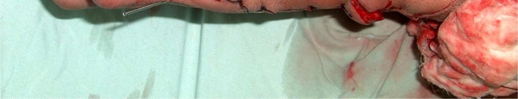


4 Plastic Surgery: An International Journal 4 Figure 1b Figure 1c Figure 1d


")


 2")
5 5 Plastic Surgery: An International Journal Figure 1e Figure 1f Figure 1: (a) A Preoperative Photo of a 26 Male Patient with Post Traumatic Ulnar Side Defect of the rt Hand, (b) Debridement of All Scarred Tissues was Done, (c) the Flap is Harvested and Inset, (d) 2 Weeks Postoperative View Showing Distal Edge Necrosis, (e &f) 2 Moths Postoperative Views Figure 2a


6 Plastic Surgery: An International Journal 6 Figure 2b Figure 2c
 2 Weeks Postoperative View, (c &d) 6 Months Antero-Posterior and Lateral Views Postoperative.")
![Discussion Despite several flaps introduced for hand reconstruction, the reversed radial forearm flap is still largely preferred for hand reconstruction [9].](/docs-images/83/87842309/images/7-1.jpg "It is considered a workhorse flap in hand reconstruction [10]. It offers many merits such as adequately large, thin and pliable and colour matched tissues.")

7 7 Plastic Surgery: An International Journal Figure 2d Figure 2: (a) 12yrs Old Male with PT Raw Area over the Ulnar Aspect of the Lt Hand with Amputated Little Finger at the Level of the MPJ He Underwent Two Debridement Operation then Coverage by PIA Flap, (b) 2 Weeks Postoperative View, (c &d) 6 Months Antero-Posterior and Lateral Views Postoperative. Discussion Despite several flaps introduced for hand reconstruction, the reversed radial forearm flap is still largely preferred for hand reconstruction [9]. It is considered a workhorse flap in hand reconstruction [10]. It offers many merits such as adequately large, thin and pliable and colour matched tissues. It offers a simple and effective one-stage method of soft tissue reconstruction. Surgery is confined to a single site and limb permitting early mobilization and rehabilitation. Moreover, vascularized bone and tendon transfer, along with the flap, are possible. If the flap pedicle was designed with proper length, it could also cover the fingers [11-15]. On the other hand, the radial forearm flap that has many disadvantages has been reported such as poor donor sit skin graft take, noticeable donor scar, bulky volar skin compared to thin dorsal skin,, hand swelling, reduced joint movement and strength, reduced sensation, coldintolerance, and radius fracture[16] [17]. Moreover, acute ischemia of the hand has been reported in spite of adequate circulation by preoperative Allen's test [18]. In a review for the clinical trials to reduce donor site morbidity, Loeffelbein et al [19] concluded that, most publications failed to provide the solid evidence characteristic of high-quality research. The reversed flow posterior interosseous artery flap (PIA) is a valuable option for reconstruction of hand defects. As it is based on the anterior-posterior interosseous artery system, it avoids sacrifice of either the radial or ulnar
8 Plastic Surgery: An International Journal 8 arteries as in case of the radial or ulnar forearm flaps. It is also possible to use this flap in circumstances where there has been damage to the palmer arch. In addition, the dorsal forearm skin is less bulky than the volar forearm skin; therefore, the PIA flap has better contour match. Furthermore, the donor site can often be closed primarily if the defect is small or by a skin graft, that will take well on the muscle bellies of the extensor carpi ulnaris and extensor digiti minimi [3]. Also in the event of flap failure, the PIA flap can be debrided and the defect skin grafted, while all other flap options remain available. This relative expendability of the PIA flap is one of its unique qualities [20]. Lastly, the flap can also be raised with a segment of the proximal third of the ulna as an osteocutaneous flap for bone defects, including thumb reconstruction [21]. In this study, 25 PIA flaps were harvested to close the ulnar side defects of the hand. The flap survival rates were comparable to other reported studies [22, 23] which supports its safety. Our results also showed a similar range of complications [23-28]. The flap can be considered as a smart and reliable option for coverage of the ulnar side of the hand, as it doesn t sacrifice the main arteries of the forearm and hand. It is a single stage procedure with no need for 2 nd stage division. It doesn t need special equipments or special training like microsurgical transfer. The flap was harvested with a skin bridge over the pedicle changing the island flap into a racquet shaped flap as described in a previous work [8]. The racquet shape modification of the flap avoids compression of the vascular pedicle which can develop in case of tunneling under skin or skin direct closure. In addition, it allows easy flap inset, enhances venous drainage as this skin bridge contains additional subcutaneous venous plexus. The skin strip can be also used for solving any additional local skin problems. Although the radial forearm flap has higher success rates than PIA flap, and has been considered the workhorse flap for hand reconstruction; it seems less satisfactory compared to PIA flap especially for ulnar side hand defects. The PIA flap seems to be a reliable option for reconstructing the ulnar side of the hand. We feel that the PIA must be considered as one of the new preferred valuable flaps for hand reconstruction. This flap must be learned well to both senior and junior staff and should be considered as another option for hand coverage when the surgeon prefers not to use free tissue transfer or the radial artery flap. Gentle pedicle dissection and avoidance of pedicle compression using the racquet shape design reduce flap necrosis. During flap elevation, we could harvest a skin paddle up to only 3-4 cm below the lateral epicondyle of the humerus that enabled us to cover defects down to the middle phalanges. In addition, anastomotic vessel dissection as described by Bayon and Pho 4 also increases flap arc of rotation. In the last decade, the senior author (Ayman shaker) as well as the other authors used the posterior interosseous flap in more than 80 cases of hand and upper limb reconstruction and they believe that the PIAF can be used efficiently to solve many problematic defects. Conclusion The PIA flap seems a smart and reliable option for coverage of the ulnar side of the hand. Further evidence- base has shown that the PIA flap should be considered as a reliable choice in hand reconstruction especially ulnar side defects. The flap should be learned to senior and junior reconstructive surgeons as a classical flap for hand reconstruction. Familiarity with the flap anatomy lessens the operative time greatly and makes surgical dissection easier. References 1. Lu, L. J., Wang, S. F., Yang, J. et al. (1986). 'The Posterior Interosseus Flap: A Report of 6 Cases,' The Second Symposium of the Chinese Association of Hand Surgery (Qing Dao city); 187e Penteado, C. V., Masquelet, A. C. & Chevrel, J. P. (1986). 'The Anatomic Basis of the Fasciocutaneous Flap of
9 9 Plastic Surgery: An International Journal the Posterior Interosseus Artery,' Surg Radiol Anat; 8: 209e Zancolli, E. A. & Angrigiani, C. (1988). Posterior Interosseous Island Forearm Flap, Journal of Hand Surgery: British & European Volume; 13-B: 130e5. 4. Bayon, P. & Pho, R. W. H. (1988). Anatomical Basis of Forearm Flap Based on Posterior Interosseus Vessels, Journal of Hand Surgery 13B: Giunta, R. E. & Lukas, B. (1998). Impossible Harvest of the Posterior Interosseus Artery Flap: A Report of an Individualised Salvage Procedure, British Journal of Plastic Surgery 51: Costa, H. & Soutar, D. S. (1988). The Distally Based Island Posterior Interosseus Flap, British Journal of Plastic Surgery 41: Brunelli, F., Giele, H. & Perrotta, R. (2000). Reverse Posterior Interosseus Flap Based on an Exteriorized Pedicle to Cover Digital Skin Defects, Journal of Hand Surgery British; 25: Nasser, S., Abdallah, Y. & Shaker, A. A. 2011). "Racquet-Shaped Modification of the Island Pedicle Flaps: A Simple Design to Increase its Versatility and to Decrease the Complication Rate, Egypt, Egyptian Journal of Plastic and Reconstructive Surgery, Vol. 35, No. 1, January: Megerle, K., Sauerbier, M. & Germann, G. (2010). The Evolution of the Pedicled Radial Forearm Flap, Hand; 5(1): Friedrich, J. B. & Pederson, W. C., Bishop, A. T., Galaviz, P. & Chang, J. (2012). New Workhorse Flaps in Hand Reconstruction, Hand 7: Soutar, D. S. & Tanner, N. S. B. (1984). The Radial Forearm Flap in the Management of Soft Tissue Injuries of the Hand, British Journal of Plastic Surgery, Volume 37, Issue 1, Jones, N. F., Jarrahy, R. & Kaufman, M. R. (2008). Pedicled and Free Radial Forearm Flaps for Reconstruction of the Elbow, Wrist, and Hand, Plastic & Reconstructive Surgery; 121(3): Taghinia, A. H., Carty, M. & Upton, J. (2010). Fascial Flaps for Hand Reconstruction, Journal of Hand Surgery; 35(8): Kim, K. S., Kim, E. S., Hwang, J. H. & Lee, S. Y. (2010). Thumb Reconstruction Using the Radial Midpalmar (Perforator Based) Island Flap (Distal Thenar Perforator-Based Island Flap), Plastic & Reconstructive Surgery; 125(2): El-Khatib, H. A. & Hammouda, A. H. (2005). Reverse Osseofasciocutaneous Radial Forearm Flap for Thumb Reconstruction: A Flap Design and Case Series, Journal of Hand Surgery; 30(6): Page, R. & Chang, J. (2006). Reconstruction of Hand Soft-Tissue Defects: Alternatives to the Radial Forearm Fasciocutaneous Flap, Journal of Hand Surgery, 31(5): Timmons, M. J., Missotten, F. E. M., Poole, M. D. & Davies, D. M. (1986). Complications of Radial Forearm Flap Donor Sites, British Journal of Plastic Surgery, Volume 39, Issue 2: Jones, B. M. & O'Brien, C. J. (1985). Acute Ischaemia of the Hand Resulting from Elevation of a Radial Forearm Flap, British Journal of Plastic Surgery, Volume 38, Issue 3: Loeffelbein, D. J., Al-Benna, S. et al. (2012). Reduction of Donor Site Morbidity of Free Radial Forearm Flaps: What Level of Evidence Is Available?, Eplasty; 12: e9.
10 Plastic Surgery: An International Journal Costa, H., Pinto, A. & Zenha, H. (2007). 'The Posterior Interosseus Flap e a Prime Technique in Hand Reconstruction,' The Experience of 100 Anatomical Dissections and 102 Clinical Cases; PRS March. 21. Hsu, C. & Chang, J. (2003). The Posterior Interosseus Artery Flap Revisited, Operative Techniques in Plastic and Reconstructive Surgery, Vol 9, No 4: p Fujiwara, M., Kawakatsu, M., Yoshida, Y. & Sumiya, A. (2003). Modified Posterior Interosseus Flap in Hand Reconstruction," Techniques in Hand and Upper Extremity Surgery, 7 (3): Balakrishnan, G., Kumar, B. S. & Hussein, S. A. (2003). Reverse Flow Posterior Interosseus Artery Flap Revisited, Plastic & Reconstructive Surgery, 111 (7): Buchler, U. & Frey, H.- P. (1991). Retrograde Posterior Interosseus Flap, Journal of Hand Surgery [Am.], 16A: Dap, F., Dautel, G., Voche, P. et al. (1993). The Posterior Interosseus Flap in Primary Repair of Hand Injuries, Journal of Hand Surgery. (Br.), 18 (4): Brunelli, F., Valenti, P., Dumonitier, C. et al. (2001). The Posterior Interosseus Reverse Flap: Experience with 113 Flaps, Annals of Plastic Surgery, 47: Koch, H., Kursumovic, A., Hubmer, M. et al. (2003). Defects on the Dorsum of the Hand - The Posterior Interosseus Flap and its Alternatives, Journal of Hand Surgery, 8: Shibata, M., Iwabuchi, Y., Kubota, S. et al. (1997). Comparison of Free and Reversed Pedicled Posterior Interosseus Flap, Plastic & Reconstructive Surgery, 99:
Evaluation of the Posterior Interosseous Artery Flap in Reconstructing Hand Defects
 POSTERIOR THE IRAQI POSTGRADUATE INTEROSSEOUS MEDICAL ARTERY JOURNAL FLAP Evaluation of the Posterior Interosseous Artery Flap in Reconstructing Hand Defects Osam Ahmed Ibraheem, Mahdi Hameed Abood, Heider
Distally Based Dorsal Forearm Fasciosubcutaneous Flap
 Distally Based Dorsal Forearm Fasciosubcutaneous Flap Kwang Seog Kim, M.D., Ph.D. Gwangju, Korea Use of a local flap is often required for the reconstruction of a skin defect on the dorsum of the hand.
The distally-based island ulnar artery perforator flap for wrist defects
 Free full text on www.ijps.org Original Article The distally-based island ulnar artery perforator flap for wrist defects Durga Karki, A. K. Singh Post Graduate Department of Plastic and Reconstructive
Reverse Adipofascial Radial Forearm Flap Surgery for Soft-Tissue Reconstruction of Hand Defects
 Reverse Adipofascial Radial Forearm Flap Surgery for Soft-Tissue Reconstruction of Hand Defects Osman Akdag, MD, a Mehtap Karamese, MD, a Muhammed NebilSelimoglu, MD, a Ahmet Akatekin, MD, a Malik Abacı,
1/13/2013. Anatomy Guy Dissection Sheet Extensor Forearm and Hand. Eastern Virginia Medical School
 Dr. Craig Goodmurphy Anatomy Guy Superficial Extensor Muscles Complete skin removal if necessary then remove the antebrachial fascia starting at the extensor retinaculum and working proximally. Define
Different modalities of soft tissue coverage of hand and wrist defects
 ifferent modalities of soft tissue coverage of hand and wrist defects Soft tissue defects of hand and wrist with exposed tendons, joints, nerves and bones represent a challenge to plastic surgeons. Such
The Anterior Interosseus Artery Perforator Flap: Anatomical Dissections and Clinical Study
 52 Anterior interosseus artery perforator flap Original Article The Anterior Interosseus Artery Perforator Flap: Anatomical Dissections and Clinical Study Nikhil S Panse*, Sheetal B Joshi2, Parag B Sahasrabudhe,
Management of Complex Avulsion Injuries of the Dorsum of the Foot and Ankle in Pediatric Patients by Using Local Delayed Flaps and Skin Grafts
 Management of Complex Avulsion Injuries of the Dorsum of the Foot and Ankle in Pediatric Patients by Using Local Delayed Flaps and Skin Grafts Ahmed Elshahat, MD Plastic Surgery Department, Ain Shams University,
Lecture 9: Forearm bones and muscles
 Lecture 9: Forearm bones and muscles Remember, the region between the shoulder and the elbow = brachium/arm, between elbow and wrist = antebrachium/forearm. Forearm bones : Humerus (distal ends) Radius
Versatility of Reverse Sural Artery Flap for Heel Reconstruction
 ORIGINAL ARTICLE Introduction: The heel has two parts, weight bearing and non-weight bearing part. Soft tissue heel reconstruction has been a challenge due to its complex nature of anatomy, weight bearing
Role Of Reverse Sural Artery Flap In Ankle, Foot And Leg Defects
 IOSR Journal of Dental and Medical Sciences (IOSR-JDMS) e-issn: 2279-0853, p-issn: 2279-0861.Volume 15, Issue 5 Ver. VIII (May. 2016), PP 64-68 www.iosrjournals.org Role Of Reverse Sural Artery Flap In
JMSCR Vol 07 Issue 01 Page January 2019
 www.jmscr.igmpublication.org Impact Factor (SJIF): 6.379 Index Copernicus Value: 79.54 ISSN (e)-2347-176x ISSN (p) 2455-0450 DOI: https://dx.doi.org/10.18535/jmscr/v7i1.36 Original Article A Study on the
ABSTRACT. Key words: Flaps, Adipofascial, Lower limb reconstruction. Khaldoun J. Haddadin, MD*, Samer Y. Haddad, MD. Introduction.
 DISTALLY BASED ADIPOFASCIAL FLAPS: A VERSATILE FLAP FOR THE RECONSTRUCTION OF LOWER LEG AND PROXIMAL FOOT DEFECTS AT THE ROYAL JORDANIAN REHABILITATION CENTER Khaldoun J. Haddadin, MD*, Samer Y. Haddad,
Nerves of the upper limb Prof. Abdulameer Al-Nuaimi. E. mail:
 Nerves of the upper limb Prof. Abdulameer Al-Nuaimi E-mail: a.al-nuaimi@sheffield.ac.uk E. mail: abdulameerh@yahoo.com Brachial plexus Median nerve After originating from the brachial plexus in the axilla,
The Boomerang Flap in Managing Injuries of the Dorsum of the Distal Phalanx
 The Boomerang Flap in Managing Injuries of the Dorsum of the Distal Phalanx Shao-Liang Chen, M.D., Trong-Duo Chou, M.D., Shyi-Gen Chen, M.D., Tian-Yeu Cheng, M.D., Tim-Mo Chen, M.D., and Hsian-Jenn Wang,
COMBINED ABDOMINAL FLAP FOR MAJOR HAND RECONSTRUCTION
 Int. J. Pharm. Med. & Bio. Sc. 2013 2014 Srinivas Somashekar et al., 2014 Research Paper ISSN 2278 5221 www.ijpmbs.com Vol. 3, No. 1, January 2014 2014 IJPMBS. All Rights Reserved COMBINED ABDOMINAL FLAP
Anatomical study. H. Costa, M. L. Gracia*, J. Vranchx?, C. Cunha, A. Conde and D. Soutar:~
 British Journal of Plastic Surgery (2001), 54, 28-33 9 2001 The British Association of Plastic Surgeons doi: 10.1054/bjps.2000.3472 PLASTIC SURGERY The posterior interosseous flap: a review of 81 clinical
Sensate First Dorsal Metacarpal Artery Flap for Resurfacing Extensive Pulp Defects of the Thumb
 ORIGINAL ARTICLE Sensate First Dorsal Metacarpal Artery Flap for Resurfacing Extensive Pulp Defects of the Thumb Shun-Cheng Chang, MD, Shao-Liang Chen, MD, Tim-Mo Chen, MD, Chia-Jueng Chuang, MD, Tian-Yeu
Variation of Superficial Palmar Arch: A Case Report
 Article ID: WMC003387 ISSN 2046-1690 Variation of Superficial Palmar Arch: A Case Report Corresponding Author: Dr. Liju S Mathew, Demonstrator, Anatomy, Gulf Medical University, 4184 - United Arab Emirates
Key Relationships in the Upper Limb
 Key Relationships in the Upper Limb This list contains some of the key relationships that will help you identify structures in the lab. They are organized by dissection assignment as defined in the syllabus.
Compartment Syndrome
 Compartment Syndrome Chapter 34 Compartment Syndrome Introduction Compartment syndrome may occur with an injury to any fascial compartment. The fascial defect caused by the injury may not be adequate to
Basics of Flap Design
 Basics of Flap Design Reconstructive Ladder (Mathes & Nahai 1982) Consider the defect, systematically Move from simple to complex Occam s Razor Sutton s Law Ladder is simple, emphasizes closure over form
17 FibulA FlAP Tor Chiu fibula flap 153
 17 Fibula Flap Tor Chiu Fibula Flap 153 Fibula Flap FLAP TERRITORY This flap includes a segment of the fibular bone with or without the overlying skin island on the peroneal/ lateral aspect of the calf.
divided by the bones ( redius and ulna ) and interosseous membrane into :
 and interosseous membrane into :") fossa Cubital Has: * floor. * roof : - Skin - superficial fasica - deep fascia ( include bicipital aponeurosis ) Structures within the roof : -cephalic and basilic veins -and between them median cubital
fossa Cubital Has: * floor. * roof : - Skin - superficial fasica - deep fascia ( include bicipital aponeurosis ) Structures within the roof : -cephalic and basilic veins -and between them median cubital
Levels of the anatomical cuts of the upper extremity RADIUS AND ULNA right
 11 CHAPTER 2 Levels of the anatomical cuts of the upper extremity AND right CUT 1 CUT 4 1 2 3 4 5 6 Isolated fixation of the radius is difficult at this level because of the anterolateral vessels and the
Viorel Nacu. The clinical anatomy of the Hand
 Viorel Nacu The clinical anatomy of the Hand The distal part of the upper limb is divided in to three regions: 1. The wrist (carpus) 2. The hand (metacarpus) 3. The digits (fingers) The landmarks of this
Ulnar and radial artery based perforator adipofascial flaps
 101 Ulnar and radial artery based perforator adipofascial flaps EEXOT Volume 59, (2):101-108, 2008 I.A. Ignatiadis 1, F.S. Giannoulis 1, A.F. Mavrogenis 2, G.N. Nomikos 2, S. Vasilas 1, S.G. Spyridonos
Split Hemianterior Tibialis Turndown Muscle Flap for Coverage of Distal Leg Wounds With Preservation of Function
 Split Hemianterior Tibialis Turndown Muscle Flap for Coverage of Distal Leg Wounds With Preservation of Function Vinay Gundlapalli, MD, a John W. Gillespie III, MD, b and Chris D. Tzarnas, MD, FACS c a
Case Report The posterior interosseous pedicle free flap in soft-tissue coverage for small-area tissue defects of the hand
 Int J Clin Exp Med 2017;10(11):15599-15605 www.ijcem.com /ISSN:1940-5901/IJCEM0047672 Case Report The posterior interosseous pedicle free flap in soft-tissue coverage for small-area tissue defects of the
STRUCTURAL BASIS OF MEDICAL PRACTICE EXAMINATION 5 October 6, 2006
 STRUCTURAL BASIS OF MEDICAL PRACTICE EXAMINATION 5 October 6, 2006 PART l. Answer in the space provided. (8 pts) 1. Identify the structures. (2 pts) B C A. _pisiform B. _ulnar artery A C. _flexor carpi
The arm: *For images refer back to the slides
 The arm: *For images refer back to the slides Muscles of the arm: deltoid, triceps (which is located at the back of the arm), biceps and brachialis (it lies under the biceps), brachioradialis (it lies
The posterior interosseous artery in the distal part of the forearm. Is the term recurrent branch of the anterior interosseous artery justified?
 The British Association of Plastic Surgeons (2004) 57, 638 644 The posterior interosseous artery in the distal part of the forearm. Is the term recurrent branch of the anterior interosseous artery justified?
TRANSPOSITIONAL ADIPOFASCIAL FLAPS FOR COMPLICATED ACUTE FINGER INJURIES
 K.B. Poon, S.H. Chien, G.T. Lin, et al TRANSPSITINAL ADIPFASCIAL FLAPS FR CMPLICATED ACUTE FINGER INJURIES Kein Boon Poon, Song-Hsiung Chien, 1 Gau-Tyan Lin, 1 and Yin-Chih Fu 1 Department of rthopaedic
Biceps Brachii. Muscles of the Arm and Hand 4/4/2017 MR. S. KELLY
 Muscles of the Arm and Hand PSK 4U MR. S. KELLY NORTH GRENVILLE DHS Biceps Brachii Origin: scapula Insertion: radius, fascia of forearm (bicipital aponeurosis) Action: supination and elbow flexion Innervation:
Nerves of Upper limb. Dr. Brijendra Singh Professor & Head Department of Anatomy AIIMS Rishikesh
 Nerves of Upper limb Dr. Brijendra Singh Professor & Head Department of Anatomy AIIMS Rishikesh 1 Objectives Origin, course & relation of median & ulnar nerves. Motor & sensory distribution Carpal tunnel
ARM Brachium Musculature
 ARM Brachium Musculature Coracobrachialis coracoid process of the scapula medial shaft of the humerus at about its middle 1. flexes the humerus 2. assists to adduct the humerus Blood: muscular branches
Anatomic Landmarks for the Radial Tunnel
 Anatomic Landmarks for the Radial Tunnel Ron Hazani, MD, Nitin J. Engineer, MD, Arian Mowlavi, MD, Michael Neumeister, MD, W.P. Andrew Lee, MD, and Bradon J. Wilhelmi, MD Division of Plastic and Reconstructive
Hand and forearm reconstruction after skin cancer ablation
 Clin Plastic Surg 31 (2004) 113 119 Hand and forearm reconstruction after skin cancer ablation Patrick J. O Neill, MD a, *, Christopher Litts, MD b a Department of Surgery, Division of Plastic Surgery,
MCQWeek2. All arise from the common flexor origin. The posterior aspect of the medial epicondyle is the common flexor origin.
 MCQWeek2. 1. Regarding superficial muscles of anterior compartment of the forearm: All arise from the common flexor origin. The posterior aspect of the medial epicondyle is the common flexor origin. Flexor
Intrinsic muscles palsies of the hand Management of Thumb Opposition with BURKHALTER s Procedure
 Intrinsic muscles palsies of the hand Management of Thumb Opposition with BURKHALTER s Procedure TRUONG LE DAO, MD, IFAAD 1 Burkhalter W.E, Cristhensen R.C, Brown P.W, Extensor Indicis Proprius opponensplasty
Gastrocnemius Myocutaneous Flap: A Versatile Option to Cover the Defect of Upper and Middle Third Leg
 Downloaded from wjps.ir at 22:25 +0330 on Sunday November 18th 28 314 Gastrocnemius flap for coverage of leg defects Original Article Gastrocnemius Myocutaneous Flap: A Versatile Option to Cover the Defect
Hand Anatomy A Patient's Guide to Hand Anatomy
 Hand Anatomy A Patient's Guide to Hand Anatomy Introduction Few structures of the human anatomy are as unique as the hand. The hand needs to be mobile in order to position the fingers and thumb. Adequate
The Forearm 2. Extensor & lateral Compartments of the Forearm
 The Forearm 2 Extensor & lateral Compartments of the Forearm 1-Lateral Fascial Compartment (at the lateral side of the forearm ) *Some books mention the lateral compartment contain just the Brachioradialis
Wrist & Hand Assessment and General View
 Wrist & Hand Assessment and General View Done by; Mshari S. Alghadier BSc Physical Therapy RHPT 366 m.alghadier@sau.edu.sa http://faculty.sau.edu.sa/m.alghadier/ Functional anatomy The hand can be divided
Johannesburg, South Africa
 NEUROVASCULAR ISLAND FLAP IN THE TREATMENT OF TROPHIC ULCERATION OF THE HEEL By ISIDORE KAPLAN, F.R.C.S., F.R.C.S.(Ed.) Johannesburg, South Africa THE transfer of skin and subcutaneous tissue on a neurovascular
Netter's Anatomy Flash Cards Section 6 List 4 th Edition
 Netter's Anatomy Flash Cards Section 6 List 4 th Edition https://www.memrise.com/course/1577581/ Section 6 Upper Limb (66 cards) Plate 6-1 Humerus and Scapula: Anterior View 1.1 Acromion 1.2 Greater tubercle
The distally-based dorsal hand flap
 British Journal of Plastic Surgery (1990), 43, 28-39 0007-1226/90/0043-0028/$10.00 1990 The Trustees of British Association of Plastic Surgeons The distally-based dorsal hand flap A. A. QUABA and P. M.
Anatomy Workshop Upper Extremity David Ebaugh, PT, PhD Workshop Leader. Lab Leaders: STATION I BRACHIAL PLEXUS
 Anatomy Workshop Upper Extremity David Ebaugh, PT, PhD Workshop Leader Lab Leaders: STATION I BRACHIAL PLEXUS A. Posterior cervical triangle and axilla B. Formation of plexus 1. Ventral rami C5-T1 2. Trunks
Outcome of Thumb Reconstruction Using the First Dorsal Metacarpal Artery Island Flap
 Ghoraba et al. 1 Original Article Outcome of Thumb Reconstruction Using the First Dorsal Metacarpal Artery Island Flap Samir M Ghoraba, Wael H Mahmoud* Plastic, Reconstructive Surgery and Burns Department,
Interesting Case Series. Reconstruction of Dorsal Wrist Defects
 Interesting Case Series Reconstruction of Dorsal Wrist Defects Maelee Yang, BS, and Joseph Meyerson, MD The Ohio State University Wexner Medical Center, Columbus Correspondence: maelee.yang@osumc.edu Keywords:
compartments of the forearm
 " forearm posterior compartment " compartments of the forearm Posterior Fascial compartment Muscles: ** The superficial group 1. Extensor carpi radialis brevis 2. Ex. digitorum 3. Ex. digiti minimi 4.
Al Hess MD NERVE REPAIR
 Al Hess MD NERVE REPAIR Historical Aspects 300 BC Hippocrates, description of nervous system 200 AD Galen of Pergamon, nerve injury, questioned possibility of regeneration 600 AD Paul of Arginia, first
The hand is full with sweat glands, activated at times of stress. In Slide #2 there was a mistake where the doctor mentioned lateral septum twice.
 We should only know: Name, action & nerve supply Layers - Skin - Superficial fascia - Deep fascia The hand is full with sweat glands, activated at times of stress. Deep fascia In Slide #2 there was a mistake
What s New in Fingertip Injuries. Gordon A. Brody, MD SOAR Redwood City
 What s New in Fingertip Injuries Gordon A. Brody, MD SOAR Redwood City Goals of Treatment Durable Sensate Aesthetic Preserve Length Preserve Mobility Goals of Treatment Pain and Worker s Compensation are
In the name of Allah, Most gracious, Most merciful
 In the name of Allah, Most gracious, Most merciful This lecture includes the following: The Palmer Oponeurosis. The Carpel tunnel. The palmaris brevis muscle. The anatomical snuffbox. The Fibrous flexor
ORIGINAL ARTICLE DISTALLY BASED PERONEUS BREVIS MUSCLE FLAP FOR DISTAL LEG DEFECTS
 DISTALLY BASED PERONEUS BREVIS MUSCLE FLAP FOR DISTAL LEG DEFECTS Peddi Manjunath 1, Ramesha K.T 2, Smitha S Segu 3, Jainath 4, Shankarappa M 5 HOW TO CITE THIS ARTICLE: Peddi Manjunath, Ramesha KT, Smitha
Interesting Case Series. Posterior Interosseous Nerve Compression
 Interesting Case Series Posterior Interosseous Nerve Compression Jeon Cha, BMedSci, MBBS, Blair York, MBChB, and John Tawfik, MBBS, BPharm, FRACS The Sydney Hospital Hand Unit, Sydney Hospital and Sydney
Interesting Case Series. Radial Tunnel Syndrome Complicated by Lateral Epicondylitis in a Middle-Aged Female
 Interesting Case Series Radial Tunnel Syndrome Complicated by Lateral Epicondylitis in a Middle-Aged Female Sumesh Kaswan, MD, a Olivier Deigni, MD, MPH, a Kashyap K. Tadisina, BS, b Michael Totten, BS,
Cubital fossa and forearm
 Cubital fossa and forearm Cubital fossa is the triangular space in front of elbow joint. - The Cubital fossa has boundaries: apex, base, roof and floor and it has contents. The base: an imaginary horizontal
Functional Anatomy of the Elbow
 Functional Anatomy of the Elbow Orthopedic Institute Daryl C. Osbahr, M.D. Chief of Sports Medicine, Orlando Health Chief Medical Officer, Orlando City Soccer Club Orthopedic Consultant, Washington Nationals
*the Arm* -the arm extends from the shoulder joint (proximal), to the elbow joint (distal) - it has one bone ; the humerus which is a long bone
, to the elbow joint (distal) - it has one bone ; the humerus which is a long bone") *the Arm* -the arm extends from the shoulder joint (proximal), to the elbow joint (distal) - it has one bone ; the humerus which is a long bone - muscles in the arm : *brachialis muscle *Biceps brachii
*the Arm* -the arm extends from the shoulder joint (proximal), to the elbow joint (distal) - it has one bone ; the humerus which is a long bone - muscles in the arm : *brachialis muscle *Biceps brachii
Chapter 4: Forearm 4.3 Forearm shaft fractures, transverse (12-D/4)
") AO Manual of ESIN in children s fractures Chapter 4: Forearm 4.3 Forearm shaft fractures, transverse (12-D/4) Title AO Manual of ESIN in children Subtitle Elastic stable intramedullary nailing (ESIN) Author
AO Manual of ESIN in children s fractures Chapter 4: Forearm 4.3 Forearm shaft fractures, transverse (12-D/4) Title AO Manual of ESIN in children Subtitle Elastic stable intramedullary nailing (ESIN) Author
Main Menu. Wrist and Hand Joints click here. The Power is in Your Hands
 1 The Wrist and Hand Joints click here Main Menu K.5 http://www.handsonlineeducation.com/classes/k5/k5entry.htm[3/23/18, 1:40:40 PM] Bones 29 bones, including radius and ulna 8 carpal bones in 2 rows of
The homodigital neurovascular antegrade island flap. for fingertip reconstruction in children and
 Acta Orthop. Belg., 2011, 77, 598-602 ORIGINAL STUDY The homodigital neurovascular antegrade island flap for fingertip reconstruction in children Bingqi WAng, Lei CHEn, Laijin LU, Zhigang LiU, Zhixin ZHAng,
The earlier clinic experience of the reverse-flow anterolateral thigh island flap
 British Journal of Plastic Surgery (2005) 58, 160 164 The earlier clinic experience of the reverse-flow anterolateral thigh island flap Gang Zhou, Qi-Xu Zhang*, Guang-Yu Chen Scar Multiple Treatment Centre,
region of the upper limb between the shoulder and the elbow Superiorly communicates with the axilla.
 1 region of the upper limb between the shoulder and the elbow Superiorly communicates with the axilla. Inferiorly, a number of important structures pass between arm & forearm through cubital fossa. 2 medial
Propeller perforator flaps for finger reconstruction
 Technical Note Page 1 of 7 Propeller perforator flaps for finger reconstruction Alexandru Valentin Georgescu, Ileana Rodica Matei Department of Plastic Surgery, University of Medicine Iuliu Hatieganu Cluj
Distally Based Sural Artery Adipofascial Flap based on a Single Sural Nerve Branch: Anatomy and Clinical Applications
 Distally Based Sural Artery Adipofascial Flap based on a Single Sural Nerve Branch: Anatomy and Clinical Applications Wan Loong James Mok 1, Yong Chen Por 1, Bien Keem Tan 2 1 Department of Plastic, Reconstructive
SUCCESSFUL TREATMENT OF soft tissue deficits at the
 SCIENTIFIC ARTICLE Vascular Perfusion of a Flexor Carpi Ulnaris Muscle Turnover Pedicle Flap for Posterior Elbow Soft Tissue Reconstruction: A Cadaveric Study Diane E. S. Payne, MD, Adam M. Kaufman, MD,
Heel Defect Reconstruction using Local Vascularized Flaps: Results and Clinical Outcomes in 16 Patients
 JFS (P) Kuldeep Singh et al ORIGINL RTICLE 10.5005/jp-journals-10040-1083 Heel Defect Reconstruction using Local Vascularized Flaps: Results and Clinical Outcomes in 16 Patients 1 Kuldeep Singh, 2 Zile
Kinesiology of The Wrist and Hand. Cuneyt Mirzanli Istanbul Gelisim University
 Kinesiology of The Wrist and Hand Cuneyt Mirzanli Istanbul Gelisim University Bones The wrist and hand contain 29 bones including the radius and ulna. There are eight carpal bones in two rows of four to
Role of free tissue transfer in management of chronic venous ulcer
 Original Article Role of free tissue transfer in management of chronic venous ulcer K. Murali Mohan Reddy, D. Mukunda Reddy Department of Plastic Surgery, Nizams Institute of Medical Sciences, India. Address
ANATOMICAL STUDY OF THE RADIAL TUNNEL BY CADAVERIC DISSECTION, FOR POSSIBLE SITES OF THE POSTERIOR INTEROSSEOUS NERVE ENTRAPMENT
 Int. J. Pharm. Med. & Bio. Sc. 2014 Bhavana Sunil Junagade and Aruna Mukherjee, 2014 Research Paper ISSN 2278 5221 www.ijpmbs.com Vol. 3, No. 2, April 2014 2014 IJPMBS. All Rights Reserved ANATOMICAL STUDY
Cross finger flap for reconstruction of complex finger defects
 Original Article Nepal Med Coll J 2015; 17(1-2): 73-77 Cross finger flap for reconstruction of complex finger defects Bista N, Shrestha KM, Bhattachan CL Department of General Surgery, Nepal Medical College
Hyperbaric oxygen therapy and surgical delay improve flap survival of reverse pedicle flaps for lower third leg and foot reconstruction
 Original Article Plastic and Aesthetic Research Hyperbaric oxygen therapy and surgical delay improve flap survival of reverse pedicle flaps for lower third leg and foot reconstruction Pradeoth Mukundan
The Elbow and the cubital fossa. Prof Oluwadiya Kehinde
 The Elbow and the cubital fossa Prof Oluwadiya Kehinde www.oluwadiya.com Elbow and Forearm Anatomy The elbow joint is formed by the humerus, radius, and the ulna Bony anatomy of the elbow Distal Humerus
Hand and wrist emergencies
 Chapter1 Hand and wrist emergencies Carl A. Germann Distal radius and ulnar injuries PEARL: Fractures of the distal radius and ulna are the most common type of fractures in patients younger than 75 years.
The free thoracodorsal artery perforator flap in head and neck reconstruction
 European Annals of Otorhinolaryngology, Head and Neck diseases (2012) 129, 167 171 Available online at www.sciencedirect.com TECHNICAL NOTE The free thoracodorsal artery perforator flap in head and neck
Primary closure of the deltopectoral flap-donor site without skin grafting
 Primary closure of the deltopectoral flap-donor site without skin grafting Received: 4/3/2013 Accepted: 14/5/2013 Introduction Reliable and simultaneous reconstruction of head-and-neck defects has been
Modified anconeus muscle transfer as treatment of failed surgical release of lateral epicondylitis of the elbow
 310 Acta Orthop. Belg., 2017, t. luyckx, 83, 310-314 a. decramer, l. luyckx, j. noyez ORIGINAL STUDY Modified anconeus muscle transfer as treatment of failed surgical release of lateral epicondylitis of
Clinical examination of the wrist, thumb and hand
 Clinical examination of the wrist, thumb and hand 20 CHAPTER CONTENTS Referred pain 319 History 319 Inspection 320 Functional examination 320 The distal radioulnar joint.............. 320 The wrist.......................
Supplied in part by the musculocutaneous nerve. Forms the axis of rotation in movements of pronation and supination
 Anatomy: Upper limb (15 questions) 1. Latissimus Dorsi: Is innervated by the dorsal scapular nerve Lies above feres major muscle Medially rotates the humerus All of the above 2. Supinator muscle is: Deep
Use of Dorsal Metacarpal Artery Flaps in Post Burn Reconstruction Two Cases Report
 Macedonian Journal of Medical Sciences. 2011 Jun 15; 4(2):180-184. doi:10.3889/mjms.1857-5773.2011.0164 Case Report OPEN ACCESS Use of Dorsal Metacarpal Artery Flaps in Post Burn Reconstruction Two Cases
Nerve Injury. 1) Upper Lesions of the Brachial Plexus called Erb- Duchene Palsy or syndrome.
 Upper Lesions of the Brachial Plexus called Erb- Duchene Palsy or syndrome.") Nerve Injury - Every nerve goes to muscle or skin so if the nerve is injured this will cause paralysis in the muscle supplied from that nerve (paralysis means loss of function) then other muscles and other
Nerve Injury - Every nerve goes to muscle or skin so if the nerve is injured this will cause paralysis in the muscle supplied from that nerve (paralysis means loss of function) then other muscles and other
Dr. Mahir Alhadidi Anatomy Lecture #9 Feb,28 th 2012
 Quick Revision: Upper arm is divided into two compartments: 1. Anterior Compartment: Contains three muscles (Biceps brachii, Coracobrachialis, Brachialis). Innervated by Musculocutaneous nerve. 2. Posterior
Closure of Chronic Heel Ulcer by Simple V-Y Flap
 Egypt, J. Plast. Reconstr. Surg., Vol. 40, No. 1, January: 97-101, 2016 Closure of Chronic Heel Ulcer by Simple V-Y lap ESA TAAN,.D.; AYAN ARHAT,.D.; OUSTAA EKY,.D. and AHOUD NASI,.D. The Department of
Medial elbow reconstruction with perforator based medial arm propeller flap
 Case Report Hand Microsurg 2018;7:58-62 doi:10.5455/handmicrosurg.250701 Medial elbow reconstruction with perforator based medial arm propeller flap Asim Uslu 1, Abdullah Surucu 1, Mehmet Ali Korkmaz 1,
Wrist and Hand Anatomy
 Wrist and Hand Anatomy Bone Anatomy Scapoid Lunate Triquetrium Pisiform Trapeziod Trapezium Capitate Hamate Wrist Articulations Radiocarpal Joint Proximal portion Distal portion Most surface contact found
Hand Trauma Update: Outline. Hand Surgeon s Area of Expertise. Orthopaedic Update 2015
 Hand Trauma Update: 2015 Orthopaedic Update 2015 March 21, 2015 Peter Tang, MD, MPH Director Hand, Upper Extremity & Microvascular Surgery Fellowship Associate Professor Drexel University College of Medicine
Ligaments of Elbow hinge: sagittal plane so need lateral and medial ligaments
 Ligaments of Elbow hinge: sagittal plane so need lateral and medial ligaments Ulnar Collateral ligament on medial side; arising from medial epicondyle and stops excess valgus movement (lateral movement)
forearm posterior compartment
 Quick revision: The anterior compartment of the forearm contains of 8 muscles... -4 superficial -1 intermediate -3 deep *All supplied by median nerve except 1 and 1/2 muscle (by ulnar N.) forearm posterior
10/10/2014. Structure and Function of the Hand. The Hand. Osteology of the Hand
 Structure and Function of the Hand 19 bones and 19 joints are necessary to produce all the motions of the hand The Hand Dorsal aspect Palmar aspect The digits are numbered 1-5 Thumb = #1 Little finger
OPEN ACCESS ATLAS OF OTOLARYNGOLOGY, HEAD & NECK OPERATIVE SURGERY
 OPEN ACCESS ATLAS OF OTOLARYNGOLOGY, HEAD & NECK OPERATIVE SURGERY BUCCINATOR MYOMUCOSAL FLAP The Buccinator Myomucosal Flap is an axial flap, based on the facial and/or buccal arteries. It is a flexible
STRUCTURAL BASIS OF MEDICAL PRACTICE EXAMINATION 5. September 30, 2011
 STRUCTURAL BASIS OF MEDICAL PRACTICE EXAMINATION 5 September 30, 2011 PART l. Answer in the space provided. (12 pts) 1. Identify the structures. (2 pts) EXAM NUMBER A. Suprascapular nerve B. Axillary nerve
THE pedicled flap, commonly used by the plastic surgeon in the reconstruction
 THE PEDICLE!) SKIN FLAP ROBIN ANDERSON, M.D. Department of Plastic Surgery THE pedicled flap, commonly used by the plastic surgeon in the reconstruction of skin and soft tissue defects, differs from the
Fascial Compartments of the Upper Arm
 Fascial Compartments of the Upper Arm The upper arm is enclosed in a sheath of deep fascia and has two fascial septa: 1- Medial fascial septum (medial intermuscular septum): attached to the medial supracondylar
Reconstruction of axillary scar contractures retrospective study of 124 cases over 25 years
 British Journal of Plastic Surgery (2003), 56, 100 105 q 2003 The British Association of Plastic Surgeons. Published by Elsevier Science Ltd. All rights reserved. doi:10.1016/s0007-1226(03)00035-3 Reconstruction
Gastrocnemius Muscle Flap Coverage of Chronically= Infected Knee Joints
 Gastrocnemius Muscle Flap Coverage of Chronically= Infected Knee Joints ABSTRACT Chronically infected open knee joints present dif cult problem. Aggressive debridement of chronically infected soft tissue
Slides of Anatomy. Spring Dr. Maher Hadidi, University of Jordan
 Slides of Anatomy Please note : These slides are Dr. Maher Hadidi s slides of spring 2016 and were edited by the Premed Academic Team to fit the slides of spring 2019. Spring 2019 Dr. Maher Hadidi, University