Neurophysiology. Danil Hammoudi.MD
|
|
|
- Oswin Chapman
- 6 years ago
- Views:
Transcription
1 Neurophysiology Danil Hammoudi.MD
2 SYNAPSES
3 Synapses The site at which neurons communicate is called a synapse, a cell junction that mediates the transfer of information a cell junction that mediates the transfer of information from one neuron to the next
4 Synapses Because signals pass across most synapses in one direction only, synapses determine the direction of information flow throughout the nervous system
5 Synapses The neuron the conducts impulses toward a synapse is The neuron the conducts impulses toward a synapse is called the presynaptic neuron
6 Synapses The neuron that conducts impulses away from the The neuron that conducts impulses away from the synapse is called the postsynaptic neuron
7 Synapses Most neurons function as presynaptic (information sending) and postsynaptic (information receiving neurons In essence they get information from some neurons and dispatch it to others
8 Synapses Most synapses occur between the axon terminals of one neuron and the dendrites d of another axons These are called axodendritic synapses
9 Synapses Less common, and far less understood, are: synapses between two axons (axoaxonic), between two dendrites (dendrodendritic) between a dendrite and a cell body (dendosomatic)
10 Types of Synapses Axodendritic synapses between the axon of one neuron and the dendrite of another Axosomatic synapses between the axon of one neuron and the soma of another Other types of synapses include: Other types of synapses include: Axoaxonic (axon to axon) Dendrodendritic (dendrite to dendrite) Dendrosomatic (dendrites to soma)
11 2 FUNCTIONAL TYPE OF SYNAPSES Electrical Synapses Electrical synapses: Are less common than chemical synapses Correspond to gap junctions found in other cell types Are important in the CNS in: Arousal from sleep Mental attention Emotions and memory Ion and water homeostasis Chemical Synapses Specialized for the release and reception of neurotransmitters Typically composed of two parts: Axonal terminal of the presynaptic neuron, which contains synaptic vesicles Receptor region on the dendrite(s) or soma of fthe postsynaptic ti neuron
12 Synapses Structurally synapses are elaborate cell junctions At the typical axodendritic synapse the presynaptic axon terminal contain synaptic vesicles
13 Synapses Synaptic vesicles are membrane bound sacs filled with molecular neurotransmitters These molecules transmit signals across the synapse
14 Synapses Mitochondria are abundant in the axon terminal as the secretion of neurotransmitters requires a great deal of energy
15 Synapses At the synapse, the plasma membranes of the two neurons are separated by a synaptic cleft lf On the under surfaces of the opposing cell membranes are dense materials; the pre- and post- synaptic densities
16 Synapses When an impulse travels along the axon of the presynaptic neuron, it signals the synaptic vesicles to fuse with the presynaptic membrane at the presynaptic density The released neurotransmitter molecules diffuse across the synaptic cleft and bind to the postsynaptic membrane at the post synaptic density
17 Synapse The binding of the two membranes changes the membrane charge on the postsynaptic neuron, influencing the generation of a nerve impulse or action potential in that neuron
18
19
20
21
22 Communication Begins with the stimulation of a neuron. One neuron may be stimulated by another, by a receptor cell, or even by some physical event such as pressure. Once stimulated, a neuron will communicate information about the causative event. Such neurons are sensory neurons and they provide info about both the internal and external environments. Sensory neurons (a.k.a. afferent neurons) will send info to neurons in the brain and spinal cord. There, association neurons (a.k.a. a interneurons) will integrate the information and then perhaps send commands to motor neurons (efferent neurons) which synapse with muscles or glands.
23 Neurotransmitters
24 Neurotransmitters Acetylcholine (AcH) Dopamine (DA) Histamine Norepinephrine (NE) Epinephrine Serotonin (5HT) Peptides GammaAminobutyric Acid (GABA) Glutamate Aspartate Glycine Neuropeptides Insulin Betaendorphin Neuropeptide Y Calcitonin
25 Types of NT 1.Acetylcholine (ACh) 1. this NT has 2 types of receptors A. nicotinic receptors: a. agonist: is nicotine b. antagonist: is curare B. muscarinic receptors: a. agonist: is muscarine b. antagonist: in atropine (deadly nightshade 2. deactivated by acetylcholinesterase (AChE) & choline is reused 3. physostigmine: i inhibits AChE (agonist) but is reversible. 4. botulinum toxin prevents the release of ACH from the terminal button (antagonist) 5. black widow spider venom causes ACH terminals to release ACH (agonist)
26 Monoamines All monoamines work thru metabotropic receptors Monoamine oxidase (MAO) & catechol-o-methyltransferase (COMT) deactivate these. 1. The drug reserpine prevents the storage of the monoamines types of monoamines: I. Indolamines: 1. Serotonin (5-HT) A. produced in the raphe nuclei in the midline of the pons & medulla 2. B. the drug parachlorophenylalanine (PCPA) blocks tryptophan hydroxylase & prevents the synthesis of 5-HT (an antagonist) C. iproniazid block MAO (agonists)
27 II. Catecholamines: The amino acid tyrosine is the precursor NT is released thru axononal varicosities: swellings on the axon 1. Norepinephrine (NE) noradrenalin A. in the CNS this is produced in the locus ceruleus (nucleus in midbrain) & is distributed though out the CNS 2. Epinephrine (E) adrenaline A. works at the same receptors as NE B. stimulates the sympathetic nervous system C. ephedrine: alpha & beta receptor agonist D. propranolol: p beta receptor blocker has antihypertensive effects 3 Dopamine (DA) A. produced in substantia nigra & ventral tegmental area (midbrain) & sent to the cortex, limbic system, hypothalamus, & basal ganglia B. implicated in movement disorders e.g., in Parkinson's s disease (L-DOPA) C. cocaine and amphetamine work by preventing reuptake D. apomorphine: stimulates only autoreceptors (an antagonist)
28 CRITERIA NT found in axon terminals NT released by action potentials Synthesis identified External application mimic normal Response Pharmacology same for normal and externally applied NT ~
29 Lock & Key Model NT binds to receptor NT = key Receptor = lock Receptor changes shape determines if EPSP or IPSP receptor subtypes NOT NT ~
30 ligand binds to receptor activation: + or - ~ NT Receptor A
31 Same NT can bind to different -R different part of NT ~ NT Receptor A Receptor B
32 Specificity of drugs Drug A NT Drug B Receptor A Receptor B
33 Acetylcholine - ACh Most abundant NT in Peripheral N.S. also found in Central N.S. Precursor = choline nutrient Degraded by acetylcholinesterase- AChE Membrane bound - pre- & postsynaptic Nicotinic receptor - ionotropic Muscarinic i receptor -metabotropic tbt ~
34 Ach - Distrubution Peripheral N.S. Excites somatic muscle Autonomic NS Ganglia Parasympathetic NS Neuroeffector junction Central N.S. - widespread Hippocampus Hypothalamus ~
35 Cholinergic Agonists Direct Muscarine Nicotine small doses Indirect AChE Inhibitors ~
36 AChE inhibitors Physostigmine Organophosphates - irreversible DFP Soman & Sarin Malathion* Agonist or Antagonist? t? indirect agonist ~
37 Cholinergic Antagonists Direct Nicotinic - Curare Muscarinic - Atropine Scopolamine Indirect Botulinum Toxin Black Widow Spider Venom ~
38 ACh Botulinum toxin BWSV A AChE N M AChE curare atropine
39 Monamines Amino acid precursors single amine group 2 groups Catecholamines - catechol ring Indolamine - indole ring Affected by many of same drugs ~
40 Monoamines Catecholamines Dopamine - DA Dopaminergic Norepinephrine - NE Noradrenergic Epinephrine - E Adrenergic ~ Indolamines Serotonin - 5-HT Serotonergic
41 Monoamines Terminated by... reuptake monoamine oxidase - MAO catechol-o-methyltranferase hl - COMT also in liver Reserpine ---> leaky vesicles depletes monoamines ~
42 Monoamines MAO Reserpine A COMT MAO
43 Indirect Monoamine Agonists MAOIs Iproniazid Reuptake blockers Ti Tricyclic antidepressants t Imipramine Desipramine Cocaine & Amphetamine ~
44 Dopamine Only in central nervous system Reward mostly inhibitory systems Schizophrenia Movement Nigrostriatal Pathway At least 5 DA-R types: D 1, D 2, etc. ~
45 Dopaminergic Drugs Agonist L-dopa Antagonists Chlorpromazine D 1 Haloperidol D 2 ~
46 Norepinephrine i Peripheral N.S. Sympathetic neuroeffector junction Adrenal glands Central N.S. Hypothalamus Locus coeruleus Alpha & Beta receptor subtypes NE α & NE β ~ β
47 Noradrenergic Drugs Agonists Mescaline Ephedrine Antagonist t Propranalol - beta receptors ~
48 Serotonin NOT a catecholamine Peripheral 98% in blood & smooth muscle Central N.S. Raphe nucleus Hypothalamus R subtypes: 5HT 1 & 5HT 2 ~
49 Sertonergic Drugs Agonists SSRIs Selective Serotonin Reuptake Inhibitors Buspirone MDMA Ecstacy ~
50 Gamma-aminobutyric aminobutyric acid GABA - GABAergic Major NT in brain inhibitory system Receptor subtypes GABA A - controls Cl- channel GABA B - controls K+ channel Precursor = glutamate ~
51 Neuropeptide Substance P - pain signaling Endorphins - analgesia, euphoria ~
52 Endorphins Opioids Dynorphin met-enkephalin leu-enkephalinenkephalin Beta-endorphin
53 Endorphins (cont.) t) Agonists morphine heroin codeine Antagonists naloxone naltrexone ~
54 Other NTs Excitatory amino acids Glutamate & Aspartate Histamine Inflammatory Response Nitric Oxide - It s a gas Anandamide
55 MODE OF ACTION
56 Postsynaptic Potentials Neurotransmitter receptors mediate changes in membrane potential according to: The amount of neurotransmitter released The amount of time the neurotransmitter is bound to receptors The two types of postsynaptic potentials are: EPSP excitatory ypostsynaptic p potentials IPSP inhibitory postsynaptic potentials order Spatial summation postsynaptic neuron is stimulated by a large number of Spatial summation postsynaptic neuron is stimulated by a large number of terminals at the same time IPSPs can also summate with EPSPs, canceling each other out
57 Excitatory Postsynaptic Potentials(EPSP) EPSPs are graded potentials that can initiate an action potential in an axon Use only chemically gated channels Na + and K + flow in opposite directions at the same time Postsynaptic membranes do not generate action potentials Inhibitory Synapses and IPSPs Neurotransmitter binding to a receptor at inhibitory synapses: ions Causes the membrane to become more permeable to potassium and chloride Leaves the charge on the inner surface negative Reduces the postsynaptic neuron s ability to produce an action potential
58 Summation A single EPSP cannot induce an action potential EPSPs must summate temporally or spatially to induce an action potential Temporal summation presynaptic neurons transmit impulses in rapidfire order Spatial summation postsynaptic neuron is stimulated by a large number of terminals at the same time IPSPs can also summate with EPSPs, canceling each other out
59 The main membrane processes involved in neural activities are: 1. resting potential: the transmembrane potential of a resting cell 2. graded d potential: ti a temporary localized li change in the resting potential, caused by a stimulus 3. action potential: an electrical impulse (produced by the graded potential) that propagates along the surface of an axon to a synapse. 4. synaptic activity: the release of neurotransmitters at the presynaptic membrane, which h produce graded d potentials ti in a postsynaptic membrane. 5. information processing: the response (integration of stimuli) of a postsynaptic cell.
60 The 3 main requirements for a transmembrane potential are: 1. A concentration gradient of ions (Na+, K+) across the cell membrane 2. The membrane be selectively permeable through membrane channels 3. Passive and active transport mechanisms maintain a difference in charge across the membrane (resting potential = -70 mv)
61 Passive forces acting across the membrane are chemical electrical.
62
63 1. Chemical gradients: -concentration gradients of ions (Na+, K+) across the membrane 2. Electrical l gradients: - the charges of positive and negative ions are separated across the membrane, resulting in a potential difference. - positive and negative charges attract one another - if charges are not separated, they will move to eliminate potential difference, resulting in an electrical current h h t b t i t i ll d it - how much current a membrane can restrict is called its resistance
64 Electrochemical gradient: 1. the sum of chemical and electrical forces acting on an ion (Na+, K+) across a cell membrane is the electrochemical gradient for that ion. 2. chemical gradient of potassium tends to move potassium out of the cell, but the electrical gradient of the cell membrane opposes this movement 3. the transmembrane potential at which there is no net movement of a particular ion across the cell membrane is the equilibrium potential for that ion (K+ = -90 mv, Na+ = +66 mv). 4. the electrochemical gradient is a form of potential energy
65 Active forces maintain the cell membrane s resting potential (-70 mv). The cell actively pumps out sodium ions (Na+), and pumps in potassium ions (K+). The sodium-potassium exchange pump (the carrier protein sodium-potassium ATPase), powered by ATP, exchanges 3 Na+ for each 2 K+, balancing the passive forces of diffusion.
66 Relative Refractory Period Could an AP be generated during the undershoot? Yes! But it would take an initial stimulus that is much, much stronger than usual. WHY? This situation is known as the relative refractory period. Imagine, if you will, a toilet. When you pull the handle, water floods the bowl. This event takes a couple of seconds and you cannot stop it in the middle. Once the bowl empties, the flush is complete. Now the upper tank is empty. If you try pulling the handle at this point, nothing happens (absolute refractory). Wait for the upper tank to begin refilling. You can now flush again, but the intensity of the flushes increases as the upper tank refills (relative refractory)
67 Action Potential Conduction If an AP is generated at the axon hillock, it will travel all the way down to the synaptic knob. The manner in which it travels depends on whether the neuron is myelinated or unmyelinated. Unmyelinated neurons undergo the continuous conduction of an AP whereas myelinated neurons undergo saltatory conduction of an AP.

68 Continuous Conduction Occurs in unmyelinated axons. In this situation, the wave of de- and repolarization simply travels from one patch of membrane to the next adjacent patch. APs moved in this fashion along the sarcolemma of a muscle fiber as well. Analogous to dominoes falling.
69 Saltatory Conduction Occurs in myelinated axons. Saltare is a Latin word meaning to leap. Recall that the myelin sheath is not completed. There exist myelin free regions along the axon, the nodes of Ranvier.


70
71 Rates of AP Conduction 1. Which do you think has a faster rate of AP conduction myelinated or unmyelinated axons? 2. Which do you think would conduct an AP faster an axon with a large diameter or an axon with a small diameter? The answer to #1 is a myelinated axon. If you can t see why, then answer this question: could you move 100ft faster if you walked heel to toe or if you bounded in a way that there were 3ft in between your feet with each step? The answer to #2 is an axon with a large diameter. If you can t see why, then answer this question: could you move faster if you walked through a hallway that was 6ft wide or if you walked through a hallway that was 1ft wide?
72 Types of Nerve Fibers 1. Group A Axons of the somatic sensory neurons and motor neurons serving the skin, skeletal muscles, and joints. Large diameters and thick myelin sheaths. How does this influence their AP conduction? 2. Group B Type B are lightly myelinated and of intermediate diameter. 3. Group C Type C are unmyelinated and have the smallest diameter. Autonomic nervous system fibers serving the visceral organs, visceral sensory fibers, and small somatic sensory fibers are Type B and Type C fibers.
73 Now we know how signals get from one end of an axon to the other, but how exactly do APs send information? Info can t be encoded in AP size, since they re all or none. In the diagram on the right, notice the effect that the size of the graded potential has on the frequency of AP s and on the quantity of NT released. The weak stimulus resulted in a small amt of NT release compared to the strong stimulus.
74 Chemical Signals One neuron will transmit info to another neuron or to a muscle or gland cell by releasing chemicals called neurotransmitters. The site of this chemical interplay is known as the synapse. An axon terminal (synaptic knob) will abut another cell, a neuron, muscle fiber, or gland cell. This is the site of transduction the conversion of an electrical signal into a chemical signal.
75 Synaptic Transmission An AP reaches the axon terminal of the presynaptic cell and causes V-gated Ca 2+ channels to open. Ca 2+ rushes in, binds to regulatory proteins & initiates NT exocytosis. NTs diffuse across the synaptic cleft and then bind to receptors on the postsynaptic p membrane and initiate some sort of response on the postsynaptic cell.
76 Effects of the Neurotransmitter Different neurons can contain different NTs. Different postsynaptic cells may contain different receptors. Thus, the effects of an NT can vary. Some NTs cause cation channels to open, which results in a graded depolarization. Some NTs cause anion channels to open, which results in a graded hyperpolarization.
77 MODE OF ACTION
78 Postsynaptic Potentials Neurotransmitter receptors mediate changes in membrane potential according to: The amount of neurotransmitter released The amount of time the neurotransmitter is bound to receptors The two types of postsynaptic potentials are: EPSP excitatory ypostsynaptic p potentials IPSP inhibitory postsynaptic potentials order Spatial summation postsynaptic neuron is stimulated by a large number of Spatial summation postsynaptic neuron is stimulated by a large number of terminals at the same time IPSPs can also summate with EPSPs, canceling each other out
79 Excitatory Postsynaptic Potentials(EPSP) EPSPs are graded potentials that can initiate an action potential in an axon Use only chemically gated channels Na + and K + flow in opposite directions at the same time Postsynaptic membranes do not generate action potentials Inhibitory Synapses and IPSPs Neurotransmitter binding to a receptor at inhibitory synapses: ions Causes the membrane to become more permeable to potassium and chloride Leaves the charge on the inner surface negative Reduces the postsynaptic neuron s ability to produce an action potential
80 Summation A single EPSP cannot induce an action potential EPSPs must summate temporally or spatially to induce an action potential Temporal summation presynaptic neurons transmit impulses in rapidfire order Spatial summation postsynaptic neuron is stimulated by a large number of terminals at the same time IPSPs can also summate with EPSPs, canceling each other out


81 EPSPs & IPSPs Typically, a single synaptic interaction will not create a graded depolarization strong enough to migrate to the axon hillock and induce the firing of an AP. However, a graded depolarization will bring the neuronal V M closer to threshold. Thus, it s often referred to as an excitatory postsynaptic potential or EPSP. Graded hyperpolarizations bring the neuronal V M farther away from threshold and thus are referred to as inhibitory postsynaptic potentials or IPSPs.
82

83 Summation One EPSP is usually not strong enough to cause an AP. However, EPSPs may be summed. Temporal summation The same presynaptic neuron stimulates the postsynaptic neuron multiple l times in a brief period. The depolarization resulting from the combination of all the EPSPs may be able to cause an AP. Spatial summation Multiple neurons all stimulate a postsynaptic neuron resulting in a combination of EPSPs which may yield an AP
84 Communication btwn neurons is not typically a one-to-one event. Sometimes a single neuron branches and its collaterals synapse on multiple target neurons. This is known as divergence. A single postsynaptic neuron may have synapses with as many as 10,000 postsynaptic neurons. This is convergence. Can you think of an advantage to having convergent and divergent circuits?
85 Neurons may also form reverberating circuits. A chain of neurons where many give off collaterals that go back and synapse on previous neurons. What might be a benefit of this arrangement?


86 Neurotransmitter Removal Why did we want to remove ACh from the neuro- muscular junction? How was ACh removed from the NMJ? NTs are removed from the synaptic cleft via: Enzymatic degradation Diffusion Reuptake
87


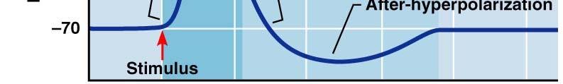
88 Phases of the Action Potential 1 resting state 2 depolarization phase 3 repolarization phase 4 hyperpolarization Figure 11.12


89 Absolute and Relative Refractory Periods Figure 11.15
90 Axonal Conduction Depolarization Threshold Axon Hillock Na ions rush in resulting in: Action potential; All or none phenomenon, high frequency Afterpotentials; hyperpolarizing, depolarizing; slow frequency Changes in membrane permeabilities Propagation Refractory yperiod
91 Postsynaptic Potentials Neurotransmitter receptors mediate changes in membrane potential according to: The amount of neurotransmitter released The amount of time the neurotransmitter is bound to receptors The two types of postsynaptic potentials are: EPSP excitatory ypostsynaptic p potentials IPSP inhibitory postsynaptic potentials order Spatial summation postsynaptic neuron is stimulated by a large number of Spatial summation postsynaptic neuron is stimulated by a large number of terminals at the same time IPSPs can also summate with EPSPs, canceling each other out
92 Excitatory Postsynaptic Potentials(EPSP) EPSPs are graded potentials that can initiate an action potential in an axon Use only chemically gated channels Na + and K + flow in opposite directions at the same time Postsynaptic membranes do not generate action potentials Inhibitory Synapses and IPSPs Neurotransmitter binding to a receptor at inhibitory synapses: ions Causes the membrane to become more permeable to potassium and chloride Leaves the charge on the inner surface negative Reduces the postsynaptic neuron s ability to produce an action potential
93 Summation A single EPSP cannot induce an action potential EPSPs must summate temporally or spatially to induce an action potential Temporal summation presynaptic neurons transmit impulses in rapidfire order Spatial summation postsynaptic neuron is stimulated by a large number of terminals at the same time IPSPs can also summate with EPSPs, canceling each other out
94 Synaptic Transmission Post-synaptic potentials (PSP's); Excitatory Inhibitory Interaction Summation/Integration i temporal spatial decremental conduction on dendrites and soma axon hillock is critical area at which threshold must be reached After release of neurotransmitter, reuptake degradation Functional Synaptic Units
95
 neuron, the membrane is polarized")

96 Signals Carried by Neurons In a resting (unstimulated) neuron, the membrane is polarized which means that the inner cytoplasmic side is negatively charged with respect to its outer, extracellular side
97 Signals Carried by Neurons When a neuron is stimulated the permeability of the plasma membrane changes at the site of the stimulus, allowing positive ions to rush in. As a result, the inner face of the membrane becomes less negative or depolarized
98 Signals Carried by Neurons Any part of the neuron depolarizes if stimulated, but at the axon alone this can result in the triggering of a nerve impulse or action potential
99 Signals Carried by Neurons When a nerve impulse or action potential develops the membrane is not only depolarized, but its polarity is completely reversed so it becomes negative externally and positive internally
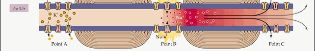
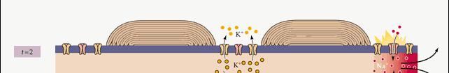
100 Signals Carried by Neurons Once begun, the nerve impulse travels rapidly down the entire length of the axon without decreasing in strength
101 Signals Carried by Neurons After the impulse has passed the membrane repolarizes itself
102 Graded Potential In humans, natural stimuli are not applied directly to axons, but to dendrites and the cell body which constitute the receptive zone of the neuron When the membrane of this receptive zone is stimulated it does not undergo a polarity reversal ersal Instead it undergoes a local depolarization in which the inner surface of the membrane merely becomes less negative
103 Graded Potential This local depolarization is called a graded potential which spreads from the receptive zone to the axon hillock (trigger zone) decreasing in strength as it travels If this depolarizing signal is strong enough when it reaches the initial segment of the axon, it acts as the trigger that initiates an action potential in the axon Signals from the receptive zone determine if the axon will fire an impulse
104 Synaptic Potential Most neurons in the body do not receive stimuli directly from the environment but are stimulated only by signals received at synapses from other neurons Synaptic input influences impulse generation through either excitatory or inhibitory synapses
105 Synaptic Potential In excitatory synapses, neurotransmitters released by presynaptic neurons alter the permeability of the postsysnaptic membrane to certain ions, this depolarizes the postsynapatic membrane and drives the postsynaptic neuron toward impulse generation
106 Synaptic Potential Inhibitory synapses cause the external surface of the postsynaptic membrane to become even more positive, thereby reducing the ability of the postsynaptic neuron to generate an action potential Thousands of excitatory and inhibitory synapses act on every neuron, competing to determine whether or not that neuron will generate an impulse
107 Neural Integration The organization of the nervous system is hierarchical The parts of the system must be integrated into a smoothly functioning whole Neuronal pools represent some of the basic patterns of communication with other parts of the nervous system
108 Neuronal Pools Neuronal pools are functional groups of neurons that process and integrate incoming information from other sources and transmit it forward One incoming presynaptic fiber synapses with Several different neurons in the pool. When Incoming fiber is excited it will excite some Postsynaptic neurons and facilitate others.
109 Neuronal Pools Neurons most likely to generate impulses are those most closely l associated with the incoming fiber because they receive the bulk of the synaptic contacts t These neurons are in the discharge zone Discharge Zone


110 Neuronal Pools Neurons farther away from the center are not excited to threshold by the incoming fiber, but are facilitated and can easily brought to threshold by stimuli from another source The periphery of the pool is the facilitated zone Facilitated zone
111 Neuronal Pools Note: The illustrations presented are a gross oversimplification of an actual neuron pool Most neuron pools consist of thousands of neurons and include inhibitory as well as excitatory neurons
Portions from Chapter 6 CHAPTER 7. The Nervous System: Neurons and Synapses. Chapter 7 Outline. and Supporting Cells
 CHAPTER 7 The Nervous System: Neurons and Synapses Chapter 7 Outline Neurons and Supporting Cells Activity in Axons The Synapse Acetylcholine as a Neurotransmitter Monoamines as Neurotransmitters Other
The Nervous System. Nervous System Functions 1. gather sensory input 2. integration- process and interpret sensory input 3. cause motor output
 The Nervous System Nervous System Functions 1. gather sensory input 2. integration- process and interpret sensory input 3. cause motor output The Nervous System 2 Parts of the Nervous System 1. central
NEURAL TISSUE (NEUROPHYSIOLOGY) PART I (A): NEURONS & NEUROGLIA
 PART I (A): NEURONS & NEUROGLIA") PART I (A): NEURONS & NEUROGLIA Neural Tissue Contains 2 kinds of cells: neurons: cells that send and receive signals neuroglia (glial cells): cells that support and protect neurons Neuron Types Sensory
PART I (A): NEURONS & NEUROGLIA Neural Tissue Contains 2 kinds of cells: neurons: cells that send and receive signals neuroglia (glial cells): cells that support and protect neurons Neuron Types Sensory
3) Most of the organelles in a neuron are located in the A) dendritic region. B) axon hillock. C) axon. D) cell body. E) axon terminals.
 Most of the organelles in a neuron are located in the A) dendritic region. B) axon hillock. C) axon. D) cell body. E) axon terminals.") Chapter 48 Neurons, Synapses, and Signaling Multiple-Choice Questions 1) A simple nervous system A) must include chemical senses, mechanoreception, and vision. B) includes a minimum of 12 ganglia. C) has
Chapter 48 Neurons, Synapses, and Signaling Multiple-Choice Questions 1) A simple nervous system A) must include chemical senses, mechanoreception, and vision. B) includes a minimum of 12 ganglia. C) has
Chapter 11: Nervous System and Nervous Tissue
 Chapter 11: Nervous System and Nervous Tissue I. Functions and divisions of the nervous system A. Sensory input: monitor changes in internal and external environment B. Integrations: make decisions about
Chapter 7. Objectives
 Chapter 7 The Nervous System: Structure and Control of Movement Objectives Discuss the general organization of the nervous system Describe the structure & function of a nerve Draw and label the pathways
Neurons. Pyramidal neurons in mouse cerebral cortex expressing green fluorescent protein. The red staining indicates GABAergic interneurons.
 Neurons Pyramidal neurons in mouse cerebral cortex expressing green fluorescent protein. The red staining indicates GABAergic interneurons. MBL, Woods Hole R Cheung MSc Bioelectronics: PGEE11106 1 Neuron
Chapter 7. The Nervous System: Structure and Control of Movement
 Chapter 7 The Nervous System: Structure and Control of Movement Objectives Discuss the general organization of the nervous system Describe the structure & function of a nerve Draw and label the pathways
Synaptic transmission
 Outline Synaptic transmission Sompol Tapechum M.D., Ph.D. Department of Physiology Faculty of Medicine Siriraj Hospital, Bangkok, Thailand. sisth@mahidol.ac.th 2 Structure of synapse Modes of synaptic
Chapter 11 Introduction to the Nervous System and Nervous Tissue Chapter Outline
 Chapter 11 Introduction to the Nervous System and Nervous Tissue Chapter Outline Module 11.1 Overview of the Nervous System (Figures 11.1-11.3) A. The nervous system controls our perception and experience
Thursday, January 22, Nerve impulse
 Nerve impulse Transmembrane Potential caused by ions moving through cell membrane at different rates Two main ions of concern Na + - Sodium K + - potassium Cell membrane not freely permeable therefore
Outline. Neuron Structure. Week 4 - Nervous System. The Nervous System: Neurons and Synapses
 Outline Week 4 - The Nervous System: Neurons and Synapses Neurons Neuron structures Types of neurons Electrical activity of neurons Depolarization, repolarization, hyperpolarization Synapses Release of
The Nervous System -The master controlling and communicating system of the body
 The Nervous System -The master controlling and communicating system of the body Functions: -Sensory input -Integration -Motor output Organization of the Nervous System Central nervous system (CNS) -Brain
Chapter 11: Functional Organization of Nervous Tissue
 Chapter 11: Functional Organization of Nervous Tissue I. Functions of the Nervous System A. List and describe the five major nervous system functions: 1. 2. 3. 4. 5. II. Divisions of the Nervous System
BIOLOGY 2050 LECTURE NOTES ANATOMY & PHYSIOLOGY I (A. IMHOLTZ) FUNDAMENTALS OF THE NERVOUS SYSTEM AND NERVOUS TISSUE P1 OF 5
 FUNDAMENTALS OF THE NERVOUS SYSTEM AND NERVOUS TISSUE P1 OF 5") P1 OF 5 The nervous system controls/coordinates the activities of cells, tissues, & organs. The endocrine system also plays a role in control/coordination. The nervous system is more dominant. Its mechanisms
P1 OF 5 The nervous system controls/coordinates the activities of cells, tissues, & organs. The endocrine system also plays a role in control/coordination. The nervous system is more dominant. Its mechanisms
Nervous Tissue and Neurophysiology
 Nervous Tissue and Neurophysiology Objectives Describe the two major divisions of the nervous system and their characteristics. Identify the structures/functions of a typical neuron. Describe the location
Introduction to Neurobiology
 Biology 240 General Zoology Introduction to Neurobiology Nervous System functions: communication of information via nerve signals integration and processing of information control of physiological and
NERVOUS SYSTEM 1 CHAPTER 10 BIO 211: ANATOMY & PHYSIOLOGY I
 BIO 211: ANATOMY & PHYSIOLOGY I 1 Ch 10 A Ch 10 B This set CHAPTER 10 NERVOUS SYSTEM 1 BASIC STRUCTURE and FUNCTION Dr. Lawrence G. Altman www.lawrencegaltman.com Some illustrations are courtesy of McGraw-Hill.
1. Name the two major divisions of the nervous system and list the organs within each. Central Nervous System Peripheral Nervous System
 CHAPTER 10: NERVOUS SYSTEM I OBJECTIVES 1. Name the two major divisions of the nervous system and list the organs within each. Central Nervous System Peripheral Nervous System Brain Spinal Cord Cranial
Neurons Chapter 7 2/19/2016. Learning Objectives. Cells of the Nervous System. Cells of the Nervous System. Cells of the Nervous System
 Learning Objectives Neurons Chapter 7 Identify and describe the functions of the two main divisions of the nervous system. Differentiate between a neuron and neuroglial cells in terms of structure and
Nervous System. Master controlling and communicating system of the body. Secrete chemicals called neurotransmitters
 Nervous System Master controlling and communicating system of the body Interacts with the endocrine system to control and coordinate the body s responses to changes in its environment, as well as growth,
Chapter 4 Neuronal Physiology
 Chapter 4 Neuronal Physiology V edit. Pg. 99-131 VI edit. Pg. 85-113 VII edit. Pg. 87-113 Input Zone Dendrites and Cell body Nucleus Trigger Zone Axon hillock Conducting Zone Axon (may be from 1mm to more
9/28/2016. Neuron. Multipolar Neuron. Astrocytes Exchange Materials With Neurons. Glia or Glial Cells ( supporting cells of the nervous system)
") Neuron Multipolar Neuron https://www.youtube.com/watch?v=lw-psbnu5xago to :38 Glia or Glial Cells ( supporting cells of the nervous system) 10X more numerous than neurons but one-tenth the size make up
Neuron Multipolar Neuron https://www.youtube.com/watch?v=lw-psbnu5xago to :38 Glia or Glial Cells ( supporting cells of the nervous system) 10X more numerous than neurons but one-tenth the size make up
MOLECULAR AND CELLULAR NEUROSCIENCE
 MOLECULAR AND CELLULAR NEUROSCIENCE BMP-218 November 4, 2014 DIVISIONS OF THE NERVOUS SYSTEM The nervous system is composed of two primary divisions: 1. CNS - Central Nervous System (Brain + Spinal Cord)
Chapter 2: Cellular Mechanisms and Cognition
 Chapter 2: Cellular Mechanisms and Cognition MULTIPLE CHOICE 1. Two principles about neurons were defined by Ramón y Cajal. The principle of connectional specificity states that, whereas the principle
Neurotransmitter Systems III Neurochemistry. Reading: BCP Chapter 6
 Neurotransmitter Systems III Neurochemistry Reading: BCP Chapter 6 Neurotransmitter Systems Normal function of the human brain requires an orderly set of chemical reactions. Some of the most important
Communication within a Neuron
 Neuronal Communication, Ph.D. Communication within a Neuron Measuring Electrical Potentials of Axons The Membrane Potential The Action Potential Conduction of the Action Potential 1 The withdrawal reflex
BIPN100 F15 Human Physiology 1 Lecture 3. Synaptic Transmission p. 1
 BIPN100 F15 Human Physiology 1 Lecture 3. Synaptic Transmission p. 1 Terms you should know: synapse, neuromuscular junction (NMJ), pre-synaptic, post-synaptic, synaptic cleft, acetylcholine (ACh), acetylcholine
Chapter 12 Nervous Tissue. Copyright 2009 John Wiley & Sons, Inc. 1
 Chapter 12 Nervous Tissue Copyright 2009 John Wiley & Sons, Inc. 1 Terms to Know CNS PNS Afferent division Efferent division Somatic nervous system Autonomic nervous system Sympathetic nervous system Parasympathetic
Communication Between
 Communication Between Neurons Bởi: OpenStaxCollege The electrical changes taking place within a neuron, as described in the previous section, are similar to a light switch being turned on. A stimulus starts
Nervous System. Chapter 9 Pages
 Nervous System Chapter 9 Pages 211-257 Chapter 9 Wordbytes 1. af- = toward 11. -ferrent = carried 2. arachn- = spider 12. gangli- = swelling 3. astro- = star 13. -glia = glue 4. auto- = self 14. mening-
Neuron types and Neurotransmitters
 Neuron types and Neurotransmitters Faisal I. Mohammed. PhD, MD University of Jordan 1 Transmission of Receptor Information to the Brain the larger the nerve fiber diameter the faster the rate of transmission
Omar Ismail. Dana Almanzalji. Faisal Mohammad
 11 Omar Ismail Dana Almanzalji Faisal Mohammad Neuronal classification: Neurons are responsible for transmitting the action potential to the brain. The speed at which the action potential is transmitted
NEUROCHEMISTRY Brief Review
 NEUROCHEMISTRY Brief Review UNIVERSITY OF PNG SCHOOL OF MEDICINE AND HEALTH SCIENCES DISCIPLINE OF BIOCHEMISTRY AND MOLECULAR BIOLOGY PBL MBBS YEAR V SEMINAR VJ Temple 1 Membrane potential Membrane potential:
Neural Communication. Central Nervous System Peripheral Nervous System. Communication in the Nervous System. 4 Common Components of a Neuron
 Neural Communication Overview of CNS / PNS Electrical Signaling Chemical Signaling Central Nervous System Peripheral Nervous System Somatic = sensory & motor Autonomic = arousal state Parasympathetic =
Neurons, Synapses and Signaling. Chapter 48
 Neurons, Synapses and Signaling Chapter 48 Warm Up Exercise What types of cells can receive a nerve signal? Nervous Organization Neurons- nerve cells. Brain- organized into clusters of neurons, called
Organization of the nervous system. [See Fig. 48.1]
![Organization of the nervous system. [See Fig. 48.1]](/thumbs/90/103926552.jpg "Organization of the nervous system. [See Fig. 48.1]") Nervous System [Note: This is the text version of this lecture file. To make the lecture notes downloadable over a slow connection (e.g. modem) the figures have been replaced with figure numbers as found
Nervous System [Note: This is the text version of this lecture file. To make the lecture notes downloadable over a slow connection (e.g. modem) the figures have been replaced with figure numbers as found
Study Guide Answer Key Nervous System
 Biology 12 Human Biology Textbook: BC Biology 12 Study Guide Answer Key Nervous System 1. Draw a neuron, label 3 parts and give the function of those parts. Dendrite: carry signals to the cell body Cell
Introduction to Physiological Psychology
 Introduction to Physiological Psychology Review Kim Sweeney ksweeney@cogsci.ucsd.edu www.cogsci.ucsd.edu/~ksweeney/psy260.html Today n Discuss Final Paper Proposal (due 3/10) n General Review 1 The article
Notes are online at The Neuron
 Notes are online at http://cogsci.ucsd.edu/~clovett/neuronotescogs17.pdf A. What is a neuron? The Neuron 1. A neuron is a type of cell that receives and transmits information in the Central Nervous System
What is Anatomy and Physiology?
 Introduction BI 212 BI 213 BI 211 Ecosystems Organs / organ systems Cells Organelles Communities Tissues Molecules Populations Organisms Campbell et al. Figure 1.4 Introduction What is Anatomy and Physiology?
10.1: Introduction. Cell types in neural tissue: Neurons Neuroglial cells (also known as neuroglia, glia, and glial cells) Dendrites.
 Dendrites.") 10.1: Introduction Copyright The McGraw-Hill Companies, Inc. Permission required for reproduction or display. Cell types in neural tissue: Neurons Neuroglial cells (also known as neuroglia, glia, and glial
10.1: Introduction Copyright The McGraw-Hill Companies, Inc. Permission required for reproduction or display. Cell types in neural tissue: Neurons Neuroglial cells (also known as neuroglia, glia, and glial
The Nervous System Mark Stanford, Ph.D.
 The Nervous System Functional Neuroanatomy and How Neurons Communicate Mark Stanford, Ph.D. Santa Clara Valley Health & Hospital System Addiction Medicine and Therapy Services The Nervous System In response
5-Nervous system II: Physiology of Neurons
 5-Nervous system II: Physiology of Neurons AXON ION GRADIENTS ACTION POTENTIAL (axon conduction) GRADED POTENTIAL (cell-cell communication at synapse) SYNAPSE STRUCTURE & FUNCTION NEURAL INTEGRATION CNS
LECTURE STRUCTURE ASC171 NERVOUS SYSTEM PART 1: BACKGROUND 26/07/2015. Module 5
 LECTURE STRUCTURE PART 1: Background / Introduction PART 2: Structure of the NS, how it operates PART 3: CNS PART 4: PNS Why did the action potential cross the synaptic junction? To get to the other side
Neurons, Synapses, and Signaling
 Chapter 8 Neurons, Synapses, and Signaling PowerPoint Lectures for Biology, Eighth Edition Overview: Lines of Communication The cone snail kills prey with venom that disables neurons Neurons are nerve
Neurophysiology scripts. Slide 2
 Neurophysiology scripts Slide 2 Nervous system and Endocrine system both maintain homeostasis in the body. Nervous system by nerve impulse and Endocrine system by hormones. Since the nerve impulse is an
Section: Chapter 5: Multiple Choice. 1. The structure of synapses is best viewed with a(n):
:") Section: Chapter 5: Multiple Choice 1. The structure of synapses is best viewed with a(n): p.155 electron microscope. light microscope. confocal microscope. nissle-stained microscopic procedure. 2. Electron
Section: Chapter 5: Multiple Choice 1. The structure of synapses is best viewed with a(n): p.155 electron microscope. light microscope. confocal microscope. nissle-stained microscopic procedure. 2. Electron
NEURONS COMMUNICATE WITH OTHER CELLS AT SYNAPSES 34.3
 NEURONS COMMUNICATE WITH OTHER CELLS AT SYNAPSES 34.3 NEURONS COMMUNICATE WITH OTHER CELLS AT SYNAPSES Neurons communicate with other neurons or target cells at synapses. Chemical synapse: a very narrow
Physiology of synapses and receptors
 Physiology of synapses and receptors Dr Syed Shahid Habib Professor & Consultant Clinical Neurophysiology Dept. of Physiology College of Medicine & KKUH King Saud University REMEMBER These handouts will
Synapses. Objectives. Synaptic Relationships Between Neurons. Structure of a Chemical Synapse. Structure of a Chemical Synapse
 bjectives Synapses s Temporal & Spatial Summation EPSP & IPSP Coding Memory Synapses a nerve signal AP travels to the end of the axon triggers the release of a neurotransmitter stimulates a new wave of
Chapter 2. The Cellular and Molecular Basis of Cognition Cognitive Neuroscience: The Biology of the Mind, 2 nd Ed.,
 Chapter 2. The Cellular and Molecular Basis of Cognition Cognitive Neuroscience: The Biology of the Mind, 2 nd Ed., M. S. Gazzaniga, R. B. Ivry, and G. R. Mangun, Norton, 2002. Summarized by B.-W. Ku,
Division Ave. High School AP Biology. cell body. signal direction
 signal direction Nervous system cells Neuron a nerve cell dendrites myelin sheath axon cell body dendrite cell body axon Structure fits function many entry points for signal one path out transmits signal
1. What are the two basic types of cells in the nervous system? Neurons and Glial Cells
 Biological Psychology Basic Structure of a Neuron 1. What are the two basic types of cells in the nervous system? Neurons and Glial Cells a. Cells that process incoming signals and respond by sending out
Neurons, Synapses, and Signaling
 Overview: Lines of Communication Chapter 8 Neurons, Synapses, and Signaling Fig. 8- The cone snail kills prey with venom that disables neurons Neurons are nerve s that transfer information within the body
Na + K + pump. The beauty of the Na + K + pump. Cotransport. The setup Cotransport the result. Found along the plasma membrane of all cells.
 The beauty of the Na + K + pump Na + K + pump Found along the plasma membrane of all cells. Establishes gradients, controls osmotic effects, allows for cotransport Nerve cells have a Na + K + pump and
Summarized by B.-W. Ku, E. S. Lee, and B.-T. Zhang Biointelligence Laboratory, Seoul National University.
 Chapter 2. The Cellular l and Molecular Basis of Cognition Cognitive Neuroscience: The Biology of the Mind, 3 rd Ed., M. S. Gazzaniga, R. B. Ivry, and G. R. Mangun, Norton, 2008. Summarized by B.-W. Ku,
Nervous System. Nervous system cells. Transmission of a signal 2/27/2015. Neuron
 Nervous System 2007-2008 signal direction Neuron a nerve cell Nervous system cells dendrites axon cell body Structure fits function many entry points for signal one path out transmits signal signal direction
Mohammad Tarek. Wahab Al-tekreeti Tamer Barakat. Faisal Mohammad
 15 Mohammad Tarek Wahab Al-tekreeti Tamer Barakat Faisal Mohammad Things to remember Types of synapse: Neuron types and neurotransmitters When it happens between an axon and dendrites it is called axodendritic
Biopsychology 2012 sec 003 (Dr. Campeau)
") Biopsychology 2012 sec 003 (Dr. Campeau) Study Guide for First Midterm What are some fun facts about the human brain? - there are approximately 100 billion neurons in the brain; - each neuron makes between
Biopsychology 2012 sec 003 (Dr. Campeau) Study Guide for First Midterm What are some fun facts about the human brain? - there are approximately 100 billion neurons in the brain; - each neuron makes between
Axon Nerve impulse. Axoplasm Receptor. Axomembrane Stimuli. Schwann cell Effector. Myelin Cell body
 Nervous System Review 1. Explain a reflex arc. 2. Know the structure, function and location of a sensory neuron, interneuron, and motor neuron 3. What is (a) Neuron Axon Nerve impulse Axoplasm Receptor
Ch. 45 Continues (Have You Read Ch. 45 yet?) u Central Nervous System Synapses - Synaptic functions of neurons - Information transmission via nerve
 u Central Nervous System Synapses - Synaptic functions of neurons - Information transmission via nerve") Ch. 45 Continues (Have You Read Ch. 45 yet?) u Central Nervous System Synapses - Synaptic functions of neurons - Information transmission via nerve impulses - Impulse may be blocked in its transmission
Ch. 45 Continues (Have You Read Ch. 45 yet?) u Central Nervous System Synapses - Synaptic functions of neurons - Information transmission via nerve impulses - Impulse may be blocked in its transmission
Ameen Alsaras. Ameen Alsaras. Mohd.Khatatbeh
 9 Ameen Alsaras Ameen Alsaras Mohd.Khatatbeh Nerve Cells (Neurons) *Remember: The neural cell consists of: 1-Cell body 2-Dendrites 3-Axon which ends as axon terminals. The conduction of impulse through
STRUCTURAL ELEMENTS OF THE NERVOUS SYSTEM
 STRUCTURAL ELEMENTS OF THE NERVOUS SYSTEM STRUCTURE AND MAINTENANCE OF NEURONS (a) (b) Dendrites Cell body Initial segment collateral terminals (a) Diagrammatic representation of a neuron. The break in
BIPN140 Lecture 8: Synaptic Transmission II
 BIPN140 Lecture 8: Synaptic Transmission II 1. Postsynaptic Receptors: Metabotropic & Ionotropic 2. Postsynaptic Responses (Postsynaptic Potentials, PSPs) 3. Neurotransmitters Su (FA16) Chemical Synapse:
Chapter 48-49: The Nervous System & Neurons
 Invertebrates Chapter 48-49: The Nervous System & Neurons Radial Symmetry - Nerve net Cnideria Bilateral Symmetry double, ventral, solid nerve cord brain (cephalization) Vertebrates Dorsal, single, hollow,
Chapter 2. The Cellular and Molecular Basis of Cognition
 Chapter 2. The Cellular and Molecular Basis of Cognition Cognitive Neuroscience: The Biology of the Mind, 2 nd Ed., M. S. Gazzaniga,, R. B. Ivry,, and G. R. Mangun,, Norton, 2002. Summarized by B.-W. Ku,
2/27/2019. Functions of the Nervous System. Nervous Tissue and Neuron Function. Fundamentals Of The Nervous System And Nervous Tissue
 Nervous Tissue and Neuron Function Fundamentals Of The Nervous System And Nervous Tissue Learn and Understand 1. Like muscle cells, neurons use membrane polarity upset (AP) as a signal therefore keeping
Chapter 7 Nerve Cells and Electrical Signaling
 Chapter 7 Nerve Cells and Electrical Signaling 7.1. Overview of the Nervous System (Figure 7.1) 7.2. Cells of the Nervous System o Neurons are excitable cells which can generate action potentials o 90%
The Nervous System. Chapter 4. Neuron 3/9/ Components of the Nervous System
 Chapter 4 The Nervous System 1. Components of the Nervous System a. Nerve cells (neurons) Analyze and transmit information Over 100 billion neurons in system Four defined regions Cell body Dendrites Axon
EE 791 Lecture 2 Jan 19, 2015
 EE 791 Lecture 2 Jan 19, 2015 Action Potential Conduction And Neural Organization EE 791-Lecture 2 1 Core-conductor model: In the core-conductor model we approximate an axon or a segment of a dendrite
Nervous System Dr. Naim Kittana Department of Biomedical Sciences Faculty of Medicine & Health Sciences An-Najah National University
 Nervous System Department of Biomedical Sciences Faculty of Medicine & Health Sciences An-Najah National University Declaration The content and the figures of this seminar were directly adopted from the
Fundamentals of the Nervous System and Nervous Tissue: Part C
 PowerPoint Lecture Slides prepared by Janice Meeking, Mount Royal College C H A P T E R 11 Fundamentals of the Nervous System and Nervous Tissue: Part C Warm Up What is a neurotransmitter? What is the
QUIZ YOURSELF COLOSSAL NEURON ACTIVITY
 QUIZ YOURSELF What are the factors that produce the resting potential? How is an action potential initiated and what is the subsequent flow of ions during the action potential? 1 COLOSSAL NEURON ACTIVITY
ANATOMY AND PHYSIOLOGY OF NEURONS. AP Biology Chapter 48
 ANATOMY AND PHYSIOLOGY OF NEURONS AP Biology Chapter 48 Objectives Describe the different types of neurons Describe the structure and function of dendrites, axons, a synapse, types of ion channels, and
Dania Ahmad. Tamer Barakat + Dania Ahmad. Faisal I. Mohammed
 16 Dania Ahmad Tamer Barakat + Dania Ahmad Faisal I. Mohammed Revision: What are the basic types of neurons? sensory (afferent), motor (efferent) and interneuron (equaled association neurons). We classified
The Nervous System AP Biology
 The Nervous System 2005-2006 Neuron (nerve cell) signal direction dendrites cell body Structure fits function, it have many entry points for signal one path out transmits signal Nodes of Ranvier axon signal
Nervous System. Nervous System. Organization of the Nervous System. Histology of Nerve Tissue. Motor Division: Two Main Parts
 Nervous System The master controlling and communicating system of the body Functions: Sensory input monitoring stimuli occurring inside and outs1ide the body Nervous System Integration interpretation of
Biology 201-Worksheet on Nervous System (Answers are in your power point outlines-there is no key!)
") Bio 201 Tissues and Skin 1 March 21, 2011 Biology 201-Worksheet on Nervous System (Answers are in your power point outlines-there is no key!) 1. The study of the normal functioning and disorders of the
Bio 201 Tissues and Skin 1 March 21, 2011 Biology 201-Worksheet on Nervous System (Answers are in your power point outlines-there is no key!) 1. The study of the normal functioning and disorders of the
Neurotransmitters. Chemical transmission of a nerve signal by neurotransmitters at a synapse
 Neurotransmitters A chemical released by one neuron that affects another neuron or an effector organ (e.g., muscle, gland, blood vessel). Neurotransmitters are small molecules that serve as messengers
Branches of the Nervous System
 The Nervous System Branches of the Nervous System There are 2 main branches of the nervous system Central Nervous System Brain Spinal Cord Peripheral Nervous System All nerves leading to rest of body Anatomy
Chapter 2 The Brain or Bio Psychology
 Chapter 2 The Brain or Bio Psychology 1 2 3 1 Glial Cells Surround neurons and hold them in place Make Myelin (covering for neurons) Manufacture nutrient chemicals neurons need Absorb toxins and waste
Chapter 12 Nervous Tissue
 9/12/11 Chapter 12 Nervous Tissue Overview of the nervous system Cells of the nervous system Electrophysiology of neurons Synapses Neural integration Subdivisions of the Nervous System 1 Subdivisions of
Lecture 22: A little Neurobiology
 BIO 5099: Molecular Biology for Computer Scientists (et al) Lecture 22: A little Neurobiology http://compbio.uchsc.edu/hunter/bio5099 Larry.Hunter@uchsc.edu Nervous system development Part of the ectoderm
Autonomic Nervous System. Lanny Shulman, O.D., Ph.D. University of Houston College of Optometry
 Autonomic Nervous System Lanny Shulman, O.D., Ph.D. University of Houston College of Optometry Peripheral Nervous System A. Sensory Somatic Nervous System B. Autonomic Nervous System 1. Sympathetic Nervous
Cell communication. Gated ion channels. Allow specific ions to pass only when gates are open
 increase decrease Cell communication Gated ion channels Allow specific ions to pass only when gates are open Triggered by: potential change, chemical binding, temperature change, stretching 1 Voltage-Gated
Cell communication. Gated ion channels. Voltage-Gated Na + Channel. Allow specific ions to pass only when gates are open
 increase decrease Cell communication Gated ion channels Allow specific ions to pass only when gates are open Voltage-Gated Na + Channel Activation gate ECF Triggered by: change, chemical binding, temperature
THE NERVOUS SYSTEM. Homeostasis Strand
 THE NERVOUS SYSTEM Homeostasis Strand Introduction In general, a nervous system has three overlapping functions : 1. Sensory input conduction of signals from sensory receptors to integration centres 2.
Neurotransmitter Systems I Identification and Distribution. Reading: BCP Chapter 6
 Neurotransmitter Systems I Identification and Distribution Reading: BCP Chapter 6 Neurotransmitter Systems Normal function of the human brain requires an orderly set of chemical reactions. Some of the
Concept 48.1 Neuron organization and structure reflect function in information transfer
 Name Chapter 48: Neurons, Synapses, and Signaling Period Chapter 48: Neurons, Synapses, and Signaling Concept 48.1 Neuron organization and structure reflect function in information transfer 1. What is
Chapter 17. Nervous System Nervous systems receive sensory input, interpret it, and send out appropriate commands. !
 Chapter 17 Sensory receptor Sensory input Integration Nervous System Motor output Brain and spinal cord Effector cells Peripheral nervous system (PNS) Central nervous system (CNS) 28.1 Nervous systems
Biol 219 Lec 12 Fall 2016
 Cell-to-Cell: Neurons Communicate at Synapses Electrical synapses pass electrical signals through gap junctions Signal can be bi-directional Synchronizes the activity of a network of cells Primarily in
NERVOUS SYSTEM. Somatic (SNS) Fibers - transmit impulses from CNS to control voluntary action of skeletal muscle
 Fibers - transmit impulses from CNS to control voluntary action of skeletal muscle") NERVOUS SYSTEM The master controlling and communicating system of the body --- cells communicate via electrical and chemical signals. Signals are rapid, specific and cause almost immediate responses. Functions
NERVOUS SYSTEM The master controlling and communicating system of the body --- cells communicate via electrical and chemical signals. Signals are rapid, specific and cause almost immediate responses. Functions
Neurophysiology. Corresponding textbook pages: ,
 Neurophysiology Corresponding textbook pages: 436-440, 442-455 Organization Helps maintain homeostasis in the body Nervous system and endocrine system Nervous system is faster due to nerve impulses 1 Fig.
Unit Three. I. General Functions of the Nervous System. I. General Functions of the Nervous System
 10 Refer to the following URLs. It is a good idea to print them and bring them to class. Be sure to study these along with your book. http://www.sirinet.net/~jgjohnso/nervous.html http://faculty.washington.edu/chudler/ap.html
 PSY 302 Lecture 6: The Neurotransmitters (continued) September 12, 2017 Notes by: Desiree Acetylcholine (ACh) CoA + Acetate Acetyl-CoA (mitochondria) (food, vinegar) + Choline ChAT CoA + ACh (lipids, foods)
Action Potentials and Synaptic Transmission. BIO 219 Napa Valley College Dr. Adam Ross
 Action Potentials and Synaptic Transmission BIO 219 Napa Valley College Dr. Adam Ross Review of action potentials Nodes of Ranvier Nucleus Dendrites Cell body In saltatory conduction, the nerve impulses
What effect would an AChE inhibitor have at the neuromuscular junction?
 CASE 4 A 32-year-old woman presents to her primary care physician s office with difficulty chewing food. She states that when she eats certain foods that require a significant amount of chewing (meat),
Learning expectations for BIOL 131. Chapters 11, Nervous System Overview Read Chapter 11. You should be able to:
 NOTE The quiz will have question ONLY from the material we get through on Tuesday. The first midterm will cover all material from day one until the lecture before the second midterm. Learning expectations
COGS 269. Lecture 1 Spring 2018
 COGS 269 Lecture 1 Spring 2018 Psychological Experience Methods of Cognitive Neuroscience Dissociation experiments (patients with brain damage) Neuroimaging experiments Computational modeling Brain damage