TITLE: Pharmacological and Behavioral Enhancement of Neuroplasticity in the MPTP- Lesioned Mouse and Nonhuman Primate.
|
|
|
- Vivian Simpson
- 5 years ago
- Views:
Transcription
1 AD Award Number: W81XWH TITLE: Pharmacological and Behavioral Enhancement of Neuroplasticity in the MPTP- Lesioned Mouse and Nonhuman Primate. PRINCIPAL INVESTIGATOR: Giselle M. Petzinger, M.D. CONTRACTING ORGANIZATION: University of Southern California Los Angeles, CA, REPORT DATE: May 2006 TYPE OF REPORT: Annual PREPARED FOR: U.S. Army Medical Research and Materiel Command Fort Detrick, Maryland DISTRIBUTION STATEMENT: Approved for Public Release; Distribution Unlimited The views, opinions and/or findings contained in this report are those of the author(s) and should not be construed as an official Department of the Army position, policy or decision unless so designated by other documentation.
2 REPORT DOCUMENTATION PAGE Form Approved OMB No Public reporting burden for this collection of information is estimated to average 1 hour per response, including the time for reviewing instructions, searching existing data sources, gathering and maintaining the data needed, and completing and reviewing this collection of information. Send comments regarding this burden estimate or any other aspect of this collection of information, including suggestions for reducing this burden to Department of Defense, Washington Headquarters Services, Directorate for Information Operations and Reports ( ), 1215 Jefferson Davis Highway, Suite 1204, Arlington, VA Respondents should be aware that notwithstanding any other provision of law, no person shall be subject to any penalty for failing to comply with a collection of information if it does not display a currently valid OMB control number. PLEASE DO NOT RETURN YOUR FORM TO THE ABOVE ADDRESS. 1. REPORT DATE (DD-MM-YYYY) REPORT TYPE Annual 3. DATES COVERED (From - To) 1 May Apr TITLE AND SUBTITLE 5a. CONTRACT NUMBER Pharmacological and Behavioral Enhancement of Neuroplasticity in the MPTP- Lesioned Mouse and Nonhuman Primate. 5b. GRANT NUMBER W81XWH c. PROGRAM ELEMENT NUMBER 6. AUTHOR(S) Giselle M. Petzinger, M.D. gpetzinger@surgery.usc.edu 5d. PROJECT NUMBER 5e. TASK NUMBER 5f. WORK UNIT NUMBER 7. PERFORMING ORGANIZATION NAME(S) AND ADDRESS(ES) 8. PERFORMING ORGANIZATION REPORT NUMBER University of Southern California Los Angeles, CA, SPONSORING / MONITORING AGENCY NAME(S) AND ADDRESS(ES) 10. SPONSOR/MONITOR S ACRONYM(S) U.S. Army Medical Research and Materiel Command Fort Detrick, Maryland SPONSOR/MONITOR S REPORT NUMBER(S) 12. DISTRIBUTION / AVAILABILITY STATEMENT Approved for Public Release; Distribution Unlimited 13. SUPPLEMENTARY NOTES 14. ABSTRACT: The purpose of this proposal is to investigate the mechanisms involving pharmacological and behavioral enhanced neuroplasticity of the injured basal ganglia. Our central hypothesis is that exercise and pharmacological intervention, specifically the administration of a D2 dopamine-receptor agonist, enhances neuroplasticity by modulating glutamatedopamine interactions. This proposal has two components. Using the MPTP C57BL/6 mouse Component One will test the hypothesis that exercise enhances plasticity of the MPTP-injured basal ganglia through glutamate by modulating dopamine biosynthesis. This hypothesis will be tested through changes in dopamine, and proteins involved in dopamine biosynthesis and uptake (tyrosine hydroxylase and dopamine transporter) and changes in glutamatergic synapses and receptor subtype. This hypothesis will be tested through determining whether exercise-enhanced neuroplasticity may be attenuated with a glutamate antagonist. Using the MPTP-lesioned non-human primate Component Two will test the hypothesis that the D2 receptor agonist (Pramipexole) enhances neuroplasticity of the MPTP-injured basal ganglia through its effect on pre- and postsynaptic dopamine biosynthesis, uptake and receptor expression as well as glutamatergic synapses. This hypothesis will be tested through changes in dopamine and its metabolites, proteins involved in dopamine biosynthesis, uptake, and storage (tyrosine hydroxylase, dopamine transporter, and vesicular monoamine transporter), changes in dopamine receptor subtypes and their respective neuropeptides, and changes in glutamatergic synapses. 15. SUBJECT TERMS MPTP, neuroplasticity, models of injury, repair, basal ganglia 16. SECURITY CLASSIFICATION OF: 17. LIMITATION OF ABSTRACT a. REPORT U b. ABSTRACT U 18. NUMBER OF PAGES c. THIS PAGE U UU 60 19a. NAME OF RESPONSIBLE PERSON USAMRMC 19b. TELEPHONE NUMBER (include area code) Standard Form 298 (Rev. 8-98) Prescribed by ANSI Std. Z39.18
3 TABLE OF CONTENTS Cover... SF Introduction... 4 Body... 4 Key Research Accomplishments...9 Reportable Outcomes Conclusions Appendices
4 Introduction The primary focus of this research proposal is to determine the underlying mechanisms responsible for neuroplasticity in the injured adult basal ganglia. For these studies we utilize the neurotoxicant MPTP that selectively destroys nigrostriatal dopaminergic neurons and leads to the depletion of striatal dopamine as well as the development of parkinsonian features. In the squirrel monkey these features include slowness of movement, balance impairment and diminished hand dexterity. In our laboratory we utilize both the MPTP-lesioned C57BL6 mouse and the MPTPlesioned squirrel monkey. Both models show intrinsic plasticity through either striatal dopamine return (mouse) and/or behavioral recovery (squirrel monkey). In this proposal we were particularly interested in understanding whether exercise (mouse) or dopamine replacement therapy (monkey) might enhance intrinsic neuroplasticity of the injured basal ganglia. For this purpose, the proposal was divided into two components, a mouse exercise study and a squirrel monkey dopamine replacement study. These studies were designed to be complementary in that both nonpharmacological and pharmacological effects of neuroplasticity are being investigated. In the following sections are included the abstract, introduction and specific aims from the original proposal. This is followed by the accomplishments and research outcomes from year one. This annual report also includes manuscripts in the form of appendices. Abstract (From the Original Application) The purpose of this proposal is to investigate the molecular mechanisms involving pharmacological and behavioral (exercise) enhanced neuroplasticity of the injured basal ganglia. Our central hypothesis is that exercise and pharmacological intervention, specifically the administration of a D2 dopaminereceptor agonist, enhances neuroplasticity by modulating glutamate-dopamine interactions. The following proposal has two complementary components using two animal models to address the molecular mechanisms underlying exercise- and pharmacologically-enhanced neuroplasticity. Using the MPTP C57BL/6 mouse Component One will test the hypothesis that exercise enhances plasticity of the MPTP-injured basal ganglia through glutamate by modulating dopamine biosynthesis. This hypothesis will be tested through changes in dopamine, and proteins involved in dopamine biosynthesis and uptake (tyrosine hydroxylase and dopamine transporter) and changes in glutamatergic synapses and receptor subtype. This hypothesis will be further tested through determining whether exercise-enhanced neuroplasticity may be attenuated with a glutamate antagonist. Using the MPTPlesioned non-human primate Component Two will test the hypothesis that the administration of a D2 receptor agonist (Pramipexole) enhances neuroplasticity of the MPTP-injured basal ganglia through its effect on pre- and post- synaptic dopamine biosynthesis, uptake and receptor expression as well as glutamatergic synapses. This hypothesis will be tested through changes in dopamine and its metabolites, proteins involved in dopamine biosynthesis, uptake, and storage (tyrosine hydroxylase, dopamine transporter, and vesicular monoamine transporter), changes in dopamine receptor subtypes and their respective neuropeptides, and changes in glutamatergic synapses. By elucidating the role of exercise and pharmacological manipulation in neuroplasticity of the injured brain we hope to identify novel therapeutic targets for the treatment of brain injury and neurotoxic insult. Since military personnel are at risk for a wide range of brain injuries including head trauma, neurotoxic exposure (from pesticides, hostile enemy poisoning, viral and biological weapon based agents) it is imperative that medical strategies be made available to reverse the debilitating neurological deficits. 4
5 D: STATEMENT OF WORK From the original Application The brain s capacity for recovery from damage is far greater than previously recognized. It is now understood that neuroplasticity can be modulated through activity-dependent processes including exercise and environmental enrichment, and through pharmacological manipulation. Most of our understanding of exercise and pharmacological enhanced neuroplasticity is derived from studies in the cortex and the hippocampus, but there is mounting evidence that the same phenomenon occurs in the injured basal ganglia. The molecular mechanisms for this phenomenon are not well understood. Using two animal models of injury induced neuroplasticity in the basal ganglia (the MPTP-lesioned mouse and MPTP-lesioned non-human primate) we propose to examine two modes of intervention to enhance neuroplasticity. These include exercise in the MPTP-lesioned mouse model and pharmacological intervention in the MPTP-lesioned non-human primate. Our central hypothesis is that exercise and pharmacological intervention, specifically the administration of a D2 dopamine-receptor agonist, enhances neuroplasticity by modulating glutamate-dopamine interactions. The following proposal has two complementary components using both animal models to address the molecular mechanisms underlying exercise- and pharmacologically-enhanced neuroplasticity. Using the MPTP C57BL/6 mouse Component One will test the hypothesis that exercise enhances plasticity of the MPTP-injured basal ganglia through glutamate by modulating dopamine biosynthesis. This hypothesis will be tested through changes in dopamine, and proteins involved in dopamine biosynthesis and uptake (tyrosine hydroxylase and dopamine transporter) and changes in glutamatergic synapses and receptor subtype. This hypothesis will be further tested through determining whether exercise-enhanced neuroplasticity may be attenuated with a glutamate antagonist. Using the MPTP-lesioned non-human primate Component Two will test the hypothesis that the administration of a D2 receptor agonist (pramipexole) enhances neuroplasticity of the MPTP-injured basal ganglia through its effect on pre- and post- synaptic dopamine biosynthesis, uptake and receptor expression as well as glutamatergic synapses. This hypothesis will be tested through changes in dopamine and its metabolites, proteins involved in dopamine biosynthesis, uptake, and storage (tyrosine hydroxylase, dopamine transporter, and vesicular monoamine transporter), changes in dopamine receptor subtypes and their respective neuropeptides, and changes in glutamatergic synapses. By elucidating the role of exercise and pharmacological manipulation in neuroplasticity of the injured brain we will identify new therapeutic targets for the treatment of traumatic brain injury and neurotoxic insult, two high-risk morbidities that are common to military personnel. Component One: To test the hypothesis that exercise enhances neuroplasticity of the MPTPlesioned mouse through glutamate by modulating dopamine biosynthesis. Component One will utilize the following 4 treatment groups for Study1 through Study 4: (1) Saline-injected; (2) MPTP-injected; (3) Saline-injected + exercise; (4) MPTP-injected + exercise. Study 5 will utilize the following glutamate antagonists: AMPA antagonist (GYKI-52466) and the NMDA antagonist (MK-801) in the following 8 treatment groups: (1) Saline-injected + GYKI-52466; (5) Saline-injected + MK801; (2) MPTP-injected + GYKI-52466; (6) MPTP-injected + MK801; 5
6 (3) Saline-injected + exercise + GYKI-52466; (7) Saline-injected + exercise + MK801; (4) MPTP-injected + exercise + GYKI-52466; (8) MPTP-injected + exercise +MK801. Exercise will be performed on a motorized rodent treadmill. Brain tissue will be collected after 30 days of running. Study 1: The level of striatal dopamine and its metabolites will be determined using HPLC analysis comparing exercise versus non-exercise groups in the MPTP-lesioned mouse. Study 2: The pattern of expression of striatal tyrosine hydroxylase (TH), dopamine transporter (DAT), camp-responsive enhancer binding protein (CREB), phospho~creb, and dopamine- and adenosine- 3':5'-monophosphate-regulated phosphoprotein (DARPP-32), and phospho~darpp-32 protein and their mrna transcripts in surviving dopaminergic neurons will be determined using immunohistochemistry, western immunoblotting, in situ hybridization and correlated with striatal dopamine return. Pilot data shows attenuation of the return of DAT protein, and TH mrna by exercise in MPTP-lesioned mice. Study 3: The effect of exercise on glutamatergic synapses in the striatum after injury will be determined using ultrastructural immunohistochemical staining with electron microscopy. Pilot data shows altered glutamatergic synapses using immuno-electron microscopy. Study 4: The pattern of expression of subunits for both the NMDA and AMPA receptor subtypes and their phosphorylated state will be determined using western immunoblotting, immunocytochemistry and in situ hybridization histochemistry. Study 5: We will test the hypothesis that exercise induced neuroplasticity can be attenuated through the administration of either a NMDA or AMPA receptor antagonist. After MPTP-lesioning mice will be subjected to exercise while receiving either the NMDA receptor antagonist MK-801 or the AMPA receptor antagonist GYKI Behavioral recovery will be compared between groups. Brain tissue will be analyzed for alteration in dopaminergic function (dopamine, DAT and TH expression). Pilot studies show that both glutamate receptor antagonists GYKI and MK-801 can be administered in this model of MPTP-lesioning. Component Two: To test the hypothesis that the administration of a D2 receptor agonist (pramipexole) enhances neuroplasticity of the MPTP-lesioned non-human primate through its effect on dopamine (biosynthesis, uptake, and receptor expression) and glutamatergic synapses. Component Two will utilize the following treatment groups (n = 4 per group): (1) Saline-injected harvested at 6 weeks after the last injection; (2) Saline-injected harvested at 16 weeks after the last injection; (3) MPTP-injected harvested at 6 weeks after the last injection; (4) MPTP-injected harvested at 16 weeks after the last injection; (5) Saline-injected + pramipexole harvested at 6 weeks after the last injection; (6) Saline-injected + pramipexole harvested at 16 weeks after the last injection; (7) MPTP-injected + pramipexole harvested at 6 weeks after the last injection; (8) MPTP-injected + pramipexole harvested at 16 weeks after the last injection. Study 1: The behavioral recovery of saline injected and MPTP-lesioned squirrel monkeys will be compared with and without the administration of pramipexole. Animal behavior will be monitored using both a cage side clinical rating scale and a personal activity monitor. 6
7 Study 2: The pattern of expression of proteins and mrna transcripts important for dopaminergic function, (including TH, DAT, VMAT2) at the level of the SNpc and CPu will be determined. Preliminary data supports our ability to carry out western immunoblotting, immunocytochemistry and in situ hybridization in the MPTP-lesioned non-human primate. Study 3: The pattern of expression of the dopamine receptors D1, D2, and D3 will be determined in both the SNpc and CPu. The level of protein expression will be determined western immunoblotting, immunohistochemistry, while the level of mrna transcript expression will be determined using in situ hybridization histochemistry. Double labeling techniques will be used to co-localize the dopamine receptor changes with other enkephalin or substance P containing neurons. Preliminary data supports our ability to use these techniques in the non-human primate. Study 4: The effect of pramipexole on glutamatergic synapses in the striatum after injury will be determined using ultrastructural immunohistochemical staining with electron microscopy. Pilot data shows our ability to quantify glutamatergic synapses using immuno-electron microscopy. At the conclusion of these studies we will have a better understanding on the role of exercise and dopamine agonist (pramipexole) treatment in enhancing neuroplasticity of the injured basal ganglia in the mouse and the non-human primate. This may then identify important therapeutic targets (through glutamate and dopamine) for the treatment of brain injury. 7
8 Table 1: Timeline of Experimental Design for Component One (Exercise in the MPTP-Lesioned Mouse Model). Component One: Exercise in the MPTP-lesioned Mouse Study 1: Analysis of Dopamine and its metabolites Study 2: Analysis of TH, DAT, CREB, and DARPP-32 Study 3: Analysis of striatal glutamate synapses Study 4: Analysis of NMDA and AMPA receptor subtypes Study 5: Attenuate neuroplasticity with NMDA and AMPA receptor antagonists Year 1 Year 2 Year 3 Year 4 HPLC Immunocytochemistry, In Situ Hybridization, Western Immunoblotting Immuno-electron microscopy Immunocytochemistry, In Situ Hybridization, Western Immunoblotting Immunocytochemistry, In Situ Hybridization, Western Immunoblotting, Immuno-electron microscopy Component 2: Pharmacological Enhancement of Neuroplasticity in the MPTP-lesioned Non- Human Primate Model. Time Line: Animal Acquisition Acclimation Quarantine Baseline Behavioral Assessment MPTP-Lesioning Post-Lesioning Behavioral Assessment 4 weeks 2 weeks 2 weeks 12 weeks 6 or 16 weeks Tissue Harvesting after 6 or 16 weeks Pramipexole administered daily Table 2: Specific Aim Year 1 Year 2 Year 3 Year 4 Study 1 Study 2 Study 3 Study 4 Lesion animals and administer Pramipexole Behavioral analysis TH, DAT, VMAT mrna and protein using WIB, ICC and ISH Dopamine Receptor D1, D2, and D3 using WIB, ICC, and ISH Analysis of glutamatergic synapses using immuno-em 8
9 Key Research Accomplishments for Year One and Year Two Component One: Enhancement of neuroplasticity in the MPTP-lesioned mouse (i) Intensive treadmill exercise improves motor performance of both MPTP-lesioned and saline treated mice. Specifically exercised animals run faster and for a longer duration. In addition, new behavioral data demonstrates that exercised animals also perform better on the accelerated rotarod, compared to non-exercised animal. This benefit is seen in both MPTP and saline treated mice. (ii) Intensive treadmill exercise suppresses the intrinsic return of striatal dopamine transporter protein. On further analysis tyrosine hydroxylase protein does not appear to be significantly altered by exercise in the MPTP-lesioned mouse. (iii) Intensive treadmill exercise suppresses the expression of dopamine transporter mrna transcripts in both saline + exercise and MPTP + exercise mice. On further analysis there does not appear to be a significant reduction for the tyrosine hydroxylase mrna transcript after exercise. (iv) Intensive treadmill exercise causes a normalization of synaptic glutamate to levels seen in nonlesioned mice without exercise. (v) The administration of AMPA and NMDA receptor antagonists altered the pattern of expression of tyrosine hydroxylase and dopamine transporter mrna transcription in nigrostriatal dopaminergic neurons as well as the pattern of expression of striatal tyrosine hydroxylase. (vi) Electrophysiological analysis of dopamine release using fast-cyclic voltammetry indicates increased release of dopamine in the MPTP-lesioned mouse undergoing intensive treadmill exercise. (vii) Intensive treadmill running increases the D2 receptor mrna transcript expression but does not alter the expression of D1 receptor subtype (viii) Preliminary data suggests that exercise has no effect on the number of SNpc neurons. (viii) Exercise animals demonstrate decreased and normalized glutamate receptor terminal density. This finding supports but does not confirm that exercise may facilitate glutamate terminal release. (ix) Preliminary data suggests that exercise alters GluR2 expression. (x) Golgi staining and qrt-pcr studies have been initiated. Component Two: Enhancement of neuroplasticity in the MPTP-lesioned nonhuman primate. 9
10 (i) The administration of the dopamine agonist Pramipexole induces dyskinesia. This occurs to a lesser degree than that observed with Sinemet. Statistically analysis is ongoing. (ii) There is no detectable enhancement of intrinsic behavioral recovery within the first 6 weeks after MPTP-lesioning with either Sinemet or Pramipexole. (iii) Pramipexole and Sinemet slightly increases dopamine levels in both the MPTP-lesioned mouse and MPTP-lesioned squirrel monkey. These changes are most evident in the ventral striatum. (iv) The addition of microdialysis studies supports the neurochemical HPLC analysis. Specifically treated animals that have undergone repeated microdialysis studies demonstrate an increase in amphetamine induced dopamine release after termination of either Sinemet or Pramipexole treatment. This amphetamine effect was not seen in the MPTP saline animals. (v) Preliminary western blot analysis demonstrates a slight increase in TH protein expression in the dorsal caudate in Pramipexole-treated animals, but no change in DAT expression. Studies are ongoing to examine subregions of the striatum for changes in TH, DAT and VMAT protein expression both by western and immunocytochemistry techniques. (vi) Preliminary neurophysiological studies show no difference in dopamine release using Fast-scan cyclic voltammetry. Methodological differences between microdialysis and voltammetry may explain why no increase in dopamine was observed in the drug treated groups using voltammetry. (vi) Preliminary neurophysiological studies have demonstrated a low AMPA to NMDA ratio in normal medium spiny neurons of the nonhuman primates. After MPTP lesioning this ratio appears to diverge into two distinct AMPA/NMDA ratio characteristics. In general the population of medium spiny cells appear to diverge either to increase AMPA to NMDA ratio and another medium spiny cell type decreases AMPA to NMDA ration. These neurophysiological studies will be added to any additional nonhuman primate added to the study. (vii) MPTP-lesioned animals demonstrate increased glutamate terminal density relative to saline treated animals. After treatment with either Sinemet or Pramipexole, there is reduced glutamate terminal density occupancy. This finding would support but not confirm that drug treatment facilitates the release of glutamate within corticostriatal terminals. Given the presence of dyskinesia in these treated animals this finding may also support the hypothesis that increased dyskinesia may be in part related to glutamate release. (viii) q RT-PCR studies have been initiated. 10
11 Reportable Outcomes Component 1: To test the hypothesis that exercise enhances neuroplasticity of the MPTPlesioned mouse through glutamate by modulating dopamine biosynthesis. Study 1: The level of striatal dopamine and its metabolites will be determined using HPLC analysis comparing exercise versus non-exercise groups in the MPTP-lesioned mouse. This Aim has been completed in Year 1. A manuscript describing these studies was sent for review to Brain Research. However, the recent addition of new data from our electrophysiological studies (see below) and additional behavioral tests has resulted in a much stronger manuscript that we have decided to submit to the Journal of Neuroscience. Briefly, an important question to be addressed in this proposal is the effect of intensive treadmill exercise in the MPTP-lesioned mouse model of basal ganglia injury on the level of striatal dopamine and its metabolites. Mice were lesioned with MPTP in a series of 4 injections at a concentration of 20 mg/kg (free-base) while another group of mice were administered saline. Mice from both the MPTP-lesioned and saline groups were subjected to either intensive treadmill exercise (1 hour per day) or no exercise for 28 days. During the exercise paradigm brain tissues were harvested at either 7 or 28 days of exercise. Striatal tissues were analyzed for dopamine and its metabolites (HVA and DOPAC) as well as the pattern of expression of protein for tyrosine hydroxylase (TH) and the dopamine transporter (DAT). Since our previous manuscript published in Year 1 of this proposal (attached as Appendix 2 of this proposal) showed that the return of DAT protein expression in the striatum is actually suppressed through intensive treadmill exercise. Another manuscript published in Year 1 of this proposal (and included as Appendix 1 in this Report) showed that there is intrinsic return of both TH and DAT protein expression 2 to 3 months after MPTP-lesioning. Initially, we expected that intensive treadmill exercise would enhance the return of these markers of basal ganglia intensity since it enhanced (accelerated) behavioral recovery (based on treadmill running duration and velocity). The suppression of DAT protein expression was in fact an unexpected outcome that we continue to pursue. Since this outcome acted as an indicator of the effect of our intensive treadmill exercise paradigm we now routinely use it to validate our intervention. The outcome of this study is that the intensive treadmill exercise paradigm does not accelerate the return of striatal dopamine. Measurement at both 7 and 28 days of exercise showed levels of striatal dopamine not to be significantly different from MPTP-lesioned non-exercise animals. Therefore, these results indicate that the enhancement of behavioral recovery in the MPTP-lesioned mouse is not apparently due to the return of striatal dopamine. Analysis of striatal glutamate using immuno-electron microscopy showed elevation in glutamate indicating that another neurotransmitter system (in this case originating from the corticostriatal pathway) plays an important role in behavioral recovery. These results are described in a manuscript (Fisher et al 2004, Appendix 2). 11
12 There are no deviations from Study 1. However, to strengthen and further understand this phenomenon of alterations in dopamine with exercise, we have added two important studies: (i) fastcyclic voltammetry; and (ii) accelerated mouse rotarod. Our initial finding from Aim 1 is that there is no significant enhancement of the return of total absolute level of striatal dopamine in the MPTP-lesioned mouse with exercise, as determined using HPLC. The HPLC technique involved the homogenization of total dissected striatal tissue. Measurement of dopamine using HPLC reflects the total pool of dopamine both synaptic and intracellular. Therefore, this technique may miss alterations in intracellular levels of dopamine within remaining surviving terminals. To address this possible alteration within intracellular dopamine storage, we established a collaboration with Dr. John Walsh to use fast-cyclic voltammetry in striatal brain slices, which measures the amount of dopamine released from terminals after stimulation. This technique is able to directly measure dopamine release from surviving dopaminergic terminals (1,2). The analysis of dopamine release were carried out in slice cultures derived from brain of mice from all four groups including (i) Saline, (ii) Saline + exercise, (iii) MPTP, and (iv) MPTP + exercise. We observed enhanced dopamine release in MPTP-lesioned animals after exercise compared compared to mice that did not go through the intensive treadmill exercise. These data are shown in Figure 2 and are included in the manuscript to be submitted to the Journal of Neuroscience. 12

13 Figure 2: The summary of the electrophysiological (voltammetry studies). The graph to the left demonstrates reduced dopamine release from nigrostriatal terminals throughout the striatum after MPTP administration. The graph to the right demonstrates increased release of dopamine from surviving terminals after treadmill exercise. 13
14 In response to our critique on our manuscript submitted for review to Brain Research and the fact that we wished to extend our motor behavioral testing repertoire beyond the motorized treadmill, we added the accelerating rotarod (Columbus Instruments, Columbus, OH). Using this approach we detected differences in the ability of exercised mice (MPTP or saline) to sustain a position on the Figure 3 rotarod for a longer period of time compared with non-exercised mice (MPTP or saline). Mice from all groups were tested once each week for the duration of the exercise period. At the start of the exercise regimen, all groups performed the behavior test equally. However, as shown in Figure 3 the MPTP+exercise mice performed better than the MPTP-non-exercise group. This indicates that treadmill running leads to improve performance on another motor behavior task and acts as a second measure of motor behavior. These data have been included in a manuscript to be submitted to the Journal of Neuroscience. Study 2: The pattern of expression of striatal tyrosine hydroxylase (TH), dopamine transporter (DAT), camp-responsive enhancer binding protein (CREB), phospho~creb, and dopamine- and adenosine- 3':5'-monophosphate-regulated phosphoprotein (DARPP-32), and phospho~darpp-32 protein and their mrna transcripts in surviving dopaminergic neurons will be determined using immunohistochemistry, western immunoblotting, in situ hybridization and correlated with striatal dopamine return. Pilot data shows attenuation of the return of DAT protein, and TH mrna by exercise in MPTP-lesioned mice. As mentioned in Study 1 (and reported in Fisher et al 2004), both striatal TH and DAT protein return after MPTP-lesioning are attenuated by intensive treadmill exercise. We now routinely use this indicator as a marker to validate the treadmill exercise paradigm. After completion of the exercise brain tissues are analyzed for the expression of striatal DAT and TH proteins comparing MPTP and 14

15 MPTP + exercise animals. Outcomes from Study 2 on the effect of exercise on DAT protein expression in the striatum are reported in a manuscript (See Fisher et al 2004, Appendix 2). As part of these studies we have analyzed the pattern of expression of TH and DAT mrna transcripts in surviving nigrostriatal dopaminergic neurons in mice with and without MPTP-lesioning and with and without exercise. Studies were carried out using in situ hybridization histochemistry with deoxyribonucleotide probes specific for either TH or DAT. The relative degree of mrna expression is determined by grain counting using computer assisted image analysis of emulsion-dipped sections with cell body staining. Results from this study are shown in Figure 4 following 28 days of exercise and are included in a manuscript to be submitted for review very soon. Briefly, we observed that exercise has an effect on DAT mrna expression by down-regulating the degree of transcript expression in the surviving SNpc neurons. Down-regulation of DAT transcript may account, in part, for the downregulation of DAT protein as previously published by our lab. Unlike DAT, exercise does not appear to significantly alter the expression of TH mrna. The mechanisms responsible for this observation of mrna suppression are not yet known but studies outlined in Component 1 of this proposal, which examine the administration of glutamate antagonists in exercised animals, may elucidate the role in which glutamate and its receptors play in the expression of DAT and TH. With respect to postsynaptic effects of exercise in the MPTP-lesioned mouse, we are currently analyzing changes in the pattern of expression of CREB and DARPP-32 and their phosphorylated states using immunocytochemistry and western immunoblotting. Figure 4: In situ hybridization histochemistry analysis of TH and DAT mrna in midbrain nigrostriatal dopaminergic neurons. 15
16 Since we observe down-regulation of DAT, we are conducting unbiased stereological cell counting of TH positive cells in all groups of animals to determine whether exercise may have a detrimental effect on remaining nigrostriatal dopaminergic neurons. Our initial findings indicate that there is no statistical significant difference in the number of TH-positive neurons between exercise and non-exercised animals either in the MPTP or saline groups. Exercise effects on DAT protein and transcript expression are therefore unlikely to be related to cell death. These findings are to be reported in the manuscript to be submitted to the Journal of Neuroscience. Findings thus far from study 1 and study 2 suggest that while the synthesis and absolute levels of dopamine do not change with exercise, exercise does appear to effect the handling of dopamine by facilitating its release (voltammetry), and by increasing its availability in the synapse (through the down-regulation of DAT). In addition, increased dopamine released is accompanied by an increase expression of D2 receptor, but with no change in D1. One next question to address is do changes in dopamine handling, through increased phasic dopamine release alter the expression of glutamate receptors (either NMDA or AMPA), as may occur through D1 and/or D2 mediated mechanism(s)? Are alterations in glutamate receptor subtype expression mediated in part through altered DARPP-32 and CREB expression or their phosphorylated forms? These questions are currently being addressed in Study 4. Study 3: The effect of exercise on glutamatergic synapses in the striatum after injury will be determined using ultrastructural immunohistochemical staining with electron microscopy. Pilot data shows altered glutamatergic synapses using immuno-electron microscopy. Results from this study using immuno-electron microscopy with an antibody against glutamate are presented in the published manuscript Fisher et al 2004 included as an Appendix in this report. Our published results demonstrate that exercise facilitates the release of striatal glutamate in MPTPlesioned mouse. Given the extensive glutamatergic innervation of the striatum from the cortex, the most likely source of this glutamatergic release is from the cortico-striatal pathway. This effect appears specific to the striatum, since these changes were not observed in the hippocampus. This result provides an important foundation to carryout studies 4 and 5 in this component. Experiments will be pursued to determine if there are alterations in the pattern of expression of AMPA and NMDA receptor subunits and their phosphorylated states in the exercise paradigm. In addition Study 5 will determine if the application of AMPA or NMDA receptor antagonists will alter (1) dopamine release or handling as observed in study 1; (2) dopamine-dependent down-stream effector molecules (such as DARPP-32); (3) effect dendritic spine density, (Golgi studies). Study 4: The pattern of expression of subunits for both the NMDA and AMPA receptor subtypes and their phosphorylated state will be determined using western immunoblotting, immunocytochemistry and in situ hybridization histochemistry. We have initiated this study using in situ hybridization histochemistry and immunohistochemistry analysis of striatal tissues in brains of mice from all 4 groups. Probes used for mrna transcript analysis include the AMPA receptor subunits GluR1 and GluR2 and the NMDA receptor subunits NMDA-NR1, NMDA-NR2A, NMDA-NR2B. Probes for the analysis of striatal proteins also include the previous list plus probes specific for the phosphorylated forms. Initial results from in situ hybridization histochemistry studies do not detect significant differences in the pattern of expression of AMPA receptor subunit GluR1 between all 4 groups when analyzed at the completion of 16

 and")
17 Figure 5 the exercise regimen. These tissues are continuing to be analyzed to determine if any subset of striatal neurons show alterations in transcript expression. Preliminary studies from immunohistochemistry staining suggest altered expression of AMPA receptor GluR2. This observation needs to be confirmed by both in situ hybridization histochemistry and western immunoblotting. To further study the contribution of NMDA and AMPA receptor expression after exercise, we have added two molecular approaches, including quantitative real-time polymerase chain reaction (qrt-pcr) and Golgi staining. We added qrt-pcr to our studies to complement and confirm the insitu studies. We have recently designed primer sets for this approach using sequences published in the literature and available through genebank data bases. We are now able to perform this technique and hope to gather meaningful data over the next several months using tissues harvested from brains of mice in all 4 groups. In order to interpret alterations in glutamate receptor pattern of expression, we have initiated studies employing Golgi stain. Studies have shown that glutamate activation, possibly through neurotrophic factors like BDNF, may influence dendritic spine density, which may in turn influence receptor expression. This approach will allow us to determine the dendritic spine density and branching of striatal medium spiny neurons. These morphological characteristics are influenced by glutamatergic neurotransmission. We hope to gather meaningful data and determine if there are any significant differences between exercise and non-exercise MPTP-lesioned and saline mice. Figure 5 is a representation of Golgi staining in striatal tissue. This is a slight deviation from the initial aim of this proposal but it is an important complementary component in our understanding of glutamate receptor expression changes in the context of exercise. For example, this approach has been based on findings by other investigators showing morphological alterations in the hippocampus of environmentally enriched animals. Study 5: We will test the hypothesis that exercise induced neuroplasticity can be attenuated through the administration of either a NMDA or AMPA receptor antagonist. After MPTP-lesioning mice will 17
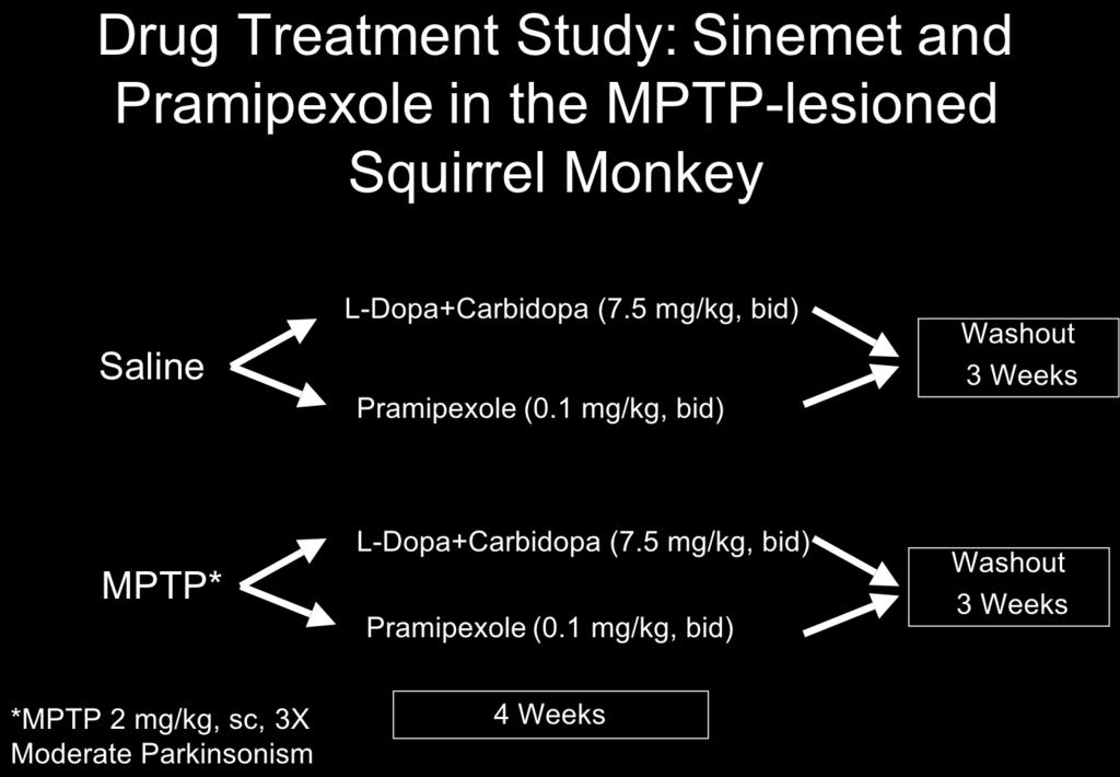
18 be subjected to exercise while receiving either the NMDA receptor antagonist MK-801 or the AMPA receptor antagonist GYKI Behavioral recovery will be compared between groups. Brain tissue will be analyzed for alteration in dopaminergic function (dopamine, DAT and TH expression). Pilot studies show that both glutamate receptor antagonists GYKI and MK-801 can be administered in this model of MPTP-lesioning. This Aim is designed to be pursued in Year 2.5 through 4. However, experiments addressing this Study have been initiated to validate this approach and to prepare for completion of Study 5. Both saline and MPTP-lesioned mice were administered either the AMPA receptor antagonist GYKI (0.5 or 5.0 mg/kg) or the NMDA receptor antagonist MK-801 (1 mg/kg). Mice were administered these antagonists starting 4 days after the last injection of MPTP for a period of 30 days. No adverse effects on behavior were observed. The brains from animals in all groups were harvested and analyzed for striatal expression of TH and DAT protein and for expression of TH and DAT mrna transcripts in nigrostriatal neurons. Data from these experiments show a differential effect of using an AMPA receptor antagonist compared to an NMDA receptor antagonist. For example, comparison of the expression of TH mrna transcripts in AMPA antagonist treated MPTP-lesioned mice show elevated TH expression in surviving nigrostriatal dopaminergic neurons. Studies have been carried out using immuno-electron microscopy with an antibody against glutamate showing no significant alteration in glutamate synaptic occupancy in mice administered the AMPA receptor antagonists GYKI or the NMDA receptor antagonists MK-801. To extend these studies we have collaborated with Dr. Frank Menneti at Pfizer, who has provided us with more specific AMPA and NMDA receptor antagonists targeting GluR1 and NMDA- NR2A subunits. These studies will be pursued in year 3. Component 2: Pharmacological Enhancement of Neuroplasticity in the MPTP-lesioned Non- Human Primate Model. Study 1: The behavioral recovery of saline injected and MPTP-lesioned squirrel monkeys will be compared with and without the administration of Pramipexole. Animal behavior will be monitored using both a cage side clinical rating scale and a personal activity monitor. In Year One of this proposal we have carried out the lesioning and behavioral analysis of MPTP-lesioned nonhuman primates in the groups utilizing the 6-week time frame. These are groups 1, 3, 5, and 7 in the Specific Aims Component 2. These squirrel monkeys were lesioned with MPTP in a series of 6 injections of neurotoxicant at a concentration of 2 mg/kg (free-base) administered once every 2 weeks for a total of 12 mg/kg per animal. The typical timeline for this protocol is outlined in Figure 1 showing that acquisition, quarantine (60 days), baseline acclimation and behavioral assessment (30 days), MPTP-lesioning (12 weeks), followed by behavioral assessment in the 6 weeks after the last injection of MPTP. The total time period for this stage is approximately 8 months. The behavioral assessment of MPTP-lesioned squirrel monkeys with and without Pramipexole in the early (6 week) time point has been completed. In study 1 we have added a Sinemet (L-DOPA/ Carbidopa) group for comparison with Pramipexole. This deviation is based on the scientific rationale that L-dopa, unlike Pramipexole, is metabolized and stored by dopaminergic terminals and therefore may have a more direct effect on the regulation of endogenous dopamine production and behavioral recovery and offers an interesting comparison to a compound that is not taken up by terminals. 18
19 One important outcome in Study 1 was the unexpected induction of dyskinesia in the MPTP-lesioned animals administered Pramipexole. This new finding has not been reported in the literature by other 19
20 20

21 investigators and we are preparing a manuscript reporting this novel finding (Figure 6). This may indicate that Sinemet and Pramipexole may both induced dyskinesias through mechanisms that include the down regulation of the dopamine transporter. Experiments to specifically address this mechanism will be carried out in years 2-3 of this proposal. Figure 6 21
.")
22 The behavioral assessment in this first group of animals carried out up to 8 weeks after the last injection of MPTP showed a slight enhancement of behavioral recovery in both the Sinemet and Pramipexole groups versus the saline treated group (Figure 7). Statistical analysis is being completed for both behavioral studies (dyskinesia and motor impairment) Figure 7 Nonhuman primates that were initially to be used for the long-term behavioral studies, were used for (1) adding a L-dopa group (2) conducting microdialysis studies to extend our findings in Study 2 (neurochemistry); and (3) to examine alterations in electrophysiolgical properties of corticostriatal neurons and voltammetry. See Sections below. 22
 at")
23 Study 2: Analysis of brain tissue from MPTP-lesioned squirrel monkeys administered Pramipexole or L-dopa/carbidopa or saline. This analysis included neurochemistry and molecular studies that examined the pattern of expression of proteins and mrna transcripts important for dopaminergic function (including TH, DAT, VMAT2) at the level of the SNpc and CPu. Total dopamine levels and metabolites were analyzed from all groups of animals, using HPLC. Brains were removed from all groups at completion of 4 weeks of drug or saline treatment, followed by 3 weeks of drug washout. Microdialysis of the putamen was added to this study to complement and support findings from out HPLC analysis of tissue. We found there was a slight increase in dopamine levels in the ventral caudate and putamen of animals receiving Pramipexole or Sinemet. These results are shown in Figure 8. Figure 8: Analysis of dopamine and its metabolites in the MPTP-lesioned squirrel monkey. Squirrel monkeys were MPTP-lesioned and then treated one week after the last injection of MPTP with either Sinemet (10 mg/kg twice daily), or Pramipexole (1 mg/kg twice daily). Animals were treated for four weeks. On each week animals received drug for three days (Tue,Wed, Thurs)) and then saline for four days (Fri, Sat, Sun, Mon). Animals were rated each day for parkinsonian features and for dyskinesia. Drug was washed out for 3 weeks and then animals were euthanized. Brain tissue was collected and striatal tissue dissected 8 weeks (1 week monitoring + 4 weeks drug treatment + 3 weeks washout) after MPTP. HPLC analysis showed that Pramipexole and Sinemet (L-dopa + carbidopa) treated animals had a slight increase in striatal dopamine, especially in the ventral putamen, compared to MPTP + saline treated nonhuman primates. 23

24 The following figure shows the timeline of microdialysis studies. 24
25 Figure 9 above shows a representative microdialysis experiment with the same animal used as its own control and undergoing repeated microdialysis studies. Our studies show that Pramipexole or Sinemet treated animals have greater amphetamine-induced dopamine release. 25


26 Figure 10: Western immunoblot analysis shows that Pramipexole treated animals have a slightly grater degree of TH protein expression compared to Sinemet treated and MPTP-lesioned alone. There was no change in the pattern of expression of DAT protein in the treatment groups. Immunohistochemistry and additional western immunoblot analysis is currently underway to determine regional differences in the pattern of expression of TH and VMAT-2 in the caudate nucleus and putamen as well as within the SNpc. We have also added physiological studies on a subset of animals to complement our current findings. Fast-scan cyclic voltammetry did not show detectable dopamine release in either the MPTPlesioned or treated groups. The slight differences between microdialysis and voltammetry may be due to the differences in sampling where microdialysis has a greater sampling area compared to voltammetry. Electrophysiological studies also showed changes in the ratio of AMPA: NMDA contribution to the EPSPs of corticostriatal synapses after MPTP, and this ratio is altered again in treated groups. 26
27 Study 3: The pattern of expression of the dopamine receptors D1, D2, and D3 will be determined in both the SNpc and CPu. The level of protein expression will be determined western immunoblotting, immunohistochemistry, while the level of mrna transcript expression will be determined using in situ hybridization histochemistry. Double labeling techniques will be used to co-localize the dopamine receptor changes with other enkephalin or substance P containing neurons. Preliminary data supports our ability to use these techniques in the non-human primate. To pursue this Study we have designed and tested appropriate primer sets for quantitative realtime PCR (qrt-pcr) using a newly acquired Eppendorf RT-PCR thermocycler. Brain tissues have now been collected from animals in all groups and studies including immunohistochemistry, western blotting, and in situ hybridization histochemistry will be carried out in Year 3 of this proposal. Study 4: The effect of Pramipexole on glutamatergic synapses in the striatum after injury will be determined using ultrastructural immunohistochemical staining with electron microscopy. Pilot data shows our ability to quantify glutamatergic synapses using immuno-electron microscopy. In collaboration with Dr. Charles Meshul (Oregon Health Sciences University, Portland, OR) perfusion fixed brain tissues were harvested from a nonhuman primate from each group for analysis using immuno-electron microscopy with an antibody against glutamate. These results are summarized in the Figure below. Following MPTP-lesioning there is an increase in the relative density of striatal glutamate immunolabeling (second bar) within corticostriatal terminals. After treating MPTP-lesioned animals with Pramipexole or Sinemet the relative density of glutamate immunolabeling is reduced. Increased density of striatal glutamate within the terminal is thought to reflect decreased glutamate release. Our study would suggest that Sinemet increases glutamate release to a slightly greater extent than Pramipexole. This increased glutamate release may be one means by which dyskinesia is elicited to a greater extent in Sinemet treated animals than Pramipexole treated animals. 27
28 Reportable Outcomes For Years One and Two Abstracts: (1) Hogg, E, M. W. Jakowec, K. L. Nixon, A. T. Abernathy, P. Arevalo, B. E. Fisher, M. Liker, and G. M. Petzinger. (2004) Behavioral recovery in the MPTP-lesioned nonhuman primate: Altered dopamine biosynthesis and storage. Society for Neuroscience, San Diego, CA. (2) Society for Neuroscience Annual Meeting, Atlanta 2006 ABSTRACT #1 Exercise induced behavioral recovery and plasticity in the MPTP-mouse model of Parkinson s disease. Jakowec, M. W., P. Arevalo, M. Vuckovic, P. Turnquist, E. Hogg, J. Walsh #, G. Akopian #, C. Meshul*, A. Abernathy, M. Ramirez, B. Fisher and G. M. Petzinger. Dept. Neurology; Davis School of Gerontology # ; Dept. Biokinesiology and Physical Therapy; University of Southern California, Los Angeles, CA.VA Medical Center*, OHSU, Portland, OR. The adult brain possesses a tremendous capacity for activity-dependent neuroplasticity. Following injury to the brain, physical therapy plays an important role in promoting recovery. In neurodegenerative disorders such as Parkinson s disease, physical activity improves motor function and may lead to alterations in disease progression. To better understand the role of activity-dependent plasticity in brain repair we are investigating the application of intensive treadmill exercise training in the MPTP mouse model of basal ganglia injury and dopamine depletion. Mice were administered MPTP (4 20 mg/kg each) and subjected to intensive treadmill running for 30 days starting 4 days after the last injection of MPTP (when cell death is complete) at a speed up to 20 meters/minute for 1 hour. During the exercise paradigm, mice were investigated for improvement in behavioral motor features and learning. Harvested brain tissues were analyzed by HPLC for levels of dopamine and its metabolites, and glutamate and the pattern of expression of genes and proteins for tyrosine hydroxylase, dopamine transporter, dopamine receptors D1 and D2, and AMPA and NMDA glutamate receptors using western immunoblotting, immuno histochemistry, and in situ hybridization histochemistry. Electrophysiological analysis of dopamine release was determined using fast cyclic voltammetry on brain slices. Our findings indicated that there was an enhancement of both motor behavior recovery and rotarod learning in exercised mice despite no change in the number of SNpc dopaminergic neurons and the striatal levels of dopamine. Molecular analysis showed down-regulation of DAT and TH, and significant changes in the pattern of expression of ionotropic glutamate receptors in the cortex and striatum. In addition, exercise resulted in an increase in dopamine release compared to MPTP-lesioned mice without exercise. These findings demonstrate that intensive exercise can induce dramatic neuroplasticity in an animal model of basal ganglia injury and provides a valuable framework for supporting exercise in patients with Parkinson s disease. Supported by grants to J. Walsh (RO1 AG21937), M. Jakowec (RO1 NS44327) and G. Petzinger (US Army NETRP W81XWH ). 28
29 (3) Society for Neuroscience Annual Meeting, Atlanta 2006 ABSTRACT #2 Changes in dopamine and glutamate electrophysiology in the MPTP-treated non-human primate and the exercised MPTP-treated mouse. J Walsh*, G Akopian*, M, Jakowec, G, Petzinger. USC Neuroscience Program, USC Davis School of Gerontology*, Department of Neurology - USC Keck School of Medicine. We tested the hypotheses that dopamine (DA) and glutamate physiology are altered in the MPTP-treated squirrel monkey using electrophysiological methods. Fast cyclic voltammetry analysis of the monkey putamen revealed that MPTP treatment (6 weeks earlier) resulted in a dramatic loss in DA released in response to intra-putamen stimulation (bipolar tungsten wire electrode, 0.1 msec µa stimulus). Saline injected monkeys showed greater DA release in the lateral versus medial putamen. To determine if excitatory amino acid receptor-mediated physiology is altered in the MPTPtreated monkey putamen we applied whole cell voltage clamp techniques and examined the relative contribution of AMPA and NMDA receptors to corticostriatal synaptic events. Saline injected monkeys showed a relatively uniform NMDA/AMPA receptor ratio, while data from MPTP-treated monkeys suggested that two new populations emerged; one with a reduced NMDA/AMPA ratio and another with an enhanced NMDA/AMPA ratio. We applied a similar strategy to examine the impact of MPTP toxicity on DA and glutamate physiology in the mouse and, more importantly, to determine if changes striatal DA or glutamate physiology tracked the behavioral recovery induced by exercise in the MPTP-treated mouse. Cyclic voltammetry revealed a dramatic reduction in evoked DA release in the striatum of mice treated a month earlier with MPTP. A parallel group of mice were treated with MPTP and exercised daily on a treadmill. MPTP treated mice were significantly compromised in treadmill performance initially but achieved the same performance as saline injected mice by the end of one month of training. The exercise- mediated enhancement of motor skills transferred to a rotorod task. Prior work demonstrated exercise-induced suppression in striatal DAT immunocytochemistry in the MPTP-treated mouse (Fisher et al, 2004, J Neur Res 77:378), but voltammetry revealed a significant exercise-induced increase in DA release in the MPTP treated mouse. These data demonstrate emergent dopaminergic and glutamatergic plasticity created in the striatum following exposure to the neurotoxin MPTP. We hypothesize these forms of synaptic plasticity underlie both behavioral deficits created early as well as recovery seen later in the MPTP model. This research is supported by grants to J Walsh (RO1 AG21937), M Jakowec (RO1 NS44327) and G Petzinger (US Army NETRP W81XWH ), and the Zumberge Foundation. Publications: (1) Jakowec M.W., K. Nixon, E. Hogg, T. McNeill, and G. M. Petzinger (2004) Tyrosine hydroxylase and dopamine transporter expression following 1-methyl-4-phenyl-1,2,3,6-tetrahydropyridine-induced neurodegeneration of the mouse nigrostriatal pathway. J. Neurosci. Res. 76 (4) (2) Fisher, B.E., G. M. Petzinger, K. Nixon, E. Hogg, S. Bremmer, C. K. Meshul, and M. W. Jakowec (2004) Exercise-Induced behavioral recovery and neuroplasticity in the 1-methyl-4-phenyl-1,2,3,6- tetrahydropyridine-lesioned mouse basal ganglia. Journal Neuroscience Research 77:
30 (3) Jakowec, M.W., and G.M. Petzinger (2004) The MPTP-Lesioned Model of Parkinson s Disease with Emphasis on Mice and Nonhuman Primates. Comparative Medicine 54 (5) (4) Petzinger, GM, and M. W. Jakowec (2005) Animal Models of Basal Ganglia Injury and Degeneration and their Application to Parkinson s Disease Research. In Parkinson s Disease, eds M. Ebadi and R. F. Pfeiffer, CRC Press, Boca Raton, FL. (5) Petzinger, G.M., K. Nixon, B. E. Fisher, A. Abernathy, and M. W. Jakowec. (2006) Behavioral Recovery in the MPTP (1-methyl-4-phenyl-1,2,3,6-tetrahydropyridine)-lesioned Squirrel Monkey (Saimiri sciureus): Analysis of Striatal Dopamine and the Expression of Tyrosine Hydroxylase and Dopamine Transporter Proteins. J. Neurosci. Res. 83: (6) Petzinger, G. M., and M. W. Jakowec (2006) Animal Model of Parkinson s Disease. In: Handbook of Parkinson s Disease, eds. R. Pawha and K. Lyons, Marcel Dekker, Inc., NY, NY. (7) Petzinger, G. M., B. E. Fisher, E. Hogg, A. Abernathy, P. Arevalo, K. Nixon, A. Chua, and M. W. Jakowec (2005) Neuroplasticity and Treadmill Exercise in the 1-methyl-4-phenyl-1,2,3,6- tetrahydropyridine-(mptp)-lesioned Mouse Model: Analysis of Striatal Dopamine, Tyrosine Hydroxylase, and Dopamine Transporter Expression. To be submitted to Journal of Neuroscience. Presentations: (1) Petzinger, Giselle MD Neuroplasticity in the MPTP-lesioned Nonhuman Primate, Plasticity and Repair in Neurodegenerative Disorders, Lake Arrowhead, California, Workshop, Feb19-22, (2) Jakowec, Michael PhD The Role of Exercise in Enhancing Neuroplasticity in the MPTP-lesioned mouse, Plasticity and Repair in Neurodegenerative Disorders, Lake Arrowhead, California, Workshop, Feb19-22, (3) Petzinger, Giselle MD Enhancing Neuroplasticity in models of Basal Ganglia Injury, Van Der Muelen Symposium, University of Southern California, Keck School of Medicine, April 1, Conclusions: The MPTP-lesioned mouse and squirrel monkey are valuable models for investigating neuroplasticity of the injured basal ganglia. Studies of year one from this proposal indicate that intensive treadmill exercise can enhance motor behavioral recovery and alter the time course of intrinsic neuroplasticity. Our data indicates that alterations in striatal dopamine is not the sole factor responsible for this enhanced recovery. Preliminary data supports the role of the glutamatergic system in exercise related effects on either the injured or normal basal ganglia. Therefore glutamatedopaminergic interactions may serve as a therapeutic target for enhancing repair. In the MPTP-lesioned primate we have not observed any enhancement of early (8 week) behavioral recovery through exogenous dopamine replacement therapy either in the form of Sinemet or 30
31 the dopamine agonist, Pramipexole. Interestingly we have seen a modest increase in striatal (putamen) dopamine in the Pramipexole treated group. Molecular analysis are examining alterations in proteins involved in the biosynthetic pathway of dopamine. One unexpected finding was the development of dyskinesia during treatment with Pramipexole. This behavioral finding has not been previously reported. Dyskinesia was noted to develop at a slightly less then that observed in Sinemet treated animals We believe that one possible mechanism for the development of dyskinesia in both Sinemet and Pramipexole treated animals is due to the down regulation of the dopamine transporter which may allow greater diffusion away from the synaptic cleft and a greater interaction with altered glutamate receptors. This hypothesis is currently being investigated using the same studies outlined in our proposal. Studies are underway to examine alterations in long -term behavioral recovery using dopamine replacement therapy. Appendices: Attached. Manuscript: Fisher, B.E., G. M. Petzinger, K. Nixon, E. Hogg, S. Bremmer, C. K. Meshul, and M. W. Jakowec. (2004) Exercise-Induced behavioral recovery and neuroplasticity in the 1-methyl-4-phenyl- 1,2,3,6-tetrahydropyridine-lesioned mouse basal ganglia. Journal Neuroscience Research 77: Manuscript: Petzinger, G.M., K. Nixon, B. E. Fisher, A. Abernathy, and M. W. Jakowec. (2005) Behavioral Recovery in the MPTP (1-methyl-4-phenyl-1,2,3,6-tetrahydropyridine)-lesioned Squirrel Monkey (Saimiri sciureus): Analysis of Striatal Dopamine and the Expression of Tyrosine Hydroxylase and Dopamine Transporter Proteins. J. Neurosci. Res. 83: Abstract: Society for Neuroscience, Atlanta, October, 2006 Abstract: Society for Neuroscience, Atlanta, October, 2006 References: (1) Schulte A, Chow R.H. A simple method for insulating carbon-fiber microelectrodes using anodic electrophoretic deposition of paint. Anal Chem (1996), 68: (2) Miles P.R., Mundorf M.L., Wightman M. Release and uptake of catecholamines in the bed nucleus of the stria terminalis measured in the mouse brain slice. Synapse (2002)44:
32 Journal of Neuroscience Research 83: (2006) Behavioral Motor Recovery in the 1-Methyl-4-Phenyl-1,2,3,6- Tetrahydropyridine-Lesioned Squirrel Monkey (Saimiri sciureus): Changes in Striatal Dopamine and Expression of Tyrosine Hydroxylase and Dopamine Transporter Proteins Giselle M. Petzinger, 1,2 * Beth Fisher, 2 Elizabeth Hogg, 1 Avery Abernathy, 1 Pablo Arevalo, 1 Kerry Nixon, 1 and Michael W. Jakowec 1,2 1 George and MaryLou Boone Parkinson s Disease and Movement Disorders Research Center, Department of Neurology, University of Southern California, Los Angeles, California 2 Department of Biokinesiology and Physical Therapy, University of Southern California, Los Angeles, California The neurotoxicant 1-methyl-4-phenyl-1,2,3,6-tetrahydropyridine (MPTP) provides an excellent opportunity to study repair and response to injury in the basal ganglia. Administration to mammals leads to the destruction of nigrostriatal dopaminergic neurons and depletion of striatal dopamine. In the squirrel monkey (Saimiri sciureus), MPTP-lesioning results in parkinsonian motor symptoms including bradykinesia, postural instability, and rigidity. Over time animals display motor behavioral recovery. To better understand this mechanism we employed a lesioning regimen of two or six subcutaneous injections of MPTP (2.0 mg/kg, free-base) to generate mild or moderate parkinsonism. Brain tissue was harvested at 6 weeks or 9 months after the last injection and analyzed for dopamine and its metabolites by high performance liquid chromatography (HPLC), and by immunohistochemical staining and Western immunoblotting for the expression of tyrosine hydroxylase (TH), dopamine transporter (DAT), and dopamine- and campresponsive protein phosphatase of 32 kda (DARPP-32), an effector molecule enriched in striatal medium spiny neurons. Several months after MPTP-lesioning, when squirrel monkeys displayed full motor behavioral recovery, striatal dopamine levels remained low with a greater return in the ventral striatum. This finding is consistent with other reports using neurotoxicantlesioning models of the basal ganglia in rodents and other species of nonhuman primates. Elevated dopamine turnover ratio and decreased DAT expression appeared in early behavioral recovery at the 6-week time point in both mild- and moderate-parkinsonian monkeys. Tyrosine hydroxylase and DAT expression was increased in late stage recovery even within dopamine-depleted regions and supports sprouting. Altered DARPP-32 expression suggests a role of medium spiny neurons in recovery. VC 2005 Wiley-Liss, Inc. Key words: basal ganglia; Parkinson s disease; nonhuman primate; neuroplasticity The neurotoxicant 1-methyl-4-phenyl-1,2,3,6-tetrahydropyridine (MPTP) provides an excellent opportunity to study repair and the response to injury in the basal ganglia. MPTP is a meperidine derivative that can be administered systemically, and the pre-toxin form crosses the blood brain barrier and is converted to the toxic form 1-phenyl-4-phenylpiperidium (MPP þ ) by astrocytic monoamine oxidase B (Chiba et al., 1985). MPP þ acts as a false-substrate for the dopamine transporter (DAT) and accumulates in dopaminergic neurons where it targets mitochondrial complex I leading to energy depletion and the formation of reactive oxygen Contract grant sponsor: Baxter Foundation; Contract grant sponsor: Zumberge Foundation; Contract grant sponsor: Parkinson s Disease Foundation; Contract grant sponsor: US Army NETRP; Contract grant number: W81XWH ; Contract grant sponsor: National Institutes for Health; Contract grant number: RO1 NS *Correspondence to: Giselle M. Petzinger, MD, Department of Neurology, Keck School of Medicine, 1333 San Pablo St., MCH-148, Los Angeles, CA, gpetzinger@surgery.usc.edu Received 17 August 2005; Revised 20 October 2005; Accepted 20 October 2005 Published online 29 December 2005 in Wiley InterScience (www. interscience.wiley.com). DOI: /jnr ' 2005 Wiley-Liss, Inc.
33 Recovery in MPTP Nonhuman Primate 333 species (Dauer and Przedborski, 2003). This results in the selective destruction of nigrostriatal dopaminergic neurons and the depletion of the striatal neurotransmitter dopamine similar to that seen in Parkinson s disease (PD) (Jakowec and Petzinger, 2004). Dopamine depletion results in behavioral motor deficits in mice and nonhuman primates. Motor behavioral changes in mice tend to be subtle and require specific behavioral testing including treadmill, paw-reach, and rotorod balancing to become evident (Sedelis et al., 2000, 2001; Tillerson et al., 2001; Fisher et al., 2004). In nonhuman primates, motor behavioral deficits resembling those seen in humans with PD or drug addicts who self-administered MPTP are evident and include akinesia, bradykinesia, postural instability, freezing, and in some species a resting tremor (Burns et al., 1983; Langston et al., 1983, 1984). MPTP has been administered to a variety of different nonhuman primates using several different regimens. In our laboratory we utilize the squirrel monkey (Saimiri sciureus), a New World monkey. We have developed a lesioning regimen that consists of either a series of two or six injections of MPTP at a concentration of 2.0 mg/ kg (free-base) with 2 weeks between injections. With this lesioning regimen we avoid high animal mortality often experienced with MPTP use. Behavioral analysis using a clinical rating scale (CRS) we designed specifically for the MPTP-lesioned squirrel monkey, documents either mild (two injections) or moderate (six injections) parkinsonian motor behavior. Interestingly, over time, animals display motor recovery. By 6 weeks post-mptp, animals show partial motor recovery and by 9 months post-mptp both groups of monkeys show motor behavior that is indistinguishable from nonlesioned animals. Specifically, recovered animals no longer display akinesia or bradykinesia and show normal spontaneous movement (climbing and jumping), normal hand dexterity, and balance. Motor behavioral recovery has been observed in several other species of nonhuman primates including the marmoset, vervet, and macaque (Rose et al., 1989b; Rothblat and Schneider, 1994; Schneider et al., 1998; Elsworth et al., 2000). It has been shown that motor behavioral recovery takes place with the incomplete return of striatal dopamine. Neurotoxicant lesioning of the dopaminergic system in rodents, cats, and nonhuman primates suggest that there are preand post-synaptic alterations within remaining dopaminergic neurons and their targets, respectively, that may compensate for the deficient return of striatal dopamine and may account for motor behavioral recovery. Presynaptic indices include increased dopamine synthesis, turnover, and release, altered dopamine uptake, and sprouting of surviving dopaminergic neurons (Zigmond et al., 1990). Alterations in dopamine synthesis may be reflected by either an increase in the activity or total protein level of tyrosine hydroxylase (TH), the rate limiting enzyme of dopamine biosynthesis. In addition, alteration in dopamine uptake may reflect the level of dopamine transporter (DAT) protein because DAT is a key component in regulating dopamine synaptic occupancy (Gainetdinov et al., 2002). Post-synaptic indices include altered dopamine receptor number and function and their influence on neuropeptide expression, second messengers, transcription factors, and down-stream effector molecules especially within striatal medium-spiny neurons (Greengard et al., 1999; Gerfen, 2000; Vallone et al., 2000). This study examines the relationship between motor behavioral recovery and pre- and post-synaptic indices of dopaminergic function, including striatal dopamine in the MPTP-lesioned squirrel monkey. We examined squirrel monkeys that were rendered either mildly or moderately parkinsonian and during early and late periods of recovery. Specifically, squirrel monkeys were administered MPTP as a series of either two or six injections 2 weeks between injections to generate either a mild or moderate parkinsonian group, respectively. Brain tissue was harvested at 6 weeks or 9 months after the last injection of MPTP. For neurochemistry and behavior, analyses were designed to compare individual groups. Molecular studies focused on the expression of two important pre-synaptic proteins, tyrosine hydroxylase and the dopamine transporter. As an initial approach to investigate post-synaptic changes, the pattern of expression of dopamine- and camp-responsive protein phosphatase of 32 kda (DARPP-32), a molecule important for the down-stream action of dopamine neurotransmission in striatal medium spiny neurons, was examined. For molecular studies, analyses were designed to compare changes in the mild-lesioned group (two injections of MPTP) at the early and late time point, and changes in the moderate-lesioned group (six injections of MPTP) at the early and late time point. MATERIALS AND METHODS Animals Twenty-four young adult male squirrel monkeys (Saimiri sciureus) weighing 900 1,200 g each were used in these studies (Osage, St. Louis, MO). All procedures utilizing the nonhuman primate strictly followed guidelines set forth by the National Institutes for Health for the humane treatment of animals in research and had the approval of the University of Southern California Institutional Animal Care and Use Committee (IACUC). Animals were housed individually in a home cage. After quarantine, animals were acclimated to the facility for 30 days before behavioral analysis in their home cages. MPTP-Lesioning MPTP (Sigma, St. Louis, MO) was administered in a series of subcutaneous injections set 2 weeks apart at a concentration of 2.0 mg/kg (free-base) dissolved in sterile water and made up fresh from a new 100-mg bottle each time. One group of animals (n ¼ 8) received a series of two injections (for a total of 4.0 mg/kg MPTP, free-base). Another group of animals (n ¼ 8) received a series of six injections of MPTP (for a total of 12.0 mg/kg, free-base). A saline injected group (n ¼ 8) acted as control. Animals were divided into the following six groups with n ¼ 4 per group: (1) saline-injected Journal of Neuroscience Research DOI /jnr
34 334 Petzinger et al. TABLE I. Parkinsonian Clinical Rating Scale Modified for Squirrel Monkey Parkinsonian motor feature 1. Spatial hypokinesia (movement around cage) Rating 0- Normal (uses entire cage space) 1- Utilizes most of the cage (at least 75% of cage space), but may be slow. 2- Definitely slowed, but uses more than 50% of cage space. 3- Definitely slowed, using less than 50% of cage space. 4- Does not move from a confined area, with little or no movement 2. Body bradykinesia 0- Normal movement around cage or bars 1- Slow or deliberate body movements, could be normal for age. 2- Moderately slow, intermittent limb dragging, moves without provocation. 3- Marked slowness, requires provocation to move arms or legs. 4- Frozen, little or no body movements regardless of provocation. 3. Manual dexterity (right arm) 0- Normal 1- Mildly slow or some loss of maneuverability of food items, could be normal for age. 2- Moderate slowness, noticeable effort needed to grab or maneuver food. 3- Marked slowness, with multiple attempts needed to grab food, may use both hands, may drop food. 4- Severe slowness, with inability to grab or maneuver food, may need to be hand fed. 4. Manual dexterity (left arm) 0- Normal 1- Mildly slow or some loss of maneuverability of food items, could be normal for age. 2- Moderate slowness, noticeable effort needed to grab or maneuver food. 3- Marked slowness, with multiple attempts needed to grab food, may use both hands, may drop food. 4- Severe slowness, with inability to grab or maneuver food, may need to be hand fed. 5. Balance 0- Normal 1- Slight tendency to hold on to cage, may be normal for age or no falls 2- Uses both hands intermittently for support or rare occasional falls 3- Uses both hands for support at all times or frequent falls 4- Continually hanging on for support or falls with any attempt to move 6. Freezing (observation over 4 minute clinical evaluation) 0- None, no freezing ever observed 1- Occasional mild (<5 sec) freezing episodes 2- Mild freezing episodes <5 sec duration or rare severe (>5 sec) 3- Frequent severe freezing observed >5 sec 4- Frozen most of the time harvested at 6 weeks; (2) saline-injected harvested at 9 months; (3) MPTP-injected 2-times harvested at 6 weeks after the last injection; (4) MPTP injected 2-times harvested at 9 months after the last injection; (5) MPTP-injected 6-times harvested at 6 weeks after the last injection; and (6) MPTP-injected 6- times harvested at 9 months after the last injection. Clinical Rating Scale for Motor Behavioral Analysis Behavioral motor features were determined using a cage-side clinical rating scale (CRS) based on the Unified Parkinson s Disease Rating Scale (UPDRS) and modified for the squirrel monkey. Table I outlines the features of the clinical rating scale. The CRS consisted of six items including: (1) spatial hypokinesia (movement around cage); (2) body bradykinesia; (3) manual dexterity in left arm; (4) manual dexterity in right arm; (5) balance; and (6) freezing. Each item has a score of 0 4 resulting in a maximum motor score deficit of 24 points. A CRS score of <4 points is within the range of normal. Two investigators, blinded to the animal and treatment group, assessed motor features three times per week in the morning before feeding time. Baseline behavior was determined in the 2-week period before the first injection of MPTP or saline. Parkinsonian motor features using the CRS were first determined 2 weeks after the last injection of MPTP or saline, and continued on a weekly basis until tissue harvesting at 6 weeks or 9 months. Data for statistical analysis were carried out at baseline and post-injection time points of 2 weeks, 6 weeks, and 9 months. At each of these four time points a nonparametric Kruskal-Wallace analysis with Mann- Whitney post-hoc was carried out comparing saline, 2-time, and 6-time injected groups. Brain Tissue Harvest and Preparation Brain tissue was harvested from four animals in each group at either 6 weeks or 9 months after the last injection of MPTP for a total of 24 animals. Animals were sedated with ketamine (0.3 ml of 100 mg/ml) followed by a lethal dose of sodium pentobarbital (2 ml of 50 mg/ml solution) and monitored for eye reflex, breathing, and heartbeat. On cessation of vital signs, brains were quickly removed, briefly cooled on wet ice, and sectioned at 3-mm thickness in the coronal plane using an acrylic brain block designed specifically for the squirrel monkey brain starting at a position approximately 3 mm rostral to the midbrain. This resulted in six rostral coronal slices of 3-mm thickness. One fresh slice through the midstriatum (at level A15 A12 in the anterior posterior plane) of each animal was used to free-hand dissect out the entire caudate nucleus or putamen, sectioned into four pieces in the dorsal ventral plane, and snap frozen on dry ice to be used for either high performance liquid chromatography (HPLC) analysis of dopamine and its metabolites or protein analysis using western immunoblotting (Emmers and Akert, 1963). The remaining caudal brain, which included the midbrain, was split at the mid-plane into two equal halves. Brain slices Journal of Neuroscience Research DOI /jnr
35 Recovery in MPTP Nonhuman Primate 335 were immersion fixed in 4% paraformaldehyde in phosphate buffered saline (PBS) (ph 7.4) for 48 hr, cryoprotected in 20% sucrose in phosphate buffer (PB) (ph 7.4) for 24 hr, and frozen onto microscope slides using immersion into isopentane on dry ice. For immunohistochemical staining, tissue was cut at 30-lm thickness using a Leica 1950 cryostat. Sections were placed free-floating in PBS (ph 7.4) containing 0.01% thimerosal and refrigerated until used. HPLC Analysis of Dopamine and Its Metabolites Neurotransmitter concentrations were determined according to an adaptation of Irwin et al. (1992) of the method of Kilpatrick et al. (1986). Tissues for analysis were homogenized in 0.4 N perchloric acid and centrifuged at 12,000 3 g to separate precipitated protein. The protein pellet was resuspended in 0.5 N NaOH and the total protein concentration determined using the Coomassie Plus protein assay system (Pierce, Inc., Rockford, IL) using a Biotek Model Elx800 microplate reader and the software KCjunior. The concentrations of dopamine, 3,4-dihydroxyphenylacetic acid (DOPAC), homovanillic acid (HVA), were assayed by HPLC with electrochemical detection (ECD). Samples were injected with an ESA autosampler. Dopamine and its metabolites were separated by a 150 mm mm reverse phase 3-lm diameter C-18 column (ESA, Chelmsford, MA) regulated at 288C. The mobile phase MD-TM (from ESA) consisted of acetyl nitrile in PB and an ion-pairing agent delivered at a rate of 0.6 ml/min. The electrochemical detector was an ESA model Coularray 5600A with a 4-channel analytical cell with set potentials at 100 mv, 50 mv, and 220 mv. The HPLC was integrated with a Dell GX-280 computer with analytical programs including ESA Coularray for Windows software and the statistics package InStat (San Diego, CA). For neurochemistry, four separate basal ganglia regions were analyzed (ventral putamen, dorsal putamen, ventral caudate, or dorsal caudate) using multiple one-way ANOVA. Data were compared from the following six groups: (1) saline-injected harvested at 6 weeks; (2) saline-injected harvested at 9 months; (3) MPTP-injected 2-times harvested at 6 weeks after the last injection; (4) MPTP injected 2-times harvested at 9 months after the last injection; (5) MPTP-injected 6-times harvested at 6 weeks after the last injection; and (6) MPTP-injected 6-times harvested at 9 months after the last injection. Independent t-test showed no differences between the saline groups for each of the independent measures and as such, the two saline groups were collapsed into a single group for further analysis. Posthoc contrasts with Bonferroni correction were carried out to determine the locus of any significant differences. For all statistical tests significance was set at P < Stereologic Analysis of Nigrostriatal Neurons To verify the degree of lesioning in the 2-time and 6- time injection groups at the time of harvest (9 months) the number of nigrostriatal dopaminergic neurons in the substantia nigra pars compacta (SNpc) was determined using unbiased stereology with the computer-imaging program BioQuant (BioQuant Image Analysis Inc., Nashville, TN) on a Dell computer and an Olympus BX-50 microscope equipped with a motorized stage and Retiga CCD camera. The total number of SNpc dopaminergic neurons was determined based on the method of Gunderson and Jensen (1987). The hemi-sectioned brain (total of six animals: two each from saline, 2-time, and 6-time MPTP-injected collected at 9 months after the last injection of MPTP) with its most rostral sections starting at level A9.0 based on the brain atlas for the squirrel monkey (Emmers and Akert, 1963) was mounted in the cryostat at a temperature of 188C. Tissue was sliced at 30-lm thickness and every sixth section collected starting just before the rostral aspect of the substantia nigra (at level A8.0) completely through to the most caudal aspect when emergence of the pontine nucleus (at level A3.5) occurs. Tissue sections were stained for TH-immunoreactivity and counterstained for Nissl substance. The SNpc was delineated from the rest of the brain based on TH-immunoreactivity of remaining SNpc neurons because the MPTP-lesioning regimen of two or six injections used in these studies leads to 30 40% of remaining THimmunoreactive dopaminergic neurons (in the case of the 6-time MPTP injections regimen) compared to saline (see Fig. 3). In addition, anatomic landmarks at the rostral and caudal aspects of the SNpc were also used for delineation including the cerebral peduncle, third nerve, and pontine nucleus as described in the atlas for the squirrel monkey (Emmers and Akert, 1963). Each stained ventral mesencephalon section was viewed at low magnification (103 objective) and the SNpc outlined and delineated from the ventral tegmental-immunoreactive neurons using the third nerve and cerebral peduncle as landmarks. Neurons were viewed at high magnification (803 objective) and counted if they displayed TH-immunoreactivity and had a clearly defined nucleus, cytoplasm, and nucleolus. Immunohistochemical Analysis of Striatal TH, DAT, and DARPP-32 The antibodies used in these studies were all purchased from Chemicon, Inc. (Temecula, CA) and included anti-tyrosine hydroxylase (polyclonal made in rabbit or monoclonal made in mouse), anti-dopamine transporter protein (monoclonal made in rat), and anti-darpp-32 (polyclonal made in rabbit). Sections were rinsed in TBS (50 mm Tris ph 7.4 with 0.9% NaCl), exposed to primary antibody (concentration of 1:1,000) for 48 hr at 48C, rinsed in TBS, and then exposed to secondary antibody made against the species of the primary antibody (ABC Elite Kit, Vectastain, Burlingame, CA). Antibody staining was visualized by development in diaminobenzoic acid/h 2 O 2. To ensure that differences in staining intensity were due to differences in antigen expression, multiple sections from each treatment group and time point were carried out concurrently under identical staining conditions. Specificity of antibody probes was verified by methods that eliminated staining including: (1) omitting primary antibody; (2) omitting secondary antibody; or (3) omitting both primary and secondary antibodies. The relative intensity of labeling between stained sections in the different treatment groups was determined using computer assisted image analysis. Slides were simultaneously scanned on a high-resolution transmission light scanner (UMAX Powerlook 1120) and digitized images analyzed using NIH Image (version 1.66). Slide and section background (using a region of interest through the corpus cal- Journal of Neuroscience Research DOI /jnr
36 336 Petzinger et al. losum) were captured. Because sections for comparison were stained simultaneously within an experiment, variability in background was minimal. Comparison of immunostaining based on the relative optical density was determined by analyzing images using a region of interest within the quadrant of the dorsal putamen (for DAT and DARPP-32 analysis). The dorsal putamen was selected for analysis because it plays a prominent role in motor behavior. At least six to eight sections from two or three animals were used for analysis. For statistical analysis, a one-way ANOVA comparing the individual groups was carried out. The saline groups were collapsed because an independent t-test showed no differences between the saline groups for each of the independent measures. Posthoc contrasts with Bonferroni correction were carried out to determine the locus of any significant differences. For all statistical tests significance was set at P < Western Immunoblot Analysis of Striatal TH and DAT Western blotting was used to determine the relative protein expression of TH and DAT in the basal ganglia. The immunoblotting technique was described previously (Jakowec et al., 1995). Tissue dissected from the dorsal putamen was homogenized in buffer (25 mm Tris ph 7.4, 1 mm EDTA, 100 lm PMSF, 0.1% SDS). The dorsal putamen was selected for analysis because it plays a prominent role in motor behavior. Protein concentration was determined by the BCA method (Pierce, Inc., Rockford, IL). Equal amounts of protein (25 lg) were separated by the method of Laemmli (1970). Proteins were transferred to nitrocellulose filters by electroblotting in Towbin buffer (Towbin et al., 1979). Filters were blocked in TS-Blotto (50 mm Tris ph 7.4, 0.9% NaCl, 5% non-fat milk), then primary antibody (1:2,000), exposed to secondary antibody and visualized by chemiluminescence (Pierce, Inc.). Filters were apposed to film (Hyperfilm ECL, Amersham, Inc.) and processed in X-OMAT developer. Images were scanned into a computer using a Personal Laser Densitometer (Molecular Dynamics, Sunnyvale, CA) and the intensity of bands determined using computer assisted image analysis (NIH Image). The intensity of bands from Western blot autoradiographs was expressed as relative optical density with the saline injected group normalized to 100% because there was no significant difference between 6 weeks and 9 months. For statistical analysis, a one-way ANOVA comparing the individual groups was carried out. The saline groups were collapsed because an independent t-test showed no differences between the saline groups for each of the independent measures. Post-hoc contrasts with Bonferroni correction were carried out to determine the locus of any significant differences. For all statistical tests significance was set at P < RESULTS Motor Behavioral Deficits After MPTP-Lesioning and the Time Course of Motor Behavioral Recovery Motor behavior was assessed using a Clinical Rating Scale (CRS) as outlined in Table I and results shown in Figure 1. The baseline CRS scores before saline or MPTP administration for all groups of animals ranged Fig. 1. Time course of motor behavior using the CRS. Baseline behavior was determined in the 2-week period before the first injection of MPTP or saline. Parkinsonian motor features using the CRS were first determined 2 weeks after the last injection of MPTP or saline, and continued on a weekly basis until tissue harvesting at 6 weeks or 9 months. Data for statistical analysis were carried out at baseline and post-injection time points of 2 weeks, 6 weeks, and 9 months. At each of these four time points a nonparametric Kruskal- Wallace analysis with Mann-Whitney post-hoc was carried out comparing saline, 2-times, and 6-times injected groups. *Indicates statistical significance compared to the saline CRS score at P < #Indicates statistical significance compared to the 2-times MPTPinjected group CRS score at P < from 1 2 of a total score of 24. Throughout the entire study, saline-injected animals had a CRS score of After completion of MPTP administration, all lesioned animals showed parkinsonian features including akinesia, bradykinesia, postural instability, and freezing. Two weeks after the last injection of MPTP, animals that received two injections of MPTP were mildly parkinsonian ( ), whereas animals that received six injections of MPTP were moderately parkinsonian ( ). The difference between the saline, 2-time, and 6-time MPTP-injected groups 2 weeks after the last injection of MPTP was significant At 6-weeks postinjection, there remained a significant differences between groups (v 2 ¼ 5.40, df ¼ 2, P < 0.05). The 6-time MPTP-injected group remained significantly more parkinsonian than the saline group (CRS of and , respectively, P < 0.014). The 2-time MPTPinjected group was different than the saline group but did not reach significance (CRS of and , respectively, P < 0.2). At the 9-month time point, both the 2-time and 6-time MPTP-injected groups displayed complete motor behavioral recovery (CRS score Journal of Neuroscience Research DOI /jnr

37 Recovery in MPTP Nonhuman Primate 337 Fig. 2. Immunohistochemical staining for TH protein in the SNpc. Representative sections are shown through the mid-snpc immunostained for TH and Nissl substance comparing saline-injected and 6- time MPTP-injected groups 9 months after the last injection. Upper panels: Photomicrographs at low magnification. Lower panels: Photomicrographs at high magnification. Note the severe depletion of TH-immunoreactive cells in the SNpc in the MPTP-lesioned SNpc despite the complete return of motor behavior. Scale bar ¼ 800 lm (upper panels); 40 lm (lower panels). SNpc, substantia nigra pars compacta. of and , respectively) and were not significantly different from the saline group. Stereologic Counting of SNpc Dopaminergic Neurons Unbiased stereologic counting methods were used to determine the degree of MPTP-lesioning based on number of TH-immunoreactive midbrain neurons at the 9-month time point. Saline animals had a total of 59, ,440 TH-immunoreactive midbrain neurons, whereas the 2-time MPTP-injected group had 64.5% depletion (to 21, ,500), and the 6-time MPTPinjected group had an 81% depletion in cell number (to 11, ). Figure 2 is a representative image staining for TH and Nissl in the mid SNpc showing the large degree of dopaminergic cell loss even at 9 months after the last injection of MPTP in the 6-time injection group. HPLC Analysis of Dopamine and Its Metabolites HPLC analysis of dopamine and its metabolites (HVA and DOPAC) was carried out on the dorsal putamen, dorsal caudate nucleus, ventral putamen, and ventral caudate nucleus of at least 3 animals in each group. The animal groups consisted of (1) saline control at 6 weeks and 9 months; (2) 2-times MPTP at 6 weeks; (3) 6-times MPTP at 6 weeks; (4) 2-times MPTP at 9 months; (5) 6-times MPTP at 9 months. Analysis was carried out to determine differences between all groups in the various anatomic regions. Because there was no significant difference between the saline groups at 6 weeks and 9 months for each anatomic region, these data were combined for analysis. Levels of dopamine and turnover ratio, defined as ([DOPAC þ HVA]/dopamine) are shown in Table II and represented in Figure 3. Dopamine levels in the dorsal putamen. Dopamine levels were significantly different in the dorsal putamen (F ¼ 23.2, df ¼ 12, P < 0.001). These data are represented in Figure 3A. Dopamine levels were significantly lower in the 2-time injected at 6 weeks ( ng dopamine/mg protein, 3.4% of control, P < 0.001), 6-time injected at 6 weeks ( ng dopamine/mg protein, 0.7% of saline control, P < 0.001), 2- time injected at 9 months ( ng dopamine/ mg protein, 17.5% of saline control, P < 0.001), 6-time injected at 9 months ( ng dopamine/mg protein, 1.6% of saline control, P < 0.001) compared to saline ( mg ng dopamine/mg protein). There was no statistically significant difference between any MPTP-injected groups although there was a slight elevation of dopamine in the 2-time MPTP-injected group at 9 months that did not reach statistical significance. Dopamine turnover ratio in the dorsal putamen. In the dorsal putamen, the dopamine turnover ratio was significantly different between groups (F ¼ 27.73, df ¼ 12, P < ). These data are represented in Figure 3B. The 2-time injected at 6 weeks ( , P < ) and 6-time injected at 6 weeks ( , P < 0.01) groups were significantly greater than saline control ( ). The turnover ratio in the 2- time MPTP-injected group was significantly higher than all other groups (P < 0.01). Dopamine levels in the ventral putamen. Dopamine levels were different in the ventral putamen between groups (F ¼ 6.1, df ¼ 12, P < 0.01). These data are shown in Table I. These differences reached significance between the 2-time MPTP injected at 6 weeks ( ng dopamine/mg protein, 12.4% of saline control) compared to saline (P < 0.05) and the 6-time MPTPinjected group at 6 weeks ( ng dopamine/mg protein, 8.7% of saline control) compared to saline control (P < 0.05). Dopamine turnover ratio in the ventral putamen. In the ventral putamen, the turnover ratio was different between groups (F ¼ 7.3, df ¼ 12, P < 0.05). These data are shown in Table I. This difference reached significance in the 2-time MPTP-injected group at 6 weeks ( , P < 0.05) compared to saline control ( ) and all other groups. Dopamine levels in the dorsal caudate nucleus. Dopamine levels were significantly different in the Journal of Neuroscience Research DOI /jnr
38 338 Petzinger et al. TABLE II. HPLC Analysis of Dopamine and its Metabolites y Treatment Time point Region Dopamine (ng/mg protein) DOPAC (ng/mg protein) HVA (ng/mg protein) Turnover ratio Saline Dorsal caudate (n ¼ 6) Ventral caudate (n ¼ 6) Dorsal putamen (n ¼ 6) ** Ventral putamen (n ¼ 6) ** 2 3 MPTP 6 weeks Dorsal caudate (n ¼ 3) * * Ventral caudate (n ¼ 3) * * Dorsal putamen (n ¼ 3) * * * Ventral putamen (n ¼ 3) * * 9 months Dorsal caudate (n ¼ 3) * Ventral caudate (n ¼ 3) Dorsal putamen (n ¼ 3) * ** Ventral putamen (n ¼ 3) ** 6 3 MPTP 6 weeks Dorsal caudate (n ¼ 3) * * Ventral caudate (n ¼ 3) * Dorsal putamen (n ¼ 3) * * *, ** Ventral putamen (n ¼ 3) * ** 9 months Dorsal caudate (n ¼ 3) * Ventral caudate (n ¼ 3) Dorsal putamen (n ¼ 3) * ** Ventral putamen (n ¼ 3) ** y Specific anatomical regions (dorsal caudate, ventral caudate, dorsal putamen, or ventral putamen) from individual groups were analyzed using a one-way ANOVA with a Bonferroni post-hoc analysis. *Significantly different from the saline group (P < 0.05). **Significantly different from the 2 3 MPTP/6 weeks group (P < 0.05). dorsal caudate nucleus (F ¼ 66.5, df ¼ 12, P < 0.001). These data are represented in Figure 3C. Dopamine levels were significantly lower in the 2-time injected at 6 weeks ( ng dopamine/mg protein, 6.9% of control, P < 0.001), 6-time injected at 6 weeks ( ng dopamine/mg protein, 0.6% of saline control, P < 0.001), 2-time injected at 9 months ( ng dopamine/mg protein, 25.4% of saline control, P < 0.001), 6-time injected at 9 months ( ng dopamine/mg protein, 8.4% of saline control, P < 0.001) compared to saline ( mg ng dopamine/mg protein). There was no statistically significant difference between any MPTP-injected groups although there was a slight elevation of dopamine in the 2-time MPTPinjected group at 9 months, which did not reach statistical significance. Dopamine turnover ratio in the dorsal caudate nucleus. In the dorsal caudate nucleus, the dopamine turnover ratio was significantly different between groups (F ¼ 5.8, df ¼ 12, P < 0.05). These data are represented in Figure 3D. The 2-time injected at 6 weeks ( , P < 0.05) and 6-time injected at 6 weeks ( , P < 0.05) groups were significantly greater than saline control ( ). Dopamine levels in the ventral caudate nucleus. Dopamine levels were different in the ventral caudate nucleus between groups (F ¼ 6.1, df ¼ 12, P < 0.02). These data are shown in Table I. These differences reached significance between the 2-time MPTP injected at 6 weeks ( ng dopamine/mg protein, 12.4% of saline control, P < 0.05) and the 6-time MPTP injected at 6 weeks ( ng dopamine/mg protein, 4.4% of saline control, P < 0.05) compared to saline ( ng dopamine/mg protein). Dopamine turnover ratio in the ventral caudate nucleus. In the ventral caudate, the dopamine turnover ratio was significantly different between groups (F ¼ 4.7, df ¼ 12, P < 0.05). These data are shown in Table I. The 2-time injected at 6 weeks ( , P < 0.05) was significantly different from saline control ( ). Analysis of TH Protein Expression The pattern of expression of TH protein in the caudate nucleus and putamen of animals from all groups was determined using both immunocytochemistry and western immunoblotting (Fig. 4). In saline injected animals, TH-immunoreactivity (TH-ir) at the level of the mid-striatum showed intense staining throughout the entire caudate nucleus and putamen as seen in coronal sections at low magnification (Fig. 4A). At high magnification the staining appeared as a dense fibrous network with staining in both puncta and dark fibers of various thicknesses (Fig. 4B). At 6 weeks after the last MPTP injection, tissues from the 2-time and 6-time injected groups showed severe depletion of TH-ir. Remaining TH-ir appeared as light background staining with dark staining TH-ir fibers. The TH-ir fibers were most apparent in the 2-time compared to the 6-time injected groups (compare Fig. 4D and 4H). Examination of THir at 9 months after the last injection of MPTP showed a Journal of Neuroscience Research DOI /jnr
39 Recovery in MPTP Nonhuman Primate 339 Fig. 3. Neurochemical analysis of dopamine and its metabolites. Data are shown for the analysis of dopamine from (A) the dorsal putamen and (C) the dorsal caudate nucleus from at least three animals per group except saline where N ¼ 6. Saline groups consist of pooled data from tissues collected at both the 6-week and 9-month time points because statistical analysis showed no difference. Tissue for analysis was collected from the 2-time and 6-time MPTP-injected groups at either 6 weeks or 9 months after the last injection of MPTP. Dopamine turnover defined as (DOPAC þ HVA)/DA, is depicted in (B) for the dorsal putamen and (D) the dorsal caudate nucleus. *Indicates statistical significance compared to saline (P < 0.05). #Indicates statistical significance compared to the 2-time MPTP-injected group at 6 weeks (P < 0.05). Journal of Neuroscience Research DOI /jnr

40 340 Petzinger et al. Fig. 4. Immunohistochemical staining for TH protein in the caudate nucleus and putamen. Upper panels: Representative images at low magnification of hemi-sections in the coronal plane at the level of the mid-striatum at the anterior posterior plane between mm. Lower panels: High magnification images obtained from the most dorsal quadrant of the caudate nucleus. A: TH-ir is seen as dark immuno-staining throughout the caudate nucleus and putamen. B: At higher magnification dark TH-ir appears as a thick fibrous network. C: At 6-weeks after two injections of MPTP there is a significant reduction in TH-ir throughout the caudate nucleus and putamen and (D) only a light fibrous TH-ir network remains. E: At 9 months after the last of the 2-time MPTP injections TH-ir increases throughout the striatum and (F) an increase in the fibrous network is observed. G: At 6 weeks after the last injection in the 6- time MPTP regimen results in a severe depletion of TH-ir (H), which appears as very light staining with a small number of distinct fibers remaining. I: At 9 months after the last of the series of six injections of MPTP there is an increase in the degree of TH-ir (J) corresponding to an increase in the degree of TH-ir fiber density. CN, caudate nucleus; Pu, putamen; cc, corpus callosum; ctx, cortex. Scale bar ¼ 0.5 mm (I, images in the top row); 15 lm (J, images in the bottom row). partial return of TH-ir in both the 2-time (compare Fig. 4C and 4E) and 6-time injected groups (compare Fig. 4G and 4I). The degree of TH-ir return was greater in the 2-time injected group (compare Fig. 4E and 4I) with the 2-time injected group showing a greater intensity of TH-ir (compare Fig. 4F and 4J), in the amount of fibers and neuropil background. Western immunoblotting analysis was carried out to determine the relative degree of TH protein expression in the dorsal putamen from animals in all groups. A representative immunoblot is shown in Figure 5. There was no difference in the level of TH protein in saline animals harvested at 6 weeks or 9 months. Therefore, the results from the saline animals were pooled. The relative amount of TH protein was significantly lower compared to saline control in the 2-time injected at 6 weeks ( % of saline control, P < 0.01), the 2- time injected at 9 months ( % of saline control, P < 0.01), 6-time injected at 6 weeks ( % of saline control, P < 0.01), and the 6-time injected at 9 months ( % of saline control, P < 0.01). The relative amount of TH protein in the 2-time MPTP injected at 9-months and the 6-time MPTP injected at 9 months showed a slight increase compared to the other MPTP-injected groups. This increase, however, did not reach significance. Analysis of DAT Protein Expression The pattern of expression of DAT protein in the caudate nucleus and putamen from animals in all groups was determined using immunohistochemistry (Fig. 6). In saline injected animals, DAT-immunoreactivity (DATir) in coronal sections at the level of the mid-striatum showed intense staining throughout as shown at low magnification (Fig. 6A). At high magnification the staining appeared as a dense fibrous network similar to that seen with TH-ir with staining in both puncta and dark fibers of various thicknesses (Fig. 6B). Tissues from Journal of Neuroscience Research DOI /jnr

41 Recovery in MPTP Nonhuman Primate 341 both the 2-time and 6-time MPTP injection groups at 6 weeks post-injection of MPTP showed significant depletion of DAT-ir. Remaining DAT-ir appeared as a light background staining with dark staining fibers. The degree of DAT-ir was greater in the 2-time injection group compared to the 6-time injected group (compare Fig. 6D and 6H). At 9 months after the last injection of MPTP, analysis of DAT-ir showed a partial return in both the 2-time injected (compare Fig. 6C and 6E) and 6-time injected groups (compare Fig. 6G and 6I). The degree of DAT-ir return was greater in the 2-time injected group (compare Fig. 6E and 6I) with the 2-time injected group showing a greater intensity of DAT-ir (compare Fig. 6F and 6J). Western immunoblotting analysis was carried out to determine the relative degree of DAT protein expression in the dorsal putamen from animals in all groups. There was no difference in the level of DAT protein in saline animals harvested at 6 weeks or 9 months. Therefore, the results from the saline animals were pooled. The relative amount of DAT protein was significantly lower compared to saline control in the 2-time injected at 6 weeks ( % of saline control, P < 0.05), 2- time injected at 9 months ( % of saline control. P < 0.05), 6-time injected at 6 weeks ( % of saline control, P < 0.05), and 6-time injected at 9 months ( % of saline control, P < 0.05). DAT expression was greater significantly, however, in the 2-time injected at 9 months and the 6-time injected at 9 months compared to the MPTP-injected groups at 6 weeks (P < 0.05). Fig. 5. Western immunoblot for TH protein in the caudate nucleus. Upper panel: Representative immunoblot comparing the degree of TH protein expression in the caudate nucleus from tissues collected from the 2-time and 6-time MPTP-injected groups at 6 weeks and 9 months after the last injection. Note the highest amount of expression in the saline group, depletion at 6 weeks in both MPTPlesioned groups and partial return in both MPTP-lesion groups at 9 months. Lower panel: Relative optical density (OD) of immunoblot data derived from two samples from three different animals from each group. *MPTP-lesioned groups statistically significant from saline (P < 0.05). Analysis of DARPP-32 Protein Expression The pattern of expression of DARPP-32 protein in coronal sections of the mid-caudate nucleus and putamen of animals from all groups was determined using immunocytochemistry (Fig. 7). The highest degree of immunoreactivity was seen in cell bodies throughout the striatum, with some neuropil staining. After MPTP administration, the 2-time injected at 6 weeks and the 6-time injected at 6 weeks showed reduction in the intensity of cell body and neuropil staining. The degree of immunoreactivity returned to levels that were similar to that of the saline control groups in the 2-time injected at 9 months and 6-time injected at 9 months. The relative optical density of DARPP-32 protein immunoreactivity was carried out in the dorsal region of these sections from all groups (Fig. 7). Optical density is reported as a % relative to saline. There was no difference in the optical density of DARPP-32 protein in saline animals harvested at 6 weeks or 9 months. Therefore, the results from the saline animals were pooled. The optical density of DARPP-32 protein was significantly lower in the 2-time injected at 6 weeks ( % of saline) and the 6-time injected at 6 weeks ( % of saline) compared to saline (P < 0.001). However the 2-time injected at 9 months and the 6- time injected at 9 months were not significantly different from the saline group ( % and %, respectively). DISCUSSION The systemic administration of the neurotoxicant MPTP in the nonhuman primate squirrel monkey (Saimiri sciureus) leads to the selective destruction of dopaminergic neurons in the substantia nigra pars compacta and ventral tegmental area and the depletion of dopamine in the caudate nucleus and putamen (Burns et al., 1983; Langston et al., 1984). In addition to the neurochemical changes, MPTP-lesioned nonhuman primates display profound deficits in motor behavior that resemble features of parkinsonism including akinesia, bradykinesia (slowness), postural instability (balance), and freezing (Petzinger and Langston, 1998). Similar to patients with idiopathic PD, MPTP-lesioned animals respond to dopamine replacement therapy (levodopa [L-DOPA] þ carbidopa) with improvement of motor features and also display L- Journal of Neuroscience Research DOI /jnr

42 342 Petzinger et al. Fig. 6. Immunohistochemical staining for dopamine transporter in the caudate nucleus and putamen. Upper panel: Series of photomicrographs images shows coronal sections through the mid-striatum at level in the anterior posterior plane at mm were stained for DAT-ir and representative sections photographed at low magnification (12.53 in upper panels; scale bar ¼ 0.2 mm) and at high magnification (4003 in lower panels; scale bar ¼ 6 lm). A: Sections from saline injected animals showed intense dark staining throughout the caudate nucleus and putamen and (B) appeared as a dark fibrous network with a high background. C: At 6 weeks after the last injection in the 2-time MPTP regimen DAT-ir was significantly reduced with (D) a small number of dark DAT-ir fibers remaining on a lightly immunoreactive background. E: After 9 months the 2-time MPTP injection regimen showed the return of DAT-ir that (F) appeared as a fibrous network of various sizes with increased DAT-ir background. G: At 6 weeks after the regimen of six injections of MPTP DAT-ir was reduced significantly throughout the caudate nucleus and putamen with (H) a small number of DAT-ir fibers remaining. I: At 9 months after the regimen of six injections of MPTP there was a slight return of DAT-ir that (J) appeared as an increase in the staining of fibers of various sizes resulting in increased background staining. Lower panel: Relative optical density for Western immunoblot analysis of DAT expression in the dorsal putamen. *Indicates statistical significance compared to the saline control group (P < 0.05). #Indicates statistical significance compared to the 2-time MPTP-injected group at the 9-month time point (P < 0.05). &Indicates statistical significance compared to the 6-time MPTP-injected group at the 9-month time point (P < 0.05). Journal of Neuroscience Research DOI /jnr

43 Recovery in MPTP Nonhuman Primate 343 Fig. 7. Immunohistochemical staining for DARPP-32 protein in the caudate nucleus and putamen. Upper panel: Series of photomicrographs images shows coronal sections through the mid-striatum were stained for DARPP-32 immunoreactivity and representative sections photographed at low magnification (12.53 in the upper panels; scale bar ¼ 0.25 mm) and at high magnification in a region representing the dorsal putamen adjacent to the corpus callosum (4003 in the lower panels; scale bar ¼ 7 lm). A: Saline animals showed immunoreactivity throughout the striatum at low magnification, (B) which appeared as staining predominantly within cell nuclei and to a lesser extent in the neuropil. At 6 weeks after MPTP-lesioning the 2-time and 6-time injected animals showed a reduction in the intensity of overall immunoreactivity (C,G), which appeared as reduced staining within both cell bodies themselves and to a lesser degree the neuropil (D,H). Immunostaining was less intense in the 6-time injected group compared to the 2-time injected group (D,H). At 9 months after MPTP-lesioning immuno-staining in the 2-time injection group was similar to that seen in saline animals (E,F). Immuno-staining at 9 months after MPTP-lesioning in the 6-time injection group had immuno-staining (I and J) that was less intense then both the saline and 2-time injected animals at the same time point. Lower panel: Relative optical density of DARPP-32 immuno-staining in the dorsal putamen in all treatment groups (n ¼ 8 sections/group). *Indicates statistical significance compared to the saline control group (P < 0.05). CN, caudate nucleus; Pu, putamen; cc, corpus callosum. Journal of Neuroscience Research DOI /jnr
44 344 Petzinger et al. DOPA-related motor complications, including dyskinesia (Schneider, 1989; Boyce et al., 1990a,b; Petzinger et al., 2001) İn our study, as in others, the degree of motor behavioral deficits is dependent on the severity of MPTP lesioning because monkeys administered two injections of MPTP (2.0 mg/kg, free-base per injection) have mild symptoms whereas animals receiving a series of six injections of MPTP display more severe parkinsonism. Additionally, we observed that motor recovery was apparent by 6 weeks post-mptp and was complete in mild and severely lesioned animals by 9 months, with animals appearing indistinguishable from normal non-lesioned animals. Fully recovered animals display normal spontaneous movements (jumping and climbing throughout cage), as well as normal hand dexterity and balance. Behavioral recovery has been reported in several species of nonhuman primates after MPTP-lesioning (Rose et al., 1989a,b; Elsworth et al., 1990; Kurlan et al., 1991; Elsworth et al., 2000). Interestingly, the reported degree of recovery is variable and may be dependent on several factors including the time after lesioning, the species, and the mode of behavioral assessment. For example, studies in the MPTP-lesioned marmoset showed recovery with respect to gross akinesia by 3 4 months post-lesioning but lack of spontaneous movement and poor coordination of activities were still evident and persisted at 18 months post-mptp-lesioning (Rose et al., 1989a,b; Ueki et al., 1989). Using our MPTP lesioning regimen, we saw significant dopamine depletion in the caudate nucleus and putamen at 6 weeks post-lesioning that persisted at 9 months despite motor behavioral recovery. This degree of depletion, however, did show regional differences (dorsal versus ventral caudate nucleus and putamen). For example, at 6 weeks after the last injection of MPTP dopamine levels in the dorsal putamen and dorsal caudate nucleus were depleted >93% compared to dopamine levels in saline control. At 9 months after the last injection of MPTP, when full motor recovery was observed, dopamine levels in the dorsal putamen and dorsal caudate nucleus remained low. This was in contrast to both the ventral putamen and ventral caudate nucleus where the degree of depletion was less than that seen in the respective dorsal regions at 6 weeks and the return of dopamine at 9 months after MPTP-lesioning was greater. Our findings are similar to other nonhuman primate studies that have reported reversal of parkinsonian features despite persistent dopamine loss (Rose et al., 1989a; Schneider and Rothblat, 1991; Gnanalingham et al., 1995; Rothblat and Schneider 1995). These studies also reported greater return of dopamine in the ventral striatum, the greatest extent measured in the nucleus accumbens, the most ventral aspect of the striatum (Rose et al., 1989b; Gnanalingham et al., 1995). Analogous to the MPTP-lesioned nonhuman primate, studies reporting behavioral recovery despite extensive dopamine depletion have been made in the MPTP-lesioned cat model examining the reversal of akinesia, bradykinesia, and sensorimotor neglect and in the 6-OHDA-lsioned rat examining the reversal of sensorimotor neglect, aphagia, adipsia, and bradykinesia (Ungerstedt, 1971; Zigmond and Stricker, 1973; Marshall and Teitelbaum, 1974; Marshall and Gotthelf, 1979; Schneider et al., 1994). Pre- and post-synaptic mechanisms may play a role in compensating for the incomplete return of dopamine, and may underlie behavioral recovery after neurotoxicant injury to the basal ganglia. The pre-synaptic mechanisms include increased dopamine biosynthesis due to altered affinity of TH for its cofactors, increased expression of TH protein, increased dopamine release, and decreased synaptic dopamine uptake. These pre-synaptic adaptations in remaining dopaminergic neurons are thought to lead to normalization of extracellular levels of dopamine especially in animals with lesions resulting in <80% depletion of total striatal intracellular and extracellular dopamine pools (Robinson and Wishaw, 1988; Zhang et al., 1988; Altar and Marien 1989; Abercrombie et al., 1990; Castaneda et al., 1990). Animals with >80% total dopamine depletion show only partial normalization of extracellular striatal dopamine (Castaneda et al., 1990). The relative contribution of post-synaptic changes in targets of dopaminergic neurons to behavioral recovery is thought to be dependent on the extent of dopamine depletion within the extracellular space (Zigmond et al., 1984, 1990; Zhang et al., 1988). These post-synaptic changes include altered dopamine receptor number and function and their influence on the expression of neuropeptides, second messengers, transcription factors, and down-stream effector molecules, especially within striatal medium-spiny neurons (Creese et al., 1977; Zigmond et al., 1984). In the MPTP-lesioned squirrel monkey, we observed an increase in dopamine metabolites relative to dopamine, defined as the turnover ratio ([DOPAC þ HVA]/dopamine), during the early motor behavioral recovery period (at 6 weeks). Turnover ratio showed regional differences within the striatum and was highest in the dorsal putamen and dorsal caudate nucleus. Consistent with our study, others have reported elevated turnover ratio highest in those areas of the striatum (dorsal region) with the greatest amount of dopamine depletion (Onn et al., 1986; Schneider and Rothblat, 1991; Elsworth et al., 2000). At 9 months after MPTP-lesioning when motor behavioral recovery was complete, however, turnover ratio was diminished in all regions of the striatum compared to the 6-week time point. This observation of a decline in turnover ratio during motor recovery has also been observed in the MPTP marmoset (Gnanalingham et al., 1995). Changes in turnover ratio is thought to be due to changes in the rate and firing of dopaminergic neurons or increase in dopamine release from surviving dopaminergic neurons (Hollerman and Grace, 1990). The result of elevated turnover ratio is to attempt to maintain extracellular levels of dopamine. The level of extracellular dopamine is also influenced by the DAT, a protein localized to nigrostriatal terminals (Gainetdinov et al., 2002). Decreased DAT Journal of Neuroscience Research DOI /jnr
45 Recovery in MPTP Nonhuman Primate 345 expression that was observed after MPTP-lesioning may lead to increased synaptic levels of dopamine and increase in the diffusion of dopamine to depleted regions (Schneider et al., 1994; Rothblat and Schneider, 1999) during the early period of motor behavioral recovery in the squirrel monkey. At 9 months, when motor behavioral recovery was complete, DAT expression increased throughout the striatum, even in the dorsal striatum where dopamine levels remained persistently low. An increased DAT expression is likely due to the presence of sprouting in surviving nigrostriatal dopaminergic neurons (Finkelstein et al., 2000; Jakowec et al., 2004). Similar to striatal dopamine and its metabolites, there were regional differences in DAT expression, with the greatest return noted in the ventral striatum (data not shown). The return of striatal DAT during motor recovery seems to vary among studies and may depend on the degree and method of lesioning, animal species, neurotoxicant (MPTP vs. 6-OHDA), and the time point of analysis. Most studies suggest that sprouting may rely on some degree of surviving dopaminergic neurons to serve as a template, because partial lesioning shows more robust expression of markers for sprouting in comparison to near-complete lesioning (Frohna et al., 1997; Mitsumoto et al., 1998; Finkelstein et al., 2000). Tyrosine hydroxylase is an enzyme important in the biosynthesis of dopamine and is often considered a marker of nigrostriatal neuron sprouting (Bezard et al., 2000; Rothblat et al., 2001; Jakowec et al., 2004). At 6 weeks post-mptp lesioning, when animals are displaying early motor behavioral recovery, dopamine levels are low and TH-immunoreactivity is severely depleted. At 9 months when animals are recovered fully, however, TH expression shows partial return. This alteration in TH is analogous to DAT, and may also be an indicator of neuronal sprouting. Interestingly, increased TH expression was observed within the dorsal putamen, despite persistently low dopamine levels (up to 98% depletion). Similar to the conclusions of others, our study suggests that sprouting of TH immunoreactive fibers may precede the biosynthesis and return of significant levels of striatal dopamine (Jakowec et al., 2004; Mitsumoto et al., 1998). One post-synaptic marker of the functional integrity of striatal medium spiny neurons is DARPP-32. DARPP-32 is a down-stream effector molecule that regulates a number of enzymatic and receptor mediated cellular processes that are essential for normal basal ganglia function (Ouimet et al., 1984; Anderson and Reiner, 1991; Nishi et al., 1997). The activity of DARPP-32 can be regulated by its phosphorylated state as well as expression levels, which may be influenced by dopaminergic or glutamatergic neurotransmission (Greengard et al., 1999; Nishi et al., 2005). Most studies on DARPP-32 expression have examined its phosphorylation state shortly after injury and not total protein levels (Raisman-Vozari et al., 1990). In our study, the relative DARPP-32 protein levels, as indicated by a pan-specific antibody, were measured by optical density of striatal immunostained tissues. DARPP-32 immunostaining was initially reduced in cells during the early period of behavioral recovery (6 weeks post-mptp) in the 2-time and 6-time MPTP-injected groups. However, by 9 months and after motor behavioral recovery, DARPP-32 immunostaining had fully returned despite regional areas of persistent and marked dopamine depletion. Given the dichotomy between the return of DARPP-32 protein expression and continued dopamine depletion suggests that other non-dopaminergic systems, such as the glutamatergic, may make a more significant contribution to the function of medium spiny neurons during motor behavioral recovery in the MPTP-lesioned squirrel monkey (Greengard et al., 1999; Wang et al., 2004; Nishi et al., 2005). We are currently investigating this hypothesis in our lab through the administration of specific glutamate receptor antagonists. In conclusion, our study indicates that several months after MPTP-lesioning, squirrel monkeys display full motor behavioral recovery using a clinical rating scale that measures parkinsonian features. Interestingly, the return of striatal dopamine appears incomplete with more pronounced return in the ventral striatum in comparison to the dorsal striatum. This finding is consistent with other reports using neurotoxicant-lesioning models of the basal ganglia in both rodents and other species of nonhuman primates. Elevated dopamine turnover ratio and decreased DAT expression seem to be two pre-synaptic mechanisms that may be important for the compensation of dopamine depletion in early behavioral recovery. The increase of both TH and DAT expression during the time when motor behavioral recovery is complete suggests neuronal sprouting as an important mechanism in late stage recovery even within dopamine depleted regions. In addition, post-synaptic mechanisms within the medium spiny neurons, as indicated by altered DARPP-32 expression, seem to be potentially important targets for investigation. ACKNOWLEDGMENTS These studies were made possible through the support of friends of the USC Parkinson s Research Program. Special thanks to G. and M.L. Boone, M. Lew, E. Kirkman, D. Casebolt, T. McNeill, and L. Weiner for their support. REFERENCES Abercrombie ED, Bonatz AE, Zigmond MJ Effects of L-DOPA on extracellular dopamine in striatum of normal and 6-hydroxydopamione-treated rats. Brain Res 525: Altar CA, Marien MR Preservation of dopamine release in the denervated striatum. Neurosci Lett 96: Anderson KD, Reiner A Immunohistochemical localization of DARPP-32 in striatal projection neurons and striatal interneurons: implications for the localization of D1-like dopamine receptors on different types of striatal neurons. Brain Res 568: Bezard E, Dovero S, Imbert C, Boraud T, Gross CE Spontaneous long-term compensatory dopaminergic sprouting in MPTP-treated mice. Synapse 38: Journal of Neuroscience Research DOI /jnr
46 346 Petzinger et al. Boyce S, Rupniak MJ, Steventon MJ, Iverson SD. 1990a. Nigrostriatal damage is required for induction of dyskinesias by L-DOPA in squirrel monkeys. Clin Neuropharmacol 13: Boyce S, Rupniak NMJ, Steventon MJ, Iverson SD. 1990b. Characterization of dyskinesias induced by L-dopa in MPTP-treated squirrel monkeys. Psychopharmacology 102: Burns RS, Chiueh CC, Markey SP, Ebert MH, Jacobowitz DM, Kopin IJ A primate model of parkinsonism: selective destruction of dopaminergic neurons in the pars compacta of the substantia nigra by N-methyl-4-phenyl-1, 2,3,6-tetrahydropyridine. Proc Natl Acad Sci USA 80: Castaneda E, Whishaw IQ, Robinson TE Changes in striatal dopamine neurotransmission assessed with microdialysis following recovery from a bilateral 6-OHDA lesion: variation as a function of lesion size. J Neurosci 10: Chiba K, Peterson LA, Castagnoli KP, Trevor AJ, Castagnoli N Studies on the molecular mechanism of bioactivation of the selective nigrostriatal toxin 1-methyl-4-phenyl-1, 2,3,6-tetrahydropyridine. Drug Metab Dispos 13: Creese I, Burt DR, Snyder SH Dopamine receptor binding enhancement accompanies lesion-induced behavioral sensitivity. Science 197: Dauer W, Przedborski S Parkinson s disease. Mechanisms and models. Neuron 39: Elsworth JD, Deutch AY, Redmond DE, Sladek JR, Roth RH MPTP-induced parkinsonism: relative changes in dopamine concentration in subregions of substantia nigra, ventral tegmental area and retrorubal field of symptomatic and asymptomatic vervet monkeys. Brain Res 513: Elsworth JD, Taylor JR, Sladek JR, Collier TJ, Redmond DE, Roth RH Striatal dopaminergic correlates of stable parkinsonism and degree of recovery in old-world primates one year after MPTP treatment. Neuroscience 95: Emmers R, Akert K A stereotaxic atlas of the brain of the squirrel monkey (Saimiri sciureus). Madison: The University of Wisconsin Press. Finkelstein DI, Stanic D, Parish CL, Tomas D, Dickson K, Horne MK Axonal sprouting following lesions of the rat substantia nigra. Neuroscience 97: Fisher B, Petzinger GM, Nixon K, Hogg E, Bremmer S, Meshul C, Jakowec MW Exercised induced behavioral recovery and neuroplasticity in the 1-methyl-4-phenyl-1, 2,3,6-tetrahydropyridine-lesioned mouse basal ganglia. J Neurosci Res 77: Frohna PA, Neal-Beliveau BS, Joyce JN Delayed plasticity of the mesolimbic dopamine system following neonatal 6-OHDA lesions. Synapse 25: Gainetdinov RR, Sotnikova TD, Caron MG Monoamine transporter pharmacology and mutant mice. Trends Pharmacol Sci 23: Gerfen CR Molecular effects of dopamine on striatal-projection pathways. Trends Neurosci 23(Suppl):S64 S70. Gnanalingham KK, Milkowski NA, Smith LA, Hunter AJ, Jenner P, Marsden CD Short- and long-term changes in striatal and extrastriatal dopamine uptake sites in the MPTP-treated common marmoset. Eur J Pharmacol 277: Greengard P, Allen PB, Nairn AC Beyond the dopamine receptor: the DARPP-32/protein phosphatase-1 cascade. Neuron 23: Gundersen HJ, Jensen EB The efficiency of systematic sampling in stereology and its prediction. J Microsc 147: Hollerman JR, Grace AA The effects of dopamine-depleting brain lesions on the electrophysiological activity of rat substantia nigra dopamine neurons. Brain Res 533: Irwin I, Finnegan KT, DeLanney LE, Di Monte D, Langston JW The relationship between aging, monoamine oxidase, striatal dopamine and the effects of MPTP in C57BL/6 mice: a critical reassessment. Brain Res 572: Jakowec MW, Nixon K, Hogg L, McNeill T, Petzinger GM Tyrosine hydroxylase and dopamine transporter expression following 1-methyl-4-phenyl-1, 2,3,6-tetrahydropyridine-induced neurodegeneration in the mouse nigrostriatal pathway. J Neurosci Res 76: Jakowec MW, Petzinger GM Methyl-4-phenyl-1, 2,3,6-tetrahydropyridine-lesioned model of Parkinson s disease, with emphasis on mice and nonhuman primates. Comp Med 54: Kilpatrick IC, Jones MW, Phillipson OT A semiautomated analysis method for catecholamines, indoleamines, and some prominent metabolites in microdissected regions of the nervous system: an isocratic HPLC technique employing coulometric detection and minimal sample preparation. J Neurochem 46: Kurlan R, Kim MH, Gash DM The time course and magnitude of spontaneous recovery of parkinsonism produced by intracarotid administration of 1-methyl-4-phenyl-1, 2,3,6-tetrahydropyridine to monkeys. Ann Neurol 29: Laemmli UK Cleavage of structural proteins during the assembly of the head of the bacteriophage T4. Nature 227: Langston JW, Ballard P, Tetrud JW, Irwin I Chronic parkinsonism in humans due to a product of meperidine-analog synthesis. Science 219: Langston JW, Forno S, Rebert CS, Irwin I Selective nigral toxicity after systemic administration of 1-methyl-4-phenyl-1, 2,5,6,-tetrahydropyridine (MPTP) in the squirrel monkey. Brain Res 292: Marshall JF, Gotthelf T Sensory inattention in rats with 6-hydroxydopamine-induced degeneration of ascending dopaminergic neurons: apomorphine-induced reversal of deficits. Exp Neurol 65: Marshall JF, Teitelbaum P Further analysis of sensory inattention following lateral hypothalamic damage in rats. J Comp Physiol Psychol 86: Mitsumoto Y, Watanabe A, Mori A, Koga N Spontaneous regeneration of nigrostriatal dopaminergic neurons in MPTP-treated C57BL/ 6 mice. Biochem Biophys Res Comm 248: Nishi A, Snyder GL, Greengard P Bidirectional regulation of DARPP-32 phosphorylation by dopamine. J Neurosci 17: Nishi A, Watanabe Y, Higashi H, Tanaka M, Nairn AC, Greengard P Glutamate regulation of DARPP-32 phosphorylation in neostriatal neurons involves activation of multiple signaling cascades. Proc Natl Acad Sci USA 102: Onn S, Berger TW, Stricker EM, Zigmond MJ Effects of intraventricular 6-hydroxydopamine on the dopaminergic innervation of striatum: histochemical and neurochemical analysis. Brain Res 376: Ouimet CC, Miller PE, Hemmings HC, Walaas SI, Greengard P DARPP-32, a dopamine- and adenosine 3 0 :5 0 -monophosphate-regulated phosphoprotein enriched in dopamine-innervated brain regions. III. Immunocytochemical localization. J Neurosci 4: Petzinger GM, Langston JW The MPTP-lesioned non-human primate: a model for Parkinson s disease. In: Marwah J, Teitelbaum H, editors. Advances in neurodegenerative disease. Volume I. Parkinson s disease. Scottsdale, AZ: Prominent Press. p Petzinger GM, Quik M, Ivashina E, Jakowec MW, Jakubiak M, Di Monte D, Langston JW Reliability and validity of a new global dyskinesia rating scale in MPTP-lesioned non-human primate. Mov Disord 16: Raisman-Vozari R, Girault JA, Moussaoui S, Feuerstein C, Jenner P, Marsden CD, Agid Y Lack of change in striatal DARPP-32 levels following nigrostriatal dopaminergic lesions in animals and in parkinsonian syndromes in man. Brain Res 507: Robinson TE, Wishaw IQ Normalization of extracellular dopamine in striatum following recovery from a partial unilateral 6-OHDA lesion of the substantia nigra: a microdialysis study in freely moving rats. Brain Res 450: Journal of Neuroscience Research DOI /jnr
47 Recovery in MPTP Nonhuman Primate 347 Rose S, Nomoto M, Jenner P, Marsden CD. 1989a. Transient depletion of nucleus accumbens dopamine content may contribute to initial akinesia induced by MPTP in common marmosets. Biochem Pharmacol 38: Rose S, Nomoto M, Kelly E, Kilpatrick G, Jenner P, Marsden CD. 1989b. Increased caudate dopamine turnover may contribute to the recovery of motor function in marmosets treated with the dopaminergic neurotoxin MPTP. Neurosci Lett 101: Rothblat DS, Schneider JS Spontaneous functional recovery from parkinsonism is not due to reinnervation of the dorsal striatum by residual dopaminergic neurons. Brain Res Bull 34: Rothblat DS, Schneider JS Repeated exposure to MPTP does not produce a permanent movement disorder in cats recovered from MPTP-induced parkinsonism. Neurodegeneration 4: Rothblat DS, Schneider JS Regional differences in striatal dopamine uptake and release associated with recovery from MPTPinduced parkinsonism: an in vivo electrochemical study. J Neurochem 72: Rothblat DS, Schroeder JA, Schneider JS Tyrosine hydroxylase and dopamine transporter expression in residual dopaminergic neurons: Potential contributors to spontaneous recovery from experimental parkinsonism. J Neurosci Res 65: Schneider JS Levodopa-induced dyskinesias in parkinsonian monkeys: relationship to extent of nigrostriatal damage. Pharmacol Biochem Behav 34: Schneider JS, Rothblat DS Neurochemical evaluation of the striatum in symptomatic and recovered MPTP-treated cats. Neuroscience 44: Schneider JS, Rothblat DS, DiStefano L Volume transmission of dopamine over large distances may contribute to recovery from experimental parkinsonism. Brain Res 643: Schneider JS, Schroeder JA, Rothblat DS Differential recovery of sensorimotor function in GM1 ganglioside-treated vs. spontaneously recovered MPTP-treated cats: partial striatal dopaminergic reinnervation vs. neurochemical compensation. Brain Res 813: Sedelis M, Hofele K, Auburger GW, Morgan S, Huston JP, Schwarting RK MPTP susceptibility in the mouse: behavioral, neurochemical, and histological analysis of gender and strain differences. Behav Genet 30: Sedelis M, Schwarting RK, Huston JP Behavioral phenotyping of the MPTP mouse model of Parkinson s disease. Behav Brain Res 125: Tillerson JL, Cohen AD, Philhower J, Miller GW, Zigmond MJ, Schallert T Forced limb-use effects on the behavioral and neurochemical effects of 6-hydroxydopamine. J Neurosci 21: Towbin H, Staehelin T, Gordon J Electrophoretic transfer of proteins from polyacrylamide gels to nitrocellulose sheets. Procedures and some applications. Proc Natl Acad Sci USA 76: Ueki A, Chong PN, Albanese A, Rose S, Chivers JK, Jenner P, Marsden CD Further treatment with MPTP does not produce parkinsonism in marmosets showing behavioral recovery from motor deficits induced by an earlier exposure to the toxin. Neuropharmacology 28: Ungerstedt U Postsynaptic supersensitivity after 6-hydroxydopamine induced degeneration of the nigro-striatal dopamine system. Acta Physiol Scand (Suppl) 367: Vallone D, Picetti R, Borrelli E Structure and function of dopamine receptors. Neurosci Biobehav Rev 24: Wang WW, Cao R, Rao ZR, Chen LW Differential expression of NMDA and AMPA receptor subunits in DARPP-32-containing neurons of the cerebral cortex, hippocampus and neostriatum of rats. Brain Res 998: Zhang WQ, Tilson HA, Nanry KP, Hudson PM, Hong JS, Stachowiak MK Increased dopamine release from striata of rats after unilateral nigrostriatal bundle damage. Brain Res 461: Zigmond MJ, Abercrombie ED, Berger TW, Grace AA, Sticker EM Compensations after lesions of central dopaminergic neurons: some clinical and basic implications. Trends Neurosci 13: Zigmond MJ, Acheson AL, Stachowiak MK, Strickerman EM Neurochemical compensation after nigrostriatal bundle injury in an animal models of preclinical parkinsonism. Arch Neurol 41: Zigmond MJ, Stricker EM Recovery of feeding and drinking by rats after intraventricular 6-hydroxydopamine or lateral hypothalamic lesions. Science 182: Journal of Neuroscience Research DOI /jnr
48 Journal of Neuroscience Research 77: (2004) Exercise-Induced Behavioral Recovery and Neuroplasticity in the 1-Methyl-4-Phenyl- 1,2,3,6-Tetrahydropyridine-Lesioned Mouse Basal Ganglia Beth E. Fisher, 2 Giselle M. Petzinger, 1 Kerry Nixon, 1 Elizabeth Hogg, 1 Samuel Bremmer, 3 Charles K. Meshul, 3,4 and Michael W. Jakowec 1 * 1 Department of Neurology, University of Southern California, Los Angeles, California 2 Department of Biokinesiology and Physical Therapy, University of Southern California, Los Angeles, California 3 Department of Behavioral Neuroscience, Oregon Health and Science University, Portland, Oregon 4 VA Medical Center, Portland, Oregon Physical activity has been shown to be neuroprotective in lesions affecting the basal ganglia. Using a treadmill exercise paradigm, we investigated the effect of exercise on neurorestoration. The 1-methyl-4-phenyl-1,2,3,6- tetrahydropyridine (MPTP)-lesioned mouse model provides a means to investigate the effect of exercise on neurorestoration because 30 40% of nigrostriatal dopaminergic neurons survive MPTP lesioning and may provide a template for neurorestoration to occur. MPTPlesioned C57 BL/6J mice were administered MPTP (four injections of 20 mg/kg free-base, 2 hr apart) or saline and divided into the following groups: (1) saline; (2) saline exercise; (3) MPTP; and (4) MPTP exercise. Mice in exercise groups were run on a motorized treadmill for 30 days starting 4 days after MPTP lesioning (a period after which MPTP-induced cell death is complete). Initially, MPTP-lesioned exercise mice ran at slower speeds for a shorter amount of time compared to saline exercise mice. Both velocity and endurance improved in the MPTP exercise group to near normal levels over the 30-day exercise period. The expression of proteins and genes involved in basal ganglia function including the dopamine transporter (DAT), tyrosine hydroxylase (TH), and the dopamine D1 and D2 receptors, as well as alterations on glutamate immunolabeling were determined. Exercise resulted in a significant downregulation of striatal DAT in the MPTP exercise compared to MPTP nonexercised mice and to a lesser extent in the saline exercised mice compared to their no-exercise counterparts. There was no significant difference in TH protein levels between MPTP and MPTP exercise groups at the end of the study. The expression of striatal dopamine D1 and D2 receptor mrna transcript was suppressed in the saline exercise group; however, dopamine D2 transcript expression was increased in the MPTP exercise mice. Immunoelectron microscopy indicated that treadmill exercise reversed the lesionedinduced increase in nerve terminal glutamate immunolabeling seen after MPTP administration. Our data demonstrates that exercise promotes behavioral recovery in the injured brain by modulating genes and proteins important to basal ganglia function Wiley-Liss, Inc. Key words: tyrosine hydroxylase; dopamine transporter; dopamine receptors; glutamate; MPTP; Parkinson s disease It has been established that neuroplasticity, as characterized by neurogenesis, synaptogenesis, and molecular adaptations, exists in the human nervous system. Animal models of brain injury have provided a means to both investigate and manipulate neuroplasticity. A heightened area of interest is the role that exercise plays in facilitating neuroplasticity in either the noninjured or injured brain (Fisher et al., 2001). Studies employing a variety of animal models of injury have shown that exercise can promote neuroplasticity and behavioral recovery in the hippocampus, cortex, and spinal cord (Kempermann et al., 2000). In Contract grant sponsor: Lisette and Norman Ackerberg Foundation; Contract grant sponsor: Parkinson s Disease Foundation; Contract grant sponsor: Parkinson s Alliance; Contract grant sponsor: Team Parkinson LA; Contract grant sponsor: National Institutes of Health; Contract grant sponsor: NINDS; Contract grant number: RO1 NS ; Contract grant sponsor: US Army NETRP; Contract grant sponsor: Department of Veterans Affairs. *Correspondence to: Michael W. Jakowec, PhD, Department of Neurology, University of Southern California, 1333 San Pablo Street. MCH-148, Los Angeles, CA mjakowec@surgery.usc.edu Received 9 November 2004; Revised 4 March 2004; Accepted 25 March 2004 Published online 20 May 2004 in Wiley InterScience (www. interscience.wiley.com). DOI: /jnr Wiley-Liss, Inc.
49 Exercise in the MPTP-Lesioned Mouse 379 rodent models of basal ganglia injury, exercise has been shown to be neuroprotective (Tillerson et al., 2001, 2003; Tillerson and Miller, 2002). By restraining the unimpaired limb immediately after injury and forcing use of the impaired upper limb, behavioral and neurochemical sparing were demonstrated in the 6-hydroxydopamine (6-OHDA)-lesioned rat. This suggested that forced use of the impaired limb protected dopaminergic cells from the neurotoxic effects of 6-OHDA. In addition, Tillerson et al. (2003) reported behavioral improvement after treadmill exercise in two rodent models of basal ganglia injury (the 6-OHDA rat and 1-methyl-4-phenyl-1,2,3,6-tetrahydropyridine [MPTP]- lesioned mouse) (Tillerson et al., 2003). Immediate exposure to treadmill training within 12 hr of injury was associated with attenuation of dopamine loss. The investigators concluded that exercise might work largely through neuroprotective mechanisms because exercise was started within 12 hr of lesioning, and MPTP and 6-OHDA may take several days to complete cell death (Sauer and Oertel, 1994; Jackson-Lewis et al., 1995). The implication of these neuroprotective studies is that exercise may be helpful in delaying or preventing Parkinson s disease in healthy individuals (Sasco et al., 1992). The interest in our laboratory is to investigate the role of exercise in promoting repair of the injured basal ganglia. We define this as neurorestoration, which is the capacity of surviving dopaminergic neurons to adapt after injury with potential behavioral benefits. The MPTP-lesioned mouse model of basal ganglia injury provides a means to investigate neurorestoration because 30 40% of the substantia nigra pars compacta (SNpc) dopaminergic neurons survive the lesioning regimen. Despite a 90% loss of striatal dopamine, these mice display robust and reproducible return of striatal dopamine, tyrosine hydroxylase (TH) protein, and dopamine transporter (DAT) protein 2 3 months after MPTP lesioning. This suggests that surviving nigrostriatal dopaminergic neurons provide a template for neurorestoration and would therefore provide a means to investigate the effect of exercise on facilitating neurorestoration. In addition, MPTP-lesioned mice manifest motor behavioral deficits that can be monitored throughout the recovery process (Tillerson et al., 2003). The purpose of our study was to investigate the effect of treadmill exercise on neurorestoration using the MPTP-lesioned mouse model of basal ganglia injury by introducing exercise 4 days after MPTP lesioning, a time point well after cell death is complete. Because TH, DAT, dopamine (D1 and D2) receptors and glutamate storage have been shown to be altered in the MPTP-lesioned mouse model (Jakowec et al., 2004), we chose to examine these same parameters in our MPTP exercise paradigm. Exercise was continued for 30 days to parallel the 30-day recovery period when TH is beginning to return. MATERIALS AND METHODS Housing and Acclimation of Mice Young adult (8 10 weeks old) male C57BL/6J mice supplied from Jackson Laboratory (Bar Harbor, ME) were used for this study. There were four treatment groups: (1) saline injected; (2) saline exercise; (3) MPTP lesioned; and (4) MPTP lesioned exercise. Three cohorts of mice, consisting of four groups of 10 mice/group were used (total n 120 mice). Animals were housed six to a cage and acclimated to a 12-hr shift in light/dark cycle so that exercise occurred during the animals normal wake period. MPTP Lesioning MPTP (Sigma, St. Louis, MO) was administered in a series of four intraperitoneal injections of 20 mg/kg (free-base) at 2-hr intervals for a total administration of 80 mg/kg. This regimen leads to a 60 70% loss of nigrostriatal neurons (as determined by unbiased stereologic techniques for both TH staining and Nissl substance in our laboratory) and an 80 90% depletion of striatal dopamine levels (Jackson-Lewis et al., 1995). Using this regimen, nigrostriatal cell loss is complete by Day 3 after MPTP administration and persists beyond 30 days postlesioning as determined by either unbiased stereologic counting techniques (Petzinger et al., in preparation) or section-sampling techniques (Jackson-Lewis et al., 1995). Selection of Mice and Exercise Protocol Figure 1A outlines the experimental design of animal groups. Before MPTP lesioning, a baseline treadmill running assessment was conducted to insure that all animals performed similarly on the treadmill task before MPTP lesioning. Forty animals that could maintain a forward position on the 2.5-m treadmill belt for 5 min at 10 m/min were assigned randomly to the four groups. A non-noxious stimulus (metal-beaded curtain) was used as a tactile incentive to prevent animals from drifting back on the treadmill. Consequently, shock-plate incentive was not used and stress related to the activity was minimized. Exercise was initiated 4 days after saline or MPTP lesioning. For exercise training, a motorized, 10-lane rodent treadmill (Fig. 1B) was used at incremental speeds to a goal treadmill speed between m/min (Fukai et al., 2000). All 10 mice from each of the two exercise groups (MPTP exercise and saline exercise) were run at the same time in the 10-lane treadmill (see Fig. 1B). Exercise duration was incrementally increased to reach the goal duration of 2 30 min/day (60 min), 5 days/week (with a 2-min warmup period) for a total of 30 days of exercise. Treadmill speed for each group was increased when all 10 mice within each group maintained a forward position on the 2.5-m treadmill belt for 75% of the running period. When all 10 mice within each of the two running groups maintained a forward position 100% of the time, duration for that group was increased. Mice were exercised as a group of 10 for two 30-min sessions (total 1 hr) per day with a 30-min rest period between sessions. To control for any nonexercise effects of treadmill running (handling, novel environment, noise, and vibration) nonexercised groups were placed on the top of the treadmill apparatus for a time period equivalent to
.")
50 380 Fisher et al. Fig. 1. A: Summary of experimental design with group assignment and exercise protocol. B: The 10-lane motorized treadmill used for exercising the mice. exercise training (Fukai et al., 2000; Kojda et al., 2001). At the end of the 30-day running period, all animals from the four groups (exercise and non-exercise, with and without MPTP) were run to compare running speed capability. Initial treadmill velocity was set at the same speed at which initial pre-exercise baseline running capability was determined (i.e., 10 m/min). Maximum velocity for each group was defined as the velocity at which the mice, as a group of 10, could maintain a forward position on the treadmill for 75% of a 5-min running trial. Collection of Brain Tissue Brain tissue was collected at 4 days post-mptp lesioning to examine the reduction in the degree of DAT and TH immunoreactivity. Brain tissue was collected from all groups at the conclusion of the 30 days of treadmill exercise, (35 days post-mptp lesioning). Tissue for immunohistochemical analysis was fixed by transcardial perfusion with 50 ml of ice-cold saline followed by 50 ml of 4% paraformaldehyde/phosphatebuffered saline (PFA/PBS) ph 7.2. Brains were removed, postfixed in 4% PFA/PBS for 48 hr, cryoprotected in 20% sucrose for 24 hr, and then quickly frozen in isopentane on dry ice. Tissues for Western immunoblotting and in situ hybridization were harvested fresh after cervical dislocation. All procedures used in these studies adhered to the guidelines of the Institutional Animal Care and Use Committee (IACUC) and the National Institutes of Health. To maximize the utilization of brain tissues each technique involving either fresh tissues (for Western immunoblotting), quick frozen tissues (for in situ hybridization histochemistry), or perfusion fixed (for immunoelectron microscopy or immunohistochemistry) consisted of at least 6 to as many as 10 mice from each group in a single experimental cohort. This assured a large enough n to detect changes within each experimental design. Immunohistochemistry Fixed tissue from at least six mice from each group was cut at 30- m thickness, placed in phosphate buffer, and used immediately for immunohistochemistry. Commercially available antibodies included rabbit polyclonal anti-th (Chemicon, Temecula, CA), and mouse monoclonal anti-dat (Chemicon). Tissue sections were washed in Tris-buffered saline (TBS; 50 mm Tris ph 7.4 and 0.9% NaCl) and exposed to antibody (1:1,000) for 48 hr at 4 C. Sections were washed in TBS, and exposed to horseradish peroxidase (HRP)-conjugated secondary antibody using the ABC Elite kit (Vector Labs, Burlingame, CA). Antibody staining was visualized by development in DAB/ H 2 O 2. To ensure that differences in staining intensity were due to differences in antigen expression, multiple sections from each of the different treatment groups were handled concurrently in identical staining conditions. Control experiments excluding either primary antibody or secondary antibody were also carried out to verify staining specificity. Determination of the relative expression of TH immunoreactivity (ir) and DAT-ir in the striatum using immunohistochemistry from different mouse groups was based on published validity studies (Burke et al., 1990). For image analysis, three or four animals per treatment group and sections per animal, spanning the midstriatum rostral to the anterior commissure (Bregma mm) were used. Striatal images were captured at low magnification and digitized. The relative optical density (OD) (expressed as arbitrary units within the linear range of detection) of the dorsal lateral striatum was determined by subtracting the relative optical density of the corpus callosum as background. To ensure that the gray values represented an OD within the nonsaturated range of the image analysis, a Kodak Photographic step tablet (density rage to 255 OD units) captured by the CCD camera was used. Maximal tissue immunostaining relative OD units did not exceed the relative OD units of the tablet. Western Immunoblotting Tissue for Western blot analysis was dissected from the mid-striatum (a 3-mm thick section between Bregma ) of at least six mice from each group and homogenized in buffer (25 mm Tris ph 7.4, 1 mm EDTA, 100 M phenylmethylsulfonylfluoride [PMSF]). Protein concentration was determined by the BCA method (Pierce, Inc.). Proteins (10 g) were
51 Exercise in the MPTP-Lesioned Mouse 381 separated by polyacrylamide gel electrophoresis (PAGE) using the method of Laemmli (1970) and transferred to nitrocellulose filters by electroblotting in Towbin buffer (Towbin et al., 1979). Filters were blocked in TS-Blotto (50 mm Tris ph 7.4, 0.9% NaCl, 5% nonfat milk) for 1 hr, then exposed to primary antibody (1:2,000) in TS for 2 hr. Filters were then washed in TS, and exposed to secondary antibody in TS-Blotto for 1 hour. After a final wash in TS, antibody binding was visualized by chemiluminescence (Pierce, Inc.) and apposing filters to film (Hyperfilm ECL; Amersham) and processed in X-OMAT developer. Images were scanned and the relative OD of bands (expressed as arbitrary units within the linear range of film) was determined using Bioquant Nova Prime, a computer-assisted image analysis program (Bioquant Imaging, Nashville, TN). Electron Microscopy/Immunocytochemistry Electron microscopic immunolabeling for glutamate was carried out on mice from the saline group (n 6), saline exercise group (n 10), MPTP group (n 7), and MPTP exercise group (n 9). Anesthetized mice were perfused transcardially with 6 ml of heparin (1,000 U/ml) in HEPES buffer (ph 7.3) followed by 50 ml of 2.5% glutaraldehyde/0.5% paraformaldehyde in HEPES (ph 7.3) containing 0.1% picric acid. The brain was removed and post-fixed overnight at 4 C. Vibratome sections (200-mm thick) were cut in the coronal plane through the striatum and the dorsal hippocampus. A 2 2mm 2 piece of the dorsolateral striatum (site of the major input of the corticostriatal pathway) and the CA1 subregion of the hippocampus (used as a control area to look for nonspecific effects of exercise), were dissected, washed in HEPES buffer, incubated at room temperature in the dark in aqueous 1% osmium tetroxide/1.5% potassium ferricyanide, washed in deionized water and en block stained with aqueous 0.5% uranyl acetate at room temperature for 30 min. The tissue was dehydrated, embedded in Embed 812/Spurr s (EMS; Fort Washington, PA) and sections were cut and stained. Post-embedding immunogold electron microscopy was carried out according to a modified method of Phend (Phend et al., 1992; Tillerson et al., 2003). Thin sections (light gold interface color) were cut and placed on 200-mesh nickel coated grids double coated previously with a solution from a Coat-Quick G pen (Kiyota International, Elk Grove, IL.), air dried for several hours, and washed for 5 min in TBS with Triton X-100 (TBST; 0.05 M Tris, ph 7.6, 0.9% NaCl, and 0.1% Triton X-100). The grids were transferred to the primary antibody solution and incubated overnight in a moist chamber. The glutamate antibody (nonaffinity purified, rabbit polyclonal; Sigma, St. Louis, MO), as characterized previously by Hepler et al. (1988), was diluted 1:400,000 in TBST 7.6. Aspartate (1 mm) was added to the glutamate antibody mixture 24 hr before incubation with the thin-sectioned tissue to prevent any cross-reactivity with aspartate within the tissue. The grids were incubated for 1.5 hr at room temperature in goat anti-rabbit IgG conjugated to 10- nanometer gold (diluted 1:50 in TBST 8.2; Amersham). Photographs (10/animal) were taken randomly throughout the section containing the caudate nucleus or the CA1 region of the hippocampus (1 section/grid, one photograph per grid square) at a final magnification of 40,000 within the area of the neuropil (location of the greatest number of synapses) by an individual blinded to the particular experimental group and then captured on the computer using an AMT (2K 2K) digital camera (Danvers, MA). The number of gold particles per nerve terminal associated with an asymmetrical (glutamate) synaptic contact and the area of the nerve terminal was determined using Image Pro Plus imaging software (Media Cybernetics, Tacoma, WA). The gold particles contacting the synaptic vesicles within the nerve terminal were counted and considered part of the vesicular or neurotransmitter pool by previously determined methods (Meshul et al., 1998; Tillerson et al., 2003). The specificity of immunolabeling for the glutamate antibody was established by incubating the antibody overnight with 3 mm glutamate. Preabsorption of the glutamate antibody resulted in a total lack of tissue labeling. The density of glutamate immunolabeling within the mitochondria associated with the presynaptic terminal was determined also as a means of investigating changes in the presynaptic metabolic pool of glutamate. The mean density of gold particles/ m 2 SEM) was determined within each treatment group. Stereologic analysis was not carried out for the nerve terminal glutamate immunolabeling study, because synapse density was not being determined, only the density of gold particles per identified nerve terminal making an asymmetrical synaptic contact. In Situ Hybridization Brains for in situ hybridization were removed quickly and frozen in isopentane on dry ice. Sections were cut 14- m thick on a Jung 1850 cryostat (Leica, Inc.) and thaw mounted onto poly-l-lysine-coated microscope slides, dried on a 55 C slide warmer, and fixed in 4% paraformaldehyde/phosphate-buffered saline (PBS), ph 7.2. Sections were dehydrated in successive ethanol washes (30, 60, 80, 95, and 100%), deprotonated in triethanolamine/acetic anhydride, delipidated in chloroform, and dehydrated in ethanol. Slides containing tissue sections were exposed to hybridization buffer containing 4 standard sodium citrate (1 SSC is 0.15 M NaCl, M sodium citrate, ph 7.0), 50% formamide, 1 Denhardt s Solution, 250 g/ml trna, 500 g/ml salmon sperm DNA, and 10% dextran sulfate with cpm of probe. Sections were cover-slipped and incubated overnight at 44 C. Tissue sections labeled with ribonucleotide probes were washed first in 2 SSC/50% formamide/0.1% -mercaptoethanol at 37 C for 30 min, then 20 g/ml RNase in 0.5 M NaCl/10 mm Tris ph7.4 at 37 C for 30 min, followed by 2 SSC/50% formamide/0.1% -mercaptoethanol at 60 C for 1 hr, 0.1 SSC/0.1% -mercaptoethanol at 65 C for 1 hour, and finally rinsed in ethanol before air drying. Slides were placed against highresolution film (Hyperfilm B-max; Amersham) with radioactive standards (Amersham, Inc.). Selected slides were dipped in NTB-2 (Kodak) photographic emulsion, developed in D-19 developer and counter stained with eosin. To minimize potential sources of variation between different experiments, slides that were to be compared were processed in the same experiment using identical hybridization cocktail, probe concentration, probe preparation, wash regimen, and film exposure. The computerized image analysis program Bioquant was used to

52 382 Fisher et al. determine the number of emulsion grains above specific anatomic regions within the substantia nigra pars compacta. Statistical Analysis Linear regression was carried out to compare the rate of change in velocity and endurance of treadmill running between the two groups. Treatment groups were compared using one-way analysis of variance (ANOVA), followed by the Fisher post hoc test for comparison of multiple means for the following measures: DAT, TH, dopamine D1 and D2 receptors, and glutamate immunogold labeling. All analyses were carried out with SPSS software. Statistical significance was accepted at P RESULTS Exercise-Induced Changes in Behavior Figure 1 outlines the four animal groups used in this study. Two groups (saline exercise and MPTP exercise) were subjected to treadmill exercise for 30 days and changes in running duration and velocity were measured. Duration increased over the 30-day exercise period for both groups (see Fig. 2A). Both the saline exercise and MPTP exercise groups were capable initially of running for a duration of 30 min on Day 1, which increased to a maximal duration of 60 min. The saline exercise group reached maximal duration by Day 12, however, whereas the MPTP exercise group did not reach maximal duration until Day 26. As such, we compared the rate of increase in exercise duration over the first 12 days of running. The rate of change of duration for the saline exercise group was significantly greater than that seen in the MPTP exercise group (P 0.05). Similar to duration, running velocity increased in both the saline exercise and MPTP exercise groups over the 30-day period (see Fig. 2B). The saline group ran at a velocity of 13.3 m/min at Day 1 and increased to 23.0 m/min by Day 30. The MPTP group ran at a velocity of 6.3 m/min at day 1 and increased to 21.7 m/min by day 30. There was a significant difference in velocity at Day 1 between the two groups (saline exercise, 13.3 m/min; MPTP exercise Fig. 2. Exercise-induced changes in behavior. A: Change in running duration over the 30-day running period for the saline exercise group (gray bars) and MPTP exercise group (black bars). The bars represent the performance of all 10 mice/group running at the same time. The increase over days of running reflects that all 10 mice met the criteria for increasing running duration. No statistical analysis was carried out because each bar represents all 10 mice/group as a single data point. B: Change in running velocity (in m/min) over the 30-day running period for the saline exercise group (triangles) and MPTP exercise group (circles). Symbols represent the performance of all 10 mice in each running group; increase over days of running reflects that all 10 mice met the criteria for increasing running velocity. C: Compares running velocity between the four groups (saline, white bar; MPTP, light gray bar; saline exercise, black bar; and MPTP exercise, dark gray bar) at the conclusion of the running program on Day 30. The bars represent performance of all 10 mice/group from the four groups running at the same time.
 after MPTP lesioning and between different treatment groups was determined using immunohistochemistry.")
53 Exercise in the MPTP-Lesioned Mouse 383 Fig. 3. Exercise-induced changes in striatal dopamine transporter immunolabeling. The relative expression of striatal dopamine transporter protein (DAT) after MPTP lesioning and between different treatment groups was determined using immunohistochemistry. The upper panels show low-magnification (10 ) images of coronal sections at the level of the midstriatum stained with an antibody against DAT protein. The lower panels show high-magnification (400 ) images from a region corresponding to the dorsal lateral striatum from respective sections in the upper panels. MPTP lesioning leads to a reduction in DAT-ir (comparing saline in A and B and MPTP in C and D at Day 4). MPTP exercise showed reduced DAT-ir (K and L) compared to MPTP without exercise (I and J). Saline exercise (G and H) showed a slight reduction in DAT-ir compared to saline alone (E and F). Scale bar 0.5 mm (in K, for upper panels); 50 m (in H, for lower panels). group, 6.3 m/min; 7.0 m/min difference between groups at Day 1; P ). This difference reduced to 1.3 m/min by Day 30 (Fig. 2B). The change in velocity over days of running resulted in a significantly different rate of change between the two groups (P ). The MPTP exercise group increased treadmill velocity by 5 m/min per day compared to 3.1 m/min per day for the saline exercise group (Fig. 2B). At the end of the 30-day running period, all animals from the four groups (saline, saline exercise, MPTP, and MPTP exercise) were tested on the treadmill to compare running speed capability (Fig. 2C). The MPTP and MPTP exercise groups had a treadmill velocity of 7.5 and 21.7 m/min, respectively. Interestingly, the running velocity of the MPTP group at 35 days post-mptp lesioning (7.5 m/min) was similar to the MPTP exercise group at Day 1 (6.3 m/min) of their treadmill exercise program. Taken together, these findings indicate that there was no spontaneous increase in running velocity in the MPTP nonexercised group. The intensity of our training regimen was substantiated by the fact that there was an effect of exercise in the saline exercise group compared to the saline nonexercised group. The comparison of running speed capability (Fig. 2C) at the end of the exercise program demonstrated differences in running velocity between the saline groups (saline exercise, 23 m/min; saline nonexercised, 15 m/min). Exercise-Induced Changes in Striatal Dopamine Transporter Protein Analysis of the pattern of expression of DAT protein in the midstriatum (Bregma level 1.00) in both the saline (Fig. 3A,B) and MPTP groups (Fig. 3C,D) at Day 4 showed a significant reduction in DAT-ir due to MPTP lesioning. After the exercise regimen (Day 35 postlesioning) the saline group showed the highest degree of striatal DAT-immunoreactivity (DAT-ir) compared to that in all other groups (Fig. 3E,F). Interestingly, the saline exercise group had reduced DAT-ir compared to the saline group, which suggests that exercise itself reduced DAT-ir (compare Fig. 3E,F with Fig. 3G,H). This reduction was shown to be significant (saline, %; saline exercise, %; P 0.006) (see Fig. 4). In addition, the MPTP exercise group demonstrated sig-


54 384 Fisher et al. Fig. 4. Analysis of exercise-induced expression of dopamine transporter immunolabeling: The relative striatal DAT-ir was determined by measuring the relative optical density of the dorsal lateral quadrant (in at least 12 sections) correcting for staining background within sections by measuring the relative OD of the corpus callosum. This data represents relative staining for representative sections from the saline and MPTP groups at Day 4 post-mptp lesioning and the four treatment groups collected after completion of the exercise program at post-mptp lesioning Day 35. For comparisons, the Day 4 saline group was arbitrarily set as 100% (mean SEM, %) and the relative optical densities of all other groups were normalized against it. Comparison of DAT-ir at Day 4 post-mptp lesioning ( %) showed a significant reduction in DAT-ir. The asterisks and cross indicate statistically significant differences between the saline and saline exercise and the MPTP and MPTP exercise groups, respectively. This reduction was significant (saline, %; saline exercise, %; P 0.006). In addition, the MPTP exercise group demonstrated significantly reduced DAT-ir compared to the MPTP group (MPTP, %; MPTP exercise, %; P 0.028). nificantly reduced DAT-ir compared to the MPTP group (MPTP, %; MPTP exercise, %; P 0.028) (see Fig. 4 and compare Fig. 3I,J with Fig. 3K,L). Exercise-Induced Changes in Tyrosine Hydroxylase Striatal Protein Western immunoblotting with an antibody recognizing TH protein was carried out on striatal tissue at post-lesioning Day 4 and at the end of the exercise program (35 days after MPTP lesioning) (Fig. 5). Comparison of the Day 4 saline with the Day 4 MPTP lesioning showed a significant reduction in striatal TH protein due to MPTP lesioning (saline, % compared to MPTP, %). Comparison of the level of TH protein at the end of the running regimen (at Day 35) showed that there was no significant difference in TH immunoreactivity between any of the groups (saline, %; saline exercise, %; MPTP, %; MPTP exercise, %, P 3.72). Although not significant (35 days after MPTP lesioning), MPTP groups (MPTP and MPTP exercise) Fig. 5. Analysis of the relative striatal TH-protein expression by Western immunoblotting. The relative optical density measurement of the saline group at Day 4 post-lesioning was set arbitrarily at % for comparison between different treatment groups. Analysis of THprotein levels at 4 days after MPTP lesioning showed a significant reduction ( %) compared to that in saline controls. There were no significant differences between groups in TH-ir at 35 days post-mptp lesioning (saline, %; saline exercise, %; MPTP, %; MPTP exercise, %; P 3.72). These data, however, indicate a small, nonsignificant reduction in the relative density of TH-ir in the MPTP groups compared to that in the saline groups. In addition, TH-ir was reduced slightly in the MPTP exercise group compared to MPTP alone. showed a slight reduction in TH immunoreactivity compared to the saline groups. In addition, TH immunoreactivity was reduced slightly in the MPTP exercise group compared to that in the MPTP group. Exercise-Induced Changes in Dopamine D1 and D2 Receptors In situ hybridization histochemistry with probes recognizing either the D1 or D2 subtype of dopamine receptor was carried out on dorsal striatal tissue of mice from all groups. Compared to the saline group, expression of D1 mrna was reduced as a result of exercise, MPTP lesioning, or both (saline, %; saline exercise, %; MPTP, %; MPTP exercise, %; P ) (Fig. 6A). Comparison of the MPTP with the MPTP exercise group showed no difference in the level of dopamine D1 mrna expression. The expression of dopamine D2 mrna was also reduced as a result of either exercise (saline exercise) or MPTP lesioning (MPTP group) compared to that in the saline group (saline, %; saline exercise, %; MPTP, %; P 0.002) (Fig. 6B). The combination of MPTP lesioning and exercise (MPTP exercise group), however, resulted in no difference in dopamine D2 mrna expression when compared to that in the saline group, but was increased significantly when compared to that in the MPTP group (MPTP exercise, %; P 0.005).
55 Exercise in the MPTP-Lesioned Mouse 385 Fig. 6. Analysis of the relative striatal dopamine D1 and D2 receptor mrna using in situ hybridization histochemistry. The relative optical density of autoradiographic grains above the dorsal striatum were determined from at least three mice from each treatment group using at least 12 sections/mouse. For comparison, the saline group was set arbitrarily at 100% and all other groups normalized against it for both dopamine receptors D1 and D2 mrna. A: Compared to the saline group, expression of D1 mrna was reduced significantly with exercise and MPTP lesioning (saline, %; saline exercise, %; MPTP, %; MPTP exercise, %; P ). B: The expression of dopamine D2 mrna was reduced as a result of either exercise (saline exercise) or MPTP lesioning (MPTP group) compared to that in saline group (saline, %; saline exercise, %; MPTP, %; P 0.002). The expression of dopamine D2 mrna was increased significantly in the MPTP exercise group when compared to that in the MPTP group (MPTP exercise, %; P 0.005). Exercise-Induced Changes in Nerve Terminal Glutamate Immunolabeling Immunogold electron microscopy was used to determine the density of nerve terminal glutamate immunolabeling in mice from all groups at completion of the exercise program. Figure 7 shows representative images of asymmetrical (excitatory) synaptic contacts labeled for the neurotransmitter glutamate in mice from the saline group, saline exercise group, MPTP group, and the MPTP exercise group (Fig. 7A D, respectively). There was a significant increase in the density of nerve terminal glutamate immunolabeling in the MPTP group compared to saline, (values are mean number of gold particles/ m 2 SEM: saline, ; MPTP, ; P 0.05) (Fig. 8). Additionally, there was a significant decrease in the density of nerve terminal glutamate immunolabeling in the MPTP exercise group compared to the MPTP group (MPTP, ; MPTP exercise, ; P 0.05). This decrease reached levels that were similar to the saline groups (mean number of gold particles/ m 2 SEM: saline, ; saline exercise, ; MPTP, ; MPTP exercise, ). To determine the specificity of the change in the density of glutamate immunolabeling within the nerve terminal, the density of labeling within the presynaptic mitochondrial pool was quantified. There was no difference between any of the experimental groups (data not shown). Additionally, there were no changes in nerve terminal area between groups (data not shown). Glutamate immunolabeling of the CA1 in the hippocampus was analyzed to confirm that the observed differences between groups in glutamate immunolabeling were specific to the striatum. This brain area was chosen because it also receives a significant glutamatergic input and is associated with spatial learning as opposed to the dorsolateral striatum that is associated primarily with motor function. There were no differences in the density of nerve terminal glutamate immunolabeling between any of the groups as shown in Figure 8B (values are mean number of gold particles/ m 2 SEM; saline, ; saline exercise, ; MPTP, ; MPTP exercise, ; P 0.49). DISCUSSION The purpose of this study was to examine the effect of exercise on restoration of surviving dopaminergic neurons after completion of MPTP-induced cell death. The MPTP lesioning regimen used in our studies involves a series of four injections of 20 mg/kg (free-base) leading to a 60 70% loss of nigrostriatal dopaminergic neurons and a 90 95% depletion of striatal dopamine (Jackson-Lewis et al., 1995). Using this regimen, nigrostriatal cell loss is complete by Day 3 after MPTP administration and shows no further decline 30 days post-lesioning as determined by either unbiased stereologic counting techniques (Petzinger et al., in preparation) or section-sampling techniques (Jackson-Lewis et al., 1995). Despite the extent of cell loss, MPTP-lesioned mice display robust and reproducible return of striatal function 2 3 months after injury (Ricaurte et al., 1986; Jakowec et al., 2003). The levels of TH and DAT immunoreactivity (TH-ir and DAT-ir) are de-
 with an underlying dendritic spine (DS).")
56 386 Fisher et al. Fig. 7. Electron photomicrographs using the immunogold technique to localize an antibody against the neurotransmitter, glutamate, within the dorsolateral striatum. A: Saline group. Three nerve terminals are seen making an asymmetrical synaptic contact (arrows) with an underlying dendritic spine (DS). Within the nerve terminal are numerous 10-nm gold particles, indicating the location of the antibody. These gold particles are found overlying the round synaptic vesicles. B: Saline group that was exercised for 30 days, starting 4 days after the injection of saline. Note that the density of nerve terminal glutamate immunolabeling seems similar to that seen in the saline-treated group in A. C: MPTPtreated group was given an acute injection of the toxin (20 mg/kg 4 injections every 2 hr) and then perfused with fixative 34 days later. Note the increase in the density of immunogold particles in all three nerve terminals compared to that observed in the saline group shown in A. D: MPTP-treated group that was exercised for 30 days, starting 4 days after the acute toxin treatment. Note that the density of glutamate immunogold labeling is similar to that seen in the salinetreated group in A. Scale bar 0.25 m. creased to 30% of pre-mptp-lesioned levels by Day 7 and return to 50 60% or more of pre-mptp-lesioned levels within days (Jakowec et al., 2003, 2004). These changes are part of molecular alterations underlying intrinsic neuroplasticity in this model (Jakowec et al., 2004) and are represented in this study by the MPTP nonexercised group. The saline group in this study served two purposes: (1) to show that the intensity of the exercise regimen was sufficient to induce a behavioral effect in non-lesioned controls; and (2) to compare the effect of exercise on the noninjured and injured brain. Using a high-intensity (high velocity and duration) treadmill exercise paradigm in the MPTP-lesioned mouse, we have shown that exercise leads to behavioral recovery, specifically amelioration of initial deficits in running speed and duration compared to nonexercised MPTP-lesioned animals. Importantly, as would be expected by an effective training paradigm, the non-lesioned animals that exercised demonstrated enhanced performance compared to their non-lesioned, nonexercised counterparts. Specifically these behavioral differences between exercised and nonexercised mice consisted of the capability of the exercised saline mice to run at higher velocities after a 30-day treadmill-training program. We showed that MPTPlesioned and non-lesioned mice could be forced to run at progressively faster speeds and longer durations and learn to associate a sensory stimulus with a behavioral response (i.e., maintaining a specific position on the treadmill). Over time, sensory feedback was no longer necessary for the animals to maintain a forward position, indicating that learning had occurred. Although the MPTP-lesioned mouse displays subtle motor behavioral deficits that may not be evident under normal caging conditions, motor deficits become evident under specific task or environmental manipulations (Sedelis et al., 2001; Tillerson et al., 2002). Bradykinesia (slowness of movement) and fatigability (decreased endurance) were two behavioral deficits we observed initially in treadmill-exercised mice after MPTP lesioning. MPTPlesioned mice were not capable of running at the same treadmill velocity as the saline exercise mice in the first 18 days of running. By the end of the 30-day exercise program, the MPTP exercise mice were running at a velocity near that of the saline exercise group and greater than that of the saline (nonexercised) group (see Fig. 2C). The rate of change of velocity was greater in the
TITLE: Pharmacological and Behavioral Enhancement of Neuroplasticity in the MPTP- Lesioned Mouse and Nonhuman Primate
 AD Award Number: W81XWH-04-1-0444 TITLE: Pharmacological and Behavioral Enhancement of Neuroplasticity in the MPTP- Lesioned Mouse and Nonhuman Primate PRINCIPAL INVESTIGATOR: Giselle Petzinger, M.D. Michael
CONTRACTING ORGANIZATION: University of California Lawrence Berkeley National Laboratory Berkeley, CA 94720
 AD Award Number: W81XWH-05-1-0526 TITLE: Effects of Extracellular Matix on DNA Repair in Vivo PRINCIPAL INVESTIGATOR: Aylin Rizki, Ph.D. CONTRACTING ORGANIZATION: University of California Lawrence Berkeley
TITLE: Adipose Estrogen and Increased Breast Cancer Risk in Obesity: Regulation by Leptin and Insulin
 AD Award Number: W81XWH-05-1-0497 TITLE: Adipose Estrogen and Increased Breast Cancer Risk in Obesity: Regulation by Leptin and Insulin PRINCIPAL INVESTIGATOR: Fahumiya Samad CONTRACTING ORGANIZATION:
TITLE: Dietary Genistein and Prostate Cancer Chemoprevention
 AD AWARD NUMBER: DAMD17-02-1-0662 TITLE: Dietary Genistein and Prostate Cancer Chemoprevention PRINCIPAL INVESTIGATOR: Coral A. Lamartiniere, Ph.D. CONTRACTING ORGANIZATION: The University of Alabama at
TITLE: Neural Protein Synuclein (SNCG) in Breast Cancer Progression
 in Breast Cancer Progression") AD AWARD NUMBER: DAMD17-01-1-0352 TITLE: Neural Protein Synuclein (SNCG) in Breast Cancer Progression PRINCIPAL INVESTIGATOR: Yangfu Jiang, M.D., Ph.D. CONTRACTING ORGANIZATION: Long Island Jewish Medical
AD AWARD NUMBER: DAMD17-01-1-0352 TITLE: Neural Protein Synuclein (SNCG) in Breast Cancer Progression PRINCIPAL INVESTIGATOR: Yangfu Jiang, M.D., Ph.D. CONTRACTING ORGANIZATION: Long Island Jewish Medical
TITLE: Genes Associated with Food Allergy and Eosinophilic Esophagitis
 AD Award Number: W81XWH-1-1-75 TITLE: Genes Associated with Food Allergy and Eosinophilic Esophagitis PRINCIPAL INVESTIGATOR: Dr. David Broide CONTRACTING ORGANIZATION: University of California, San Diego
TITLE: New Advanced Technology to Improve Prediction and Prevention of Type 1 Diabetes
 AD Award Number: DAMD17-01-1-0009 TITLE: New Advanced Technology to Improve Prediction and Prevention of Type 1 Diabetes PRINCIPAL INVESTIGATOR: Robert A. Vigersky CONTRACTING ORGANIZATION: Children s
TITLE: A PSCA Promoter Based Avian Retroviral Transgene Model of Normal and Malignant Prostate
 AD Award Number: DAMD17-03-1-0163 TITLE: A PSCA Promoter Based Avian Retroviral Transgene Model of Normal and Malignant Prostate PRINCIPAL INVESTIGATOR: Robert Reiter, M.D. CONTRACTING ORGANIZATION: The
Detection of Prostate Cancer Progression by Serum DNA Integrity
 AD Award Number: TITLE: Detection of Prostate Cancer Progression by Serum DNA Integrity PRINCIPAL INVESTIGATOR: Dave S.B. Hoon, Ph.D. CONTRACTING ORGANIZATION: John Wayne Cancer Institute Santa Monica,
TITLE: Harnessing GPR17 Biology for Treating Demyelinating Disease
 AD Award Number: W81XWH-10-1-0723 TITLE: Harnessing GPR17 Biology for Treating Demyelinating Disease PRINCIPAL INVESTIGATOR: Qing Lu, Ph.D. CONTRACTING ORGANIZATION: University of Texas Southwestern Medical
Fort Detrick, Maryland
 Award Number: W81XWH-15-1-0636 TITLE: Effect of Diet on Gulf War Illness: A Pilot Study PRINCIPAL INVESTIGATOR: Ashok Tuteja, M.D. M.P.H CONTRACTING ORGANIZATION: Western Institute for Biomedical Research
TITLE: 64Cu-DOTA-trastuzumab PET imaging in women with HER2 overexpressing breast cancer
 AD Award Number: W81XWH-10-1-0824 TITLE: 64Cu-DOTA-trastuzumab PET imaging in women with HER2 overexpressing breast cancer PRINCIPAL INVESTIGATOR: Joanne Mortimer, M.D. CONTRACTING ORGANIZATION: City of
CONTRACTING ORGANIZATION: North Eastern Ohio Universities Rootstown OH 44202
 AD Award Number: DAMD17-03-1-0082 TITLE: Prevalence and Outcomes of Restless Legs Syndrome among Veterans PRINCIPAL INVESTIGATOR: Claire C. Bourguet, Ph.D. CONTRACTING ORGANIZATION: North Eastern Ohio
Award Number: W81XWH
 Award Number: W81XWH-14-2-0193 TITLE: Prevention of Bone Loss after Acute SCI by Zoledronic Acid: Durability, Effect on Bone Strength, and Use of Biomarkers to Guide Therapy PRINCIPAL INVESTIGATOR: Thomas
AWARD NUMBER: W81XWH TITLE: Androgen Deprivation Therapy and Cognitive Impairment. PRINCIPAL INVESTIGATOR: Robert N. Pechnick, Ph.D.
 AWARD NUMBER: W81XWH-16-1-0429 TITLE: Androgen Deprivation Therapy and Cognitive Impairment PRINCIPAL INVESTIGATOR: Robert N. Pechnick, Ph.D. CONTRACTING ORGANIZATION: Western University of Health Sciences
AD (Leave blank) TITLE: Proteomic analyses of nipple fluid for early detection of breast cancer
 TITLE: Proteomic analyses of nipple fluid for early detection of breast cancer") AD (Leave blank) Award Number: DAMD17-03-1-0575 TITLE: Proteomic analyses of nipple fluid for early detection of breast cancer PRINCIPAL INVESTIGATOR: Judy Garber CONTRACTING ORGANIZATION: Dana-Farber
AD (Leave blank) Award Number: DAMD17-03-1-0575 TITLE: Proteomic analyses of nipple fluid for early detection of breast cancer PRINCIPAL INVESTIGATOR: Judy Garber CONTRACTING ORGANIZATION: Dana-Farber
TITLE: Inhibitors of Histone Deacetylases for Radiosensitization of Prostate Cancer
 AD Award Number: W81XWH-04-1-0170 TITLE: Inhibitors of Histone Deacetylases for Radiosensitization of Prostate Cancer PRINCIPAL INVESTIGATOR: Mira O. Jung, Ph.D. CONTRACTING ORGANIZATION: Georgetown University
CONTRACTING ORGANIZATION: Regents of the University of Michigan Ann Arbor, MI 48109
 AWARD NUMBER: W81XWH-13-1-0463 TITLE: The Ketogenic Diet and Potassium Channel Function PRINCIPAL INVESTIGATOR: Dr. Geoffrey Murphy CONTRACTING ORGANIZATION: Regents of the University of Michigan Ann Arbor,
CONTRACTING ORGANIZATION: Western Institute For Biomedical Research Salt Lake City, UT
 AD Award Number: W81XWH-10-1-0593 TITLE: Probiotic (VSL#3) for Gulf War Illness PRINCIPAL INVESTIGATOR: Ashok Tuteja, M.D., M.P.H. CONTRACTING ORGANIZATION: Western Institute For Biomedical Research Salt
PREPARED FOR: U.S. Army Medical Research and Materiel Command Fort Detrick, Maryland
 Award Number: W81XWH-10-1-0593 TITLE: Probiotic (VSL#3) for Gulf War Illness PRINCIPAL INVESTIGATOR: Ashok Tuteja, M.D. M.P.H. CONTRACTING ORGANIZATION: Western Institute for Biomedical Research Salt Lake
TITLE: Evaluation of DNA Methylation as a Target for Intraductal Therapy for Ductal Carcinoma in Situ of the Breast
 AD AWARD NUMBER: DAMD17-02-1-0569 TITLE: Evaluation of DNA Methylation as a Target for Intraductal Therapy for Ductal Carcinoma in Situ of the Breast PRINCIPAL INVESTIGATOR: Kristin A. Skinner, M.D. CONTRACTING
TITLE: Investigation of the Akt/PKB Kinase in the Development of Hormone-Independent Prostate Cancer
 AD Award Number: TITLE: Investigation of the Akt/PKB Kinase in the Development of Hormone-Independent Prostate Cancer PRINCIPAL INVESTIGATOR: Linda A. degraffenried, Ph.D. CONTRACTING ORGANIZATION: The
CONTRACTING ORGANIZATION: Sloan-Kettering Institute for Cancer Research New York, NY 10021
 AD Award Number: DAMD17-94-J-4376 TITLE: Position Emitter I124 Iododeoxyuridine as a Tracer to Follow DNA Metabolism on Scans and in Tumor Samples in Advanced Breast Cancer: Comparison of 18F 2-Fluror-2-Deoxy-(D)-Glucose,
PREPARED FOR: U.S. Army Medical Research and Materiel Command Fort Detrick, Maryland
 AD Award Number: TITLE: PRINCIPAL INVESTIGATOR: Jeremy Chien, PhD CONTRACTING ORGANIZATION: Mayo Clinic, REPORT DATE: September 2012 TYPE OF REPORT: Annual PREPARED FOR: U.S. Army Medical Research and
TITLE: Enhancing the Anti-Tumor Activity of ErbB Blockers with Histone Deaccetylase (HDAC) Inhibition in Prostate Cancer Cell Lines
 Inhibition in Prostate Cancer Cell Lines") AD Award Number: W81XWH-05-1-0040 TITLE: Enhancing the Anti-Tumor Activity of ErbB Blockers with Histone Deaccetylase (HDAC) Inhibition in Prostate Cancer Cell Lines PRINCIPAL INVESTIGATOR: Prakash Chinnaiyan,
AD Award Number: W81XWH-05-1-0040 TITLE: Enhancing the Anti-Tumor Activity of ErbB Blockers with Histone Deaccetylase (HDAC) Inhibition in Prostate Cancer Cell Lines PRINCIPAL INVESTIGATOR: Prakash Chinnaiyan,
AWARD NUMBER: W81XWH TITLE: Treatment of Pain and Autonomic Dysreflexia in Spinal Cord Injury with Deep Brain Stimulation
 AWARD NUMBER: W81XWH-12-1-0559 TITLE: Treatment of Pain and Autonomic Dysreflexia in Spinal Cord Injury with Deep Brain Stimulation PRINCIPAL INVESTIGATOR: Jonathan R. Jagid, M.D. CONTRACTING ORGANIZATION:
TITLE: Targeting the Immune System s Natural Response to Cell Death to Improve Therapeutic Response in Breast Cancers
 AD Award Number: W81XWH-13-1-0158 TITLE: Targeting the Immune System s Natural Response to Cell Death to Improve Therapeutic Response in Breast Cancers PRINCIPAL INVESTIGATOR: Rebecca S. Cook CONTRACTING
TITLE: Investigating the Role of TBX2 in the Inhibition of Senescence in Prostate Cancer
 AD Award Number: W81XWH-07-1-0155 TITLE: Investigating the Role of TBX2 in the Inhibition of Senescence in Prostate Cancer PRINCIPAL INVESTIGATOR: Srinivas Nandana CONTRACTING ORGANIZATION: Vanderbilt
Approved for public release; distribution unlimited
 Award Number: W81XWH-16-1-0763 TITLE: Increasing Bone Mass and Bone Strength in Individuals with Chronic Spinal Cord Injury: Maximizing Response to Therapy PRINCIPAL INVESTIGATOR: Thomas J. Schnitzer,
TITLE: Oxidative Stress, DNA Repair and Prostate Cancer Risk. PRINCIPAL INVESTIGATOR: Hua Zhao, Ph.D.
 AD Award Number: W81XWH-08-1-0416 TITLE: Oxidative Stress, DNA Repair and Prostate Cancer Risk PRINCIPAL INVESTIGATOR: Hua Zhao, Ph.D. CONTRACTING ORGANIZATION: Health Research Inc Buffalo, NY 14263 REPORT
Award Number: W81XWH TITLE: Characterizing an EMT Signature in Breast Cancer. PRINCIPAL INVESTIGATOR: Melanie C.
 AD Award Number: W81XWH-08-1-0306 TITLE: Characterizing an EMT Signature in Breast Cancer PRINCIPAL INVESTIGATOR: Melanie C. Bocanegra CONTRACTING ORGANIZATION: Leland Stanford Junior University Stanford,
PREPARED FOR: U.S. Army Medical Research and Materiel Command Fort Detrick, Maryland
 AD Award Number: TITLE: Suppression of Breast Cancer Progression by Tissue Factor PRINCIPAL INVESTIGATOR: Wolfram Ruf, M.D. CONTRACTING ORGANIZATION: The Scripps Research Institute La Jolla, CA 92037 REPORT
TITLE: Prevention and Treatment of Neurofibromatosis Type 1-Associated Malignant Peripheral Nerve Sheath Tumors
 AWARD NUMBER: W81XWH-14-1-0073 TITLE: Prevention and Treatment of Neurofibromatosis Type 1-Associated Malignant Peripheral Nerve Sheath Tumors PRINCIPAL INVESTIGATOR: Kevin A. Roth, MD, PhD CONTRACTING
CONTRACTING ORGANIZATION: Mount Sinai School of Medicine New York, New York
 AD AWARD NUMBER: W81XWH-05-1-0475 TITLE: Restoration of Epithelial Polarity in Metastatic Tumors PRINCIPAL INVESTIGATOR: Sergei Sokol, Ph.D. CONTRACTING ORGANIZATION: Mount Sinai School of Medicine New
CONTRACTING ORGANIZATION: Oregon Health & Science University Portland OR 97239
 AWARD NUMBER: W81XWH-16-1-0300 TITLE: Metformin Therapy for Fanconis Anemia PRINCIPAL INVESTIGATOR: Markus Grompe CONTRACTING ORGANIZATION: Oregon Health & Science University Portland OR 97239 REPORT DATE:
TITLE: Systemic Oncolytic Cytokine HSV Therapy of Prostate Cancer. CONTRACTING ORGANIZATION: Massachusetts General Hospital Boston, MA
 AD Award Number: W81XWH-05-1-0367 TITLE: Systemic Oncolytic Cytokine HSV Therapy of Prostate Cancer PRINCIPAL INVESTIGATOR: Susan Varghese, Ph.D. CONTRACTING ORGANIZATION: Massachusetts General Hospital
TITLE: Prostate Expression Databases: Gene Expression Resources for Comparative Studies of Prostate Carcinogenesis
 AD Award Number: W81XWH-05-1-0110 TITLE: Prostate Expression Databases: Gene Expression Resources for Comparative Studies of Prostate Carcinogenesis PRINCIPAL INVESTIGATOR: Peter S. Nelson, Ph.D. CONTRACTING
PREPARED FOR: U.S. Army Medical Research and Materiel Command Fort Detrick, Maryland X Approved for public release; distribution unlimited
 AD (Leave blank) Award Number: W81XWH-07-1-0244 TITLE: The Root Cause of Post-traumatic and Developmental Stress Disorder PRINCIPAL INVESTIGATOR: Keith A. Young, PhD CONTRACTING ORGANIZATION: Texas A&M
PREPARED FOR: U.S. Army Medical Research and Materiel Command Fort Detrick, Maryland
 AD Award Number: W81XWH-4-1-82 TITLE: Effect of Saw Palmetto on the Development and Progression of Prostate Carcinoma in TRAMP Mice PRINCIPAL INVESTIGATOR: Charles E. Roselli CONTRACTING ORGANIZATION:
TITLE: Development of Antigen Presenting Cells for adoptive immunotherapy in prostate cancer
 AD Award Number: W8XWH-5-- TITLE: Development of Antigen Presenting Cells for adoptive immunotherapy in prostate cancer PRINCIPAL INVESTIGATOR: Mathias Oelke Ph.D. CONTRACTING ORGANIZATION: Johns Hopkins
TITLE: Induction of Ephs/Ephrins-Mediated Tumor Cells-Endothelial Cells Repulsion as an Anti-Cancer Therapeutic Approach
 AD Award Number: W81XWH-04-1-0661 TITLE: Induction of Ephs/Ephrins-Mediated Tumor Cells-Endothelial Cells Repulsion as an Anti-Cancer Therapeutic Approach PRINCIPAL INVESTIGATOR: Gerald Batist, M.D. CONTRACTING
PREPARED FOR: U.S. Army Medical Research and Materiel Command Fort Detrick, MD
 AD Award Number: W81XWH-11-1-0126 TITLE: Chemical strategy to translate genetic/epigenetic mechanisms to breast cancer therapeutics PRINCIPAL INVESTIGATOR: Xiang-Dong Fu, PhD CONTRACTING ORGANIZATION:
TITLE: Overcoming Resistance to Inhibitors of the Akt Protein Kinase by Modulation of the Pim Kinase Pathway
 AWARD NUMBER: W81XWH-12-1-0560 TITLE: Overcoming Resistance to Inhibitors of the Akt Protein Kinase by Modulation of the Pim Kinase Pathway PRINCIPAL INVESTIGATOR: Andrew S. Kraft, MD CONTRACTING ORGANIZATION:
PREPARED FOR: U.S. Army Medical Research and Materiel Command Fort Detrick, Maryland
 AWARD NUMBER: W81XWH-13-1-0486 TITLE: Early Recognition of Chronic Traumatic Encephalopathy Through FDDNP PET Imaging PRINCIPAL INVESTIGATOR: Charles Bernick, MD, MPH CONTRACTING ORGANIZATION: Cleveland
TITLE: "Impact of Obesity on Tamoxifen Chemoprevention in a Model of Ductal Carcinoma in Situ"
 AD Award Number: W81XWH-09-1-0720 TITLE: "Impact of Obesity on Tamoxifen Chemoprevention in a Model of Ductal Carcinoma in Situ" PRINCIPAL INVESTIGATOR: Sarah Dunlap-Smith, Ph.D. CONTRACTING ORGANIZATION:
PREPARED FOR: U.S. Army Medical Research and Materiel Command Fort Detrick, Maryland
 AWARD NUMBER: W81XWH-11-1-0626 TITLE: Treatment of Fragile X Syndrome with a Neuroactive Steroid PRINCIPAL INVESTIGATOR: Randi Hagerman, M.D. CONTRACTING ORGANIZATION: University of California, Davis Sacramento,
TITLE: The Role Of Alternative Splicing In Breast Cancer Progression
 AD Award Number: W81XWH-06-1-0598 TITLE: The Role Of Alternative Splicing In Breast Cancer Progression PRINCIPAL INVESTIGATOR: Klemens J. Hertel, Ph.D. CONTRACTING ORGANIZATION: University of California,
AWARD NUMBER: W81XWH TITLE: Gulf War Illness as a Brain Autoimmune Disorder. PRINCIPAL INVESTIGATOR: Apostolos Georgopoulos, MD, PhD
 AWARD NUMBER: W81XWH-15-1-0520 TITLE: Gulf War Illness as a Brain Autoimmune Disorder PRINCIPAL INVESTIGATOR: Apostolos Georgopoulos, MD, PhD CONTRACTING ORGANIZATION: Regents University of Minnesota Minneapolis,
TITLE: Computerized Tailored Interventions for Behavioral Sequelae of Post-Traumatic Stress Disorder in Veterans
 AD (Leave blank) Award Number: W81XWH-09-2-0106 TITLE: Computerized Tailored Interventions for Behavioral Sequelae of Post-Traumatic Stress Disorder in Veterans PRINCIPAL INVESTIGATOR: Sarah D. Miyahira,
TITLE: Identification of New Serum Biomarkers for Early Breast Cancer Diagnosis and Prognosis Using Lipid Microarrays.
 AD Award Number: W81XWH-06-1-0690 TITLE: Identification of New Serum Biomarkers for Early Breast Cancer Diagnosis and Prognosis Using Lipid Microarrays. PRINCIPAL INVESTIGATOR: Guangwei Du, Ph.D. CONTRACTING
PREPARED FOR: U.S. Army Medical Research and Materiel Command Fort Detrick, Maryland
 AWARD NUMBER: W81XWH-13-1-0463 TITLE: The Ketogenic Diet and Potassium Channel Function PRINCIPAL INVESTIGATOR: Dr. Geoffrey Murphy CONTRACTING ORGANIZATION: University of Michigan Ann Arbor, MI 48109
U.S. Army Medical Research and Materiel Command Fort Detrick, Maryland
 AWARD NUMBER: W81XWH-14-1-0503 TITLE: Effect of Diabetes and Obesity on Disparities in Prostate Cancer Outcomes PRINCIPAL INVESTIGATOR: Bettina F. Drake, MPH, PhD CONTRACTING ORGANIZATION: Washington University
TITLE: Role of ADAM15 in Tumor/Endothelial Interactions Prostate Cancer Regression
 AD Award Number: W81XWH-07-1-0030 TITLE: Role of ADAM15 in Tumor/Endothelial Interactions Prostate Cancer Regression PRINCIPAL INVESTIGATOR: Mark L. Day CONTRACTING ORGANIZATION: University of Michigan
PREPARED FOR: U.S. Army Medical Research and Materiel Command Fort Detrick, Maryland
 AD Award Number: W81XWH-06-1-0093 TITLE: An Epigenetic Link to Prostate Cancer PRINCIPAL INVESTIGATOR: Raphael F Margueron, Ph.D. CONTRACTING ORGANIZATION: University of Medicine and Dentistry of New Jersey
La Jolla, CA Approved for Public Release; Distribution Unlimited
 AD Award Number: TITLE: Suppression of Breast Cancer Progression by Tissue Factor PRINCIPAL INVESTIGATOR: Wolfram Ruf, M.D. CONTRACTING ORGANIZATION: The Scripps Research Institute La Jolla, CA 92037 REPORT
Promote Adjustment during Reintegration following Deployment
 Award Number: W81XWH-13-2-0001 TITLE: Development of Cognitive Bias Modification (CBM) Tools to Promote Adjustment during Reintegration following Deployment PRINCIPAL INVESTIGATOR: Professor Yair Bar-Haim
TITLE: Responsiveness of a Neuromuscular Recovery Scale for Spinal Cord Injury: Inpatient and Outpatient Rehabilitation
 Award Number: W81XWH-10-1-0959 TITLE: Responsiveness of a Neuromuscular Recovery Scale for Spinal Cord Injury: Inpatient and Outpatient Rehabilitation PRINCIPAL INVESTIGATOR: Andrea Behrman CONTRACTING
Award Number: W81XWH
 AD Award Number: W81XWH-08-2-0050 TITLE: PT073853: Mild TBI Following Exposure to Explosive Devices: Device Characteristics, Neuropsychological Functioning, and Symptoms of Post-Traumatic Stress Disorder
PREPARED FOR: U.S. Army Medical Research and Materiel Command Fort Detrick, Maryland
 AD Award Number: W81XWH-06-1-0524 TITLE: Elucidating and Modeling Irradiation Effects on Centrosomal and Chromosomal Stability within Breast Cancer PRINCIPAL INVESTIGATOR: Christopher A. Maxwell, Ph.D.
Award Number: W81XWH TITLE: A Novel Membrane-Permeable, Breast-Targeting, Pro-Apoptotic Peptide for Treatment of Breast Cancer
 AD Award Number: W81XWH-04-1-0705 TITLE: A Novel Membrane-Permeable, Breast-Targeting, Pro-Apoptotic Peptide for Treatment of Breast Cancer PRINCIPAL INVESTIGATOR: Bin Guo, Ph.D. CONTRACTING ORGANIZATION:
TITLE: Prazosin for Treatment of Patients With PTSD and Comorbid Alcohol Dependence
 AD Award Number: W81XWH-08-2-0075 TITLE: Prazosin for Treatment of Patients With PTSD and Comorbid Alcohol Dependence PRINCIPAL INVESTIGATOR: Ismene Petrakis CONTRACTING ORGANIZATION: Yale University New
PREPARED FOR: U.S. Army Medical Research and Materiel Command Fort Detrick, Maryland
 AWARD NUMBER: W81XWH-14-1-0546 TITLE: Assessing EphA2 and Ephrin-A as Novel Diagnostic and Prognostic Biomarkers of Prostate Cancer PRINCIPAL INVESTIGATOR: Carvell Tran Nguyen, MD PhD CONTRACTING ORGANIZATION:
CONTRACTING ORGANIZATION: Vanderbilt University Medical Center Nashville, TN
 AD Award Number: W81XWH-04-1-0867 TITLE: A Myc-Driven in Vivo Model of Human Prostate Cancer PRINCIPAL INVESTIGATOR: Simon W. Hayward, Ph.D. CONTRACTING ORGANIZATION: Vanderbilt University Medical Center
Award Number: W81XWH TITLE: Global Positioning Systems (GPS) Technology to Study Vector-Pathogen-Host Interactions
 Technology to Study Vector-Pathogen-Host Interactions") Award Number: W81XWH-11-2-0175 TITLE: Global Positioning Systems (GPS) Technology to Study Vector-Pathogen-Host Interactions PRINCIPAL INVESTIGATOR: Timothy P. Endy MD, MPH CONTRACTING ORGANIZATION: SUNY
Award Number: W81XWH-11-2-0175 TITLE: Global Positioning Systems (GPS) Technology to Study Vector-Pathogen-Host Interactions PRINCIPAL INVESTIGATOR: Timothy P. Endy MD, MPH CONTRACTING ORGANIZATION: SUNY
TITLE: A Tissue Engineering Approach to Study the Progression of Breast Tumor Metastasis in Bone
 AD AWARD NUMBER: W81XWH-04-1-0749 TITLE: A Tissue Engineering Approach to Study the Progression of Breast Tumor Metastasis in Bone PRINCIPAL INVESTIGATOR: Mingxin Che, M.D., Ph.D. Daotai Nie, Ph.D. CONTRACTING
TITLE: Targeted Eradication of Prostate Cancer Mediated by Engineered Mesenchymal Stem Cells
 AD AWARD NUMBER: TITLE: Targeted Eradication of Prostate Cancer Mediated by Engineered Mesenchymal Stem Cells PRINCIPAL INVESTIGATOR: CONTRACTING ORGANIZATION: Louisiana State University New Orleans, Louisiana
PREPARED FOR: U.S. Army Medical Research and Materiel Command Fort Detrick, Maryland
 AD Award Number: W81XWH-13-2-0032 TITLE: Increasing Treatment Seeking Among At-Risk Service Members Returning from Warzones PRINCIPAL INVESTIGATOR: Tracy Stecker, PhD CONTRACTING ORGANIZATION: Dartmouth
CONTRACTING ORGANIZATION: West Virginia University Morgantown, West Virginia
 AD AWARD NUMBER: DAMD17-98-1-8513 TITLE: Exercise and Bone Density: Meta-Analysis PRINCIPAL INVESTIGATOR: George A. Kelley, M.D. CONTRACTING ORGANIZATION: West Virginia University Morgantown, West Virginia
Sphingosine-1-Phosphate Receptor Subtype 3: A Novel Therapeutic Target of K-Ras Mutant Driven Non-Small Cell Lung Carcinoma
 AWARD NUMBER: W81XWH-14-1-0346 TITLE: Sphingosine-1-Phosphate Receptor Subtype 3: A Novel Therapeutic Target of K-Ras Mutant Driven Non-Small Cell Lung Carcinoma PRINCIPAL INVESTIGATOR: Lee, Menq-Jer,
CONTRACTING ORGANIZATION: Cold Spring Harbor Laboratory Cold Spring Harbor, NY 11724
 AD Award Number: W81XWH-10-1-0450 TITLE: The role of NF1 in memory retrieval PRINCIPAL INVESTIGATOR: Yi Zhong, Ph.D. CONTRACTING ORGANIZATION: Cold Spring Harbor Laboratory Cold Spring Harbor, NY 11724
Award Number: W81XWH TITLE: Dietary Fish Oil in Reducing Bone Metastasis of Breast Cancer
 AD Award Number: W81XWH-04-1-0693 TITLE: Dietary Fish Oil in Reducing Bone Metastasis of Breast Cancer PRINCIPAL INVESTIGATOR: Nandini Ghosh-Choudhury, Ph.D. CONTRACTING ORGANIZATION: University of Texas
PREPARED FOR: U.S. Army Medical Research and Materiel Command Fort Detrick, Maryland
 Award Number: W81XWH-12-2-0122 TITLE: Pediatric Susceptibility to Organophosphate-Induced Seizures and Effectiveness of Anticonvulsant Treatments PRINCIPAL INVESTIGATOR: Francis Edward Dudek CONTRACTING
Page 1 AWARD NUMBER: W81XWH TITLE: A Novel Pleiotropic Anti-Inflammatory Drug to Reduce ARDS Incidence. PRINCIPAL INVESTIGATOR: Gary Nieman
 Page 1 AWARD NUMBER: W81XWH-16-1-0288 TITLE: A Novel Pleiotropic Anti-Inflammatory Drug to Reduce ARDS Incidence PRINCIPAL INVESTIGATOR: Gary Nieman CONTRACTING ORGANIZATION: Upstate Medical University
PREPARED FOR: U.S. Army Medical Research and Materiel Command Fort Detrick, Maryland
 Award Number: W81XWH-13-1-0188 TITLE: CaMKK2 Inhibition in Enhancing Bone Fracture Healing PRINCIPAL INVESTIGATOR: Uma Sankar, Ph.D. CONTRACTING ORGANIZATION: Indiana University Bloomington, IN 47405 REPORT
U.S. Army Medical Research and Materiel Command Fort Detrick, Maryland
 AWARD NUMBER: W81XWH-14-1-0503 TITLE: Effect of Diabetes and Obesity on Disparities in Prostate Cancer Outcomes PRINCIPAL INVESTIGATOR: Bettina F. Drake, MPH, PhD CONTRACTING ORGANIZATION: Washington University
TITLE: Properties of Leukemia Stem Cells in a Novel Model of CML Progression to Lymphoid Blast Crisis
 AD Award Number: W81XWH-05-1-0608 TITLE: Properties of Leukemia Stem Cells in a Novel Model of CML Progression to Lymphoid Blast Crisis PRINCIPAL INVESTIGATOR: Craig T. Jordan, Ph.D. CONTRACTING ORGANIZATION:
Approved for public release; distribution unlimited
 Award Number: W81XWH-10-1-1021 TITLE: Post-traumatic Headache and Psychological Health: Mindfulness Training for Mild Traumatic Brain Injury PRINCIPAL INVESTIGATOR: Sutapa Ford, PhD CONTRACTING ORGANIZATION:
PREPARED FOR: U.S. Army Medical Research and Materiel Command Fort Detrick, Maryland
 AWARD NUMBER: W81XWH-13-1-0421 TITLE: The Fanconi Anemia BRCA Pathway as a Predictor of Benefit from Bevacizumab in a Large Phase III Clinical Trial in Ovarian Cancer PRINCIPAL INVESTIGATOR: Elizabeth
CONTRACTING ORGANIZATION: Johns Hopkins Bloomberg School of Public Health
 AD Award Number: W81XWH-12-1-0170 TITLE: Prospective Evaluation of Intraprostatic Inflammation and Focal Atrophy as a Predictor of Risk of High-Grade Prostate Cancer and Recurrence after Prostatectomy
Expression and Promoter Methylation of P16INK4A During Estrogen-Induced Mammary Carcinogenesis in the ACI Rat. Omaha, NE
 AD Award Number: DAMD17-03-1-0466 TITLE: Expression and Promoter Methylation of P16INK4A During Estrogen-Induced Mammary Carcinogenesis in the ACI Rat PRINCIPAL INVESTIGATOR: Dr. Lois M. Bartsch CONTRACTING
TITLE: Treatment of Dengue Virus Infection with Repurposed Pharmaceuticals that Inhibit Autophagy
 AWARD NUMBER: W81XWH-16-1-0074 TITLE: Treatment of Dengue Virus Infection with Repurposed Pharmaceuticals that Inhibit Autophagy PRINCIPAL INVESTIGATOR: Karla Kirkegaard, Ph.D. CONTRACTING ORGANIZATION:
AD (Leave blank) Award Number: W81XWH TITLE: Targeting Autophagy for the Treatment of TSC and LAM. PRINCIPAL INVESTIGATOR: Elizabeth Henske
 Award Number: W81XWH TITLE: Targeting Autophagy for the Treatment of TSC and LAM. PRINCIPAL INVESTIGATOR: Elizabeth Henske") AD (Leave blank) Award Number: W81XWH-12-1-0578 TITLE: Targeting Autophagy for the Treatment of TSC and LAM PRINCIPAL INVESTIGATOR: Elizabeth Henske CONTRACTING ORGANIZATION: Brigham and Women s Hospital
AD (Leave blank) Award Number: W81XWH-12-1-0578 TITLE: Targeting Autophagy for the Treatment of TSC and LAM PRINCIPAL INVESTIGATOR: Elizabeth Henske CONTRACTING ORGANIZATION: Brigham and Women s Hospital
TITLE: Long Term Outcomes of BRCA1/BRCA2 Mutation Testing. CONTRACTING ORGANIZATION: Georgetown University Washington, DC
 AD AWARD NUMBER: DAMD17-03-1-0553 TITLE: Long Term Outcomes of BRCA1/BRCA2 Mutation Testing PRINCIPAL INVESTIGATOR: Marc D. Schwartz, Ph.D. CONTRACTING ORGANIZATION: Georgetown University Washington, DC
TITLE: The Role of HOX Proteins in Androgen-Independent Prostate Cancer
 AD Award Number: W81XWH-6-1-64 TITLE: The Role of HOX Proteins in Androgen-Independent Prostate Cancer PRINCIPAL INVESTIGATOR: Sunshine Daddario, B.A. CONTRACTING ORGANIZATION: University of Colorado Health
TITLE: Maximizing Energy After Traumatic Brain Injury: A Novel Intervention
 AD Award Number: W81XWH-10-1-0920 TITLE: Maximizing Energy After Traumatic Brain Injury: A Novel Intervention PRINCIPAL INVESTIGATOR: Ketki D. Raina,PhD, OTR/L CONTRACTING ORGANIZATION: University of Pittsburgh
Award Number: W81XWH TITLE: CYP1B1 Polymorphism as a Risk Factor for Race-Related Prostate Cancer
 AD Award Number: W81XWH-04-1-0579 TITLE: CYP1B1 Polymorphism as a Risk Factor for Race-Related Prostate Cancer PRINCIPAL INVESTIGATOR: Yuichiro Tanaka, Ph.D. Rajvir Dahiya, Ph.D. CONTRACTING ORGANIZATION:
TITLE: Post-Traumatic Headache and Psychological Health: Mindfulness Training for Mild Traumatic Brain Injury
 AD Award Number: W81XWH-10-1-1021 TITLE: Post-Traumatic Headache and Psychological Health: Mindfulness Training for Mild Traumatic Brain Injury PRINCIPAL INVESTIGATOR: Sutapa Ford, Ph.D. CONTRACTING ORGANIZATION:
CONTRACTING ORGANIZATION: University of Mississippi Medical Center Jackson, MS 39216
 AD Award Number: W81XWH-12-1-0201 TITLE: Role of long non-coding RNAs in prostate cancer PRINCIPAL INVESTIGATOR: Yin-Yuan Mo CONTRACTING ORGANIZATION: University of Mississippi Medical Center Jackson,
TITLE: Homeostatic and Circadian Abnormalities in Sleep and Arousal in Gulf War Syndrome
 Award Number: W81XWH-10-2-0129 TITLE: Homeostatic and Circadian Abnormalities in Sleep and Arousal in Gulf War Syndrome PRINCIPAL INVESTIGATOR: Timothy M. Juergens, M.D. CONTRACTING ORGANIZATION: University
TITLE: Role of CDK5 as a Tumor Suppressor Gene in Non-Small Cell Lung Cancer
 AWARD NUMBER: W81XWH-13-1-0157 TITLE: Role of CDK5 as a Tumor Suppressor Gene in Non-Small Cell Lung Cancer PRINCIPAL INVESTIGATOR: Barry D. Nelkin, Ph.D. CONTRACTING ORGANIZATION: Johns Hopkins University
CONTRACTING ORGANIZATION: Veterans Medical Research Foundation San Diego, CA
 Award Number: W81XWH-11-2-0001 TITLE: Role of Sleep Deprivation in Fear Conditioning and Extinction: Implications for Treatment of PTSD PRINCIPAL INVESTIGATOR: Victoria Risbrough, PhD CONTRACTING ORGANIZATION:
TITLE: Short-Term Exercise and Prostate Cancer Prevention in African American Men. CONTRACTING ORGANIZATION: Howard University Washington DC 20059
 AD Award Number: W81XWH-05-1-0366 TITLE: Short-Term Exercise and Prostate Cancer Prevention in African American Men PRINCIPAL INVESTIGATOR: Teletia R. Taylor, Ph.D. CONTRACTING ORGANIZATION: Howard University
PREPARED FOR: U.S. Army Medical Research and Materiel Command Fort Detrick, Maryland
 AD AWARD NUMBER: DAMD17-02-C-0134 TITLE: Integrative Medicine Distance-Learning Program PRINCIPAL INVESTIGATOR: Howard Silverman, M.D. CONTRACTING ORGANIZATION: University of Arizona Tucson, Arizona 85722
Award Number: W81XWH TITLE: Regenerative Stem Cell Therapy for Breast Cancer Bone Metastasis
 AD Award Number: W81XWH-11-1-593 TITLE: Regenerative Stem Cell Therapy for Breast Cancer Bone Metastasis PRINCIPAL INVESTIGATOR: Selvarangan Ponnazhagan, Ph.D. CONTRACTING ORGANIZATION: University of Alabama
TITLE: Selective Oncolytic Therapy for Hypoxic Breast Cancer Cells. CONTRACTING ORGANIZATION: Ordway Research Institute Albany, New York 12208
 AD Award Number: W81XWH-06-1-0586 TITLE: Selective Oncolytic Therapy for Hypoxic Breast Cancer Cells PRINCIPAL INVESTIGATOR: Michael T. Fasullo, Ph.D. CONTRACTING ORGANIZATION: Ordway Research Institute
CONTRACTING ORGANIZATION: Cleveland Clinic Foundation Cleveland, OH 44195
 AD (Leave blank) Award Number: W81XWH-08-2-0211 TITLE: Deep Brain Stimulation Deep Brain Stimulation of Treatment of Traumatic Brain Injury PRINCIPAL INVESTIGATOR: Imad Najm, MD CONTRACTING ORGANIZATION:
TITLE: MicroRNA in Prostate Cancer Racial Disparities and Aggressiveness
 AWARD NUMBER: W81XWH-13-1-0477 TITLE: MicroRNA in Prostate Cancer Racial Disparities and Aggressiveness PRINCIPAL INVESTIGATOR: Cathryn Bock CONTRACTING ORGANIZATION: Wayne State University REPORT DATE:
CONTRACTING ORGANIZATION: Fred Hutchinson Cancer Research Center Seattle, WA 98109
 AWARD NUMBER: W81XWH-10-1-0711 TITLE: Transgenerational Radiation Epigenetics PRINCIPAL INVESTIGATOR: Christopher J. Kemp, Ph.D. CONTRACTING ORGANIZATION: Fred Hutchinson Cancer Research Center Seattle,
PREPARED FOR: U.S. Army Medical Research and Materiel Command Fort Detrick, Maryland
 AD Award Number: W81XWH-12-1-0212 TITLE: Wnt/beta-Catenin, Foxa2, and CXCR4 Axis Controls Prostate Cancer Progression PRINCIPAL INVESTIGATOR: Xiuping Yu CONTRACTING ORGANIZATION: Vanderbilt University