Premalignant lesions may expose to a promoting. factor & may be induced to undergo malignant. Carcinoma in situ displays the cytologic features of
|
|
|
- Lawrence George
- 5 years ago
- Views:
Transcription

1 بسم رلاهللا
2 Def. Premalignant lesions may expose to a promoting factor & may be induced to undergo malignant transformation. Carcinoma in situ displays the cytologic features of malignancy without invasion of the basement membrane
3 Premalignant lesions include: 1. Leukoplakia 2. Candidal leukoplakia 3.Erythroplakia 4.Oral Submucous filrosis.


4 Leukoplakia
5 Definition It is a clinical term, and the lesion is defined as a white patch or plaque, firmly attached to the oral mucosa, that cannot be classified as any other disease entity. It is a precancerous lesion
6 Etiology The exact etiology remains unknown. predisposing factors 1.Tobacco 2.Alcohol 3.chronic local friction 4. Candida albicans 5. Human papilloma virus (HPV) 6.biting the cheek 7. rough, uneven teeth 8. dentures (especially if improperly fitted)
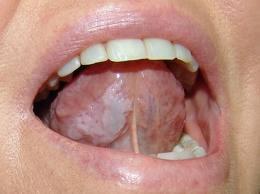
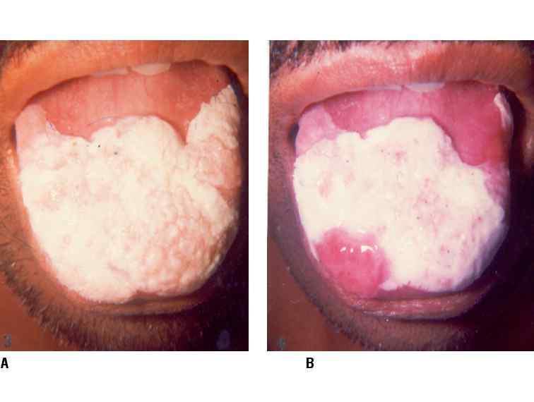
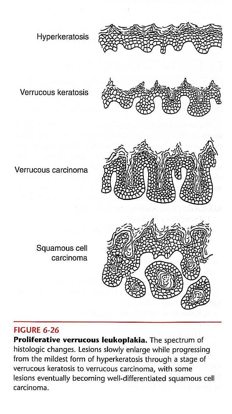
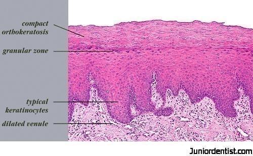
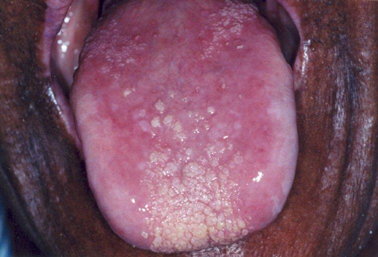
7 Clinical classification 1.Homogeneous (common) 2.Nodular leukoplakia 3. speckled (less common) 4. Proliferative verrucous (rare).


8 Clinical Features Site: mainly on buccal mucosa Features: uniformly white plaques Prognosis: Low malignant transformation potential
9
10


11 Features: 1.Small aggregated hemispherical red or white surface alterations 2. red background or substrate Prognosis Stronger risk of dysplasia or malignant potential than in homogeneous leukoplakia


12 Def. Regarded as a combination of or a transition between leukoplakia and erythroplakia. It is less common Clinical Features Site: buccal mucosa, tongue, floor of the mouth, gingiva S&S: 1.white flecks 2. fine nodules on an atrophic erythematous base Prognosis: Stronger malignant potential than homogeneous leukoplakia
13 Def. Diffuse white and /or papillary ( warty ) areas of the oral mucosa resulting from varying degrees of epithelial hyperplasia;has the potential to develop into verrucous carcinoma or well-differentiated.it is the least common type squamous cell carcinoma.
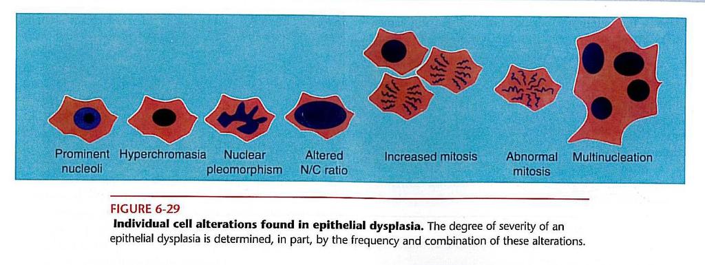
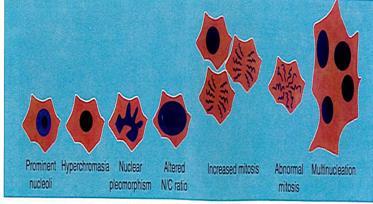
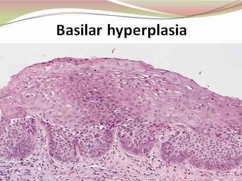
14
15 Prognosis: High risk of intervening dysplasia and carcinoma developing High rate of recurrence and histological progression toward carcinoma

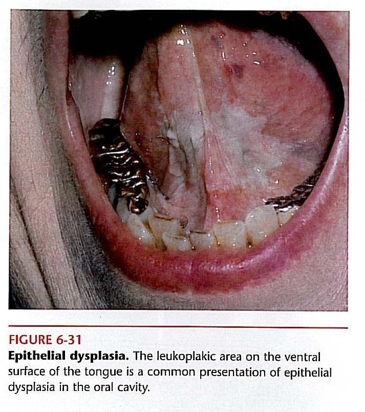
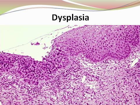
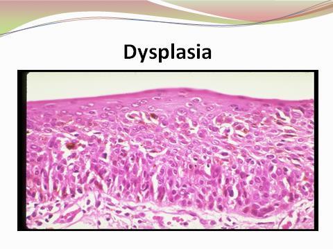
16 epithelial dysplasia
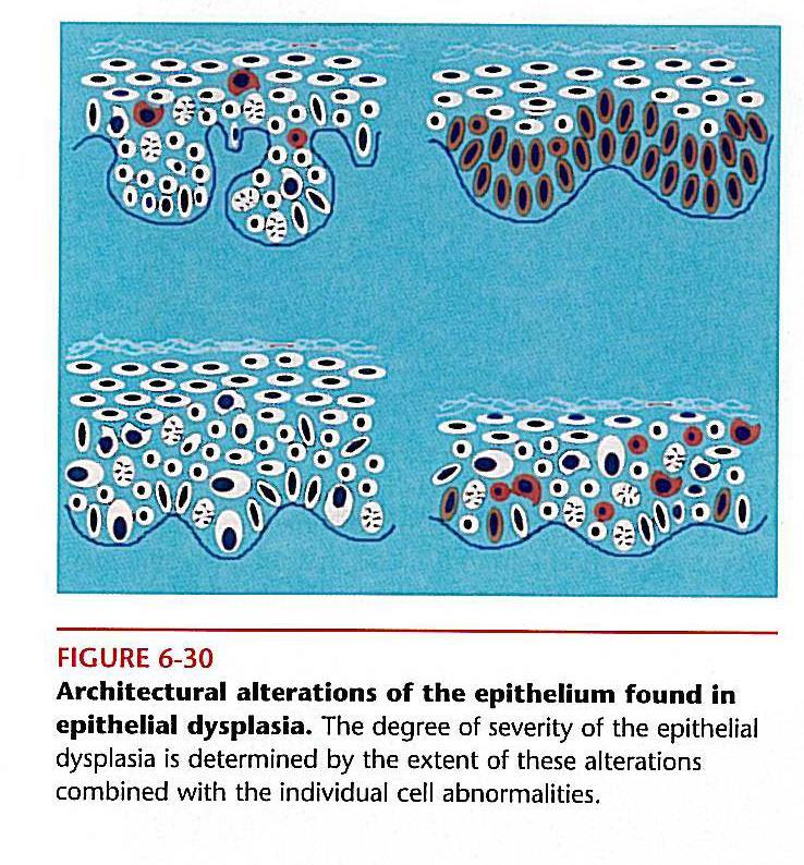
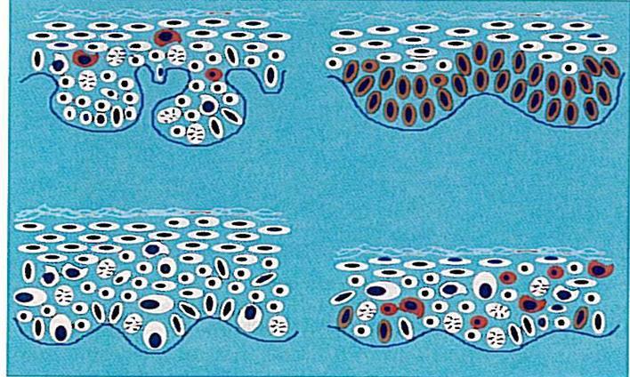
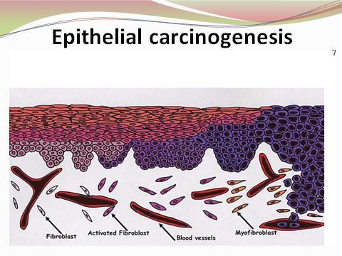
17 Epithelial atrophy Cellular atypia Epithelial dysplasia Carcinoma in situ Reduction in the Atypia is A premalignant change The most severe stage of normal thickness of individual cellular in epithelium epithelial dysplasia, epithelium that changes in characterized by a involving the entire involves less than the dysplastic combination of cellular thickness of the entire thickness of epithelium which and architectural epithelium, with the the epithelium reflect alterations epithelial basement abnormalities in membrane remaining proliferation, intact. maturation, and differentiation of epithelial cells.
18
19
20 1.The histological appearances reflect varying degrees of keratosis 2.Changes in epithelial thickness 3.Epithelial dysplasia 4. Diffuse chronic inflammatory cell infiltration of varying severity in the lamina propria.
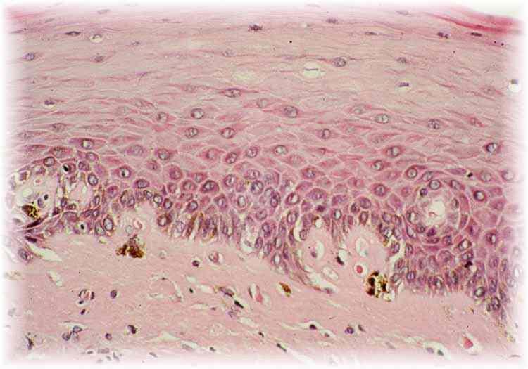
21 1. Nuclear and cellular pleomorphism (Nuclei and cells show different size and shape). 2. Increase in the nuclear/cytoplasmic ratio by either area or volume. 3. Nuclear hyperchromatism (Deeply stained Nucleus). 4. Prominent nucleoli. 5. Increased and abnormal mitoses. Mitoses may be increased in number, occur higher up in epithelium than is usual (i.e. away from the basal layer 6. Distributed polarity of the basal cells or loss of cellular orientation (The cells in the basal layer have no definable long axis and the nuclei have no regular polarity. 7. Basal cell hyperplasia. The presence of several layers of cells of basaloid appearance. 8. Drop-shaped rete pegs(the rete pegs are wider at their deeper part than they are more superficially). 9.Irregular epithelial stratification or distributed maturation. The cells no longer show a proper sequence of morphological and maturational changes as they pass from the basal layer to the surface. 10.Abnormal keratinization. Keratinization occurring below the normal keratin layer, either as individual cell keratinization with in the stratum spinosum or as disturbed maturation of groups of cells resulting in the formation of intraepithelial keratin pearls. 11-Loss or reduction of intercellular adhesion (or cohesion). This may be difficult to distinguish from intercellular oedema.
22
23
24
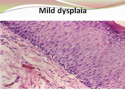
25 Types: 1.Mild epithelial dysplasia 2. Moderate epithelial dysplasia 3.Severe epithelial dysplasia

26 Alterations limited principally to the basal and parabasal layers.

27 Demonstrates involvement from the basal layer to the midportion of the spinous layer.

28 Demonstrates alterations from the basal layer to above midpoint of the epithelium.
29
30
31
32

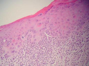
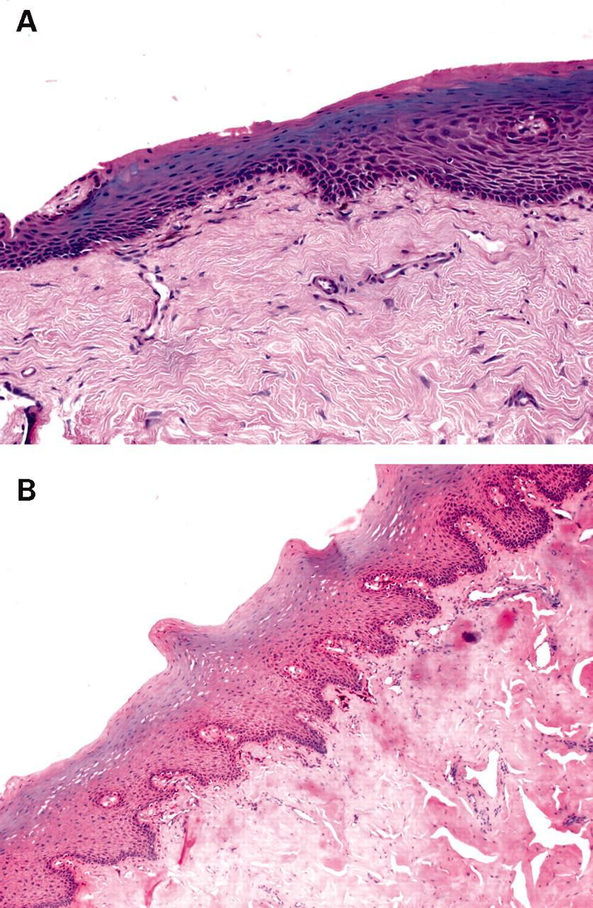
33 Def. Carcinoma in situ is defined as dysplastic epithelial cells that extend from the basal layer to the surface of the mucosa " top to bottom" change. Histopathology 1.There may or may not be a thin layer of parakeratin on the surface. 2. The epithelium may be hyperplastic or atrophic. 3. the entire thickness of the epithelium is involved 4. No invasion has occurred despite the fact that a typical epithelial- cell look exactly like those of squamous cell carcinoma. 5. Keratin pearl formation is rare in carcinoma in situ and may indicate the presence of a focus of invasive squamous cell carcinoma
34
35
36
37 1.Leukoplakia is generally diagnosed with an oral exam. 2. During a physical exam, your dentist can confirm if the patches are leukoplakia. 3. Other tests may be needed to confirm the cause. 4. Biopsy ( A small tissue sample is sent to a pathologist for diagnosis. The goal is to rule out the possibility of oral cancer). D.D Oral thrush is a yeast infection of the mouth. The patches it causes are usually softer than leukoplakia patches. They may bleed more easily. With treatment, you may be able to prevent future patches from developing.

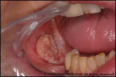
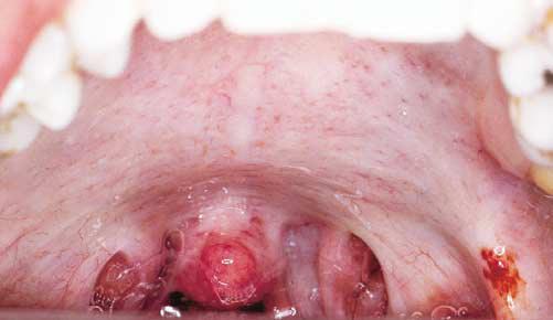
38 Erythroplakia
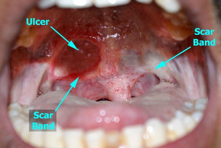
 years old Site: floor of mouth, tongue, and soft palate S&S: 1. asymptomatic 2. well demarcated erythematous macule 3. plaque with a soft velvety texture. 4.")

39 Erythroplakia Def. Red - patch that cannot be clinically or pathologically diagnosed as any other condition Etiology Clinical features 1.Unknown 2.epithelial dysplasia 3.Carcinoma in situ Age: older men (65-74) years old Site: floor of mouth, tongue, and soft palate S&S: 1. asymptomatic 2. well demarcated erythematous macule 3. plaque with a soft velvety texture. 4. associated with an adjacent leukoplakia ( erythroplakia). Histopathology 1.several epithelial dysplasia 2. carcinoma in situ 3.superficially invasive squamous cell carcinoma. 4.The epithelium shows a lack of keratin production and often is atrophic, but it may be hyperplastic. 5.This lack of keratinization, especially when combined epithelial thinness allows the underlying microvasculature to show through, thereby explaining the red color. 6.The underlying connective tissue often demonstrate chronic inflammation Differential Diagnosis 1.Non specific mucsitis 2. Candidiasis 3. Vascular lesions These lesions may clinically mimic Erythroplakia Biopsy is often required to distinguish between them

40 Oral submucous fibrosis

 2, mucosal pain associated with spicy food. 3.")
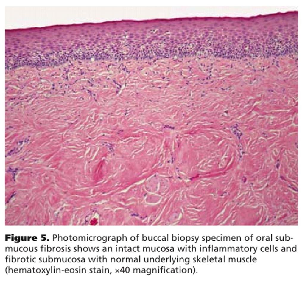
41 Oral submucous fibrosis Def. It is a chronic progressive, diffuse firm whitish areas of submucosal scarring usually caused by frequent and prolonged contact with betel nut quids,tobacco,or hot chili peppers; lesions have high-risk precancerous condition of the oral mucosa seen primarily on the Indian and in South East Asia. Etiology Clinical features Histopathology 1. tobacco 2. betel nut quids 3. hot chili peppers Site: buccal mucosa retromalar area, and the soft palate Age: young adult S&S:1. inability to open the mouth ( trismus ) 2, mucosal pain associated with spicy food. 3. The jaws may actually be inseparable in the advanced cases. 4. Vesicles, petachiae, melanosis, xerostomia 5. Generalized oral burning sensation are usually the first signs and symptoms. 6. The mucosa in these regions develops a blotchy marblelike pallor and a progressive stiffness of subepithelial tissues. 7. When the tongue is involved, it becomes rather immobile frequently diminished in size and often avoid of papillae. 1.submucosal deposition of extremely dense & a vascular collagenous connective tissue 2. variable numbers of chronic inflammatory cells. 3. Epithelial changes include hyperkeratosis with marked epithelial atrophy. 4. Epithelial dysplasia without carcinoma is found.
42
43
44
45
46
47

48 Chronic hyperplastic candidosis (candidal leukoplakia)


 4. Roughly triangular 5. Often bilateral white plaques tapering posteriorly Histopathology.1.")
49 Chronic hyperplastic candidosis (candidal leukoplakia) A clinical form of C. albicans infection consisting of white plaques or papules against an erythematous background containing hyphae in the parakeratin layer of the thickened epithelium. Etiology 1.Tobacco 2. Smoking 3. Denture wearing 4. Occlusal friction Clinical features Site: Lesions are seen most frequently on the buccal mucosa to the commissure of the lips S&S: 1. Dense, opaque white patches of irregular thickness and density with a rough or nodular surface 2. They cannot be removed by scraping,but fragments may be detached & identification of hyphae in smears of such material assists in the diagnosis. 3. Speckled leukoplakia (areas fo erythematous mucosa are present within the plaque) 4. Roughly triangular 5. Often bilateral white plaques tapering posteriorly Histopathology.1.Parakeratinized epithelium 2. Markedly hyperplastic and acanthotic epithelium 3. Many of cells in tparakeratinizedd surface of epitheliums separated by oedema 4.Micro-abscesses from numerous neutrophil leucocytes 5. Candidal hyphae invade the parakeratin more or less at right angles to the surface, but never penetrate deeper into the prickle cell layers 6.Acute and chronic inflammatory cells innprickle cell layer 7. Mixed chronic inflammatory cell infiltrate lamina propria) 8. Areas of atrophic epithelium may be present within the lesion and in these areas the superficial layers of candida infected parakeratinnmay be missing which may be responsible for the erythematous appearance seen clinically
50 Thank you
04/09/2018. Squamous Cell Neoplasia and Precursor Lesions. Agenda. Squamous Dysplasia. Squamo-proliferative lesions. Architectural features
 Squamous Cell Neoplasia and Precursor Lesions Jennifer L. Hunt, MD, MEd Aubrey J. Hough Jr, MD, Endowed Professor of Pathology Chair of Pathology and Laboratory Medicine University of Arkansas for Medical
Squamous Cell Neoplasia and Precursor Lesions
 Squamous Cell Neoplasia and Precursor Lesions Jennifer L. Hunt, MD, MEd Aubrey J. Hough Jr, MD, Endowed Professor of Pathology Chair of Pathology and Laboratory Medicine University of Arkansas for Medical
Diagnostic difficulties with lesions of the oral mucosa
 BDIAP London, November 2010 School of Clinical Dentistry University of Sheffield Diagnostic difficulties with lesions of the oral mucosa Paul M Speight Dept Oral & Maxillofacial Pathology University of
Dysplasia, Mimics and Other Controversies
 Dysplasia, Mimics and Other Controversies Mary S. Richardson, MD Dept. of Pathology Medical University of South Carolina Charleston, SC Notice of Faculty Disclosure In accordance with ACGME guidelines,
NEOPLASMS OF THE SURFACE EPITHELIUM (KERATINOCYTES)
") NEOPLASMS OF THE SURFACE EPITHELIUM (KERATINOCYTES) Papillary Lesions Precancerous Lesions Keratinocyte Proliferations Carcinomas Melanotic Lesions Melanomas Normal Mucosa Keratin layer Spinous layer Basal
NEOPLASMS OF THE SURFACE EPITHELIUM (KERATINOCYTES) Papillary Lesions Precancerous Lesions Keratinocyte Proliferations Carcinomas Melanotic Lesions Melanomas Normal Mucosa Keratin layer Spinous layer Basal
Lesions & Lifestyles
 Lesions & Lifestyles attended a 3 hour Continuing Education Seminar on Oral Pathology presented by Nancy Dewhirst, RDH,BS on (date) at (location):. Course material is directly related patient care. Notes:
الطلاوة = Leukoplakia LEUKOPLAKIA
 LEUKOPLAKIA Leukoplakia is a clinical term that refers to a predominantly white lesion of the oral mucosa that cannot be rubbed off or characterized by any other definable lesion or known disease. 130
Squamous Cell Carcinoma of the Head and Neck (SCCHN)
") Squamous Cell Carcinoma of the Head and Neck (SCCHN) Part 1 Bruce M. Wenig, M.D. Dept. of Pathology & Laboratory Medicine Continuum Health Partners New York, NY College of American Pathologists 2004. Materials
Squamous Cell Carcinoma of the Head and Neck (SCCHN) Part 1 Bruce M. Wenig, M.D. Dept. of Pathology & Laboratory Medicine Continuum Health Partners New York, NY College of American Pathologists 2004. Materials
That. Name QUIZ. 60 SEPTEMBER 2017 // dentaltown.com
 QUIZ Name That General dentists are first in the line of practitioners that patients see for an oral lesion evaluation; therefore, a sound understanding of oral mucosal diseases and their clinical presentation
Dr Rodney Itaki Lecturer Division of Pathology Anatomical Pathology Discipline
 Oral Lesions & Oral Cancer Dr Rodney Itaki Lecturer Division of Pathology Anatomical Pathology Discipline University of Papua New Guinea School of Medicine & Health Sciences Division of Pathology Overview
Diseases of oral cavity
 Diseases of oral cavity Diseases of Teeth and Supporting Structures Inflammatory/Reactive Lesions Infections Oral Manifestations of Systemic Disease Precancerous and Cancerous Lesions Odontogenic Cysts
Benign and malignant epithelial lesions: Seborrheic keratosis: A common benign pigmented epidermal tumor occur in middle-aged or older persons more
 Benign and malignant epithelial lesions: Seborrheic keratosis: A common benign pigmented epidermal tumor occur in middle-aged or older persons more common on the trunk; but extremities, head and neck are
Clinically Microscopically Pathogenesis: autoimmune not lifetime
 Vulvar Diseases: Can be divided to non-neoplastic and neoplastic diseases. The neoplastic diseases are much less common. Of those, squamous cell carcinoma is the most common. most common in postmenopausal
Histopathology: Cervical HPV and neoplasia
 Histopathology: Cervical HPV and neoplasia These presentations are to help you identify basic histopathological features. They do not contain the additional factual information that you need to learn about
Review Article- Leukoplakia: A mysterious white patch.
 International Journal Of Scientific Research And Education Volume 2 Issue 9 Pages 1824-1830 September-2014 ISSN (e): 2321-7545 Website: http://ijsae.in Review Article- Leukoplakia: A mysterious white patch.
LARYNGEAL DYSPLASIA. Tomas Fernandez M; 3 rd year ENT resident, Son Espases University Hospital
 LARYNGEAL DYSPLASIA Tomas Fernandez M; 3 rd year ENT resident, Son Espases University Hospital INTRODUCTION Laryngeal cancer constitutes 1-2% of all malignancies diagnosed worldwide Survival is related
ORAL LEUKOPLAKIA IN A SOUTH AFRICAN SAMPLE: A CLINICOPATHOLOGICAL STUDY
 ORAL LEUKOPLAKIA IN A SOUTH AFRICAN SAMPLE: A CLINICOPATHOLOGICAL STUDY Rakesh Chandran A research report submitted to the Faculty of Health Sciences, University of Witwatersrand, Johannesburg, in partial
LESIONS OF THE ORAL CAVITY ORAL CAVITY. Oral Cavity Subsites 4/10/2013 LIPS TEETH GINGIVA ORAL MUCOUS MEMBRANES PALATE TONGUE ORAL LYMPHOID TISSUES
 LESIONS OF THE ORAL CAVITY David I. Kutler, MD, FACS Associate Professor Division of Head and Neck Surgery Department of Otolaryngology HNS Weill Cornell Medical Center ORAL CAVITY LIPS TEETH GINGIVA ORAL
Histopathology: skin pathology
 Histopathology: skin pathology These presentations are to help you identify, and to test yourself on identifying, basic histopathological features. They do not contain the additional factual information
CINtec p16 INK4a Staining Atlas
 CINtec p16 INK4a Staining Atlas Rating Rating Positive The rating positive will be assigned if the p16 INK4a -stained slide shows a continuous staining of cells of the basal and parabasal cell layers of
Oral Cancer Dr Christine Goodall Consultant Oral Surgeon University of Glasgow Dental School
 Oral Cancer Dr Christine Goodall Consultant Oral Surgeon University of Glasgow Dental School christine.goodall@glasgow.ac.uk Locations Lip, mouth, oropharynx Tongue, floor of mouth, buccal mucosa, palate,
Pathology of the skin. 2nd Department of Pathology, Semmelweis University
 Pathology of the skin 2nd Department of Pathology, Semmelweis University Histology of the skin Epidermis: Stratum corneum Stratum granulosum Stratum spinosum Stratum basale Dermis: papillary and reticular
LEUKOPLAKIA Definition Epidemiology Clinical presentation
 LEUKOPLAKIA Definition Leukoplakia is the most common premalignant or "potentially malignant" lesion of the oral mucosa. Leukoplakia is a predominantly white lesion of the oral mucosa than cannot be clinicopathologically
Gastrooesophageal reflux disease. Jera Jeruc Institute of pathology, Faculty of Medicine, Ljubljana, Slovenia
 Gastrooesophageal reflux disease Jera Jeruc Institute of pathology, Faculty of Medicine, Ljubljana, Slovenia Reflux esophagitis (RE) GERD: a spectrum of clinical conditions and histologic alterations resulting
Oral Epithelial Tumors, Melanocytic Nevi, and Melanoma (I)
") Introduction: Oral Epithelial Tumors, Melanocytic Nevi, and Melanoma (I) Oral Epithelial Tumors may be: Benign tumors Sequamous cell Papilloma Malignant tumors Sequamous cell carcinoma, Basal cell carcinoma
Introduction: Oral Epithelial Tumors, Melanocytic Nevi, and Melanoma (I) Oral Epithelial Tumors may be: Benign tumors Sequamous cell Papilloma Malignant tumors Sequamous cell carcinoma, Basal cell carcinoma
OROPHYRENGEAL CANCERS
 OROPHYRENGEAL CANCERS INTRODUCTION 2 % 4 % of all malignant Tumors in west Asia India 40% Men ^ Age :Over 60 yrs 90% of all oral cancers results from Tobacco and Alcohol Pan (Betel Leaf,Nut, Lime), Reverse
Chapter 5. Oxygenated Hemoglobin Diffuse Reflectance Ratio for In Vivo Detection of oral Pre-cancer
 Chapter 5 Oxygenated Hemoglobin Diffuse Reflectance Ratio for In Vivo Detection of oral Pre-cancer This work is published in: JB0 (SPIE) 13(4):041306 (1-10), 2008 Oxygenated Hemoglobin Diffuse Reflectance
Prepared By Jocelyn Palao and Layla Faqih
 Prepared By Jocelyn Palao and Layla Faqih The structure of the suspected atypical cell should always be compared to the structure of other similar, benign, cells which are present in the smears. The diagnosis
Neoplasia 2018 Lecture 2. Dr Heyam Awad MD, FRCPath
 Neoplasia 2018 Lecture 2 Dr Heyam Awad MD, FRCPath ILOS 1. List the differences between benign and malignant tumors. 2. Recognize the histological features of malignancy. 3. Define dysplasia and understand
Papillary verrucous lesion of the oral mucosa: A need for detailed histopathological examination
 Case report DOI: http://dx.doi.org/10.18320/jimd/201502.03163 JOURNAL OF INTERNATIONAL MEDICINE AND DENTISTRY To search..to know...to share p-issn: 2454-8847 e-issn: 2350-045X Papillary verrucous lesion
Dermatopathology: The tumor is composed of keratinocytes which show atypia, increase mitoses and abnormal mitoses.
 Squamous cell carcinoma (SCC): A common malignant tumor of keratinocytes arising in the epidermis, usually from a precancerous condition: 1- UV induced actinic keratosis, usually of low grade malignancy.
Problem diagnoses. Current issues in Anatomic pathology. Problem Diagnoses in Tumors of the Oral Cavity 5/29/2009
 Current issues in Anatomic pathology Problem Diagnoses in Tumors of the Oral Cavity Richard Jordan DDS PhD FRCPath Professor of Oral Pathology & Pathology Director, UCSF Oral Pathology Diagnostic Laboratory
Oral Manifestations of Dermatologic Disease: A Focus on Lichenoid Lesions. Proceedings of the NASHNP Companion Meeting, March, 2011, San Antonio, TX
 1 Oral Manifestations of Dermatologic Disease: A Focus on Lichenoid Lesions Proceedings of the NASHNP Companion Meeting, March, 2011, San Antonio, TX Susan Müller, DMD, MS Professor Department of Pathology
HEAD AND NECK PATHOLOGY
 Bosnian-British School of Pathology November 2012 HEAD AND NECK PATHOLOGY Slide seminar: Oral Pathology Preferred Diagnoses Dr A Sandison, Slide seminar: Pathology of the Oral Cavity Page 1 of 5 1. Female
Premalignant lesion is a morphologically altered tissue in which cancer is more likely to occur, than its apparently normal counter parts.
 Oral Premalignancy Premalignant lesion is a morphologically altered tissue in which cancer is more likely to occur, than its apparently normal counter parts. Premalignant condition is a generalized state
When Immunostains Can Get You in Trouble: Gynecologic Pathology p16: Panacea or Pandora s Box?
 When Immunostains Can Get You in Trouble: Gynecologic Pathology p16: Panacea or Pandora s Box? Teri A. Longacre, MD Stanford Medicine Stanford California pi6 in Gynecologic Pathology: Panacea or Pandora
Role of the Dental Hygienist in Oral Pathology. Role of the Dental Hygienist in Oral Pathology. Cancers of the Oral Cavity.
 Gum Gardeners Study Club April 25, 2016 Early Detection of Oral Cancer Cindy Kleinegger, DDS, MS NW Oral Pathology Tigard, OR nworalpathology.com Role of the Dental Hygienist in Oral Pathology Work closely
DENIS P. LYNCH, DDS, PHD
 140 TH ANNUAL MEETING MAY 6 MAY 7, 2010 JEWEL OF THE GREAT LAKES DENIS P. LYNCH, DDS, PHD FRIDAY, MAY 7, 2010 9:00 A.M. TO 12:00 NOON ORAL CANCER AND RELATED PREMALIGNANCY Oral Cancer and Premalignancy
ACCURATE DIAGNOSIS IS THE ONLY TRUE CORNERSTONE ON WHICH RATIONAL TREATMENT CAN BE BUILT. C Noyek
 ACCURATE DIAGNOSIS IS THE ONLY TRUE CORNERSTONE ON WHICH RATIONAL TREATMENT CAN BE BUILT. C Noyek Oral diagnostics Definition of the discipline That area of dentistry, the which deals with gathering, recording
A Speckled Lesion. Angela C. Chi, DMD; Michele Carter Ravenel, DMD
 A Speckled Lesion Angela C. Chi, DMD; Michele Carter Ravenel, DMD The following Case Challenge is provided in conjunction with the American Academy of Oral and Maxillofacial Pathology. Case Summary This
Pattern of oral lesions Cytohistopathological study in tertiary care centre.
 International Journal of Current Research in Medical Sciences ISSN: 2454-5716 P-ISJN: A4372-3064, E -ISJN: A4372-3061 www.ijcrims.com Original Research Article Volume 3, Issue 10-2017 Pattern of oral lesions
New Diagnoses Need New Approaches: A Glimpse into the Near Future of Gynecologic Pathology
 New Diagnoses Need New Approaches: A Glimpse into the Near Future of Gynecologic Pathology United States and Canadian Academy of Pathology 102 nd Annual Meeting Baltimore, Maryland Christina S. Kong, M.D.
Oral cavity cancer accounts for approximately 3% of all malignancies and is a significant worldwide health problem.
 Oral cavity cancer accounts for approximately 3% of all malignancies and is a significant worldwide health problem. Majority are SCC ( 5-year survival rate only about 50-60% ) Many SCC arrive from premalignant
The Oral Cavity. Image source:
 The Oral Cavity Anatomy Image source: http://anatomyforlayla.blogspot.co.za/2007/04/blog-post.html The major structures of the oral cavity are the lips, the teeth, the alveolar ridges (bony areas that
Oral Cancer and Common Oral Lesions seen in HIV Seropositive Patients. Gwen Cohen Brown DDS, FAAOMP Professor New York City College of Technology
 Oral Cancer and Common Oral Lesions seen in HIV Seropositive Patients Gwen Cohen Brown DDS, FAAOMP Professor New York City College of Technology Program Objectives Recognize the oral health needs of the
LGM International, Inc.
 Liqui-PREP TM Cytology Atlas Preface The following pictures are examples with descriptions of cytology slides processed with the Liqui-PREP TM System.. The descriptions are reviewed by Pathologists. It
CASE REPORT PLAQUE TYPE ORAL VERRUCOUS HYPERPLASIA AND IRRITATIONAL FIBROMA: A REPORT OF CONJOINT OCCURRENCE
 CASE REPORT PLAQUE TYPE ORAL VERRUCOUS HYPERPLASIA AND IRRITATIONAL FIBROMA: A REPORT OF CONJOINT OCCURRENCE Alphy Alphonsa Sebastian, Hasan Subhi 1. Phd student, Department of Oral Medicine and Oral Pathology,
Evaluation and Management of Head and Neck Cancer in Patients with Fanconi anemia David I. Kutler, M.D., F.A.C.S.
 Evaluation and Management of Head and Neck Cancer in Patients with Fanconi anemia David I. Kutler, M.D., F.A.C.S. Residency Site Director Weill Cornell Medical Center Associate Professor Division of Head
Pathology of bladder cancer in Egypt; a current study.
 Pathology of bladder cancer in Egypt; a current study. Thesis Submitted for partial fulfillment of Master degree in urology By Mohamed Atef Mohamed Ahmed M.B.B.CH Supervised by Prof.Dr.: Omar Mohamed Abdel-
number Done by Corrected by Doctor Maha Shomaf
 number 16 Done by Waseem Abo-Obeida Corrected by Zeina Assaf Doctor Maha Shomaf MALIGNANT NEOPLASMS The four fundamental features by which benign and malignant tumors can be distinguished are: 1- differentiation
Leukoplakia is a white patch on the oral mucous membrane, which is undeliable and can not diagnose neither clinically nor pathologically as an other
 Leukoplakia Leukoplakia is a white patch on the oral mucous membrane, which is undeliable and can not diagnose neither clinically nor pathologically as an other disease. (Pindborg. 1978) Precancerous lesion
4Ps LUMPS AND BUMPS B.L.&T. BUMPS, LUMPS, AND TATTOOS. Most Common BUMP in the oral cavity Fibroma INTERDENTAL PAPILLAE LESIONS
 B.L.&T. BUMPS, LUMPS, AND TATTOOS LUMPS AND BUMPS DIFFERENTIAL DIAGNOSIS FOR LUMPS AND BUMPS Traumatic Fibroma Papilloma Epulis Fissuratum Inflammatory Papillary Hyperplasia Lesions of Attached Gingiva
Diseases of the breast (1 of 2)
") Diseases of the breast (1 of 2) Introduction A histology introduction Normal ducts and lobules of the breast are lined by two layers of cells a layer of luminal cells overlying a second layer of myoepithelial
Diseases of the breast (1 of 2) Introduction A histology introduction Normal ducts and lobules of the breast are lined by two layers of cells a layer of luminal cells overlying a second layer of myoepithelial
WHITE LESIONS OF THE ORAL CAVITY - diagnostic appraisal & management strategies
 WHITE LESIONS OF THE ORAL CAVITY - diagnostic appraisal & management strategies * Joshy V.R ** Hari.S * Reader, Dept of Oral Pathology, Yenepoya Dental College, Yenepoya University, Mangalore 575 018.
Head & Neck Squamous Carcinoma: Artifacts, Challenges, and Controversies. Agenda
 Head & Neck Squamous Carcinoma: Artifacts, Challenges, and Controversies Jennifer L. Hunt, MD, MEd Aubrey J. Hough Jr, MD, Endowed Professor of Pathology Chair of Pathology and Laboratory Medicine University
Demystifying Endometrial Hyperplasia
 Demystifying Endometrial Hyperplasia A review from Diagnostic Histopathology 19:7 Dr R Hadden ST5 Histopathology Derriford Hospital Plymouth Endometrium Target for sex-steroid hormones Glands Stroma Proliferate
Actinic keratosis (AK): Dr Sarma s simple guide
: Dr Sarma s simple guide") Actinic keratosis (AK): Dr Sarma s simple guide Actinic keratosis is a very common lesion that you will see in your day-to-day practice. First, let me explain the name Actinic keratosis. It means keratosis
Actinic keratosis (AK): Dr Sarma s simple guide Actinic keratosis is a very common lesion that you will see in your day-to-day practice. First, let me explain the name Actinic keratosis. It means keratosis
-1- Pathology Department (code: 0605) Final Exam for Third year students Date: Time allowed: 2 hours. Paper II (75 marks).
 Final Exam for Third year students Date: Time allowed: 2 hours. Paper II (75 marks).") -1- BENHA UNIVERSITY FACULTY OF MEDICINE Pathology Department (code: 0605) Final Exam for Third year students Date: 28-5-2011 Time allowed: 2 hours. Paper II (75 marks). Please note that this question
-1- BENHA UNIVERSITY FACULTY OF MEDICINE Pathology Department (code: 0605) Final Exam for Third year students Date: 28-5-2011 Time allowed: 2 hours. Paper II (75 marks). Please note that this question
Proliferative Verrucous Leukoplakia of the Gingiva, Report of two Cases with Malignant Transformation
 Journal of Clinical and Anatomic Pathology Case Report Open Access Proliferative Verrucous Leukoplakia of the Gingiva, Report of two Cases with Malignant Transformation Nadereh Ghanee DMD, Selene Saraf
SESSION 1: GENERAL (BASIC) PATHOLOGY CONCEPTS Thursday, October 16, :30am - 11:30am FACULTY COPY
 PATHOLOGY CONCEPTS Thursday, October 16, :30am - 11:30am FACULTY COPY") SESSION 1: GENERAL (BASIC) PATHOLOGY CONCEPTS Thursday, October 16, 2008 9:30am - 11:30am FACULTY COPY GOAL: Describe the basic morphologic (structural) changes which occur in various pathologic conditions.
SESSION 1: GENERAL (BASIC) PATHOLOGY CONCEPTS Thursday, October 16, 2008 9:30am - 11:30am FACULTY COPY GOAL: Describe the basic morphologic (structural) changes which occur in various pathologic conditions.
Epithelial dysplasia in lichenplanus
 Epithelial dysplasia in lichenplanus Girish HC* Sanjay Murgod**Savita JK*** *M.D.S, Professor & Head, **M.D.S, Professor,***M.D.S, Senior Lecturer,Dept of Oral Pathology, Raja Rajeswari Dental College
WHITE LESIONS OF THE UPPER AIRWAY
 WHITE LESIONS OF THE UPPER AIRWAY WHITE LESION CONFIGURATIONS Solitary vrs Multifocal Flat Plaque Verrucous/rippled Lacey White with red component Papular (curdled milk plaques) Pseudomembranous PLAQUES
Synonyms. Nephrogenic metaplasia Mesonephric adenoma
 Nephrogenic Adenoma Synonyms Nephrogenic metaplasia Mesonephric adenoma Definition Benign epithelial lesion of urinary tract with tubular, glandular, papillary growth pattern Most frequently in the urinary
Oral Cavity and Pharynx Cancer
 Oral Cavity and Pharynx Cancer Figure 18 Definition: Oral cancer begins in the mouth and can include the lips, cheeks, teeth, gums, the floor of the tongue, the roof of the mouth, and the front two-thirds
Oral Cancer- Improving Early Detection
 Oral Cancer- Improving Early Detection GDC Recommended Subject Aims: To give an overview of the dental team's role in detecting the early signs of oral cancer; to give an overview of the risk factors associated
5/21/2018. Prostate Adenocarcinoma vs. Urothelial Carcinoma. Common Differential Diagnoses in Urological Pathology. Jonathan I.
 Common Differential Diagnoses in Urological Pathology Jonathan I. Epstein Prostate Adenocarcinoma vs. Urothelial Carcinoma 1 2 NKX3.1 NKX3.1 3 4 5 6 Proposed ISUP Recommendations Option to use PSA as a
Diagnostic aids of oral cancer
 Diagnostic aids of oral cancer The World Health Organization has clearly indentified prevention and early detection as major objectives in the control of the oral cancer. At the present time, screening
A Rare case of Tubercular Gingivitis Case Report
 Case Report A Rare case of Tubercular Gingivitis Case Report *Dr. Ansh Chugh 1, Dr. Firoz A Hakkim 2, Dr. Rajesh. V 3, Dr. Raghava Sharma 4 1: JUNIOR RESIDENT IN GENERAL MEDICINE 2: SENIOR RESIDENT IN
Premalignant skin tumours
 Chapter 14: Premalignant skin tumours page: 434 Premalignant skin tumours page: 435 Solar keratoses (senile keratoses) Raised red and well-defined plaques with a rough surface covered in scales of varying
MECHANISMS OF HUMAN DISEASE: LABORATORY SESSION PATHOLOGY OF THE SKIN LAB. Friday, February 12, :30 am 11:00 am
 MECHANISMS OF HUMAN DISEASE: LABORATORY SESSION PATHOLOGY OF THE SKIN LAB Friday, February 12, 2012 9:30 am 11:00 am FACULTY COPY GOALS: Describe the basic clinical and morphologic features of various
Field Cancerization Of Oral Cavity - A Case Report And An Update
 ISPUB.COM The Internet Journal of Dental Science Volume 7 Number 1 Field Cancerization Of Oral Cavity - A Case Report And An Update A Mufeed, L Chatra, P Shenai Citation A Mufeed, L Chatra, P Shenai..
Rectal biopsy as an aid to cancer control in ulcerative colitis
 Rectal biopsy as an aid to cancer control in ulcerative colitis B. C. MORSON AND LILLIAN S. C. PANG From the Research Department, St. Mark's Hospital, London Gut, 1967, 8, 423 EDITORIAL COMMENT This is
SUBMUCOSA PRECEDES LAMINA PROPRIA IN INITIATING FIBROSIS IN ORAL SUBMUCOUS FIBROSIS - EVIDENCE BASED ON COLLAGEN HISTOCHEMISTRY.
 SUBMUCOSA PRECEDES LAMINA PROPRIA IN INITIATING FIBROSIS IN ORAL SUBMUCOUS FIBROSIS - EVIDENCE BASED ON COLLAGEN HISTOCHEMISTRY. *Anna P. Joseph ** R. Rajendran Abstract Oral submucous fibrosis is a chronic
RESULT OF SURVEY OF 1705 LAWSUITS* ORAL CANER: A significant Public Health Concern. ORAL CANCER: Other Epidemiologic Facts
 FAILURE TO DIAGNOSE ORAL CANCER AND OTHER PATHOLOGIC CONDITIONS OF THE ORAL CAVITY Kalu U.E. Ogbureke, BDS, MSc, DMSc, JD, FDSRCS, FDSRCPS, FDSRCSEd, FRCPath Diplomate, American Board of Oral and Maxillofacial
Diseases of the vulva
 Diseases of the vulva 1. Bartholin Cyst - Infection of the Bartholin gland produces an acute inflammation within the gland (adenitis) and may result in an abscess. Bartholin duct cysts - Are relatively
Oral histology. How is the oral mucosa different from skin?
 Oral histology Lec.13 Oral mucosa: Dr.Nada AL.Ghaban The oral mucosa is the mucous membrane lining the inside of the mouth and consists of stratified squamous epithelium termed oral epithelium and an underlying
CLINICAL SIGNIFICANCE OF BENIGN EPITHELIAL CHANGES
 Papillomas. Papillomas are composed of multiple branching fibrovascular cores, each having a connective tissue axis lined by luminal and myoepithelial cells ( Fig. 23-11 ). Growth occurs within a dilated
PACIFIC JOURNAL OF MEDICAL SCIENCES {Formerly: Medical Sciences Bulletin} ISSN:
 PACIFIC JOURNAL OF MEDICAL SCIENCES {Formerly: Medical Sciences Bulletin} ISSN: 2072 1625 Pac. J. Med. Sci. (PJMS) www.pacjmedsci.com. Email: pacjmedsci@gmail.com. BILATERAL RECURRENT SPECKLED LEUKOPLAKIA:
Red and White Tissue Reactions: A white appearance of the oral mucosa may be caused by: An increased production of keratin (hyperkeratosis).
.") Burket, chapter 4 Red and White Tissue Reactions: A white appearance of the oral mucosa may be caused by: An increased production of keratin (hyperkeratosis). An abnormal but benign thickening o stratum
Burket, chapter 4 Red and White Tissue Reactions: A white appearance of the oral mucosa may be caused by: An increased production of keratin (hyperkeratosis). An abnormal but benign thickening o stratum
SQUAMOUS CELLS: Atypical squamous cells (ASC) - of undetermined significance (ASC-US) - cannot exclude HSIL (ASC-H)
 - of undetermined significance (ASC-US) - cannot exclude HSIL (ASC-H)") SQUAMOUS CELLS: Atypical squamous cells (ASC) - of undetermined significance (ASC-US) - cannot exclude HSIL (ASC-H) ASC refers to cytologic changes suggestive of SIL, which are qualitativley or quantitatively
SQUAMOUS CELLS: Atypical squamous cells (ASC) - of undetermined significance (ASC-US) - cannot exclude HSIL (ASC-H) ASC refers to cytologic changes suggestive of SIL, which are qualitativley or quantitatively
E Queyrat in 1911 as occurring on the glans
 ERYTHROPLAKIA OF THE ORAL CAVITY WILLIAM G. SHAFER, DDS,* AND CHARLES A. WALDRON, DDS+ Erythroplakia of the oral cavity is a specific disease entity which must be differentiated from other specific or
PREVENTION OF ORAL CANCER
 PREVENTION OF ORAL CANCER Oral cancer is increasing in incidence worldwide. Throughout the world, malignant neoplasms of the mouth and pharynx rate as the fifth most common cancer in men and the seventh
Mody. AIS vs. Invasive Adenocarcinoma of the Cervix
 Common Problems in Gynecologic Pathology Michael T. Deavers, M.D. Houston Methodist Hospital, Houston, Texas Common Problems in Gynecologic Pathology Adenocarcinoma in-situ (AIS) of the Cervix vs. Invasive
4/16/2018. Bumps & Lumps. What s HPV Got To Do With It? 2 hour Oral & Pharyngeal Pathology Review Nancy Dewhirst RDH,BS
 4/6/08 Bumps & Lumps. What s HPV Got To Do With It? hour Oral & Pharyngeal Pathology Review Nancy Dewhirst RDH,BS www.nancydewhirst.com Bumps & Lumps What s HPV Got To Do With It? Patient Assessment Clinical
Basal cell carcinoma 5/28/2011
 Goal of this Presentation A practical approach to the diagnosis of cutaneous carcinomas and their mimics Thaddeus Mully, MD University of California San Francisco To review common non-melanoma skin cancers
Epithelial tumors. Dr. F.F. Khuzin, PhD Dr. M.O. Mavlikeev
 Epithelial tumors Dr. F.F. Khuzin, PhD Dr. M.O. Mavlikeev Epithelial tumors Tumors from the epithelium are the most frequent among tumors. There are 2 group features of these tumors: The presence in most
The Pathologist s Role in the Diagnosis and Management of Neoplasia in Barrett s Oesophagus Cian Muldoon, St. James s Hospital, Dublin
 The Pathologist s Role in the Diagnosis and Management of Neoplasia in Barrett s Oesophagus Cian Muldoon, St. James s Hospital, Dublin 24.06.15 Norman Barrett Smiles [A brief digression - Chair becoming
Morsicatio Mucosae Oris A Chronic Oral Frictional Keratosis, Not a Leukoplakia
 J Oral Maxillofac Surg 67:140-146, 2009 Morsicatio Mucosae Oris A Chronic Oral Frictional Keratosis, Not a Leukoplakia Sook-Bin Woo, DMD,* and Dorothy Lin Purpose: Morsicatio mucosae oris (MMO) presents
Pigmented lesions of the Oral cavity
 Oral medicine أ.م.د احسان عبد هللا كميل Pigmented lesions of the Oral cavity Pigmented oral lesions are a large group of disorders in which the dark or brown color is the essential clinical characteristic.
Contents. 3 Diagnostic Tests and Studies Introduction Examination... 27
 Contents 1 Normal Anatomy... 1 1.1 Introduction... 1 1.2 Surface Landmarks... 1 1.3 Oral Mucosa... 3 1.4 Tongue... 5 1.5 Floor of Mouth... 6 1.6 Palate... 6 1.7 Dentition... 7 1.8 Temporomandibular Joint...
Conclusion: It was concluded that laser provides good coagulation, healing, reduces surgical time and prevents high-grade infection.
 Research AHB Article Adv Hum Biol 2014; 4(2):45-49. Clinical and Histopathological Evaluation of Healing After Excision of Leukoplakia with Diode Laser Kruti A Shah 1* Hemal R Brahmkshatriya 2 Rushit J
Clinical Relevance: Identification of various cell markers may provide valuable information of diagnostic and prognostic significance.
 ORAL MEDICINE An Overview of the Prevention of Oral Cancer and Diagnostic Markers of Malignant Change: 2. Markers of Value in Tumour Diagnosis M. MACLUSKEY AND G.R. OGDEN Abstract: Earlier diagnosis of
CPC 4 Breast Cancer. Rochelle Harwood, a 35 year old sales assistant, presents to her GP because she has noticed a painless lump in her left breast.
 CPC 4 Breast Cancer Rochelle Harwood, a 35 year old sales assistant, presents to her GP because she has noticed a painless lump in her left breast. 1. What are the most likely diagnoses of this lump? Fibroadenoma
INTERNATIONAL JOURNAL OF ADVANCES IN CASE REPORTS
 INTERNATIONAL JOURNAL OF ADVANCES IN CASE REPORTS e - ISSN - 2349-8005 www.mcmed.us/journal/ijacr Case Report VERRUCOUS CARCINOMA IN ASSOCIATION WITH OSMF 2 CASE REPORTS K. Saraswathi Gopal 1 * and B.
EXPERIMENTAL THERMAL BURNS I. A study of the immediate and delayed histopathological changes of the skin.
 EXPERIMENTAL THERMAL BURNS I A study of the immediate and delayed histopathological changes of the skin. RJ Brennan, M.D. and B. Rovatti M.D. The purpose of this study was to determine the progressive
Orofacial Disease: Update For The Dental Clinical Team: 3. White Lesions
 ORAL MEDICINE Orofacial Disease: Update For The Dental Clinical Team: 3. White Lesions Crispian Scully and Stephen Porter Abstract: White lesions usually contain an increased amount of keratin. Some are
Clinical behaviour of malignant transforming oral lichen planus
 EJSO 2002; 28: 838±843 doi:10.1053/ejso.2002.1302, available online at http://www.idealibrary.com on 1 Clinical behaviour of malignant transforming oral lichen planus M. D. Mignogna*, L. Lo Russo*, S.
Epidemiological and clinicopathological study of oral leukoplakia
 Study Epidemiological and clinicopathological study of oral Minati Mishra, Janardan Mohanty*, Sujata Sengupta, Satyabrata Tripathy Department of Dermatology & Venereology, S.C.B. Medical College, *Department
Original Article- A CYTOLOGICAL STUDY OF LEUKOPLASTIC LESIONS IN ORAL CAVITY
 Original Article- A CYTOLOGICAL STUDY OF LEUKOPLASTIC LESIONS IN ORAL CAVITY I. GUJRAL*, P. SINGH**, S. SHARMA***, N. GANGANE*** ABSTRACT Oral white lesions that cannot be clinically or pathologically