TB Radiology for Nurses Garold O. Minns, MD
|
|
|
- Brendan Osborne
- 5 years ago
- Views:
Transcription
1 TB Nurse Case Management Salina, Kansas March 31-April 1, 2010 TB Radiology for Nurses Garold O. Minns, MD April 1, 2010 TB Radiology for Nurses Highway Patrol Training Center Salina, KS April 1, 2010 Garold O. Minns, MD Professor and Program Director Department of Internal Medicine KU School of Medicine-Wichita Radiology Page 1 of 32


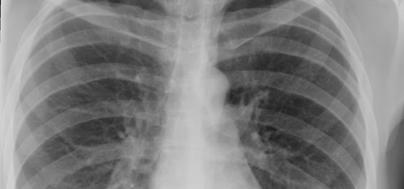
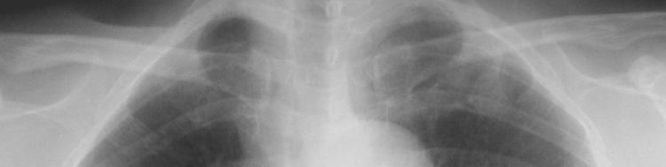
2 Frontal Technique Lateral Posteroanterior (PA) view of the chest Film Radiology Page 2 of 32


3 Anteroposterior (AP) view of the chest Film Items closer to film appear smaller! Object Film Radiology Page 3 of 32


4 Items closer to film appear smaller! Object Film PA View of the Chest Radiology Page 4 of 32


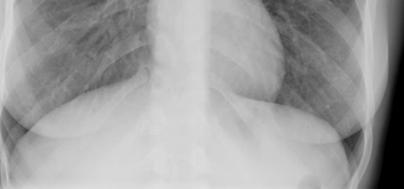
5 Anteroposterior (AP) view of the chest Film AP view of the chest Radiology Page 5 of 32


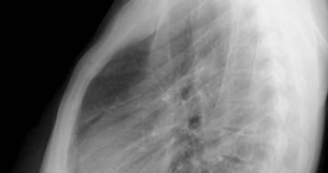
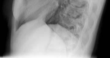
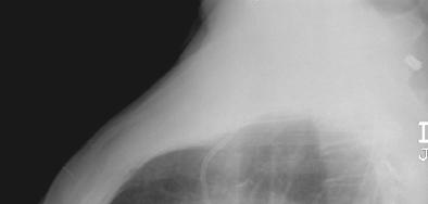
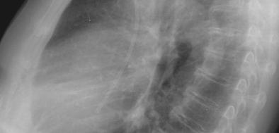
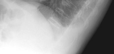
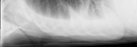
6 Lateral view of the chest Film Lateral View of the Chest Radiology Page 6 of 32


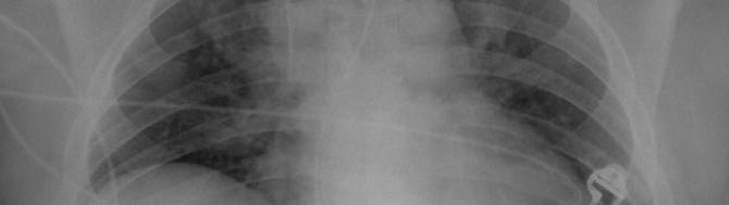
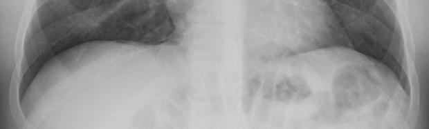
7 LLL pneumonia Pleural effusions Radiology Page 7 of 32


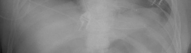
8 Other views Decubitus Apical lordotic Suspected pleural effusion Radiology Page 8 of 32


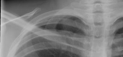
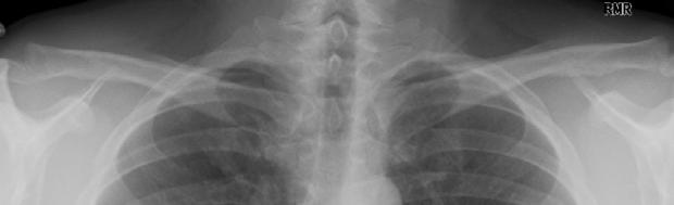
9 Apical TB Radiology Page 9 of 32


10 Apical lordotic Film Radiology Page 10 of 32


")
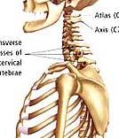
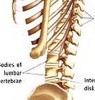
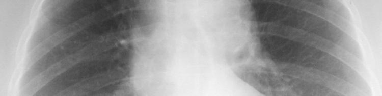
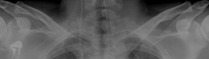
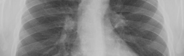
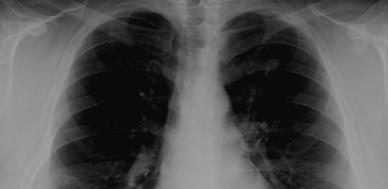
11 Assess CXR Technical Quality Inspiratory effort 9-10 posterior ribs Penetration thoracic intervertebral disc space just visible Positioning / rotation medial clavicle heads equidistant from spinous process Inspiration: ( 10 posterior ribs) Radiology Page 11 of 32


12 1st rib 3rd rib 2nd rib 1st 2nd 3rd 4th 5th 6th 7th 8th 9th 10th Radiology Page 12 of 32
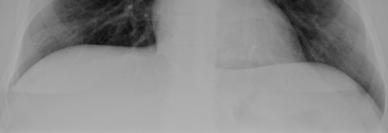
13 Poor inspiration Good inspiration Radiology Page 13 of 32
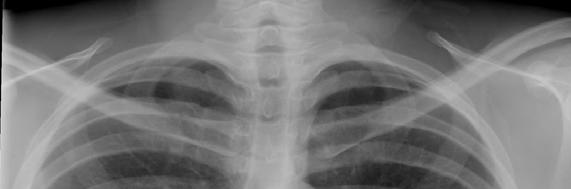
14 Overexposure Exposure Proper Exposure Intervertebral Disks Penetration Radiology Page 14 of 32

15 Over-penetrated Rotated (Oblique) Radiology Page 15 of 32


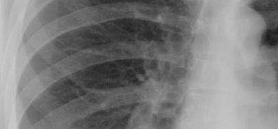
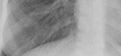
16 Apicesp Worth a Second Look Retrocardiac areas (left and right) Hilar regions Below diaphragm Radiology Page 16 of 32


17 Lobar anatomy RUL LUL RML RLL LLL Left Lung Right Lung Silhouette sign Heart RML RLL Dome of Right Hemidiaphragm Radiology Page 17 of 32


18 RML opacity obscures heart border RML RLL opacity obscures diaphragm RLL Obscured Diaphragm Clear Heart Border Radiology Page 18 of 32


19 Classification of lung pathology Alveolar Interstitial Airways Radiology Page 19 of 32


20 Alveolar Disease Consolidation Confluent opacity Fluffy around the periphery Air bronchograms Ill-defined nodules Diff dx depends on: Focal Diffuse Radiology Page 20 of 32


21 Consolidation / Air Space Opacity Caused by filling of alveoli with fluid, pus, blood, cells (tumor), etc. May be diffuse, or isolated to segments or lobes of the lung May be associated with air bronchograms (air-filled bronchus surrounded by opacified lung) Pneumonia Radiology Page 21 of 32

, sarcoidosis, infection, tumor (lymphangitic spread), etc.")
22 Interstitial Opacity Disease localized to pulmonary interstitium, i.e., the alveolar septae and connective tissues that support the alveoli Hallmarks: Lines and/or reticulation Small, well-defined nodules Miliary pattern DDX: Pulmonary edema, interstitial lung diseases (ex. idiopathic pulmonary fibrosis), sarcoidosis, infection, tumor (lymphangitic spread), etc. Interstitial Opacity: Lines & Reticulation Radiology Page 22 of 32


23 Nodules and Masses Nodule: discrete pulmonary lesion, sharply defined, nearly circular opacity cm Mass: larger than 3 cm Describe with qualifiers: Single or multiple Size Border characteristics Presence or absence of calcification Location Well-Defined Calcification Ill-Defined Mass Radiology Page 23 of 32


24 Lymphadenopathy (LAN) Non-specific terms: Mediastinal widening Hilar prominence Specific patterns: Particular station enlargement Important to know what normal should look like in order to recognize abnormal Radiology Page 24 of 32


25 Right Paratracheal & Bilateral LAN Right Hilar LAN Radiology Page 25 of 32
, filled")
 Cyst:")


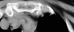
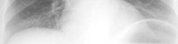
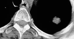
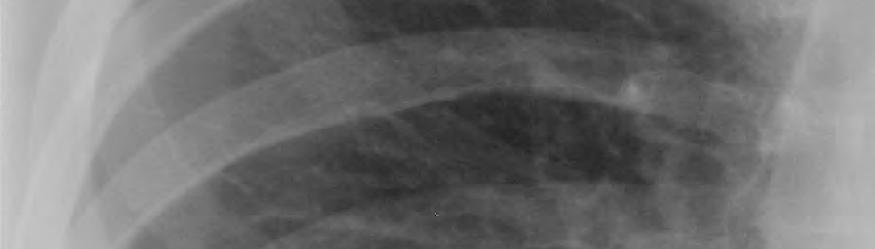
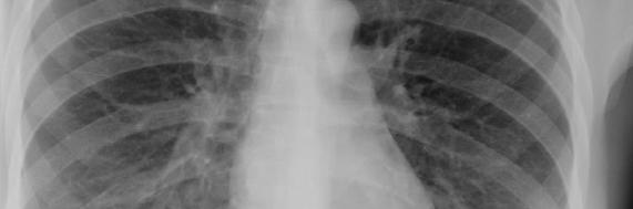
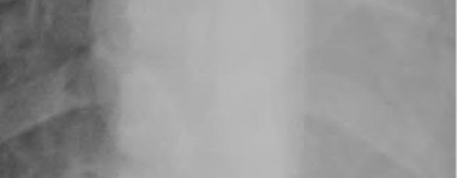
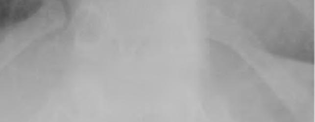
26 Cysts & Cavities Abnormal pulmonary parenchymal spaces ( holes ), filled with air and/or fluid, with a definable wall (>1 mm) Cyst: congenital or acquired Cavity: caused by tissue necrosis, (inflammatory and/or neoplastic) Characterize: Wall thickness at thickest portion Inner lining Presence/absence of air/fluid level Number and location TB or Not TB? Cysts and Cavities Are there radiographic features that suggest benign vs. malignant diagnoses? 45 yo man from China with cough, wt. loss A B C D Radiology Page 26 of 32


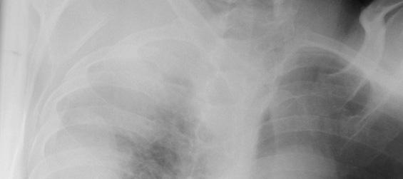
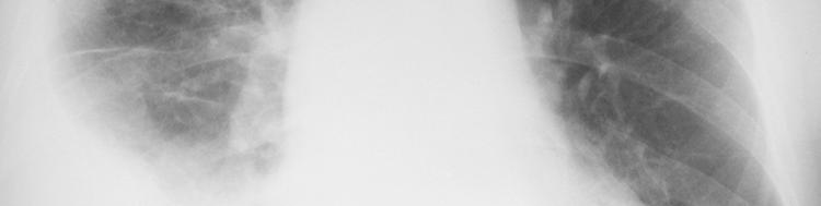
27 Pleural Disease: Basic Patterns Effusion Angle blunting to massive Thickening Mass Air Calcification Pleural Effusion Radiology Page 27 of 32


28 Can this be TB? Typical Pattern : Post-primary TB Distribution Apical / posterior segments of upper lobes Superior segments of lower lobes Isolated anterior segment involvement unusual for M.tb (think M. avium complex) Radiology Page 28 of 32
 Can this be TB?")
 Adenopathy is common (esp.")
29 Typical pattern : Post-Primary TB Patterns of disease Air-space consolidation Cavitation, cavitary nodule Endobronchial spread Miliary Bronchostenosis Tuberculoma Pleural effusions (empyema most likely in post-primary disease) Can this be TB? Atypical pattern : Primary TB Distribution : any lobe involved (slight lower lobe predominance) Air-space consolidation Cavitation is uncommon (<10%) Adenopathy is common (esp. children and HIV), predilection for right side Miliary pattern Pleural effusions Radiology Page 29 of 32


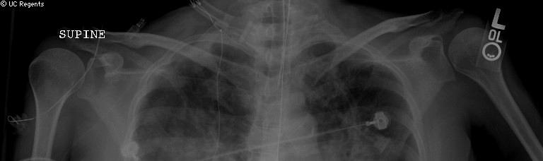
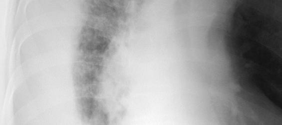
30 Miliary TB Insidious in onset, with general malaise, fever, weight loss, and sweats Typical diffuse miliary pattern often appears in the chest x-ray Sputum smear for AFB are only positive in 30% of cases Other organ involvement is not uncommon Miliary TB Radiology Page 30 of 32

31 Active pulmonary TB: HIV vs. Non-HIV Patients with HIV and TB: Normal chest x-ray, or with infiltrates in any lobes and any location No radiological appearance is pathognomonic of TB Pediatric TB Usually progression from primary TB Lower lobes Bulky lymphadenopathy LAD compression of bronchi Non-apical cavities Miliary forms Extrapulmonary Head and neck LAD (60%) Meningeal (10%) Radiology Page 31 of 32


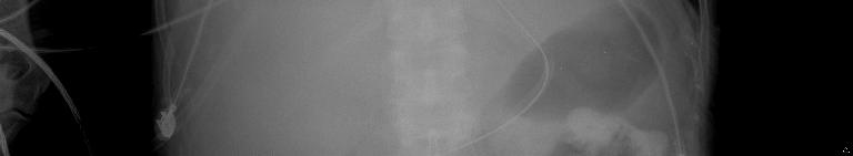
32 Pediatric TB Pleural TB Peripheral subpleural lesions pleural cavity tubercles effusion and empyema Pleural fluid: exudate with lymphocyte predominance Pleural fluid smear and culture is only positive in 1/3 Pleural tissue culture and granulomatous histology: diagnostic yield >70% Radiology Page 32 of 32
33 Radiographic Patterns: Pulmonary TB TB Pattern Infiltrate Typical (Post-Primary) 85% upper Atypical (Primary) Upper : Lower 60 : 40 Usually upper in children Cavitation Common Uncommon Adenopathy Uncommon Children common Adults ~30% Unilateral > bilateral Effusion May be present May be present CXR Pattern: Early vs. Advanced HIV Pattern Infiltrate Early HIV (CD4>200) Typical (Post-primary) Upper lobes Advanced HIV (CD4<200) Atypical (Primary) Lower lobes, multiple sites, or miliary Cavitation Common Uncommon Adenopathy Uncommon Common Effusion Uncommon More common Radiology Page 33 of 32


34 Can this be TB? Old / healed TB Ca ++ granuloma - Ghon lesion Ca ++ granuloma and hilar node calcification - Ranke complex Apical pleural thickening Fibrosis and volume loss26 Conclusions Primary TB usually involves lower lobes Reactivation TB usually involve upper lobes TB-HIV co-infected can present with normal chest x-ray, or with infiltrates in any lobes and any location No radiological appearance is pathognomonic of TB CT scan can improve chest X-ray evaluation Other image modalities such as MRI can be helpful Radiology Page 34 of 32
An Introduction to Radiology for TB Nurses
 An Introduction to Radiology for TB Nurses Garold O. Minns, MD September 14, 2017 TB Nurse Case Management September 12 14, 2017 EXCELLENCE EXPERTISE INNOVATION Garold O. Minns, MD has the following disclosures
Chest Radiology Interpretation: Findings of Tuberculosis
 Chest Radiology Interpretation: Findings of Tuberculosis Get out your laptops, smart phones or other devices pollev.com/chestradiology Case #1 1 Plombage Pneumonia Cancer 2 Reading the TB CXR Be systematic!
PULMONARY TUBERCULOSIS RADIOLOGY
 PULMONARY TUBERCULOSIS RADIOLOGY RADIOLOGICAL MODALITIES Medical radiophotography Radiography Fluoroscopy Linear (conventional) tomography Computed tomography Pulmonary angiography, bronchography Ultrasonography,
10/17/2016. Nuts and Bolts of Thoracic Radiology. Objectives. Techniques
 Nuts and Bolts of Thoracic Radiology October 20, 2016 Carleen Risaliti Objectives Understand the basics of chest radiograph Develop a system for interpreting chest radiographs Correctly identify thoracic
TB Intensive Houston, Texas
 TB Intensive Houston, Texas October 15-17, 17 2013 Diagnosis of TB: Radiology Rosa M Estrada-Y-Martin, MD MSc FCCP October 16, 2013 Rosa M Estrada-Y-Martin, MD MSc FCCP, has the following disclosures to
UERMMMC Department of Radiology. Basic Chest Radiology
 UERMMMC Department of Radiology Basic Chest Radiology PHYSICS DENSITIES BONE SOFT TISSUES WATER FAT AIR TELEROENTGENOGRAM Criteria for an Ideal Chest Radiograph 1. Upright 2. Posteroanterior View 3. Full
Radiological Aspects of Pulmonary Tuberculosis in Immunocompetent Hosts
 Nov 2003 Radiological Aspects of Pulmonary Tuberculosis in Immunocompetent Hosts Josh Rempell, Harvard Medical School Year III Tuberculosis: the captain of all (wo)men of death Overall, one third of the
Interpretation of Chest Radiographs Paul Christensen, MD 10/21/09. Diagnostic Evaluation. Medical Evaluation & CXR Interpretation.
 Diagnostic Evaluation Medical Evaluation & CXR Interpretation University of Michigan TB Consultant Washtenaw County Medical history Physical examination Testing for TB exposure (previously covered) Radiologic
Chest XRay interpretation INTERPRETATIONS Identifications: Name & Date Technical evaluation Basic Interpretations
 Chest XRay interpretation INTERPRETATIONS Identifications: Name & Date Technical evaluation Basic Interpretations TECHNICAL EVALUATION 1. Projection: AP/PA view To differentiate between AP & PA films,
Chest X-ray Interpretation
 Chest X-ray Interpretation Introduction Routinely obtained Pulmonary specialist consultation Inherent physical exam limitations Chest x-ray limitations Physical exam and chest x-ray provide compliment
Diagnosis of TB: Radiology David Finlay, MD
 TB Intensive Tyler, Texas June 2-4, 2010 Diagnosis of TB: Radiology David Finlay, MD June 3, 2010 2stages stages- Tuberculosis 1. primary infection 2. reactivation, or post primary disease 2 1 Primary
Let s Talk TB A Series on Tuberculosis, A Disease That Affects Over 2 Million Indians Every Year
 A Series on Tuberculosis, A Disease That Affects Over 2 Million Indians Every Year Barry Rabinovitch, MD, FRCP(C) Author Madhukar Pai, MD, PhD co-author and Series Editor Barry Rabinovitch is an assistant
Tuberculosis: The Essentials
 Tuberculosis: The Essentials Kendra L. Fisher, MD, PhD THORACIC TUBERCULOSIS: THE BARE ESSENTIALS Kendra Fisher MD, FRCP (C) Department of Radiology Loma Linda University Medical Center TUBERCULOSIS ()
Bronchial syndrome. Atelectasis Draining bronchus Bronchiectasis
 Bronchial syndrome Atelectasis Draining bronchus Bronchiectasis Etienne Leroy Terquem Pierre L Her SPI / ISP Soutien Pneumologique International / International Support for Pulmonology Atelectasis Consequence
Chest X-ray (CXR) Interpretation Brent Burbridge, MD, FRCPC
 Interpretation Brent Burbridge, MD, FRCPC") Chest X-ray (CXR) Interpretation Brent Burbridge, MD, FRCPC An approach to reviewing a chest x-ray will create a foundation that will facilitate the detection of abnormalities. You should create your own
Chest X-ray (CXR) Interpretation Brent Burbridge, MD, FRCPC An approach to reviewing a chest x-ray will create a foundation that will facilitate the detection of abnormalities. You should create your own
Manage TB Dr. A. Chitrakumar Madras Medical College and RGGGH Institute of Thoracic Medicine, Chennai
 Manage TB Dr. A. Chitrakumar Madras Medical College and RGGGH Institute of Thoracic Medicine, Chennai Lecture 16 Radiology in diagnosis of Tuberculosis Session 01 So, welcome to the session Radiology in
TB Intensive San Antonio, Texas November 29-December 2, 2011
 TB Intensive San Antonio, Texas November 29-December 2, 2011 Diagnosis of TB: Radiology Michael McCarthy, MD, FACR November 30, 2011 Michael McCarthy, MD, FACR has the following disclosures to make: No
4/16/2017. Learning Objectives. Interpretation of the Chest Radiograph. Components. Production of the Radiograph. Density & Appearance
 Interpretation of the Arthur Jones, EdD, RRT Learning Objectives Identify technical defects in chest radiographs Identify common radiographic abnormalities This Presentation is Approved for 1 CRCE Credit
How to Analyse Difficult Chest CT
 How to Analyse Difficult Chest CT Complex diseases are:- - Large lesion - Unusual or atypical pattern - Multiple discordant findings Diffuse diseases are:- - Numerous findings in both sides 3 basic steps
David E. Griffith, MD has the following disclosures to make:
 Diagnosis of TB: Radiology David E. Griffith, MD March 13, 2015 TB for Pulmonologist March 13, 2015 Phoenix, AZ EXCELLENCE EXPERTISE INNOVATION David E. Griffith, MD has the following disclosures to make:
CHEST & ABDOMINAL X-RAYS MALIKA IBRAHIM CORE MEDICAL TRAINEE BLACKPOOL VICTORIA HOSPITAL DATA INTERPRETATION COURSE FEB 20, 2017
 CHEST & ABDOMINAL X-RAYS MALIKA IBRAHIM CORE MEDICAL TRAINEE BLACKPOOL VICTORIA HOSPITAL DATA INTERPRETATION COURSE FEB 20, 2017 1. Sample x-rays 2. Basic chest x-ray interpretation skills 3. Chest x-ray
Chief Complain. For chemotherapy
 Chief Complain For chemotherapy Present Illness 93.12 Progressive weakness of R t arm for 1 year X-ray: peneative lesion over right proximal humorous Bone scan: multiple increased intake Biopsy of distal
Undergraduate Teaching
 Prof. James F Meaney Undergraduate Teaching Chest X-Ray Understanding the normal anatomical by reference to cross sectional imaging Radiology? It s FUN! Cryptic puzzle Sudoku (Minecraft?) It s completely
Objectives. What is a Chest X Ray? CXR Workshop. Definition (diagnostic tool/internal PE) Types. Cost
 Types. Cost") Objectives CAPA 2011 Christy Wilson, PA C Georgia Lung Associates Identify the radiographic landmarks on a chest radiograph Recognize identifiers of poor quality on the chest radiograph Outline an approach
Objectives CAPA 2011 Christy Wilson, PA C Georgia Lung Associates Identify the radiographic landmarks on a chest radiograph Recognize identifiers of poor quality on the chest radiograph Outline an approach
Pediatric TB Intensive Houston, Texas October 14, 2013
 Pediatric TB Intensive Houston, Texas October 14, 2013 Radiologic Presentation of Childhood TB Susan D. John, MD, FACR October 14, 2013 Disclosures I have no disclosures or conflicts of interest to report
Radiological Anatomy of Thorax. Dr. Jamila Elmedany & Prof. Saeed Abuel Makarem
 Radiological Anatomy of Thorax Dr. Jamila Elmedany & Prof. Saeed Abuel Makarem Indications for Chest x - A chest x-ray may be used to diagnose and plan treatment for various conditions, including: Diseases/Fractures
Introduction to Radiology for TB Nurses
 Introduction to Radiology for TB Nurses Juzar Ali, MD; FRCP(C); FCCP May 4, 2018 Essential Skills for the TB Nurse Case Manager Little Rock, AR May 3 4, 2017 Juzar Ali, MD; FRCP(C); FCCP has the following
X-Rays. Kunal D Patel Research Fellow IMM
 X-Rays Kunal D Patel Research Fellow IMM The 12-Steps } 1: Name 2: Date 3: Old films 4: What type of view(s) 5: Penetration } Pre-read 6: Inspiration 7: Rotation Quality Control 8: Angulation 9: Soft tissues
Pediatric TB Intensive Houston, Texas
 Pediatric TB Intensive Houston, Texas November 13, 2009 Radiographic Manifestations of Pediatric TB Susan D. John, MD, FACR November 13, 2009 Radiologic Presentation of Childhood TB Susan D. John, MD,
Do you want to be an excellent Radiologist? - Focus on the thoracic aorta on lateral chest image!!!
 The lateral chest radiograph: Challenging area around the thoracic aorta!!! Do you want to be an excellent Radiologist? - Focus on the thoracic aorta on lateral chest image!!! Dong Yoon Han 1, So Youn
Pulmonary TB aspects
 Pulmonary TB aspects Nodule & infiltrate Cavern Pneumonia Etienne Leroy Terquem Pierre L Her SPI / ISP Soutien Pneumologique International / International Support for Pulmonology Nodules and infiltrates
Signs in Chest Radiology
 Signs in Chest Radiology Jonathan H. Chung, MD Disclosures No pertinent disclosures Jonathan H. Chung, MD Assistant Professor Institute t of fadvanced d Biomedical Imaging National Jewish Health Denver,
Radiological Features of Mycobacterium tuberculosis TUBERCULE BACILLUS TUBERCULE BACILLUS DIAGNOSIS. Guy Richards. PATHOGENESIS of TUBERCULOSIS
 Radiological Features of Guy Richards Department of critical care Charlotte Maxeke Johannesburg Academic Hospital University of the Witwatersrand, Johannesburg, South Africa TUBERCULE BACILLUS Discovery
Approach to CXR. Terminology. 1.Identification. Greg Blecher SCH Respir Fellow. Correct patient Correct date and time Correct examination
 Approach to CXR Greg Blecher SCH Respir Fellow From Rob Posteraro http://home.earthlink.net/~rhpos/cxr_interpret.txt.html ; http://home.earthlink.net/~rhpos/cxr_main.txt.html) Approach to viewing Chest
Radiology of the respiratory disease
 Radiology of the respiratory disease [ Color index: Important Notes Extra ] [ Editing file Feedback Share your notes Shared notes ] Resources: - 435 Slides - 434 Team - 435 Notes Done by: - Mai Alageel
Introduction to Chest Radiography
 Introduction to Chest Radiography RSTH 366: DIAGNOSTIC TECHNIQUES Alan Alipoon BS, RCP, RRT Instructor Department of Cardiopulmonary Sciences 1 Introduction Discovered in 1895 by Wilhelm Roentgen Terminology
FDG PET/CT in Lung Cancer Read with the experts. Homer A. Macapinlac, M.D.
 FDG PET/CT in Lung Cancer Read with the experts Homer A. Macapinlac, M.D. Patient with suspected lung cancer presents with left sided chest pain T3 What is the T stage of this patient? A) T2a B) T2b C)
INTRATHORACIC TUBERCULOSIS IN CHILDREN
 DIAGNOSTIC ATLAS OF INTRATHORACIC TUBERCULOSIS IN CHILDREN A Guide For Low Income Countries 2003 International Union Against Tuberculosis and Lung Disease DIAGNOSTIC ATLAS OF INTRATHORACIC TUBERCULOSIS
Lecture 3. Inflammatory Processes
 Lecture 3 Inflammatory Processes Process: Increased vascular permeability Water and cellular infiltrations Results: Abscess, ulceration, cavitation Penetration, perforation and fistula formation Scarring,
Atypical radiologic appearances of pulmonary tuberculosis in non-hiv adult patients
 Atypical radiologic appearances of pulmonary tuberculosis in non-hiv adult patients Poster No.: R-0200 Congress: RANZCR-AOCR 2012 Type: Educational Exhibit Authors: K. N. Jeon, K. Bae, M. J. Park Keywords:
PATIENT DATA EVALUATION AND RECOMMENDATION: IMAGING STUDIES
 PATIENT DATA EVALUATION AND RECOMMENDATION: IMAGING STUDIES Robert Harwood, MSA, RRT-NPS Objectives At the end of this presentation the student should be able to: Describe the indications of a chest radiograph.
Chest X rays and Case Studies. No disclosures. Outline 5/31/2018. Carlo Manalo, M.D. Department of Radiology Loma Linda University Children s Hospital
 Chest X rays and Case Studies Carlo Manalo, M.D. Department of Radiology Loma Linda University Children s Hospital No disclosures. Outline Importance of history Densities delineated on radiography An approach
Man, 65 years old, heavy smoker, cough, dyspnea and weight loss. AFB negative in sputum. Is TB possible?
 Chapter 10 Man, 65 years old, heavy smoker, cough, dyspnea and weight loss. AFB negative in sputum. Is TB possible? The opacity of the left upper lobe is not cavited and looks like a tissular mass, not
Tuberculosis. By: Shefaa Q aqa
 Tuberculosis By: Shefaa Q aqa Tuberculosis is a communicable chronic granulomatous disease caused by Mycobacterium tuberculosis. It usually involves the lungs but may affect any organ or tissue in the
Dilemma of Thoracic Tuberculosis Vs. Sarcoidosis in TB Endemic Areas: An Imaging Approach
 Dilemma of Thoracic Tuberculosis Vs. Sarcoidosis in TB Endemic Areas: An Imaging Approach A. S. Bhalla, A. Das, A. GOYAL, P. NARANJE, R. GULERIA, G. C. KHILNANI ALL INDIA INSTITUTE OF MEDICAL SCIENCES
Key messages. CXR interpretation in TB/HIV setting. Training course
 Key messages CXR interpretation in TB/HIV setting Training course Normal CXR Front view and lateral view Good notions of technical conditions to obtain a good CXR Good knowledge of criteria for quality
Web Chapter 3. Image Gallery: Lesion detection on low dose chest CT
 Web Chapter 3 Image Gallery: Lesion detection on low dose chest CT Sarabjeet Singh, MD Mannudeep K. Kalra, MD *Eugene J. Mark, MD *James Stone, MD James H. Thrall, MD Department of Radiology and *Department
Pathology of pulmonary tuberculosis. Dr: Salah Ahmed
 Pathology of pulmonary tuberculosis Dr: Salah Ahmed Is a chronic granulomatous disease, caused by Mycobacterium tuberculosis (hominis) Usually it involves lungs but may affect any organ or tissue Transmission:
General History. 林陳 珠 Female 69 years old 住院期間 : ~ Chief Complaint : sudden loss of conscious 5 minutes in the morning.
 General History 林陳 珠 Female 69 years old 住院期間 : 93.5.8~93.5.15 Chief Complaint : sudden loss of conscious for 2-52 5 minutes in the morning. General History DM under regular medical control for 10 years.
Chest imaging (Case-based teaching) 胸腔病例教學 謝叔強 財團法人恩主公醫院主治醫師萬芳醫院放射科兼任主治醫師.
 胸腔病例教學 謝叔強 財團法人恩主公醫院主治醫師萬芳醫院放射科兼任主治醫師.") Chest imaging (Case-based teaching) 胸腔病例教學 謝叔強 財團法人恩主公醫院主治醫師萬芳醫院放射科兼任主治醫師 e510019@gmail.com Check List(1) 1. Check patient data, position, technical quality and normal anatomy. 2. Review systematically
Chest imaging (Case-based teaching) 胸腔病例教學 謝叔強 財團法人恩主公醫院主治醫師萬芳醫院放射科兼任主治醫師 e510019@gmail.com Check List(1) 1. Check patient data, position, technical quality and normal anatomy. 2. Review systematically
Interactive Lecture. Lecture 7 - Interactive. Radiology of cardiorespiratory disease. Editing File. Done By. Color Coding Important Notes Extra
 Lecture 7 - Interactive 436 Teams Interactive Lecture Radiology of cardiorespiratory disease Done By Team Leaders: Khalid Alshehri Hanin Bashaikh Team Members: Ghaida Alsaeed Maha Alissa Nawwaf AlHarbi
Role of imaging (images) in my practice. Dr P Senthur Nambi Consultant Infectious Diseases
 in my practice. Dr P Senthur Nambi Consultant Infectious Diseases") Role of imaging (images) in my practice Dr P Senthur Nambi Consultant Infectious Diseases Medical images: My thoughts Images are just images Subject to the intellect of the interpreter View it in conjuction
Role of imaging (images) in my practice Dr P Senthur Nambi Consultant Infectious Diseases Medical images: My thoughts Images are just images Subject to the intellect of the interpreter View it in conjuction
Interpretation of the chest radiograph Elizabeth Puddy MB ChB FCARCSI Catherine Hill MB ChB MRCP FRCR
 Interpretation of the chest radiograph Elizabeth Puddy MB ChB FCARCSI Catherine Hill MB ChB MRCP FRCR The traditional technique used in the acquisition and development of a chest radiograph uses methods
Pictorial essay of unusual radiologic manifestations of pulmonary and airway metastasis at initial presentation of lung cancer
 Pictorial essay of unusual radiologic manifestations of pulmonary and airway metastasis at initial presentation of lung cancer Poster No.: C-2297 Congress: ECR 2012 Type: Educational Exhibit Authors: Y.
Tanya Van, M.D. has the following disclosures to make:
 Imaging of Pulmonary Mycobacterial TB Infection Tanya Van, M.D. April6, 2016 TB Intensive April 5 8, 2016 San Antonio, TX EXCELLENCE EXPERTISE INNOVATION Tanya Van, M.D. has the following disclosures to
IS IT A GOOD PA CHEST X-RAY?
 1 CHEST IMAGING 1. PLAIN X-RAYS THE CHEST FILM A Chest X-ray (CXR) is normally taken erect and PA (posterior anterior) at a distance of 5 or 6 feet (150 or 200cm). The anterior chest wall is against the
Pediatric TB Radiology: It s Not Black and White Part 2
 Experiencing technical difficulties? Please call Adobe Connect for technical assistance at 1-800-422-3623 Pediatric TB Radiology: It s Not Black and White Part 2 June 18, 2018 A National Webinar June 18,
An Image Repository for Chest CT
 An Image Repository for Chest CT Francesco Frajoli for the Chest CT in Antibody Deficiency Group An Image Repository for Chest CT he Chest CT in Antibody Deficiency Group is an international and interdisciplinary
Thoracic Sarcoidosis Imaging Updated: Jul 19, 2013
 Thoracic Sarcoidosis Imaging Updated: Jul 19, 2013 Overview Radiography Computed Tomography Magnetic Resonance Imaging Nuclear Imaging Show All Multimedia Library References Overview For patients with
CLINICAL FEATURES IN PULMONARY TUBERCULOSIS
 CLINICAL FEATURES IN PULMONARY TUBERCULOSIS Dr. Amitesh Aggarwal Department of Medicine Tuberculosis Captain of all the Men of Death Great White Plague devastating effect on society 100 years ago one in
Thoracic Imaging: A Case of Metastatic Adenocarcinoma of Unknown Primary
 January 28, 2009 Thoracic Imaging: A Case of Metastatic Adenocarcinoma of Unknown Primary Kristina Mirabeau-Beale, Harvard Medical School Year III Gillian Lieberman, MD Agenda Introduce Patient RS Discuss
The Dr. Jae Yang Lecture: An Overview of the Radiographic Picture of TB
 The Dr. Jae Yang Lecture: An Overview of the Radiographic Picture of TB Harvey H. Wong, MD FRCPC MScCH Assistant Professor Department of Medicine Division of Respirology University of Toronto Financial
Interesting Cases. Pulmonary
 Interesting Cases Pulmonary 54M with prior history of COPD, hep B/C, and possible history of TB presented with acute on chronic dyspnea, and productive cough Hazy opacity overlying the left hemithorax
Tuberculosis - clinical forms. Dr. A.Torossian,, M.D., Ph. D. Department of Respiratory Diseases
 Tuberculosis - clinical forms Dr. A.Torossian,, M.D., Ph. D. Department of Respiratory Diseases 1 TB DISEASE Primary Post-primary (Secondary) Common primary forms Primary complex Tuberculosis of the intrathoracic
X-rays. Dr Will Dooley
 X-rays Dr Will Dooley Plan Chest X-Rays Abdominal X-Rays Exam approach Presentation skills EMQ EMQ- answers Chest X-Ray - Systematic Approach D R Details RIP Image Quality +/- OBVIOUS ABNORMALITY A B C
B-I-2 CARDIAC AND VASCULAR RADIOLOGY
 (YEARS 1 3) CURRICULUM FOR RADIOLOGY 13 B-I-2 CARDIAC AND VASCULAR RADIOLOGY KNOWLEDGE To describe the normal anatomy of the heart and vessels including the lymphatic system as demonstrated by radiographs,
Dr Francis Ogaro MTRH ELDORET
 Dr Francis Ogaro MTRH ELDORET TB in children often severe, disseminated and can progress rapidly and with poor outcome TB diagnosis in children has relied on clinical, imaging, microscopy and TST findings.
Systemic lupus erythematosus (SLE): Pleuropulmonary Manifestations
: Pleuropulmonary Manifestations") 08/30/10 09/26/10 Systemic lupus erythematosus (SLE): Pleuropulmonary Manifestations Camila Downey S. Universidad de Chile, School of Medicine, Year VII Harvard University, School of Medicine Sept 17,
08/30/10 09/26/10 Systemic lupus erythematosus (SLE): Pleuropulmonary Manifestations Camila Downey S. Universidad de Chile, School of Medicine, Year VII Harvard University, School of Medicine Sept 17,
Interpreting thoracic x-ray of the supine immobile patient: Syllabus
 Interpreting thoracic x-ray of the supine immobile patient: Syllabus Johannes Godt Dep. of Radiology and Nuclear Medicine Oslo University Hospital Ullevål NORDTER 2017, Helsinki Content - Why bedside chest
Pneumocystis jirovecci pneumonia: from mild disease to a real disaster. A pictorial review of the different radiologic patterns in acute settings
 Pneumocystis jirovecci pneumonia: from mild disease to a real disaster. A pictorial review of the different radiologic patterns in acute settings Poster No.: C-1425 Congress: ECR 2017 Type: Educational
ACUTE PULMNARY INFECTIONS: UNDERSTANDING THE CHEST RADIOGRAPH. Leonard E. Swischuk, M.D. University of Texas Medical Branch
 ACUTE PULMNARY INFECTIONS: UNDERSTANDING THE CHEST RADIOGRAPH Leonard E. Swischuk, M.D. University of Texas Medical Branch AUTHOR HAS NOTHING TO DECLARE LEARNING OBJETIVES Understand the pathophysiology
Tuberculosis in Patients with End-Stage Renal Disease 1
 Tuberculosis in Patients with End-Stage Renal Disease 1 Hyo-Cheol Kim, M.D., Jin Mo Goo, M.D., Myung Jin Chung, M.D., Min Hoan Moon, M.D., Young Hwan Koh, M.D., and Jung-Gi Im, M.D. Purpose: The purpose
and localized ground glass opacities, or bronchiolar focal or multifocal micronodules;
 E1 Chest CT scan and Pneumoniae_YE Claessens et al- Supplementary methods Level of CAP probability according to CT scan - definite CAP: systematic alveolar condensation, or alveolar condensation with peripheral
Pulmonary Sarcoidosis - Radiological Evaluation
 Original Research Article Pulmonary Sarcoidosis - Radiological Evaluation Jayesh Shah 1, Darshan Shah 2*, C. Raychaudhuri 3 1 Associate Professor, 2 1 st Year Resident, 3 Professor and HOD Radiology Department,
THE EARLY DIAGNOSIS OF PULMONARY TUBERCULOSIS
 THE EARLY DIAGNOSIS OF PULMONARY TUBERCULOSIS AM Edwards Lecture Rocky Mountain/ACP Internal Medicine Meeting Banff Park Lodge Banff, AB November 25, 2011 Declaration of Conflict of Interest (This is a
Lung cancer in patients with chronic empyema
 Lung cancer in patients with chronic empyema Poster No.: P-0025 Congress: ESTI 2015 Type: Scientific Poster Authors: Y. Lee, C.-K. Park; Guri/KR Keywords: Neoplasia, Biopsy, PET-CT, CT, Thorax, Lung DOI:
ARDS - a must know. Page 1 of 14
 ARDS - a must know Poster No.: C-1683 Congress: ECR 2016 Type: Authors: Keywords: DOI: Educational Exhibit M. Cristian; Turda/RO Education and training, Edema, Acute, Localisation, Education, Digital radiography,
Diagnosis and Medical Management of Latent TB Infection
 Diagnosis and Medical Management of Latent TB Infection Marsha Majors, RN September 7, 2017 TB Contact Investigation 101 September 6 7, 2017 Little Rock, AR EXCELLENCE EXPERTISE INNOVATION Marsha Majors,
Eun-Young Kang, M.D., Jae Wook Lee, M.D., Ji Yung Choo, M.D., Hwan Seok Yong, M.D., Ki Yeol Lee, M.D., Yu-Whan Oh, M.D.
 Eun-Young Kang, M.D., Jae Wook Lee, M.D., Ji Yung Choo, M.D., Hwan Seok Yong, M.D., Ki Yeol Lee, M.D., Yu-Whan Oh, M.D. Department of Radiology, Korea University Guro Hospital, College of Medicine, Korea
Case 1 : Question. 1.1 What is the intralobular distribution? 1. Centrilobular 2. Perilymphatic 3. Random
 Interesting case Case 1 Case 1 : Question 1.1 What is the intralobular distribution? 1. Centrilobular 2. Perilymphatic 3. Random Case 1: Answer 1.1 What is the intralobular distribution? 1. Centrilobular
100 Chest X Rays for Study Group. by Dr. Suneet Khurana
 100 Chest X Rays for Study Group by Dr. Suneet Khurana Approach to - Chest X Ray (shadow of the viscera on a photographic plate) Gas appears Black Fat appears Dark Grey Water Appears as Light Grey Bone
Supplemental Figure 1. Gating strategies for flow cytometry and intracellular cytokinestaining
 Supplemental Figure 1. Gating strategies for flow cytometry and intracellular cytokinestaining of PBMCs. Forward scatter area (FSC-A) versus side scatter area (SSC-A) was used to select lymphocytes followed
TUBERCULOSIS. By Dr. Najaf Masood Assistant Prof Pediatrics Benazir Bhutto Hospital Rawalpindi
 TUBERCULOSIS By Dr. Najaf Masood Assistant Prof Pediatrics Benazir Bhutto Hospital Rawalpindi Tuberculosis Infectious, Systemic, Chronic granulomatous disease caused by mycobacterium tuberculosis DEFINITION
The Imaging Analysis of Pulmonary Sarcodiosis
 www.cancercellresearch.org ISSN: 2161-2609 Article The Imaging Analysis of Pulmonary Sarcodiosis Xin He, Chuanyu Zhang* Department of Radiology, Affiliated Hospital of Qingdao University, Qingdao, China
Tuberculosis Tools: A Clinical Update
 Tuberculosis Tools: A Clinical Update CAPA Conference 2014 JoAnn Deasy, PA-C. MPH, DFAAPA jadeasy@sbcglobal.net Adjunct Faculty Touro PA Program Learning Objectives Outline the pathogenesis of active pulmonary
DIAGNOSIS AND MEDICAL MANAGEMENT OF TB DISEASE
 DIAGNOSIS AND MEDICAL MANAGEMENT OF TB DISEASE Annie Kizilbash MD, MPH Assistant Professor University of Texas Health Science Center Staff Physician, Texas Center for Infectious Diseases TB Nurse Case
The Thorax The Ever Challenging Pulmonary Patterns
 The Thorax The Ever Challenging Pulmonary Patterns Lisa G. Britt, DVM, MS, Diplomate American College of Veterinary Radiology, Clinical Assistant Professor @ University of Missouri s College of Veterinary
Man 70 years old, chronic chonic exercice dyspnea, and past history of HTA. Acute and severe dyspnea, with non purulent sputum.
 Chapter 7 Man 70 years old, chronic chonic exercice dyspnea, and past history of HTA. Acute and severe dyspnea, with non purulent sputum. Auscultation: crepitant bilateral rales. Chest Xray: cardiomegaly
Diagnosis & Medical Case Management of TB Disease. Lisa Armitige, MD, PhD October 22, 2015
 Diagnosis & Medical Case Management of TB Disease Lisa Armitige, MD, PhD October 22, 2015 Comprehensive Care of Patients with Tuberculosis and Their Contacts October 19 22, 2015 Wichita, KS EXCELLENCE
Case of the Day Chest
 Case of the Day Chest Darin White MDCM FRCPC Department of Radiology, Mayo Clinic 76 th Annual Scientific Meeting Canadian Association of Radiologists Montreal, QC April 26, 2013 2013 MFMER slide-1 Disclosures
Disclosure. Clinical Chest Radiography Interpretation Part I
 Clinical Chest Radiography Interpretation Part I Anthony M. Angelow, PhD(c), MSN, ACNPC, AGACNP-BC, CEN Associate Lecturer, Fitzgerald Health Education Associates Clinical practice Division of Trauma Surgery
X-Rays. Prepared by Prof.Dr. Magda Hassab Allah Assist.lecturer Marwa Al Hady
 X-Rays Prepared by Prof.Dr. Magda Hassab Allah Assist.lecturer Marwa Al Hady CHEST X-RAYS Normal Chest X-ray Comments on chest X ray includes examination of 1- Bony cage (ribs,clavicles &vertebral column
Primary tuberculosis in a malnourished adolescent
 Primary tuberculosis in a malnourished adolescent Mazen Zawaideh 1*, Cherng Chao 2, Patricia Poole 2, John Naheedy 3 1. School of Medicine, University of California, San Diego, La Jolla, USA 2. Radiology
September 2014 Imaging Case of the Month. Michael B. Gotway, MD. Department of Radiology Mayo Clinic Arizona Scottsdale, AZ
 September 2014 Imaging Case of the Month Michael B. Gotway, MD Department of Radiology Mayo Clinic Arizona Scottsdale, AZ Clinical History: A 57-year-old non-smoking woman presented to her physician as
Lung Cancer - Suspected
 Lung Cancer - Suspected Shared Decision Making Lung Cancer: http://www.enhertsccg.nhs.uk/ Patient presents with abnormal CXR Lung cancer - clinical presentation History and Examination Incidental finding
Financial disclosure COMMON DIAGNOSES IN HRCT. High Res Chest HRCT. HRCT Pre test. I have no financial relationships to disclose. Anatomy Nomenclature
 Financial disclosure I have no financial relationships to disclose. Douglas Johnson D.O. Cardiothoracic Imaging Gaston Radiology COMMON DIAGNOSES IN HRCT High Res Chest Anatomy Nomenclature HRCT Sampling
Case Scenario 1. The patient agreed to a CT guided biopsy of the left upper lobe mass. This was performed and confirmed non-small cell carcinoma.
 Case Scenario 1 An 89 year old male patient presented with a progressive cough for approximately six weeks for which he received approximately three rounds of antibiotic therapy without response. A chest
Children are not small adults Children are Not Small Adults Anatomic considerations Pliable bony & cartilagenous structures - Significant thoracic inj
 PEDIATRIC CHEST TRAUMA Children are not small adults Role of imaging Spectrum of injury Children are not small adults Children are Not Small Adults Anatomic considerations Pliable bony & cartilagenous