Technique Guide. VA-Locking Intercarpal Fusion System. Variable angle locking technology for mediocarpal partial arthrodesis.
|
|
|
- Clarence Atkinson
- 5 years ago
- Views:
Transcription

1 Technique Guide VA-Locking Intercarpal Fusion System. Variable angle locking technology for mediocarpal partial arthrodesis.
2
3 Table of Contents Introduction VA-Locking Intercarpal Fusion System 2 Indications 4 Surgical Technique Preparation 5 Approach and Reduction 6 Reaming Using reaming guide or reduction reaming guide 8 Option: Reaming without guide over central Kirschner wire 12 Plate Fixation 14 Product Information Implants 22 Instruments 24 Image intensifier control Warning This description alone does not provide sufficient background for direct use of the product. Instruction by a surgeon experienced in handling this product is highly recommended. Reprocessing, Care and Maintenance of Synthes Instruments For general guidelines, function control and dismantling of multi-part instruments, please refer to: VA-Locking Intercarpal Fusion System Technique Guide Synthes 1

 and 17 mm (7 holes) Available in stainless steel and titanium alloy (TAN) VA-locking")

4 VA-Locking Intercarpal Fusion System. Variable angle locking technology for mediocarpal partial arthrodesis. Low profile, variable angle locking plates Kirschner wire holes for temporary fixation Variable angle locking technology for angular stable fixation within 15 in all directions to adapt to the specific anatomy Plate sizes 15 mm (6 holes) and 17 mm (7 holes) Available in stainless steel and titanium alloy (TAN) VA-locking holes accept both 2.4 mm VA-locking screws 2.4 mm cortex screws Low profile circular plate with smooth surface and rounded edges to minimize soft tissue irritation 2 Synthes VA-Locking Intercarpal Fusion System Technique Guide


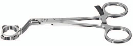
5 Innovative instrumentation Reamer with cannulation to prepare plate countersink Reaming guide / Reduction reaming guide Preliminary fixation of the carpus Optimal guidance of the reamer Soft tissue protection during reaming procedure Controlled reduction of the carpal bones Optional radiolucent plate positioning aid for placement of a central reaming guide wire Plate holder for secure plate positioning VA-Locking Intercarpal Fusion System Technique Guide Synthes 3
6 Indications Posttraumatic mediocarpal arthritis following scaphoid non-union (SNAC) or scapholunate ligament rupture (SLAC) Mediocarpal instability (final stage) 4 Synthes VA-Locking Intercarpal Fusion System Technique Guide
.")
7 Preparation 1 Preoperative planning Complete preoperative radiographic assessment should confirm the presence of sufficient bone stock for the four corner fusion procedure as well as intact joint surfaces between the radius and the lunate. Determine the extent of the lunate deformity (DISI). 2 Position patient Place the patient in a supine position with the forearm positioned on a hand table in full pronation. Use a rolled towel to support the palm of the hand. The use of tourniquet and magnifying glases is recommended. VA-Locking Intercarpal Fusion System Technique Guide Synthes 5

8 Approach and Reduction 1 Approach Make a standard dorsal longitudinal approach to the wrist. 2 Excise scaphoid, debride midcarpal joint Completely excise the scaphoid and debride the midcarpal joint. Use a small rongeur and/or chisel to remove the residual cartilage. Make sure that sclerotic and dense subchondral bone is removed down to cancellous bone. At this point, complete debridement of the midcarpal joint is mandatory. Preparation of the joint surfaces between the capitate/hamate and lunate/triquetrum is optional or may be carried out after provisional fixation. Note: Proper denudation of the cartilage down to good cancellous bone is important. 6 Synthes VA-Locking Intercarpal Fusion System Technique Guide


9 3 Reduce rotational deformity and provisionally fix the carpal bones Instrument Kirschner Wire 1.25 mm with trocar tip, length 80 mm, Stainless Steel If necessary, use a 1.25 mm Kirschner wire as a joystick to correct the position of the lunate. With a Kirschner wire temporariliy fix the lunate in neutral position from distal trough the capitate. Remove the joystick Kirschner wire. Check residual mobility. In general, more extension than flexion is recommended. Notes Verify correct alignment of the carpal bones under image intensifier control. If the reaming technique without reaming guide is used, temporary fixation with multiple intercarpal Kirschner wires is recommended. Important: Place temporary Kirschner wires as volar as possible to avoid later interference with the reamer. VA-Locking Intercarpal Fusion System Technique Guide Synthes 7


.")
10 Reaming Using reaming guide or reduction reaming guide 1 Position reaming guide Instruments Kirschner Wire 1.25 mm with trocar tip, length 80 mm, Stainless Steel Reaming Guide for VA-Locking Intercarpal Fusion Plate 15 mm Reaming Guide for VA-Locking Intercarpal Fusion Plate 17 mm Optional instruments Reduction Reaming Guide for VA-Locking Intercarpal Fusion Plate 15 mm Reduction Reaming Guide for VA-Locking Intercarpal Fusion Plate 17 mm Choose the reaming guide ( 15 mm or 17 mm) according to the selected plate and fix it temporarily with at least one 1.25 mm Kirschner wire per carpal bone over the center of the four-bone junction. If necessary, remove the volar luno-capitate Kirschner wire to avoid later interference with the reamer. Note: The shape of the reaming guide is adapted to the contour of the carpus (less height on distal part). Make sure the handle of the reaming guide is oriented to the radius as etched (see marking RADIUS SHAFT ). 8 Synthes VA-Locking Intercarpal Fusion System Technique Guide
11 Optional instrument: Reduction reaming guide Use the reduction reaming guide if reduction of the carpal bones is required. Choose the reduction reaming guide ( 15 mm or 17 mm) according to the selected plate. Open the clamp slightly and place the reduction reaming guide centered over the four bone junction. The amount of opening can be chosen according to preference. Fix the reduction reaming guide with at least one 1.25 mm Kirschner wire per carpal bone. VA-Locking Intercarpal Fusion System Technique Guide Synthes 9

12 Reaming Using reaming guide or reduction reaming guide 2 Ream plate recess Instruments Reamer for VA-Locking Intercarpal Fusion Plate 15 mm Reamer for VA-Locking Intercarpal Fusion Plate 17 mm Choose the reamer corresponding to the (reduction) reaming guide ( 15 mm or 17 mm). Ream through the reaming guide to the first laser marking line. Check for sufficient reaming depth by trial placement of the plate, ensuring that the plate edge does not project beyond the bone at any point. If necessary, continue reaming. Note: The first laser marking line on the reamer indicates a reaming depth of 6 mm. The subsequent marking lines are at increments of 2 mm reaming depth. Reaming guide Reduction reaming guide 10 Synthes VA-Locking Intercarpal Fusion System Technique Guide


13 Optional: Further intercarpal debridement With the (reduction) reaming guide in place, remove cartilage and sclerotic bone from the luno-triquetral and capitohamate intervals 4 6 mm adjacent to the midcarpal joint. Reaming guide Reduction reaming guide VA-Locking Intercarpal Fusion System Technique Guide Synthes 11
14 Reaming Option: Reaming without guide over central Kirschner wire Instruments Positioning Aid for VA-Locking Intercarpal Fusion Plate 15 mm Positioning Aid for VA-Locking Intercarpal Fusion Plate 17 mm Combi Drill Guide 1.8 for VA-Locking and Standard Kirschner Wire 1.25 mm with trocar tip, length 80 mm, Stainless Steel Choose the positioning aid ( 15 mm or 17 mm) according to the selected plate. Mount the positioning aid on the combi drill guide (click-on mechanism). Define the plate position using the radiolucent plate positioning aid under image intensifier control. Note: If needed for better visibility, fix the positioning aid temporarily on the carpus with a Kirschner wire and remove the combi drill guide from the plate positioning aid. Insert a Kirschner wire through the combi drill guide. 12 Synthes VA-Locking Intercarpal Fusion System Technique Guide
15 Ream the plate recess with the appropriate sized reamer over the Kirschner wire. Check for sufficient reaming depth by trial placement of the plate, ensuring that the plate edge does not project beyond the bone at any point. Important: The markings on the reamer should not be used as a reference for reaming depth when reaming without the reaming guide. The reaming depth from the tip to the first laser marking line is 15 mm. 6 Bone grafting Fill the space between the four bones with autogenous bone graft taken from the iliac crest or Lister s tubercle. VA-Locking Intercarpal Fusion System Technique Guide Synthes 13
.")
16 Plate Fixation 1 Place plate Instrument Plate Holder for VA-Locking Intercarpal Fusion Use the plate holder to pick up the appropriate sized plate (either 15 mm or 17 mm). Position the plate through the (reduction) reaming guide. 14 Synthes VA-Locking Intercarpal Fusion System Technique Guide
17 2 Fix plate with VA-locking screws Instruments Drill Bit 1.8 mm, with marking, length 110/85 mm, 2-flute, for Quick Coupling Combi Drill Guide 1.8 for VA-Locking and Standard Depth Gauge for Screws 2.0 to 2.7 mm, measuring range up to 40 mm Screwdriver Shaft, Stardrive, T8, self-holding Handle with Quick Coupling Torque Limiter, 0.8 Nm, with AO/ASIF Quick Coupling Optional instrument Screwdriver Shaft Stardrive 2.4, short, self-holding, for Quick Coupling Start plate fixation with the placement of VA-locking screws in the lunate. Use the variable angle part of the combi drill guide 1.8 (see marking VARIABLE ANGLE ) and fully insert it into the VA-locking hole. Drill the hole with the 1.8 mm drill bit at the desired angle. Notes It is important not to angulate more than 15 from the central axis of the screw hole. Overangulation could result in difficulty in getting the screw to lock. All VA-locking holes accept both 2.4 mm VA-locking and 2.4 mm cortex screws. The plate may be provisionally fixed with a Kirschner wire to secure it in position. VA-Locking Intercarpal Fusion System Technique Guide Synthes 15
18 Plate Fixation Measure screw length using the depth gauge. Insert the VA-locking screw using the T8 screwdriver shaft with Stardrive attached to the handle with quick coupling. Note: Do not yet fully tighten the screw to lock. For locking of VA-locking screws, refer to step Synthes VA-Locking Intercarpal Fusion System Technique Guide
19 Continue plate fixation by inserting a second VA-locking screw in the lunate. VA-Locking Intercarpal Fusion System Technique Guide Synthes 17
20 Plate Fixation 3 Optional: Apply compression Instruments Drill Bit 1.8 mm, with marking, length 110/85 mm, 2-flute, for Quick Coupling Combi Drill Guide 1.8 for VA-Locking and Standard Depth Gauge for Screws 2.0 to 2.7 mm, measuring range up to 40 mm Screwdriver Shaft, Stardrive, T8, self-holding Handle with Quick Coupling To apply compression insert a cortex screw in the distal carpal bones (e.g. capitate). Use the 1.8 mm drill bit and the combi drill guide to drill the hole for the cortex screw (see marking COMPRESSION ). 18 Synthes VA-Locking Intercarpal Fusion System Technique Guide
21 Measure screw length using the depth gauge. Insert the screw using the T8 screwdriver shaft with Stardrive attached to the handle with quick coupling. VA-Locking Intercarpal Fusion System Technique Guide Synthes 19
. 5 Lock VA-locking screws Instruments 03.110.")

22 Plate Fixation 4 Finalize plate fixation Finalize plate fixation with a combination of 2.4 mm VAlocking and/or 2.4 mm cortex screws as preferred. Note: Remove the reaming guide before final tightening of the compression screw(s). 5 Lock VA-locking screws Instruments Handle for Torque Limiters 0.4/0.8/1.2 Nm Torque Limiter, 0.8 Nm, with AO/ASIF Quick Coupling Screwdriver Shaft, Stardrive, T8, self-holding Optional instrument Screwdriver Shaft, Stardrive, 2.4, short, self-holding, for Quick Coupling 20 Synthes VA-Locking Intercarpal Fusion System Technique Guide
23 Use the 0.8 Nm torque limiter to perform the final locking step for the VA-locking screws. The torque limiter prevents over-tightening and ensures that the VA-Locking screws are securely locked into the plate. 6 Postoperative treatment Use a removable cock-up splint. According to the stability of the construct, protected range of motion exercises can be started after the postoperative swelling has subsided. Otherwise, permanent protection of the wrist in the splint is recommended until the bone has consolidated. VA-Locking Intercarpal Fusion System Technique Guide Synthes 21
 02.111.300 04.111.300 6 15 02.111.301 04.")
 201.756 401.756 6 201.757 401.")

24 Implants VA-Locking Intercarpal Fusion Plate 2.4 Stainless steel Titanium Holes Diameter (mm) Cortex Screw Stardrive 2.4 mm, self-tapping Stainless steel Titanium Length (mm) Synthes VA-Locking Intercarpal Fusion System Technique Guide
25 VA-locking Screw Stardrive 2.4 mm, self-tapping Stainless steel Titanium Length (mm) All implants are also available sterile packed. Add suffix "S" to article number. VA-Locking Intercarpal Fusion System Technique Guide Synthes 23
26 Instruments Standard instruments Reamer for VA-Locking Intercarpal Fusion Plate, 15 mm Reamer for VA-Locking Intercarpal Fusion Plate, 17 mm Reaming Guide for VA-Locking Intercarpal Fusion Plate, 15 mm Reaming Guide for VA-Locking Intercarpal Fusion Plate, 17 mm (S) Kirschner Wire 1.25 mm with trocar tip, length 80 mm, Stainless Steel 24 Synthes VA-Locking Intercarpal Fusion System Technique Guide
27 Handle with Quick Coupling Combi Drill Guide 1.8 for VA-Locking and Standard Drill Bit 1.8 mm, with marking, length 110/85 mm, 2-flute, for Quick Coupling Screwdriver Shaft, Stardrive, T8, self-holding Torque Limiter, 0.8 Nm, with AO/ASIF Quick Coupling Handle for Torque Limiters 0.4/0.8/1.2 Nm Depth Gauge for Screws 2.0 to 2.7 mm, measuring range up to 40 mm VA-Locking Intercarpal Fusion System Technique Guide Synthes 25

28 Instruments Optional instruments Screwdriver Shaft Stardrive 2.4, short, self-holding, for Quick Coupling Holding Sleeve for Screws Stardrive 2.4 mm, T8, for Screwdriver Shafts 3.5 mm, for No Plate Holder for VA-Locking Intercarpal Fusion Plate Positioning Aid for VA-Locking Intercarpal Fusion Plate 15 mm Positioning Aid for VA-Locking Intercarpal Fusion Plate 17 mm Reduction Reaming Guide for VA-Locking Intercarpal Fusion Plate 15 mm Reduction Reaming Guide for VA-Locking Intercarpal Fusion Plate, 17 mm 26 Synthes VA-Locking Intercarpal Fusion System Technique Guide
29 VA-Locking Intercarpal Fusion System Technique Guide Synthes 27
30 28 Synthes VA-Locking Intercarpal Fusion System Technique Guide
31
32 02/ Synthes, Inc. or its affiliates Subject to modification Synthes and Stardrive are trademarks of Synthes, Inc. or its affiliates All technique guides are available as PDF files at Ö öAAfä version AA
VA-Locking Intercarpal Fusion System. Variable angle locking technology for mediocarpal partial arthrodesis.
 VA-Locking Intercarpal Fusion System. Variable angle locking technology for mediocarpal partial arthrodesis. Surgical Technique This publication is not intended for distribution in the USA. Image intensifier
2.4 mm Variable Angle LCP Intercarpal Fusion System. Variable angle locking technology for mediocarpal partial arthrodesis.
 2.4 mm Variable Angle LCP Intercarpal Fusion System. Variable angle locking technology for mediocarpal partial arthrodesis. Technique Guide Instruments and implants approved by the AO Foundation Table
2.4 mm Variable Angle Locking Intercarpal Fusion System
 For Partial Wrist Arthrodesis With Variable Angle Locking Technology 2.4 mm Variable Angle Locking Intercarpal Fusion System Surgical Technique Table of Contents Introduction 2.4 mm Variable Angle Locking
Variable Angle LCP Volar Rim Distal Radius Plate 2.4. For fragment-specific fracture fixation with variable angle locking technology.
 Technique Guide Variable Angle LCP Volar Rim Distal Radius Plate 2.4. For fragment-specific fracture fixation with variable angle locking technology. Image intensifier control Warning This description
2.4 mm Variable Angle LCP Volar Extra-Articular Distal Radius System. For fragment-specific fracture fixation with variable angle locking technology.
 Technique Guide 2.4 mm Variable Angle LCP Volar Extra-Articular Distal Radius System. For fragment-specific fracture fixation with variable angle locking technology. Table of Contents Introduction 2.4
Technique Guide. Locking Attachment Plate. For treatment of periprosthetic fractures.
 Technique Guide Locking Attachment Plate. For treatment of periprosthetic fractures. Table of Contents Introduction Locking Attachment Plate 2 Indications 4 Surgical Technique Patient Positioning 5 Preparation
Technique Guide. 2.4 mm Variable Angle LCP Distal Radius System. For fragment-specific fracture fixation with variable angle locking technology.
 Technique Guide 2.4 mm Variable Angle LCP Distal Radius System. For fragment-specific fracture fixation with variable angle locking technology. Table of Contents Introduction 2.4 mm Variable Angle LCP
LCP Medial Distal Tibia Plate, without Tab. The Low Profile Anatomic Fixation System with Angular Stability and Optimal Screw Orientation.
 LCP Medial Distal Tibia Plate, without Tab. The Low Profile Anatomic Fixation System with Angular Stability and Optimal Screw Orientation. Technique Guide LCP Small Fragment System Table of Contents Introduction
LCP Wrist Fusion Set. Anatomic plates for total wrist fusion.
 LCP Wrist Fusion Set. Anatomic plates for total wrist fusion. Technique Guide This publication is not intended for distribution in the USA. Instruments and implants approved by the AO Foundation. Table
VA-LCP Anterior Clavicle Plate. The anatomically precontoured fixation system with angular stability for clavicle shaft and lateral clavicle.
 Technique Guide VA-LCP Anterior Clavicle Plate. The anatomically precontoured fixation system with angular stability for clavicle shaft and lateral clavicle. Table of Contents Introduction VA-LCP Anterior
2.4 mm Variable Angle LCP Volar Extra-Articular Distal Radius System. For fragment-specific fracture fixation with variable angle locking technology.
 2.4 mm Variable Angle LCP Volar Extra-Articular Distal Radius System. For fragment-specific fracture fixation with variable angle locking technology. Surgical Technique This publication is not intended
Technique Guide. LCP Distal Fibula Plates. Part of the Synthes locking compression plate (LCP) system.
 system.") Technique Guide LCP Distal Fibula Plates. Part of the Synthes locking compression plate (LCP) system. Table of Contents Introduction LCP Distal Fibula Plates 2 AO Principles 4 Indications 5 Surgical Technique
Technique Guide LCP Distal Fibula Plates. Part of the Synthes locking compression plate (LCP) system. Table of Contents Introduction LCP Distal Fibula Plates 2 AO Principles 4 Indications 5 Surgical Technique
LCP Low Bend Medial Distal Tibia Plates 3.5 mm. Anatomic plates with low profile head for intra- and extraarticular fractures.
 LCP Low Bend Medial Distal Tibia Plates 3.5 mm. Anatomic plates with low profile head for intra- and extraarticular fractures. Surgical Technique This publication is not intended for distribution in the
LCP Medial Proximal Tibial Plate 3.5. Part of the Synthes small fragment Locking Compression Plate (LCP) system.
 system.") LCP Medial Proximal Tibial Plate 3.5. Part of the Synthes small fragment Locking Compression Plate (LCP) system. Technique Guide This publication is not intended for distribution in the USA. Instruments
LCP Medial Proximal Tibial Plate 3.5. Part of the Synthes small fragment Locking Compression Plate (LCP) system. Technique Guide This publication is not intended for distribution in the USA. Instruments
Technique Guide. 3.5 mm LCP Low Bend Medial Distal Tibia Plates. Part of the Synthes locking compression plate (LCP) system.
 system.") Technique Guide 3.5 mm LCP Low Bend Medial Distal Tibia Plates. Part of the Synthes locking compression plate (LCP) system. Table of Contents Introduction 3.5 mm LCP Low Bend Medial Distal Tibia Plates
Technique Guide 3.5 mm LCP Low Bend Medial Distal Tibia Plates. Part of the Synthes locking compression plate (LCP) system. Table of Contents Introduction 3.5 mm LCP Low Bend Medial Distal Tibia Plates
Instrument and Implant for wrist fracture
 Instrument and Implant for wrist fracture Jansri Janpanya Product specialist The Bangkok Unitrade Co,.ltd. Objectives Type of LCP for distal radius Fx. The new LCP design for distal radius Fx. Have knowledge
The Wrist Fusion Set. Stainless Steel and Titanium TECHNIQUE GUIDE. Instruments and implants approved by the AO Foundation
 The Wrist Fusion Set Stainless Steel and Titanium TECHNIQUE GUIDE Instruments and implants approved by the AO Foundation Three Plate Options Stainless Steel or Titanium* Standard Bend Stainless Steel [242.510]
Technique Guide. PHILOS and PHILOS Long. The anatomic fixation system for the proximal humerus.
 Technique Guide PHILOS and PHILOS Long. The anatomic fixation system for the proximal humerus. Table of Contents Introduction PHILOS and PHILOS Long 2 AO Principles 4 Indications 5 Surgical Technique
LCP Proximal Radius Plates 2.4. Plates for radial head rim and for radial head neck address individual fracture patterns of the proximal radius.
 Technique Guide LCP Proximal Radius Plates 2.4. Plates for radial head rim and for radial head neck address individual fracture patterns of the proximal radius. Table of Contents Introduction LCP Proximal
LCP Distal Tibia Plate
 Surgical Technique LCP Locking Compression Plate Original Instruments and Implants of the Association for the Study of Internal Fixation AO/ASIF Table of contents Indications 3 Implants/Instruments 5 Surgical
Integra. Spider and Mini Spider Limited Wrist Fusion System SURGICAL TECHNIQUE
 Integra Spider and Mini Spider Limited Wrist Fusion System SURGICAL TECHNIQUE Table of contents Description... 02 Indications... 02 Contraindications... 02 Surgical Technique... 03 Spider Introduction-Four
LCP Superior Clavicle Plate. The anatomically precontoured fixation system with angular stability for clavicle shaft and lateral clavicle.
 Technique Guide LCP Superior Clavicle Plate. The anatomically precontoured fixation system with angular stability for clavicle shaft and lateral clavicle. Table of Contents Introduction LCP Superior Clavicle
3.5 mm Locking Attachment Plate
 For Treatment of Periprosthetic Fractures 3.5 mm Locking Attachment Plate Surgical Technique Table of Contents Introduction 3.5 mm Locking Attachment Plate 2 Indications 4 Surgical Technique Preparation
LCP Medial Proximal Tibial Plate 4.5/5.0. Part of the Synthes LCP periarticular plating system.
 LCP Medial Proximal Tibial Plate 4.5/5.0. Part of the Synthes LCP periarticular plating system. Technique Guide This publication is not intended for distribution in the USA. Instruments and implants approved
Wrist Fusion Instrument and Implant Set.
 Wrist Fusion Instrument and Implant Set. Surgical Technique Discontinued December 2016 DSEM/TRM/0815/0479(2) This publication is not intended for distribution in the USA. Instruments and implants approved
OBSOLETED. LCP Medial Distal Tibia Plate, without Tab. The Low Profile Anatomic Fixation System with Angular Stability and Optimal Screw Orientation.
 LCP Medial Distal Tibia Plate, without Tab. The Low Profile Anatomic Fixation System with Angular Stability and Optimal Screw Orientation. Surgical Technique LCP Small Fragment System This publication
LCP Distal Humerus Plates
 The anatomic fixation system for the distal humerus with angular stability Surgical technique LCP Locking Compression Plate Contents Indications and contraindications 2 Implants 3 Instruments 5 Preparation
Technique Guide. LCP Proximal Femoral Hook Plate 4.5/5.0. Part of the LCP Periarticular Plating System.
 Technique Guide LCP Proximal Femoral Hook Plate 4.5/5.0. Part of the LCP Periarticular Plating System. Table of Contents Introduction Features and Benefits 2 AO ASIF Principles 4 Indications 5 Surgical
Periarticular Aiming Arm Instruments for LCP Proximal Tibial Plate 4.5/5.0. Part of the LCP Periarticular Aiming Arm Instrument System (large).
.") Technique Guide Periarticular Aiming Arm Instruments for LCP Proximal Tibial Plate 4.5/5.0. Part of the LCP Periarticular Aiming Arm Instrument System (large). Image intensifier control Warning This description
Technique Guide Periarticular Aiming Arm Instruments for LCP Proximal Tibial Plate 4.5/5.0. Part of the LCP Periarticular Aiming Arm Instrument System (large). Image intensifier control Warning This description
Technique Guide. LCP Posterior Medial Proximal Tibial Plate 3.5. Part of the Synthes small fragment LCP system.
 Technique Guide LCP Posterior Medial Proximal Tibial Plate 3.5. Part of the Synthes small fragment LCP system. Table of Contents Introduction LCP Posterior Medial Proximal Tibial Plate 3.5 2 AO Principles
Long Volar Plates for Diaphyseal-Metaphyseal Radius Fractures LCP. Dia-Meta Volar Distal Radius Plates. Surgical Technique
 Long Volar Plates for Diaphyseal-Metaphyseal Radius Fractures LCP Dia-Meta Volar Distal Radius Plates Surgical Technique Table of Contents Introduction LCP Dia-Meta Volar Distal Radius Plates 2 AO Principles
LCP Wrist Fusion Set. Anatomic plates for total wrist fusion.
 LCP Wrist Fusion Set. Anatomic plates for total wrist fusion. Surgical Technique This publication is not intended for distribution in the USA. Instruments and implants approved by the AO Foundation. Image
Technique Guide. 3.5 mm LCP Olecranon Plates. Part of the Synthes locking compression plate (LCP) system.
 system.") Technique Guide 3.5 mm LCP Olecranon Plates. Part of the Synthes locking compression plate (LCP) system. Table of Contents Introduction 3.5 mm LCP Olecranon Plates 2 AO Principles 3 Indications 3 Clinical
Technique Guide 3.5 mm LCP Olecranon Plates. Part of the Synthes locking compression plate (LCP) system. Table of Contents Introduction 3.5 mm LCP Olecranon Plates 2 AO Principles 3 Indications 3 Clinical
Low Bend Distal Tibia Plates
 Part of the DePuy Synthes Locking Compression Plate (LCP ) System 3.5 mm LCP Low Bend Medial Distal Tibia Plates Surgical Technique Table of Contents Introduction 3.5 mm LCP Low Bend Medial Distal Tibia
Alignment Rod. For intraoperatively confirming correction of the mechanical leg axis.
 Alignment Rod. For intraoperatively confirming correction of the mechanical leg axis. Easy to use Accuracy of surgery Reduces X-ray exposure Table of Contents Introduction Alignment Rod 2 Handling Technique
Technique Guide. DHS Blade. For osteoporotic bone.
 Technique Guide DHS Blade. For osteoporotic bone. Table of Contents Introduction Features and Benefits 2 Indications and Contraindications 4 Clinical Cases 5 Surgical Technique Implantation 6 Implant
Compact Distal Radius System. Consolidated solution for Distal Radius Plating Systems
 Compact Distal Radius System. Consolidated solution for Distal Radius Plating Systems Designed to allow customized system configurations Variable Angle (VA) and Locking Compression Plate (LCP ) options
3.5 mm LCP Extra-articular Distal Humerus Plate
 Part of the DePuy Synthes Locking Compression Plate (LCP ) System 3.5 mm LCP Extra-articular Distal Humerus Plate Surgical Technique Table of Contents Introduction 3.5 mm LCP Extra-articular Distal Humerus
Technique Guide. 3.5 mm LCP Low Bend Medial Distal Tibia Plate Aiming Instruments. Part of the 3.5 mm LCP Percutaneous Instrument System.
 Technique Guide 3.5 mm LCP Low Bend Medial Distal Tibia Plate Aiming Instruments. Part of the 3.5 mm LCP Percutaneous Instrument System. Table of Contents Introduction 3.5 mm LCP Low Bend Medial Distal
USS II ILIO-SACRAL Modular System for Stable Fixation in the Sacrum and Illium
 USS II ILIO-SACRAL Modular System for Stable Fixation in the Sacrum and Illium Instruments and implants approved by the AO Foundation. This publication is not intended for distribution in the USA. TECHNIQUE
LCP Medial Proximal Tibial Plate 3.5. Part of the Synthes small fragment Locking Compression Plate (LCP) system.
 system.") LCP Medial Proximal Tibial Plate 3.5. Part of the Synthes small fragment Locking Compression Plate (LCP) system. Surgical Technique This publication is not intended for distribution in the USA. Instruments
LCP Medial Proximal Tibial Plate 3.5. Part of the Synthes small fragment Locking Compression Plate (LCP) system. Surgical Technique This publication is not intended for distribution in the USA. Instruments
LCP Ulna Osteotomy System 2.7. Low profile angular stable fixation for ulna shortening osteotomies.
 LCP Ulna Osteotomy System 2.7. Low profile angular stable fixation for ulna shortening osteotomies. Surgical Technique This publication is not intended for distribution in the USA. Instruments and implants
LCP Superior Clavicle Plate. The anatomically precontoured fixation system with angular stability for clavicle shaft and lateral clavicle.
 LCP Superior Clavicle Plate. The anatomically precontoured fixation system with angular stability for clavicle shaft and lateral clavicle. Surgical Technique This publication is not intended for distribution
LCP DISTAL TIBIA PLATE
 LCP DISTAL TIBIA PLATE Instruments and implants approved by the AO Foundation. This publication is not intended for distribution in the USA. SURGICAL TECHNIQUE Image intensifier control This description
LCP Anterolateral Distal Tibia Plate 3.5. The low profile anatomic fixation system with optimal plate placement and angular stability.
 LCP Anterolateral Distal Tibia Plate 3.5. The low profile anatomic fixation system with optimal plate placement and angular stability. Technique Guide LCP Small Fragment System Table of Contents Introduction
Technique Guide. 2.7 mm/3.5 mm LCP Distal Fibula Plates. Part of the Synthes locking compression plate (LCP) system.
 system.") Technique Guide 2.7 mm/3.5 mm LCP Distal Fibula Plates. Part of the Synthes locking compression plate (LCP) system. Table of Contents Introduction 2.7 mm/3.5 mm LCP Distal Fibula Plates 2 AO Principles
Technique Guide 2.7 mm/3.5 mm LCP Distal Fibula Plates. Part of the Synthes locking compression plate (LCP) system. Table of Contents Introduction 2.7 mm/3.5 mm LCP Distal Fibula Plates 2 AO Principles
Cannulated Angled Blade Plate 3.5 and 4.5, 90.
 Cannulated Angled Blade Plate 3.5 and 4.5, 90. Technique Guide This publication is not intended for distribution in the USA. Instruments and implants approved by the AO Foundation. Table of Contents Introduction
2.4 mm LCP Volar Column Distal Radius Plates. Part of the 2.4 mm LCP Distal Radius System.
 2.4 mm LCP Volar Column Distal Radius Plates. Part of the 2.4 mm LCP Distal Radius System. 12 anatomically shaped volar plates Multiple screw options for fixedangle support to articular surface Combi holes
LCP Anterolateral Distal Tibia Plate 3.5. The low profile anatomic fixation system with optimal plate placement and angular stability.
 LCP Anterolateral Distal Tibia Plate 3.5. The low profile anatomic fixation system with optimal plate placement and angular stability. Technique Guide LCP Small Fragment System Table of Contents Introduction
3.5 MM VA-LCP PROXIMAL TIBIA PLATE SYSTEM
 3.5 MM VA-LCP PROXIMAL TIBIA PLATE SYSTEM Part of the DePuy Synthes Variable Angle Periarticular Plating System SURGICAL TECHNIQUE TABLE OF CONTENTS INTRODUCTION 3.5 mm VA-LCP Proximal Tibial Plate 2 AO
Distal Ulnar Locking Plate
 INDEX Indications Patient Position Surgical Technique - Step 1 Approach - Step 2 Plate Contouring - Step 3 Fracture Reduction - Step 4 Distal Plate Fixation - Step 5 Confirm Proper Reconstruction - Step
LCP Extra-articular Distal Humerus Plate.
 Technique Guide LCP Extra-articular Distal Humerus Plate. The anatomically shaped and angular stable fixation system for extraarticular fractures of the distal humerus. Table of Contents Introduction
Distal Radius Plate Instrument and Implant Set. Discontinued December 2017 DSUS/TRM/0916/1063(1)
") Distal Radius Plate Instrument and Implant Set Surgical Technique Discontinued December 2017 DSUS/TRM/0916/1063(1) The Distal Radius Plates Indications For fixation of fractures and osteotomies, including
Distal Radius Plate Instrument and Implant Set Surgical Technique Discontinued December 2017 DSUS/TRM/0916/1063(1) The Distal Radius Plates Indications For fixation of fractures and osteotomies, including
LCP Proximal Radius Plates 2.4. Plates for radial head rim and for radial head neck address individual fracture patterns of the proximal radius.
 LCP Proximal Radius Plates 2.4. Plates for radial head rim and for radial head neck address individual fracture patterns of the proximal radius. Surgical Technique This publication is not intended for
LCP Anterior Ankle Arthrodesis Plates. Part of the Synthes Locking Compression Plate (LCP) System.
 System.") LCP Anterior Ankle Arthrodesis Plates. Part of the Synthes Locking Compression Plate (LCP) System. Technique Guide Instruments and implants approved by the AO Foundation Table of Contents Introduction
LCP Anterior Ankle Arthrodesis Plates. Part of the Synthes Locking Compression Plate (LCP) System. Technique Guide Instruments and implants approved by the AO Foundation Table of Contents Introduction
Technique Guide. 6.5 mm Midfoot Fusion Bolt. For intramedullary fixation of the medial column of the foot.
 Technique Guide 6.5 mm Midfoot Fusion Bolt. For intramedullary fixation of the medial column of the foot. Table of Contents Introduction 6.5 mm Midfoot Fusion Bolt 2 AO Principles 4 Indications 5 Surgical
VA LOCKING CALCANEAL PLATES 2.7
 VA LOCKING CALCANEAL PLATES 2.7 Instruments and Implants approved by the AO Foundation. This publication is not intended for distribution in the USA. SURGICAL TECHNIQUE Image intensifier control Warning
2.4 mm LCP Radial Head Plates. Part of the Synthes LCP Distal Radius Plate System.
 2.4 mm LCP Radial Head Plates. Part of the Synthes LCP Distal Radius Plate System. Technique Guide Instruments and Implants approved by the AO Foundation Table of Contents Introduction 2.4 mm LCP Radial
2.4 mm Variable Angle LCP Volar Rim Distal Radius Plates
 For Fragment-Specific Fracture Fixation With Variable Angle Locking Technology 2.4 mm Variable Angle LCP Volar Rim Distal Radius Plates Surgical Technique Table of Contents Introduction 2.4 mm Variable
LCP Proximal Radius Plates 2.4. Plates for radial head rim and for radial head neck address individual fracture patterns of the proximal radius.
 LCP Proximal Radius Plates 2.4. Plates for radial head rim and for radial head neck address individual fracture patterns of the proximal radius. Surgical Technique This publication is not intended for
LCP Distal Fibula Plates. Part of the Synthes locking compression plate (LCP) system.
 system.") LCP Distal Fibula Plates. Part of the Synthes locking compression plate (LCP) system. Surgical Technique This publication is not intended for distribution in the USA. Instruments and implants approved
LCP Distal Fibula Plates. Part of the Synthes locking compression plate (LCP) system. Surgical Technique This publication is not intended for distribution in the USA. Instruments and implants approved
Technique Guide. 3.5 mm LCP Periarticular Proximal Humerus Plate. Part of the Synthes locking compression plate (LCP) system.
 system.") Technique Guide 3.5 mm LCP Periarticular Proximal Humerus Plate. Part of the Synthes locking compression plate (LCP) system. Table of Contents Introduction 3.5 mm LCP Proximal Humerus Plate 2 AO Principles
Technique Guide 3.5 mm LCP Periarticular Proximal Humerus Plate. Part of the Synthes locking compression plate (LCP) system. Table of Contents Introduction 3.5 mm LCP Proximal Humerus Plate 2 AO Principles
LCP Metaphyseal Plates. For extra-articular fractures.
 LCP Metaphyseal Plates. For extra-articular fractures. Surgical Technique This publication is not intended for distribution in the USA. Instruments and implants approved by the AO Foundation. Image intensifier
Technique Guide. 4.5 mm LCP Proximal Tibia Plates. Part of the Synthes LCP Periarticular Plating System.
 Technique Guide 4.5 mm LCP Proximal Tibia Plates. Part of the Synthes LCP Periarticular Plating System. Table of Contents Introduction 4.5 mm LCP Proximal Tibia Plates 2 AO Principles 4 Indications 5 Surgical
LCP Distal Fibula Plates. Part of the Synthes locking compression plate (LCP) system.
 system.") LCP Distal Fibula Plates. Part of the Synthes locking compression plate (LCP) system. Surgical Technique This publication is not intended for distribution in the USA. Instruments and implants approved
LCP Distal Fibula Plates. Part of the Synthes locking compression plate (LCP) system. Surgical Technique This publication is not intended for distribution in the USA. Instruments and implants approved
Olecranon Osteotomy Nail. For simple fractures and osteotomies of the olecranon.
 Olecranon Osteotomy Nail. For simple fractures and osteotomies of the olecranon. Technique Guide Discontinued June 2016; AVAILABLE FOR IMPLANT REMOVAL PURPOSES ONLY DSEM/TRM/0517/0843 Table of Contents
VECTRA-T SURGICAL TECHNIQUE. The Translational Anterior Cervical Palate System. This publication is not intended for distribution in the USA.
 VECTRA-T The Translational Anterior Cervical Palate System This publication is not intended for distribution in the USA. SURGICAL TECHNIQUE Image intensifier control This description alone does not provide
VA-LCP Anterior Clavicle Plate. The anatomically precontoured fixation system with angular stability for clavicle shaft and lateral clavicle.
 VA-LCP Anterior Clavicle Plate. The anatomically precontoured fixation system with angular stability for clavicle shaft and lateral clavicle. Surgical Technique This publication is not intended for distribution
2.7 mm/3.5 mm LCP Distal Fibula Plate System. Part of the Synthes locking compression plate (LCP) system.
 system.") 2.7 mm/3.5 mm LCP Distal Fibula Plate System. Part of the locking compression plate (LCP) system. Anatomically contoured Multiple screw options for fixedangle support Coaxial holes minimize screw head
2.7 mm/3.5 mm LCP Distal Fibula Plate System. Part of the locking compression plate (LCP) system. Anatomically contoured Multiple screw options for fixedangle support Coaxial holes minimize screw head
Technique Guide. Orthodontic Bone Anchor (OBA) System. Skeletal implants for the orthodontic movement of the teeth.
 System. Skeletal implants for the orthodontic movement of the teeth.") Technique Guide Orthodontic Bone Anchor (OBA) System. Skeletal implants for the orthodontic movement of the teeth. Table of Contents Introduction Orthodontic Bone Anchor (OBA) System 2 Indications and
Technique Guide Orthodontic Bone Anchor (OBA) System. Skeletal implants for the orthodontic movement of the teeth. Table of Contents Introduction Orthodontic Bone Anchor (OBA) System 2 Indications and
3. Insert Tocar Sleeves Insert the NCB tissue protection sleeve assembly 1.6 to 10mm through a skin incision (Fig. 38).
.") NCB Proximal Humerus Plating System Surgical Technique 19 2. Temporary Plate Fixation The plate can be temporary fixed to the bone with 1.6mm K-wire through the proximal cannulated fixation screw of the
NCB Proximal Humerus Plating System Surgical Technique 19 2. Temporary Plate Fixation The plate can be temporary fixed to the bone with 1.6mm K-wire through the proximal cannulated fixation screw of the
3.0/3.5/4.0/4.5/6.5/7.0/7.3. Cannulated Screws. Surgical Technique
 3.0/3.5/4.0/4.5/6.5/7.0/7.3 Cannulated Screws Surgical Technique Image intensifier control This description alone does not provide sufficient background for direct use of DePuy Synthes products. Instruction
The Flower Four Corner Fusion Plate
 The Flower Four Corner Fusion Plate PROCEDURE GUIDE www.flowerortho.com The Flower Upper Extremity Application PROXIMAL HUMERUS PLATE SMALL BONE PLATES FOUR CORNER FUSION PLATE ANATOMIC DISTAL RADIUS PLATE
VA-LCP Condylar Plate 4.5/5.0. Part of the Synthes Variable Angle Periarticular Plating System.
 VA-LCP Condylar Plate 4.5/5.0. Part of the Synthes Variable Angle Periarticular Plating System. Technique Guide This publication is not intended for distribution in the USA. Instruments and implants approved
3.5 mm LCP Olecranon Plates
 Part of the DePuy Synthes Locking Compression Plate (LCP ) System 3.5 mm LCP Olecranon Plates Surgical Technique Table of Contents Introduction 3.5 mm LCP Olecranon Plates 2 AO Principles 3 Indications
Olecranon Locking Plate II
 INDEX Indications Patient Position Fracture Reduction and Fixation Surgical Technique Step 1 Surgical Approach Step 2 Implantation Step 3 Proximal Locking Screw Insertion Step 4 Distal Screw Insertion
Technique Guide. TomoFix Osteotomy System. A comprehensive plating system for stable fixation of osteotomies around the knee.
 Technique Guide TomoFix Osteotomy System. A comprehensive plating system for stable fixation of osteotomies around the knee. Table of Contents Introduction TomoFix Osteotomy System 2 AO Principles 4 Indications
LCP Condylar Plate 4.5/5.0. Part of the LCP Periarticular Plating System.
 LCP Condylar Plate 4.5/5.0. Part of the LCP Periarticular Plating System. Surgical Technique This publication is not intended for distribution in the USA. Instruments and implants approved by the AO Foundation.
Arthrodesis System 2.0/2.3, 2.5
 PRODUCT INFORMATION Arthrodesis System 2.0/2.3, 2.5 APTUS Wrist 2 Arthrodesis System 2.0/2.3, 2.5 Arthrodesis System 2.0/2.3, 2.5 Multidirectional and angular stable partial or total wrist arthrodesis
LCP Condylar Plate 4.5/5.0. Part of the LCP Periarticular Plating System.
 LCP Condylar Plate 4.5/5.0. Part of the LCP Periarticular Plating System. Surgical Technique This publication is not intended for distribution in the USA. Instruments and implants approved by the AO Foundation.
DOUBLE/TRIPLE PELVIC OSTEOTOMY PLATES For Treating Coxofemoral Joint Instability and Subluxation in Immature Dogs
 DOUBLE/TRIPLE PELVIC OSTEOTOMY PLATES For Treating Coxofemoral Joint Instability and Subluxation in Immature Dogs Instruments and implants approved by the AO Foundation. This publication is not intended
4.5 mm LCP Medial Proximal Tibia Plates
 Part of the DePuy Synthes LCP Periarticular Plating System 4.5 mm LCP Medial Proximal Tibia Plates Surgical Technique Table of Contents Introduction 4.5 mm LCP Medial Proximal Tibia Plates 2 AO Principles
3.5 mm LCP Distal Humerus Plates
 Part of the DePuy Synthes Locking Compression Plate (LCP ) System 3.5 mm LCP Distal Humerus Plates Surgical Technique Table of Contents Introduction 3.5 mm LCP Distal Humerus Plates 2 AO Principles 4 Indications
Ordering information. LCP Locking Compression Plate. Combine without compromise.
 Ordering information LCP Locking Compression Plate. Combine without compromise. Table of Contents Small Fragment System Large Fragment System LCP Instruments Emergency Instruments Implants Humerus Plates,
LCP Superior Anterior Clavicle Plate. The anatomically precontoured fixation system with angular stability for clavicle shaft and lateral clavicle.
 LCP Superior Anterior Clavicle Plate. The anatomically precontoured fixation system with angular stability for clavicle shaft and lateral clavicle. Surgical Technique This publication is not intended for
Technique Guide. VA-LCP Distal Humerus Plates 2.7/3.5. The low-profile fixation system with variable angle locking technology.
 Technique Guide VA-LCP Distal Humerus Plates 2.7/3.5. The low-profile fixation system with variable angle locking technology. Image intensifier control Warning This description alone does not provide sufficient
LCP Pediatric Hip Plate 2.7. For proximal femoral osteotomies.
 LCP Pediatric Hip Plate 2.7. For proximal femoral osteotomies. Technique Guide This publication is not intended for distribution in the USA. Instruments and implants approved by the AO Foundation. Image
The Calcaneal Plate. The Synthes non-locking solution for the Calcaneus.
 The Calcaneal Plate. The Synthes non-locking solution for the Calcaneus. Surgical Technique This publication is not intended for distribution in the USA. Instruments and implants approved by the AO Foundation.
Technique Guide. 3.5 mm LCP Proximal Tibia Plate. Part of the Synthes Small Fragment LCP System.
 Technique Guide 3.5 mm LCP Proximal Tibia Plate. Part of the Synthes Small Fragment LCP System. Table of Contents AO ASIF Principles of Internal Fixation 4 Indications/Contraindications 5 Surgical Technique
3.5 mm LCP Clavicle Hook Plates
 Part of the Synthes Locking Compression Plate (LCP ) System 3.5 mm LCP Clavicle Hook Plates Surgical Technique Table of Contents Introduction 3.5 mm LCP Clavicle Hook Plates 2 AO Principles 4 Indications
Surgical Technique Carpal Fusion
 Carpal Fusion Patent and Patent Pending CAUTION: Federal Law (USA) restricts this device to sale by or on the order of a physician. INDICATIONS FOR USE The Extremity Medical Lag Screw and X-Post System
Technique Guide. Small Fragment Locking Compression Plate (LCP) System. Stainless steel and titanium.
 System. Stainless steel and titanium.") Technique Guide Small Fragment Locking Compression Plate (LCP) System. Stainless steel and titanium. Table of Contents Introduction Small Fragment Locking Compression Plate (LCP) System 2 AO Principles
Technique Guide Small Fragment Locking Compression Plate (LCP) System. Stainless steel and titanium. Table of Contents Introduction Small Fragment Locking Compression Plate (LCP) System 2 AO Principles
MatrixNEURO. The next generation cranial plating system.
 MatrixNEURO. The next generation cranial plating system. Technique Guide CMF Matrix This publication is not intended for distribution in the USA. Instruments and implants approved by the AO Foundation
Thoracolumbar Spine Locking Plate (TSLP) System. A low-profile plating system for anterior stabilization of the thoracic and lumbar spine.
 System. A low-profile plating system for anterior stabilization of the thoracic and lumbar spine.") Thoracolumbar Spine Locking Plate (TSLP) System. A low-profile plating system for anterior stabilization of the thoracic and lumbar spine. Technique Guide Instruments and implants approved by the AO Foundation
Thoracolumbar Spine Locking Plate (TSLP) System. A low-profile plating system for anterior stabilization of the thoracic and lumbar spine. Technique Guide Instruments and implants approved by the AO Foundation
VA-LCP Olecranon Plates 2.7/3.5. The fracture-specific low-profile fixation system with variable angle locking technology.
 VA-LCP Olecranon Plates 2.7/3.5. The fracture-specific low-profile fixation system with variable angle locking technology. Surgical Technique This publication is not intended for distribution in the USA.
Zimmer Small Fragment Universal Locking System. Surgical Technique
 Zimmer Small Fragment Universal Locking System Surgical Technique Zimmer Small Fragment Universal Locking System 1 Zimmer Small Fragment Universal Locking System Surgical Technique Table of Contents Introduction
Surgical Technique. Cannulated Angled Blade Plate 3.5 and 4.5, 90
 Surgical Technique Cannulated Angled Blade Plate 3.5 and 4.5, 90 Cannulated Angled Blade Plate 3.5 and 4.5, 90 Table of contents Indications/Contraindications 2 Implants 3 Surgical technique 5 Implant
3.5 mm LCP Superior and Superior Anterior Clavicle Plates. Part of the Synthes modular clavicle plate system.
 3.5 mm LCP Superior and Superior Anterior Clavicle Plates. Part of the Synthes modular clavicle plate system. Technique Guide Instruments and implants approved by the AO Foundation Table of Contents Introduction