Eye Care for Animals Micki Armour VMD DACVO THE CORNEA
|
|
|
- Marsha Moody
- 5 years ago
- Views:
Transcription

1 Eye Care for Animals Micki Armour VMD DACVO THE CORNEA
2 ANATOMY mm thick 4 primary layers Epithelium (5-7 cell layers) Stroma (90% total thickness) Descemet s membrane Endothelium (1 layer)

3 ANATOMY- CORNEAL EPITHELIUM 25-40µm Non-keratinized squamous cells 2-3 layers Polyhedral wing cells 2-3 layers Basal cells Single layer Hydrophobic


4 ANATOMY-CORNEAL STROMA 90% corneal thickness Composed of lamellae + keratocytes Hydrophilic; takes up fluorescein dye

5 Slatter fig 10-24: Descemetocele ANATOMY-DESCEMET S MEMBRANE Produced by corneal endothelium Elastic, thickens with age Does NOT take up fluorescein dye

6 ANATOMY- ENDOTHELIUM Single layer No mitosis Source of Na+/K+ ATPase pumps

7 ANATOMY-CORNEAL INNERVATION Innervation via the long ciliary nerve Ophthalmic branch of CN V Branches enter at mid-stroma Branch anteriorly Hence Superficial cornea is innervated Deep cornea is not Corneal epithelium Corneal endothelium
8 TRANSPARENCY Dependent on 5 basic factors Avascularity Relative dehydration (deturgescence) Hydrophobic epithelium Endothelial Na+/K+ ATPase pumps Orderly arranged stromal collagen Lack of pigment Non-keratinized epithelium
 Aqueous humor (posterior")
9 NUTRITION Tears (anterior cornea) Aqueous humor (posterior cornea)
10 INDICATORS OF CORNEAL DISEASE Corneal Edema Cellular infiltrate Lipid/mineral deposits Fibrosis Pigmentation Blood vessels Precipitates


11 CORNEAL EDEMA Focal Implies superficial disease Epithelial dysfunction Corneal ulcer Diffuse Implies deep corneal/intraocular disease Endothelial dysfunction Anterior uveitis Glaucoma Deep corneal ulcer Primary endothelial defect


12

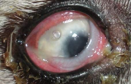
13 CELLULAR INFILTRATE Creamy, opaque infiltrate Superficial cornea Corneal stroma Presume infected!


14 LIPID/MINERAL DEPOSITS Phospholipids, Cholesterol, Calcium ground glass /chalky deposits

15 FIBROSIS Wispy, cloud-like Non-specific sign of previous injury Caused by deposition of poorly organized collagen by stromal keratocytes
 Exposure keratitis Species variable Most common in the dog 82% of all pugs!")
16 PIGMENTATION Response to chronic irritation or inflammation Keratoconjunctivitis Sicca (KCS) Chronic superficial keratitis (CSK) Exposure keratitis Species variable Most common in the dog 82% of all pugs! Reversible?

17 ASSESSMENT OF DEPTH
18 NEOVASCULARIZATION Non-specific response to corneal injury Begins w/in 2-3 days of injury Vessels grow ~1mm/day
19 NEOVASCULARIZATION Depth matters! Superficial vessels = superficial disease Branching, fine Entropion, trichiasis, distichiasis, KCS, superficial corneal ulcer Deep vessels = deep disease Dense, hedge-like Uveitis, glaucoma, deep stromal ulcer
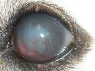
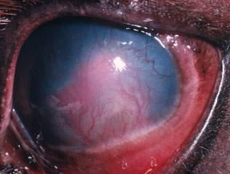
20 SUPERFICIAL NEOVASCULARIZATION


21 DEEP NEOVASCULARIZATION

22 INTRASTROMAL CORNEAL HEMORRHAGE

23

24 KERATIC PRECIPITATES Accumulated inflammatory debris on corneal endothelium Accumulate ventrally Specific sign of intraocular inflammation (uveitis)

25

26 CORNEAL ULCER - SIMPLE Heals within 5-7 days Corneal healing - Slide and divide Adjacent epithelial cells migrate to cover exposed stroma Begins w/in 1 hour of injury Basal cells reform basement membrane Mitosis of basal epithelial cells

")
27 CORNEAL ULCER COMPLICATED Persists >7 days= complicated! Underlying cause not addressed (ie ectopic cilium, distichiasis, foreign body, lagophthalmos, KCS, etc) Infected (bacterial, fungal, viral) Indolent boxer ulcer

28

29

30

31 FELINE HERPESVIRUS-ASSOCIATED KERATITIS Infects conjunctival and corneal epithelial cells Persistent and recurrent disease Blepharospasm, epiphora, chemosis, hyperemia Dendritic ulcer=pathognomonic Corneal sequestrum Eosinophilic keratitis Slatter fig 10-42


32 CORNEAL SEQUESTRUM Associated with FHV-1 Necrotic cornea Reaction of dead/dying cornea with tears Painful! Treatment=surgery


33 EOSINOPHILIC KERATITIS Associated with FHV-1 Proliferative, superficial plaque Ddx=SCC, fungal keratitis Diagnosis= cytology

34 CHRONIC SUPERFICIAL KERATITIS PANNUS German shepherd dog/gsd crosses Immune-mediated disease Fibrosis, blood vessels, pigment extend from temporal limbus toward axial cornea Bilateral Associated with UV exposure


35 CORNEAL DYSTROPHY Bilateral Central/paracentral Composed of phospholipid Non-painful No treatment necessary


36 CORNEAL DEGENERATION Calcium/lipid deposits Secondary to corneal injury or inflammation Uni- or bilateral Associated with corneal inflammation May exfoliate, resulting in corneal ulceration


37

38 MINERAL SLOUGH

39

40

41

42

43 QUESTIONS?
Around The Globe in 60 Minutes
 Around The Globe in 60 Minutes Around the GLOBE in Sixty Minutes Basic Ocular Anatomy, Examination, and Diagnostic Techniques Introduction Focusing on canine and feline ocular anatomy and basic examination
Table of Contents 1 Orbit 3 2 Eyelids 7
 Table of Contents Preface, x List of abbreviations xi Glossary xii Section I Atlas 1 1 Orbit 3 Clinical signs associated with orbital neoplasia 3 Clinical signs associated with orbital cellulitis 3 Enophthalmos
INDOLENT ULCER IN BOXER. Dr n. wet. Przemysław K. Bryla Przychodnia weterynaryjna w Warszawie INTRODUCTION
 Dr n. wet. Przemysław K. Bryla Przychodnia weterynaryjna w Warszawie brylapik@wp.pl SUMMARY A case of indolent ulcer in a Boxer is described. An indolent ulcer is a ulcer which fails to heal in the expected
n Corneal epithelium is derived from surface ectoderm n Composed of stratified squamous epith. n 5% of total corneal thickness (50-90micro m thick)
") Cornea overview Dr. Sarita Tuladhar MD, Ophthalmology Gandaki Medical College Embryology CORNEA: n Corneal epithelium is derived from surface ectoderm n Corneal stroma, descement memb, bowman s layer,
Cornea overview Dr. Sarita Tuladhar MD, Ophthalmology Gandaki Medical College Embryology CORNEA: n Corneal epithelium is derived from surface ectoderm n Corneal stroma, descement memb, bowman s layer,
Nonulcerative Keratitis (Type of Inflammation of the Cornea) Basics
 Basics") Nonulcerative Keratitis (Type of Inflammation of the Cornea) Basics OVERVIEW Keratitis is inflammation of the cornea; the cornea is the clear outer layer of the front of the eye The corneal epithelium
Nonulcerative Keratitis (Type of Inflammation of the Cornea) Basics OVERVIEW Keratitis is inflammation of the cornea; the cornea is the clear outer layer of the front of the eye The corneal epithelium
Ulcerative Keratitis (Type of Inflammation of the Cornea) Basics
 Basics") Ulcerative Keratitis (Type of Inflammation of the Cornea) Basics OVERVIEW Keratitis is inflammation of the cornea; the cornea is the clear outer layer of the front of the eye The corneal epithelium is
Ulcerative Keratitis (Type of Inflammation of the Cornea) Basics OVERVIEW Keratitis is inflammation of the cornea; the cornea is the clear outer layer of the front of the eye The corneal epithelium is
Nonulcerative Keratitis (Type of Inflammation of the Cornea) Basics
 Basics") Nonulcerative Keratitis (Type of Inflammation of the Cornea) Basics OVERVIEW Keratitis is inflammation of the cornea; the cornea is the clear outer layer of the front of the eye The corneal epithelium
Nonulcerative Keratitis (Type of Inflammation of the Cornea) Basics OVERVIEW Keratitis is inflammation of the cornea; the cornea is the clear outer layer of the front of the eye The corneal epithelium
Non-ulcerative corneal disorders in the dog and cat
 Natasha Mitchell MVB DVOphthal MRCVS Non-ulcerative corneal disorders in the dog and cat Abstract: Non-ulcerative corneal disorders include any conditions of the cornea which do not uptake fluorescein
Selected Diseases of the Cornea Dick Dubielzig July 20 th, 2009
 Selected Diseases of the Cornea Dick Dubielzig July 20 th, 2009 Canine Collagenolytic Keratitis Canine Collagenolytic Keratitis 270 cases (1.5%) Usually associated with suppurative keratitis Collagenolytic
Journal of Ophthalmic Medical Technology. Fuchs Dystrophy Amy Hischier
 Journal of Ophthalmic Medical Technology Volume 8, Number 1 October 2013 www.jomtonline.com Fuchs Dystrophy Amy Hischier Patient History: A 55 year old female complained that both of her eyes were red,
Histology of the Eye
 Histology of the Eye Objectives By the end of this lecture, the student should be able to describe: The general structure of the eye. The microscopic structure of:»cornea.»retina. EYE BULB Three coats
Corneal Ulcers. Andrew Enders, DVM Resident, Ophthalmology
 Corneal Ulcers Andrew Enders, DVM Resident, Ophthalmology Normal Corneal Anatomy Four layers Epithelium Stroma Descemet s membrane Endothelium Total thickness Canine 500-650 um Feline 500-700 um http://www.vetmed.ucdavis.edu/courses/vet_eyes/eye_path/
Photodynamic therapy for IMMK in horses
 Photodynamic therapy for IMMK in horses Overview Immune mediated keratitis in horses Traditional treatment options Sustained release implant Photodynamic therapy Use in veterinary medicine Treatment for
Degenerations. Conditions with cloudy cornea at birth or in infancy
 Dermoids The lesions are choristomas, which are congenital masses of tissue that have been dislocated from their normal position Limbal dermoids--overlapping the cornea and sclera, often inferotemporally
THE WEBINAR VET. K9 Ulcers: Drops, Cut or Refer. Guy Clare MA BVSc CertVOphthal E: 8/11/2016
 THE WEBINAR VET K9 Ulcers: Drops, Cut or Refer Guy Clare MA BVSc CertVOphthal E: info@ncvs.net.au 8/11/2016 Corneal anatomy; Corneal Ulcer Diagnosis; Corneal Ulcer Aetiology; The Boston Terrier; Indolent
Conjunctivitis in Cats
 Customer Name, Street Address, City, State, Zip code Phone number, Alt. phone number, Fax number, e-mail address, web site Conjunctivitis in Cats (Inflammation of the Moist Tissues of the Eye) Basics OVERVIEW
Proceedings of the World Small Animal Veterinary Association Sydney, Australia 2007
 Proceedings of the World Small Animal Sydney, Australia 2007 Hosted by: Next WSAVA Congress MANAGEMENT OF CORNEAL ULCERS IN SMALL ANIMALS Robin G Stanley, BVSc(Hons), FACVSc-Ophthalmology Animal Eye Care
PAINFUL PAINLESS Contact lens user BOV
 Common Causes Allergies Infections Ocular Cornea, uveitis, endophthalmitis Orbital Orbital cellulitis Inflammation Uveitis Scleritis / episcleritis Glaucomas Trauma Foreign bodies Chemical injuries History
Corneal Ulceration. Client Information Sheet Copyright Bilton Veterinary Centre All rights Reserved. What is the cornea?
 What is the cornea? Corneal Ulceration The cornea is the central clear part of the eye that is surrounded by the white of the eye called the Sclera. Looking through the cornea, you can see the coloured
CONJUNCTIVITIS IN SMALL ANIMALS: DIAGNOSING AND TREATING CASES
 Vet Times The website for the veterinary profession https://www.vettimes.co.uk CONJUNCTIVITIS IN SMALL ANIMALS: DIAGNOSING AND TREATING CASES Author : James Oliver Categories : Vets Date : April 23, 2012
Proceedings of the Southern European Veterinary Conference and Congreso Nacional de AVEPA
 www.ivis.org Proceedings of the Southern European Veterinary Conference and Congreso Nacional de AVEPA Oct. 18-21, 2012 - Barcelona, Spain Next Conference: Oct. 17-19, 2013 - Barcelona, Spain Reprinted
Eye Examination Techniques in Horses
 Eye Examination Techniques in Horses Dennis E. Brooks DVM, PhD Dip ACVO University of Florida brooksd@mail.vetmed.ufl.edu Basic Instruments How to tell the potential of vision? PLRs (retina, CN 2, chiasm,
Conjunctivitis in Dogs
 Customer Name, Street Address, City, State, Zip code Phone number, Alt. phone number, Fax number, e-mail address, web site Conjunctivitis in Dogs (Inflammation of the Moist Tissues of the Eye) Basics OVERVIEW
Fleck. Pre-Descemet Dystrophies (generally good vision and comfort) Primary Pre-Descemet Dystrophy
 Primary Pre-Descemet Dystrophy") Fleck Etiology: bilateral, sometimes asymmetric, autosomal dominant opacities located in all levels of stroma as early as 1 st decade Slit lamp: well demarcated, small round gray-white doughnut-like, wreath-like
Fleck Etiology: bilateral, sometimes asymmetric, autosomal dominant opacities located in all levels of stroma as early as 1 st decade Slit lamp: well demarcated, small round gray-white doughnut-like, wreath-like
Senile: flattening of vertical meridian, thinning of periphery, lack of luster
 Pterygia Etiology: triangular, fibrovascular, connective tissue overgrowths of bulbar conjunctiva onto cornea; distribution of ultraviolet energy- heat, wind, dust, dry atmosphere,higher prevalence nearer
Distinction layer by layer. HRT II Rostock Cornea Module
 Distinction layer by layer HRT II Rostock Cornea Module Homogenously illuminated, undistorted images Movie capture Manual Pachymetry Epithelial and intra-corneal pachymetry Full corneal thickness Post-LASIK
Differential diagnosis of the red eye. Carol Slight Nurse Practitioner Ophthalmology
 Differential diagnosis of the red eye Carol Slight Nurse Practitioner Ophthalmology The red eye Conjunctivitis HSV Keratitis Acute angle closure glaucoma Anterior Uveitis Red eye Scleritis Subconjunctival
PIGMENTARY KERATITIS IN DOGS A STUDY ON INCIDENCE IN 83 CORNEAS
 Volume 7 No. 1 January 2016 pages 9-14 Malaysian Journal of Veterinary Research RE# MJVR - 0003-2015 PIGMENTARY KERATITIS IN DOGS A STUDY ON INCIDENCE IN 83 CORNEAS ANOOP,S1, DEVANAND,C.B2, SYAM K.V3,
The Orbit. The Orbit OCULAR ANATOMY AND DISSECTION 9/25/2014. The eye is a 23 mm organ...how difficult can this be? Openings in the orbit
 The eye is a 23 mm organ...how difficult can this be? OCULAR ANATOMY AND DISSECTION JEFFREY M. GAMBLE, OD COLUMBIA EYE CONSULTANTS OPTOMETRY & UNIVERSITY OF MISSOURI DEPARTMENT OF OPHTHALMOLOGY CLINICAL
Meet Libby. Corneal Dysgenesis, Degeneration, and Dystrophies Definitions. Dr. Victor Malinovsky
 Meet Libby Corneal Dysgenesis, Degeneration, and Dystrophies 2006 Dr. Victor Malinovsky Definitions Dysgenesis: (congenital anomalies) A development disorder that results in a congenital malformation of
Specialist Referral Service Willows Information Sheets. Recurrent corneal erosions (indolent ulcers)
") Specialist Referral Service Willows Information Sheets Recurrent corneal erosions (indolent ulcers) A rabbit s cornea undergoing debridement under topical anaesthesia Recurrent corneal erosions (indolent
Specialist Referral Service Willows Information Sheets Recurrent corneal erosions (indolent ulcers) A rabbit s cornea undergoing debridement under topical anaesthesia Recurrent corneal erosions (indolent
Dry Eye Assessment and Management Study ELIGIBILITY OCULAR EVALUATION FORM
 Page 1 of 13 BEFORE COMPLETING THE OCULAR EXAMINATION, YOU MUST BE ABLE TO ANSWER YES TO THE FOLLOWING QUESTIONS: Have you done MMP9? (SVonly) The Following are done at Baseline: Have you done Tear Osmolarity?
Childhood corneal neovascularization
 Miltos Balidis PhD, FEBOphth, ICOphth Sotiria Palioura MD,PhD Childhood corneal neovascularization Opacities Cornea clarity is essential for optimal vision at any age. In childhood, loss of corneal transparency
Trifluridine Ophthalmic Solution, 1% Sterile
 Trifluridine Ophthalmic Solution, 1% Sterile DESCRIPTION Trifluridine (also known as trifluorothymidine, F 3 TdR,F 3 T), is an antiviral drug for topical treatment of epithelial keratitis caused by herpes
Dr Jo-Anne Pon. Dr Sean Every. 8:30-9:25 WS #70: Eye Essentials for GPs 9:35-10:30 WS #80: Eye Essentials for GPs (Repeated)
") Dr Sean Every Ophthalmologist Southern Eye Specialists Christchurch Dr Jo-Anne Pon Ophthalmologist Southern Eye Specialists, Christchurch Hospital, Christchurch 8:30-9:25 WS #70: Eye Essentials for GPs
Dr Sean Every Ophthalmologist Southern Eye Specialists Christchurch Dr Jo-Anne Pon Ophthalmologist Southern Eye Specialists, Christchurch Hospital, Christchurch 8:30-9:25 WS #70: Eye Essentials for GPs
Corneal specimens that influence clinical decisions
 Corneal specimens that influence clinical decisions Refractive surgery Corneal dystrophies Microbial infections J. Douglas Cameron, MD Chief, Ophthalmic Pathology Division Neuropathology Department Armed
Dr.saifalshamarti. Objective. Where is cornea? Functions of the cornea
 Cornea Dr.saifalshamarti Objective Functions Anatomy: detailed description of the 5 layers: epithelium, Bowman s layer, stroma, Descement s membrane, endothelium. Diseases of the cornea: - infection: bacterial
MANAGING MELTING EYE ULCERS
 Vet Times The website for the veterinary profession https://www.vettimes.co.uk MANAGING MELTING EYE ULCERS Author : Anna Jennings Categories : Vets Date : January 18, 2010 Anna Jennings details effective
CORNEAL CONDITIONS CORNEAL TRANSPLANTATION
 GENERAL INFORMATION CORNEAL CONDITIONS CORNEAL TRANSPLANTATION WHAT ARE CORNEAL CONDITIONS? The cornea is the clear outer layer of the eye. Shaped like a dome, it helps to protect the eye from foreign
VIROPTIC Ophthalmic Solution, 1% Sterile (trifluridine ophthalmic solution)
") VIROPTIC Ophthalmic Solution, 1% Sterile (trifluridine ophthalmic solution) PRODUCT OVERVIEW: VIROPTIC SOLUTION DESCRIPTION VIROPTIC is the brand name for trifluridine (also known as trifluorothymidine,
VIROPTIC Ophthalmic Solution, 1% Sterile (trifluridine ophthalmic solution) PRODUCT OVERVIEW: VIROPTIC SOLUTION DESCRIPTION VIROPTIC is the brand name for trifluridine (also known as trifluorothymidine,
OCULAR DISORDERS REPORT BOSTON TERRIER
 OCULAR DISORDERS REPORT BOSTON TERRIER 1991-1999 2000-2009 2010-2012 TOTAL DOGS EXAMINED 2723 6803 2004 Diagnostic Name # % # % # % GLOBE 0.110 microphthalmia 1 0.0% 1 0.0% 0 EYELIDS 20.140 ectopic cilia
ICD-10 Coding for Contact Lens Problems. The EyeCodingForum.com
 ICD-10 Coding for Contact Lens Problems The EyeCodingForum.com Jeffrey Restuccio, CPC, CPC-H, MBA Memphis TN (901) 517-1705 jeff@eyecodingforum.com www.eyecodingforum.com EyeCodingForum.com 1 Coding for
Learning Objectives:
 Viral keratitis and antivirals Learning Objectives: Recognise and distinguish different types of viral keratitis HSV HZO Adenovirus Discuss the use of antiviral agents in the treatment of herpetic infections
OOGZIEKTEN VOOR DE HUISARTS F. GOES, JR.
 OOGZIEKTEN VOOR DE HUISARTS F. GOES, JR. HET RODE OOG F. GOES, JR. Condition Signs Symptoms Causes Conjunctivitis Viral Normal vision, normal pupil size Mild to no pain, diffuse Adenovirus (most common),
Ocular Pathology. I. Congenital and/or developmental. A. Trisomy 21. Hypertelorism (widely spaced eyes) Keratoconus (cone shaped cornea)
 Keratoconus (cone shaped cornea)") I. Congenital and/or developmental Robbins Pathologic Basis of Disease, 6 th Ed. A. Trisomy 21 Hypertelorism (widely spaced eyes) Keratoconus (cone shaped cornea) Focal hypoplasia of iris Cataracts frequently
I. Congenital and/or developmental Robbins Pathologic Basis of Disease, 6 th Ed. A. Trisomy 21 Hypertelorism (widely spaced eyes) Keratoconus (cone shaped cornea) Focal hypoplasia of iris Cataracts frequently
Subject Index. Atopic keratoconjunctivitis (AKC) management 16 overview 15
 management 16 overview 15") Subject Index Acanthamoeba keratitis, see Infective keratitis Acute allergic conjunctivitis AKC, see Atopic keratoconjunctivitis Allergy acute allergic conjunctivitis 15 atopic keratoconjunctivitis 15
Subject Index Acanthamoeba keratitis, see Infective keratitis Acute allergic conjunctivitis AKC, see Atopic keratoconjunctivitis Allergy acute allergic conjunctivitis 15 atopic keratoconjunctivitis 15
a.superficial (adenoid layer).contain lymphoid tissue.
.contain lymphoid tissue.") Conjunctiva Dr. saifalshamarti Anatomy Microscopic: 1.Epithelium (non keratinized,includes goblet cell). 2.Epithelial basement membrane. 3.Stroma : a.superficial (adenoid layer).contain lymphoid tissue.
Conjunctiva Dr. saifalshamarti Anatomy Microscopic: 1.Epithelium (non keratinized,includes goblet cell). 2.Epithelial basement membrane. 3.Stroma : a.superficial (adenoid layer).contain lymphoid tissue.
Aging & Ophthalmology
 Aging & Ophthalmology Pr Jean-Marie Rakic Dr Denis Malaise January 2018 Major ocular diseases 1. Cataract 2. Age-related macular degeneration 3. Ischemic optic neuropathy 4. Horton arteritis 5. Glaucoma
Acute Eyes for ED. Enis Kocak. The Alfred Ophthalmology
 Acute Eyes for ED Enis Kocak The Alfred Ophthalmology The problem with eyes Things to cover Ocular anatomy Basic assessment Common presentations Eye first aid and procedures Ophthalmic emergencies What
Strategies for Anterior Segment Disease Management Mile Brujic, OD, FAAO 1409 Kensington Blvd Bowling Green, OH
 Strategies for Anterior Segment Disease Management Mile Brujic, OD, FAAO 1409 Kensington Blvd Bowling Green, OH 43402 brujic@prodigy.net 419-261-9161 Summary As optometry s scope of practice continues
CORNEA Anatomy, Physiology and Pathology by Joseph Bacotti, MD, FACS
 CORNEA Anatomy, Physiology and Pathology by Joseph Bacotti, MD, FACS After completion of this course the reader should be able to: 1. Describe the anatomy of the cornea and surrounding tissues 2. Describe
Codes for Medically Necessary Contact Lenses
 Codes for Medically Necessary Contact Lenses CPT Codes for Medically Necessary Prescribing Preamble for the 9231X Codes The prescription of contact lenses includes specification of optical and physical
MELTING CORNEAL ULCERS IN HORSES: DIAGNOSIS AND TREATMENT METHODS
 Vet Times The website for the veterinary profession https://www.vettimes.co.uk MELTING CORNEAL ULCERS IN HORSES: DIAGNOSIS AND TREATMENT METHODS Author : FERNANDO MALALANA Categories : Vets Date : October
Condition: Herpes Simplex Keratitis
 Condition: Herpes Simplex Keratitis Description: Herpes simplex infection is very common but usually remains latent. When the virus is reactivated it travels along the trigeminal nerve to cause local infection
OPACIFICATION IN PERFORATING CORNEAL GRAFTS*t
 Brit. J. Ophthal. (1954) 38, 10. OPACIFICATION IN PERFORATING CORNEAL GRAFTS*t BY A. G. LEIGH From the Institute of Ophthalmology, London As the success ofa grafting operation ultimately depends upon the
Unit VIII Problem 8 Anatomy: Orbit and Eyeball
 Unit VIII Problem 8 Anatomy: Orbit and Eyeball - The bony orbit: it is protecting our eyeball and resembling a pyramid: With a base directed: anterolaterally. And an apex directed: posteromedially. Notes:
Dr. D. Y. Patil Medical College, Pimpri, Pune
 Dr. D. Y. Patil Medical College, Pimpri, Pune - 411 018 Period : 04/July/16 to 22/September/16 Semester : 7 th Semester Department : Ophthalmology Lecture Lesson Plan Sr No Date Topic Learning objectives
Herpetic Eye Disease Jason Duncan, OD, FAAO Diplomate, American Board of Optometry Associate Professor, Southern College of Optometry
 Herpetic Eye Disease Jason Duncan, OD, FAAO Diplomate, American Board of Optometry Associate Professor, Southern College of Optometry I have what?! How to break the news Meet the Herpes Quick virology
02/03/2014. Average Length: 23mm (Infant ~16mm) Approximately the size of a quarter Volume: ~5mL
 Approximately the size of a quarter Volume: ~5mL") Identify the anatomy of the eye. Explain the basic physiology of the parts of the eye. Briefly discuss various surgeries related to different parts of the anatomy. Average Length: 23mm (Infant ~16mm) Approximately
Identify the anatomy of the eye. Explain the basic physiology of the parts of the eye. Briefly discuss various surgeries related to different parts of the anatomy. Average Length: 23mm (Infant ~16mm) Approximately
Affections of CONJUNCTIVA in animals
 Anatomy and physiology: Affections of CONJUNCTIVA in animals Thin and near transparent mucous membrane. Composed of an epithelial layer and a substantia propria. Two types: Bulbar and palpebral. Bulbar
THE EYE: RETINA AND GLOBE
 Neuroanatomy Suzanne Stensaas February 24, 2011, 10:00-12:00 p.m. Reading: Waxman Ch. 15. Your histology and gross anatomy books should be useful. Reading: Histology of the Eye from any histology book
Department of Ophthalmology
 Department of Ophthalmology Period : 02/July/18 to 30/August/18 Semester : 7 th Semester Lecture Lesson Plan Sr. Date Topic Lesson plan Name of Faculty No. 1 02.07.18 Lens- Lens-Anatomy, Classification
Department of Ophthalmology
 Period : 03/July/17 to 07/September/17 Semester : 7 th Semester Department of Ophthalmology Lecture Lesson Plan Sr 1 03.07.17 Uvea-Anatomy, Uvea-Anatomy, Classification of Uveitis Dr R Paranjpe Classification
DO YOU SEE WHAT I SEE? OPHTHALMIC EXAM BASICS
 DO YOU SEE WHAT I SEE? OPHTHALMIC EXAM BASICS Shelby Reinstein, DVM, MS, DACVO Veterinary Specialty and Emergency Center 301 Veterans Highway Levittown, PA 19057 Ophthalmic History Obtaining a thorough
Differential Diagnosis of Conjunctivitis and Keratoconjunctivitis
 Differential Diagnosis of Conjunctivitis and Keratoconjunctivitis Dr. Victor Malinovsky 2006 Mechanical-Physical Trauma Corneal Abrasions Abrasions (interpalpebral/variable): a focal loss of epithelium
FACING YOUR FUNDIC FEARS: EXAMINATION OF THE OCULAR FUNDUS J. Seth Eaton, VMD, DACVO Cornell University Veterinary Specialists
 FACING YOUR FUNDIC FEARS: EXAMINATION OF THE OCULAR FUNDUS J. Seth Eaton, VMD, DACVO Cornell University Veterinary Specialists The goal of a thorough fundus examination is to clinically evaluate the structures
Grid keratotomy as a treatment for superficial nonhealing corneal ulcers in 10 horses
 Veterinary Ophthalmology (2007) 10, 3, 162 167 Blackwell Publishing Inc Grid keratotomy as a treatment for superficial nonhealing corneal ulcers in 10 horses A. Brünott,* M. H. Boevé and M. A. Velden*
Small Animal Ophthalmic Atlas and Guide
 Small Animal Ophthalmic Atlas and Guide Small Animal Ophthalmic Atlas and Guide Christine C. Lim, DVM Diplomate of the American College of Veterinary Ophthalmologists Assistant professor of ophthalmology
Treating corneal ulceration in dogs part 2: deep ulcers
 Vet Times The website for the veterinary profession https://www.vettimes.co.uk Treating corneal ulceration in dogs part 2: deep ulcers Author : Mateusz Jaksz, Claudia Busse Categories : Companion animal,
Morning Report. copyright The University of Colorado. 11/25/09 Emily McCourt MD
 Morning Report 11/25/09 Emily McCourt MD HPI 46 year old presents to denver health eye clinic for urgent eval. Complains of 3 days of red eye on right No previous episodes mild pain, no fbs Has spent all
CANINE EXTRAOCULAR DISEASE. By Terri Baldwin, DVM, MS Diplomate ACVO
 CANINE EXTRAOCULAR DISEASE By Terri Baldwin, DVM, MS Diplomate ACVO Eyelid Eyelid diseases can be congenital or inherited, secondary to trauma, inflammatory, or neoplastic. When eyelid diseases occur,
founder of McDonald s Restaurants
 Press On Nothing in the world can take the place of persistence. Talent will not; nothing is more common than unsuccessful men with talent. Genius will not; unrewarded genius is almost a proverb. Education
Ocular Allergy. Phil Lieberman, MD
 Ocular Allergy Phil Lieberman, MD Disclosure Consultant/Advisory Board: Genentech, Meda, Mylan, Teva Speaker: Genentech, Meda, Merck, Mylan, Teva Learning Objectives Upon completion of this session, participants
Lamellar Keratoplasty for the Treatment of Fungal Keratitis
 Cornea 21(1): 33 37, 2002. 2002 Lippincott Williams & Wilkins, Inc., Philadelphia Lamellar Keratoplasty for the Treatment of Fungal Keratitis Lixin Xie, M.D., Weiyun Shi, M.D., Zhaosheng Liu, M.D., and
Pathology of the lens
 Pathology of the lens Carol Naranjo, LV, DACVP, DECVP, PhD IDEXX Laboratories Embryonal development Gelatt s Veterinary Ophthalmology, 5th Ed. Normal histology Lens capsule Anterior > posterior Lens cortex
Viroptic (trifluridine) solution [Monarch Pharmaceuticals, Inc.]
![Viroptic (trifluridine) solution [Monarch Pharmaceuticals, Inc.]](/thumbs/94/118693790.jpg "Viroptic (trifluridine) solution [Monarch Pharmaceuticals, Inc.]") Viroptic (trifluridine) solution [Monarch Pharmaceuticals, Inc.] Description VIROPTIC is the brand name for trifluridine (also known as trifluorothymidine, F3TdR,F3T), an antiviral drug for topical treatment
Viroptic (trifluridine) solution [Monarch Pharmaceuticals, Inc.] Description VIROPTIC is the brand name for trifluridine (also known as trifluorothymidine, F3TdR,F3T), an antiviral drug for topical treatment
10 EYE EMERGENCIES. Who goes, who you better not send! Brant Slomovic, MD, FRCPC University Health Network
 10 EYE EMERGENCIES Who goes, who you better not send! Brant Slomovic, MD, FRCPC University Health Network DISCLOSURES I have none PVD CASE 1 WHAT IS A PVD? a process of aging (45-55) liquefaction of vitreous
Basic Histology. By Mrs. Bailey
 Basic Histology By Mrs. Bailey Primary Tissues 1. Epithelial Tissue 2. Connective Tissue 3. Muscle Tissue 4. Nervous Tissue Very cellular Supported by underlying connective tissue Epithelial & connective
_ Assessment of the anterior chamber. Review of anatomy of the angle
 Assessment of the anterior chamber Dr Simon Barnard PhD BSc FCOptom FAAO DCLP Department of Optometry & Visual Science City University London, UK Review of anatomy of the angle Figure 1. Anatomical section
Keratoconjunctivitis Sicca (KCS) Dry Eye in Dogs
 Dry Eye in Dogs") Keratoconjunctivitis Sicca (KCS) Dry Eye in Dogs The eyes of dogs are beautiful orbs with intricate parts functioning together to enable each of us to see the incredible world surrounding us. Their eyes,
Keratoconjunctivitis Sicca (KCS) Dry Eye in Dogs The eyes of dogs are beautiful orbs with intricate parts functioning together to enable each of us to see the incredible world surrounding us. Their eyes,
Medical Affairs Policy
 Medical Affairs Policy Service: Corneal Treatments and Specialized Contact Lenses (Corneal remodeling, Corneal transplant, Corneal collagen crosslinking, Intrastromal Rings- INTACS, Keratoconus treatments,
Varicella-Zoster Virus Epithelial Keratitis in Herpes Zoster Ophthalmicus
 Varicella-Zoster Virus Epithelial Keratitis in Herpes Zoster Ophthalmicus Helena M. Tabery Varicella-Zoster Virus Epithelial Keratitis in Herpes Zoster Ophthalmicus In Vivo Morphology in the Human Cornea
Corneal blood staining after hyphaema
 Brit. J_. Ophthal. (I 972) 56, 589 after hyphaema J. D. BRODRICK Sheffield has been described as a rare complication of contusion injury in which a hyphaema of relatively long duration and a raised intraocular
Eye Fluids. Dr. Mohamed Saad Daoud
 Eye Fluids 1 Reference Books: Text Book of Medical physiology (Guyton and Hall) Eleventh edition 2 Fluid System of the Eye (Intraocular Fluid) The eye is filled with intraocular fluid, which maintains
The Pathology and Pathogenesis of Acute Glaucoma in Dogs. Richard R Dubielzig
 The Pathology and Pathogenesis of Acute Glaucoma in Dogs Richard R Dubielzig Overview of Glaucoma Intraocular Pressure too High to Support a Healthy Optic Nerve Terminology used in the classification of
Acute Hydrops following Penetrating Keratoplasty in a Keratoconic Patient
 Acute Hydrops following Penetrating Keratoplasty in a Keratoconic Patient Alireza Baradaran-Rafiee, MD 1 Manijeh Mahdavi, MD 2 Sepehr Feizi, MD 3 Abstract Purpose: To report a case with history of penetrating
UVEITIS. Dr. Yılmaz ÖZYAZGAN
 UVEITIS Dr. Yılmaz ÖZYAZGAN UVEITIS DEFINITION BY STRICT DEFINITION, UVEITIS IS AN INFLAMMATION OF UVEAL TRACT. BUT IN PRACTICAL, IT IS GENERALLY NOT RESTRICTED TO THE UVEA AND INVOLVES OTHER ADJACENT
Herpes simplex virus (HSV) stromal keratitis is the leading infectious cause of corneal blindness in
 stromal keratitis is the leading infectious cause of corneal blindness in") Herpes Simplex Epithelial Keratitis and Proposed Treatments Andrea De Souza, OD I. Introduction Author s Bio Dr. Andrea De Souza received her Doctor of Optometry Degree in 2012 from the New England College
Herpes Simplex Epithelial Keratitis and Proposed Treatments Andrea De Souza, OD I. Introduction Author s Bio Dr. Andrea De Souza received her Doctor of Optometry Degree in 2012 from the New England College
ICD-10-CM Cornea. Type RT LT OU SINGLE CODE UNSPECIFIED. Acute atopic conjunctivitis H10.11 H10.12 H10.13 X H10.10
 ICD-10-CM Cornea Conjunctiva Acute atopic conjunctivitis H10.11 H10.12 H10.13 H10.10 Acute chemical conjunctivitis H10.211 H10.212 H10.213 H10.219 Acute conjunctivitis, unspecified H10.31 H10.32 H10.33
PATIENT INFORMATION ON CORNEAL GRAFT
 PATIENT INFORMATION ON CORNEAL GRAFT (TRANSPLANT) SURGERY M ANANDAN What is the cornea? The clear window of the eye approximately 0.5mm thick and 12mm across. It lies in front of the fluid filled anterior
To Evaluate the Sociodemographic Factors And Etiology of Corneal Neovascularisation at out Patient Department of M.L.B Medical College, Jhansi.(U.
 IOSR Journal of Dental and Medical Sciences (IOSR-JDMS) e-issn: 2279-0853, p-issn: 2279-0861.Volume 15, Issue 6 Ver. IV (June. 2016), PP 129-134 www.iosrjournals.org To Evaluate the Sociodemographic Factors
Ocular Anatomy for the Paraoptometric
 Ocular Anatomy for the Paraoptometric Minnesota Optometric Association Paraoptometric CE Friday September 30, 2016 Lindsay A. Sicks, OD, FAAO Assistant Professor, Illinois College of Optometry lsicks@ico.edu
Relationship between limbal incisions. angle. and the structures of the anterior chamber
 Brit. _7. Ophthal. (I 973) 57, 722 Relationship between limbal incisions and the structures of the anterior chamber angle MOHAMED I. AYOUB AND AHMED H. SAID Department of Ophthalmology, Faculty of Medicine,
Funduscopic Interpretation Understanding the Fundus: is that normal?
 Funduscopic Interpretation Understanding the Fundus: is that normal? Gillian McLellan BVMS PhD DVOphthal DECVO DACVO MRCVS With thanks to Christine Heinrich and all who contributed images Fundus Retina
 Traumatic Cataract Orbital Wall Fracture Vitreous Hemorrhage Optic Disc Hemorrhage a) Amblyopia b) Strabismus c) Trauma Playing with other children Sports Fire works BB gun Injecting needles .
Case Study: Fuzz April 18th
 Case Study: Fuzz April 18th 33 year old Quarter Horse Had been battling corneal ulcer for several weeks before seeing us No foreign debris found Culture and cytology were taken. Started on topical antibiotics,
What are some common conditions that affect the cornea?
 What are some common conditions that affect the cornea? Injuries After minor injuries or scratches, the cornea usually heals on its own. Deeper injuries can cause corneal scarring, resulting in a haze
2008 Gross Ocular Pathology. Gross Pathology 2
 2008 Gross Ocular Pathology Gross Pathology 2 08rd1281 Feline T-Cell Lymphoma 08rd1300 Canine Iridociliary Adenoma Foam Cell Variant 08rd1331 Feline Feline Iridociliary Adenoma 08rd1340 Canine Retinal