CNS consists of brain and spinal cord Cephalization Evolutionary development of rostral (anterior) portion of CNS Increased number of neurons in head
|
|
|
- Joleen Morgan
- 5 years ago
- Views:
Transcription
 portion of CNS Increased number of neurons in head Highest level reached in human")
1 CNS consists of brain and spinal cord Cephalization Evolutionary development of rostral (anterior) portion of CNS Increased number of neurons in head Highest level reached in human brain 1

2 Mostly to orient view of embryonic disc Ectoderm, mesoderm, endoderm - primitive tissues from which all body organs derive Epithelia cells Ectoderm nervous system; skin 2
3 epidermis Endoderm epithelial linings of digestive, respiratory, urogenital systems; associated glands Mesenchyme cells Mesoderm everything else 2
4 After primitive embryonic tissues developed = cells committed to pathways from those tissues Ectodermal layer develops a groove and then an in-folding and then fuses into a tube 3
5 Brain and spinal cord begin as neural tube 3 primary vesicles form at anterior end Prosencephalon or forebrain Mesencephalon or midbrain Rhombencephalon or hindbrain Posterior end becomes spinal cord 4
6 Primary vesicles 5 secondary brain vesicles Forebrain telencephalon and diencephalon Midbrain remains undivided Hindbrain metencephalon and myelencephalon 5

7 Telencephalon cerebral hemispheres Diencephalon epithalamus, thalamus, hypothalamus, and retina Mesencephalon midbrain Metencephalon pons and cerebellum Myelencephalon medulla oblongata Central cavity of neural tube ventricles 6

8 Central cavity of neural tube ventricles 7
 Cerebral hemispheres double back and envelop diencephalon and midbrain while creasing and folding to increase surface area")
9 Brain grows faster than membranous skull Folds to occupy available space Forebrain moved toward brain stem (midbrain, pons, medulla oblongata) Cerebral hemispheres double back and envelop diencephalon and midbrain while creasing and folding to increase surface area 8
10
11
 4.")
12 Adult brain regions 1. Cerebral hemispheres 2. Diencephalon 3. Brain stem (midbrain, pons, and medulla) 4. Cerebellum

13 Spinal cord Central cavity surrounded by gray matter External white matter composed of myelinated fiber tracts 12
14 Meninges Function of meninges: Cover and protect CNS Protect blood vessels and enclose venous sinuses Contain cerebrospinal fluid (CSF) Form partitions in skull Consists of three layers (from external to internal): dura mater, arachnoid mater, and pia mater 13

15 Dura mater Strongest meninx Made up of two layers of fibrous connective tissue Periosteal layer attaches to inner surface of skull Found only in brain, not spinal cord Meningeal layer: true external covering of brain Extends into vertebral canal as spinal dura mater Two layers are mostly fused, but separate in certain areas to form dural venous sinuses Sinuses collect venous blood from brain, empty into jugular veins of neck 14

16 Dura mater extends inward in several areas to form flat partitions that divide cranial cavity Partitions referred to as dural septa Act to limit excessive movement of brain Three main septa: Falx cerebri: in longitudinal fissure; attached to crista galli Falx cerebelli: along vermis of cerebellum Tentorium cerebelli: horizontal dural fold over cerebellum and in transverse fissure 15
17 Arachnoid mater Middle layer with spiderweb-like extensions Separated from dura mater by subdural space Subarachnoid space contains CSF and largest blood vessels of brain Arachnoid granulations protrude through dura mater into superior sagittal sinus Permit reabsorption of CSF back into venous blood 16

18 Pia mater Delicate connective tissue that clings tightly to brain, following every convolution Contains many tiny blood vessels that feed brain 17
19 18

20 Brain (brainstem, anyway) Brainstem similar pattern to spinal cord Additional areas of gray matter in brain 19

21 Brainstem Similar pattern Additional areas of gray matter in brain 20

22 Additional areas of gray matter in brain Cerebral hemispheres and cerebellum Outer gray matter called cortex 21
23 Paired, C-shaped lateral ventricles in cerebral hemispheres Separated anteriorly by septum pellucidum Third ventricle in diencephalon Fourth ventricle in hindbrain Three openings: paired lateral apertures in side walls; median aperture in roof Connect ventricles to subarachnoid space

24 Filled with cerebrospinal fluid (CSF) Lined by ependymal cells Connected to one another and to central canal of spinal cord Lateral ventricles third ventricle via interventricular foramen Third ventricle fourth ventricle via cerebral aqueduct

25 Choroid plexus: cluster of capillaries that hangs from roof of each ventricle, enclosed by pia mater and surrounding layer of ependymal cells CSF is filtered from plexus at constant rate Ependymal cells use ion pumps to control composition of CSF and help cleanse CSF by removing wastes Cilia of ependymal cells help to keep CSF in motion Normal adult CSF volume of ~150 ml is replaced every 8 hours 24

26 Cerebrospinal fluid (CSF) forms a liquid cushion of constant volume around brain Functions Gives buoyancy to CNS structures Reduces weight of brain by 97% by floating it so it is not crushed under its own weight Protects CNS from blows and other trauma Nourishes brain and carries chemical signals Composed of watery solution formed from blood plasma, but with less protein and different ion concentrations from plasma 25

27 Five lobes Frontal Parietal Temporal Occipital Insula
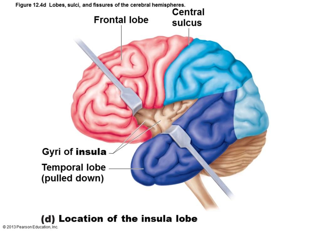
28 Insula
29 Surface markings Ridges (gyri), shallow grooves (sulci), and deep grooves (fissures)

30 Longitudinal fissure Separates two hemispheres

31 Transverse cerebral fissure Separates cerebrum and cerebellum

32 Central sulcus Separates precentral gyrus of frontal lobe and postcentral gyrus of parietal lobe Parieto-occipital sulcus Separates occipital and parietal lobes Lateral sulcus outlines temporal lobes

33 Three basic regions Cerebral cortex of gray matter superficially White matter internally Basal nuclei deep within white matter 32

34 Thin (2 4 mm) superficial layer of gray matter 40% mass of brain Site of conscious mind: awareness, sensory perception, voluntary motor initiation, communication, memory storage, understanding 33
 34")
35 Has six layers: I Molecular layer II External granular layer III External pyramidal layer IV Internal granular layer V Internal pyramidal layer VI Multiform (polymorphic layer) 34
 35")
36 Has six layers: I Molecular layer II External granular layer III External pyramidal layer IV Internal granular layer V Internal pyramidal layer VI Multiform (polymorphic layer) 35

37 Layer 1 consists mainly of apical dendrites from pyramidal cells from lower layers plus axons synapsing on those dendrites. It contains almost no neuron cell bodies. Layer 2 contains many small densely-packed pyramidal neurons giving it a granular appearance. Layer 3 contains medium-sized pyramidal neurons which send outputs to other cortical areas. Layer 4 contains many spiny stellate (excitatory) interneurons Layer 5 contains the largest pyramidal neurons, which send outputs to the brain stem and spinal cord (the pyramidal tract) Layer 6 consists of pyramidal neurons and neurons with spindle-shaped cell bodies. 36

38 A Brodmann area is a region of the cerebral cortex, in the human or other primate brain, defined by its cytoarchitecture, or histological structure and organization of cells. Wikipedia 52 original areas duplicated in hemispheres Subsequently subdivided areas as more refined techniques developed. Functional separation of parts of the cortex grossly matches cytoarchitectural differences 37
 receptive")
39 Cortical neurons interconnected in vertical columns Presumably all cells functionally related all have same precise (or very, very close) receptive fields 38
 of cortical function in hemispheres 4.")
40 1. Three types of functional areas Motor areas control voluntary movement Sensory areas conscious awareness of sensation Association areas integrate diverse information 2. Each hemisphere concerned with contralateral side of body 3. Lateralization (specialization) of cortical function in hemispheres 4. Conscious behavior involves entire cortex in some way 39
41

42 Broca's area anterior to inferior premotor area Frontal eye field within and anterior to premotor cortex; superior to Broca's area

43 In frontal lobe; control voluntary movement Primary (somatic) motor cortex in precentral gyrus Premotor cortex anterior to precentral gyrus 42

44 Allows conscious control of precise, skilled, skeletal muscle movements Betz cells very large pyramidal neurons in layer V of pre-central gyrus Next slide
 tracts of spinal")
45 Large pyramidal cells of precentral gyri Long axons pyramidal (corticospinal) tracts of spinal cord 44

46 Motor homunculi - upside-down caricatures represent contralateral motor innervation of body regions
47 Premotor Cortex Helps plan movements; staging area for skilled motor activities Controls learned, repetitious, or patterned motor skills Coordinates simultaneous or sequential actions Controls voluntary actions that depend on sensory feedback
48 Broca's Area Present in one hemisphere (usually the left) Motor speech area that directs muscles of speech production Active in planning speech and voluntary motor activities
49 Frontal Eye Field Controls voluntary eye movements
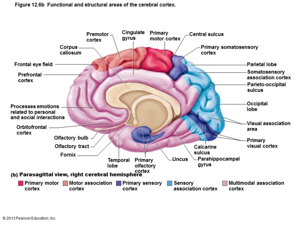
50

51 Conscious awareness of sensation Occur in parietal, insular, temporal, and occipital lobes 50

52 Primary somatosensory cortex Visual areas Auditory areas Vestibular cortex Olfactory cortex Gustatory cortex Visceral sensory area 51

53 In postcentral gyri of parietal lobe Receives general sensory information from skin, and proprioceptors of skeletal muscle, joints, and tendons Capable of spatial discrimination: identification of body region being stimulated Somatosensory homunculus upside-down caricatures represent contralateral sensory input from body regions

54 Somatosensory association cortex Posterior to primary somatosensory cortex Integrates sensory input from primary somatosensory cortex for understanding of object Determines size, texture, and relationship of parts of objects being felt 53

55 Primary visual (striate) cortex Extreme posterior tip of occipital lobe Most buried in calcarine sulcus of occipital lobe Receives visual information from retinas 54
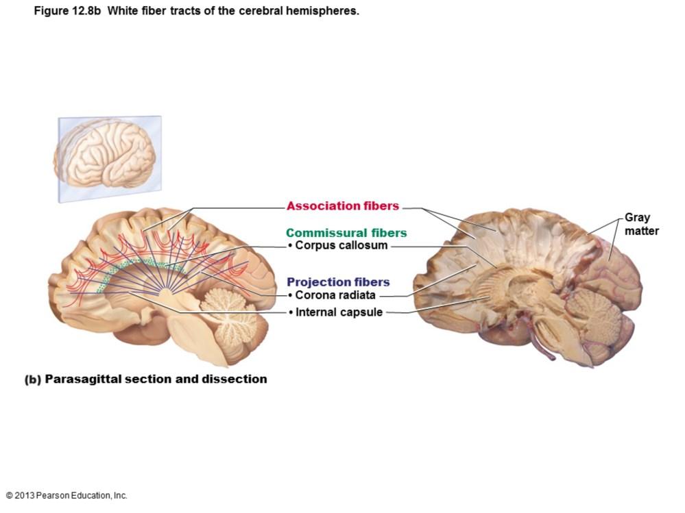
 E.g.")
56 Visual association area Surrounds primary visual cortex Uses past visual experiences to interpret visual stimuli (e.g., color, form, and movement) E.g., ability to recognize faces Complex processing involves entire posterior half of cerebral hemispheres Ventral Stream what stream Dorsal Stream where stream 55

57 Primary auditory cortex Superior margin of temporal lobes Interprets information from inner ear as pitch, loudness, and location 56

58 Auditory association area Located posterior to primary auditory cortex Stores memories of sounds and permits perception of sound stimulus 57
 58")
59 Posterior part of insula and adjacent parietal cortex Responsible for conscious awareness of balance (position of head in space) 58

60 Primary olfactory (smell) cortex Medial aspect of temporal lobes (in piriform lobes) Part of primitive rhinencephalon, along with olfactory bulbs and tracts Remainder of rhinencephalon in humans part of limbic system Region of conscious awareness of odors 59

61 In insula just deep to temporal lobe Involved in perception of taste 60

62 Posterior to gustatory cortex Conscious perception of visceral sensations, e.g., upset stomach or full bladder 61

63 Receive inputs from multiple sensory areas Send outputs to multiple areas, including premotor cortex Allows meaning to information received, store in memory, tying to previous experience, and deciding on actions Sensations, thoughts, emotions become conscious makes us who we are 62
 Posterior association area [and")
64 Three broad parts: Anterior association area (prefrontal cortex) Posterior association area [and next slide] 63

65 Limbic association area 64

66 Part of limbic system Involves cingulate gyrus, parahippocampal gyrus, and hippocampus Provides emotional impact that makes scene important and helps establish memories 65

67 Most complicated cortical region Involved with intellect, cognition, recall, and personality Contains working memory needed for abstract ideas, judgment, reasoning, persistence, and planning Development depends on feedback from social environment 66
68 Large region in temporal, parietal, and occipital lobes Plays role in recognizing patterns and faces and localizing us in space Involved in understanding written and spoken language (Wernicke's area 67
")
69 Hemispheres almost identical Lateralization - division of labor between hemispheres Cerebral dominance - hemisphere dominant for language (left hemisphere - 90% people) 68

70 Left hemisphere Controls language, math, and logic Right hemisphere Visual-spatial skills, intuition, emotion, and artistic and musical skills Hemispheres communicate almost instantaneously via fiber tracts and functional integration 69

71 Myelinated fibers and tracts Communication between cerebral areas, and between cortex and lower CNS Association fibers horizontal; connect different parts of same hemisphere Commissural fibers horizontal; connect gray matter of two hemispheres Projection fibers vertical; connect hemispheres with lower brain or spinal cord
72
 and substantia nigra")
73 Subcortical nuclei Caudate nucleus Putamen Globus pallidus Caudate nucleus + putamen = striatum Associated with subthalamic nuclei (diencephalon) and substantia nigra (midbrain)

74 Functions thought to be Influence muscle movements Role in cognition and emotion Regulate intensity of slow or stereotyped movements Filter out incorrect/inappropriate responses Inhibit antagonistic/unnecessary movements
75 Three paired structures Thalamus Hypothalamus Epithalamus Encloses third ventricle
 Contains several nuclei, named for location Nuclei project and receive fibers from cerebral")
76 80% of diencephalon Superolateral walls of third ventricle Bilateral nuclei connected by interthalamic adhesion (intermediate mass) Contains several nuclei, named for location Nuclei project and receive fibers from cerebral cortex

77 Gateway to cerebral cortex Sorts, edits, and relays ascending input Impulses from hypothalamus for regulation of emotion and visceral function Impulses from cerebellum and basal nuclei to help direct motor cortices Impulses for memory or sensory integration Mediates sensation, motor activities, cortical arousal, learning, and memory 76

78 Forms inferolateral walls of third ventricle Contains many nuclei Example: mammillary bodies Paired anterior nuclei Olfactory relay stations Infundibulum stalk that connects to pituitary gland
 Physical responses to emotions (limbic")
79 Controls autonomic nervous system (e.g., blood pressure, rate and force of heartbeat, digestive tract motility, pupil size) Physical responses to emotions (limbic system) Perception of pleasure, fear, and rage, and in biological rhythms and drives 78
 Controls endocrine system Controls secretions of anterior pituitary gland Produces posterior pituitary")
80 Regulates body temperature sweating/shivering Regulates hunger and satiety in response to nutrient blood levels or hormones Regulates water balance and thirst Regulates sleep-wake cycles Suprachiasmatic nucleus (biological clock) Controls endocrine system Controls secretions of anterior pituitary gland Produces posterior pituitary hormones 79
 extends from posterior border and secretes melatonin Melatonin helps regulate sleep-wake")
81 Epithalamus Most dorsal portion of diencephalon; forms roof of third ventricle Pineal gland (body) extends from posterior border and secretes melatonin Melatonin helps regulate sleep-wake cycle
Homework Week 2. PreLab 2 HW #2 Synapses (Page 1 in the HW Section)
") Homework Week 2 Due in Lab PreLab 2 HW #2 Synapses (Page 1 in the HW Section) Reminders No class next Monday Quiz 1 is @ 5:30pm on Tuesday, 1/22/13 Study guide posted under Study Aids section of website
Homework Week 2 Due in Lab PreLab 2 HW #2 Synapses (Page 1 in the HW Section) Reminders No class next Monday Quiz 1 is @ 5:30pm on Tuesday, 1/22/13 Study guide posted under Study Aids section of website
Anatomy and Physiology (Bio 220) The Brain Chapter 14 and select portions of Chapter 16
 The Brain Chapter 14 and select portions of Chapter 16") Anatomy and Physiology (Bio 220) The Brain Chapter 14 and select portions of Chapter 16 I. Introduction A. Appearance 1. physical 2. weight 3. relative weight B. Major parts of the brain 1. cerebrum 2.
Anatomy and Physiology (Bio 220) The Brain Chapter 14 and select portions of Chapter 16 I. Introduction A. Appearance 1. physical 2. weight 3. relative weight B. Major parts of the brain 1. cerebrum 2.
BRAIN PART I (A & B): VENTRICLES & MENINGES
: VENTRICLES & MENINGES") BRAIN PART I (A & B): VENTRICLES & MENINGES Cranial Meninges Cranial meninges are continuous with spinal meninges Dura mater: inner layer (meningeal layer) outer layer (endosteal layer) fused to periosteum
BRAIN PART I (A & B): VENTRICLES & MENINGES Cranial Meninges Cranial meninges are continuous with spinal meninges Dura mater: inner layer (meningeal layer) outer layer (endosteal layer) fused to periosteum
Chapter 18: The Brain & Cranial Nerves. Origin of the Brain
 Chapter 18: The Brain & Cranial Nerves BIO 218 Fall 2015 Origin of the Brain The brain originates from a structure called the neural tube, which arises during a developmental stage called neurulation.
Human Anatomy. Brain and Cranial Nerves
 Human Anatomy Brain and Cranial Nerves 1 Brain and Cranial Nerves An adult brain weighs between 1.35 and 1.4 kilograms (kg) (around 3 pounds) and has a volume of about 1200 cubic centimeters (cc). Brain
The Central Nervous System I. Chapter 12
 The Central Nervous System I Chapter 12 The Central Nervous System The Brain and Spinal Cord Contained within the Axial Skeleton Brain Regions and Organization Medical Scheme (4 regions) 1. Cerebral Hemispheres
b. The groove between the two crests is called 2. The neural folds move toward each other & the fuse to create a
 Chapter 13: Brain and Cranial Nerves I. Development of the CNS A. The CNS begins as a flat plate called the B. The process proceeds as: 1. The lateral sides of the become elevated as waves called a. The
Biological Bases of Behavior. 3: Structure of the Nervous System
 Biological Bases of Behavior 3: Structure of the Nervous System Neuroanatomy Terms The neuraxis is an imaginary line drawn through the spinal cord up to the front of the brain Anatomical directions are
Department of Cognitive Science UCSD
 Department of Cognitive Science UCSD Verse 1: Neocortex, frontal lobe, Brain stem, brain stem, Hippocampus, neural node, Right hemisphere, Pons and cortex visual, Brain stem, brain stem, Sylvian fissure,
Chapter 3. Structure and Function of the Nervous System. Copyright (c) Allyn and Bacon 2004
 Allyn and Bacon 2004") Chapter 3 Structure and Function of the Nervous System 1 Basic Features of the Nervous System Neuraxis: An imaginary line drawn through the center of the length of the central nervous system, from the
Chapter 3 Structure and Function of the Nervous System 1 Basic Features of the Nervous System Neuraxis: An imaginary line drawn through the center of the length of the central nervous system, from the
The neurvous system senses, interprets, and responds to changes in the environment. Two types of cells makes this possible:
 NERVOUS SYSTEM The neurvous system senses, interprets, and responds to changes in the environment. Two types of cells makes this possible: the neuron and the supporting cells ("glial cells"). Neuron Neurons
The Brain. Brain. Spinal Cord. Cauda Equina
 The Brain Brain Spinal Cord Cauda Equina The Brain Ventricles- cavities in the brain filled with cerebrospinal fluid connected to the subarachnoid space- fluid filled space surrounding the brain Brain
Embryonic Brain Development
 Chapter 14 The Brain and Cranial Nerves Largest organ in the body? Brain functions in sensations, memory, emotions, decision making, behavior 19-1 19-2 Embryonic Brain Development Principal Parts of the
Organization of The Nervous System PROF. MOUSAED ALFAYEZ & DR. SANAA ALSHAARAWY
 Organization of The Nervous System PROF. MOUSAED ALFAYEZ & DR. SANAA ALSHAARAWY Objectives At the end of the lecture, the students should be able to: List the parts of the nervous system. List the function
Unit 12a: The Nervous System The Brain. MDL231 Principle of Anatomy
 Unit 12a: The Nervous System The Brain MDL231 Principle of Anatomy The Brain - Overview Cerebrum T PP H midbrain Cerebellum pons m.o. Brain stem medulla oblongata (M.O.) pons midbrain (mesencephalon) Diencephalon
The Nervous System PART B
 7 The Nervous System PART B PowerPoint Lecture Slide Presentation by Jerry L. Cook, Sam Houston University ESSENTIALS OF HUMAN ANATOMY & PHYSIOLOGY EIGHTH EDITION ELAINE N. MARIEB The Reflex Arc Reflex
Parts of the Brain. Hindbrain. Controls autonomic functions Breathing, Heartbeat, Blood pressure, Swallowing, Vomiting, etc. Upper part of hindbrain
 Parts of the Brain The human brain is made up of three main parts: 1) Hindbrain (or brainstem) Which is made up of: Myelencephalon Metencephalon 2) Midbrain Which is made up of: Mesencephalon 3) Forebrain
CEREBRUM Dr. Jamila Elmedany Dr. Essam Eldin Salama
 CEREBRUM Dr. Jamila Elmedany Dr. Essam Eldin Salama Objectives At the end of the lecture, the student should be able to: List the parts of the cerebral hemisphere (cortex, medulla, basal nuclei, lateral
Chapter 13 Brain and Cranial Nerves
 Chapter 13 Brain and Cranial Nerves 13-1 Brain and Cranial Nerves Brain Part of CNS contained in cranial cavity Control center for many of body s functions Much like a complex computer but more Parts of
Blood supply to the brain Blood brain barrier isolates neural tissue from general circulation
 The Brain and Cranial Nerves Objectives Name the major regions of the brain and describe their functions. Discuss the formation, circulation, and functions of the CSF. List the main components of the medulla
Brain ميهاربا لض اف دمح ا د The Meninges 1- Dura Mater of the Brain endosteal layer does not extend meningeal layer falx cerebri tentorium cerebelli
 .احمد د فاضل ابراهيم Lecture 15 Brain The Meninges Three protective membranes or meninges surround the brain in the skull: the dura mater, the arachnoid mater, and the pia mater 1- Dura Mater of the Brain
Anatomy Lecture Notes Chapter 13
 I. embryonic development of the CNS A. neurulation is the formation of the CNS in the embryo invagination of dorsal ectoderm (outer layer of embryo cells) this process is induced (caused) by the notochord
14 - Central Nervous System. The Brain Taft College Human Physiology
 14 - Central Nervous System The Brain Taft College Human Physiology Development of the Brain The brain begins as a simple tube, a neural tube. The tube or chamber (ventricle) is filled with cerebrospinal
Unit Three. The brain includes: cerebrum, diencephalon, brain stem, & cerebellum. The brain lies within the cranial cavity of the skull.
 Human Anatomy & Physiology 11 Divisions of the Nervous System Karen W. Smith, Instructor Unit Three BRAIN & SPINAL CORD Refer to the following URLs. Be sure to study these along with your book. http://www.sirinet.net/~jgjohnso/nervous.html
Chapter 14: The Brain and Cranial Nerves. Copyright 2009, John Wiley & Sons, Inc.
 Chapter 14: The Brain and Cranial Nerves Development of the Brain Three to four-week embryo: prosencephalon, mesencephalon and rhombencephalon. Five-week embryo: telencephalon (cerebrum), diencephalon
PET Scans. External Appearance. The Brain: Anatomy & Functions. Cerebral Hemispheres
 PET Scans The Brain: Anatomy & Functions Click for PET Scan video Cerebral Hemispheres External Appearance a pattern of ridges and shallow grooves ridges - gyri (sing. gyrus) grooves - sulci (sing. sulcus)
Announcement. Danny to schedule a time if you are interested.
 Announcement If you need more experiments to participate in, contact Danny Sanchez (dsanchez@ucsd.edu) make sure to tell him that you are from LIGN171, so he will let me know about your credit (1 point).
Organization of The Nervous System PROF. SAEED ABUEL MAKAREM
 Organization of The Nervous System PROF. SAEED ABUEL MAKAREM Objectives By the end of the lecture, you should be able to: List the parts of the nervous system. List the function of the nervous system.
meninges Outermost layer of the meninge dura mater arachnoid mater pia mater membranes located between bone and soft tissue of the nervous system
 membranes located between bone and soft tissue of the nervous system meninges Outermost layer of the meninge dura mater middle layer of the meninges, contains no blood vessels arachnoid mater Innermost
Sheep Brain Dissection
 Sheep Brain Dissection Mammalian brains have many features in common. Human brains may not be available, so sheep brains often are dissected as an aid to understanding the mammalian brain since he general
Functional Organization of the Central Nervous System
 Functional Organization of the Central Nervous System Hierarchical orgnization CNS consists of the brain and the spinal cord The brain analyzes and interprets the information Response messages are
Good Morning! Take out your notes and vocab 1-10! Copyright 2003 Pearson Education, Inc. publishing as Benjamin Cummings
 Good Morning! Take out your notes and vocab 1-10! Functions of the Nervous System 1. Sensory input gathering information To monitor changes occurring inside and outside the body (changes = stimuli) 2.
CEREBRUM & CEREBRAL CORTEX
 CEREBRUM & CEREBRAL CORTEX Seonghan Kim Dept. of Anatomy Inje University, College of Medicine THE BRAIN ANATOMICAL REGIONS A. Cerebrum B. Diencephalon Thalamus Hypothalamus C. Brain Stem Midbrain Pons
Ch 13: Central Nervous System Part 1: The Brain p 374
 Ch 13: Central Nervous System Part 1: The Brain p 374 Discuss the organization of the brain, including the major structures and how they relate to one another! Review the meninges of the spinal cord and
The Nervous System 7PART B. PowerPoint Lecture Slide Presentation by Patty Bostwick-Taylor, Florence-Darlington Technical College
 PowerPoint Lecture Slide Presentation by Patty Bostwick-Taylor, Florence-Darlington Technical College The Nervous System 7PART B What is a reflex? What is a reflex? What is meant by the statement that
Chapter 12 The Central Nervous System Chapter Outline
 Chapter 12 The Central Nervous System Chapter Outline Module 12.1 Overview of the Central Nervous System (Figures 12.1, 12.2, 12.3) A. The central nervous system (CNS) includes the and, and is involved
ACTIVITY 7: NERVOUS SYSTEM HISTOLOGY, BRAIN, CRANIAL NERVES
 ACTIVITY 7: NERVOUS SYSTEM HISTOLOGY, BRAIN, CRANIAL NERVES LABORATORY OBJECTIVES: 1. Histology: Identify structures indicated on three different slides or images of nervous system tissue. These images
Chapter 13 Lecture Outline *
 Anatomy and Physiology, Seventh Edition Rod R. Seeley Idaho State University Trent D. Stephens Idaho State University Philip Tate Phoenix College Chapter 13 Lecture Outline * *See PowerPoint Image Slides
a) Central sulcus- shallow groove that runs across brain sagitally
 Central sulcus- shallow groove that runs across brain sagitally") KEY BRAIN Brain Gross Anatomy Terms 1) Explain each of the following in terms of structure of the brain a) Central sulcus- shallow groove that runs across brain sagitally b) Lateral fissure- deep groove
KEY BRAIN Brain Gross Anatomy Terms 1) Explain each of the following in terms of structure of the brain a) Central sulcus- shallow groove that runs across brain sagitally b) Lateral fissure- deep groove
Neuroanatomy lecture (1)
") Neuroanatomy lecture (1) Introduction: Neuroanatomy has two parts: the central and peripheral nervous system. The central nervous system is composed of brain and spinal cord. The brain has the following
Neuroanatomy lecture (1) Introduction: Neuroanatomy has two parts: the central and peripheral nervous system. The central nervous system is composed of brain and spinal cord. The brain has the following
I. Anatomy of the Brain A. Cranial Meninges and Ventricles of the Brain 1. Meninges a. Dura mater 1) Endosteal/Periosteal Layer - Outer 2) Meningeal
 Endosteal/Periosteal Layer - Outer 2) Meningeal") I. Anatomy of the Brain A. Cranial Meninges and Ventricles of the Brain 1. Meninges a. Dura mater 1) Endosteal/Periosteal Layer - Outer 2) Meningeal Layer - Inner 3) Falx cerebri a) Superior sagittal sinus
I. Anatomy of the Brain A. Cranial Meninges and Ventricles of the Brain 1. Meninges a. Dura mater 1) Endosteal/Periosteal Layer - Outer 2) Meningeal Layer - Inner 3) Falx cerebri a) Superior sagittal sinus
stored information, making decisions, and taking action. 1. It is also the center for intellect, emotions, behavior, and memory.
 Chapter 14 - Outline I. INTRODUCTION A. The brain is the center for registering sensations, correlating them with one another and with stored information, making decisions, and taking action. 1. It is
DIENCEPHALON. ..Central core of the forebrain..consists of three paired structures
 DIENCEPHALON..Central core of the forebrain..consists of three paired structures ---------- --thalamus, --------hypothalamus, -------epithalamus..encloses the third ventricle Diencephalon between cerebral
The Brain and Cranial Nerves Pg Three Main Regions of the Brain. Forebrain
 The Brain and Cranial Nerves Pg. 129 Three Main Regions of the Brain Forebrain Cerbral hemispheres Diencephalon Midbrain Brain stem Hindbrain Pons Cerebellum Medulla oblongata Interprets sensory inputs
The Nervous System PART B
 7 The Nervous System PART B PowerPoint Lecture Slide Presentation by Jerry L. Cook, Sam Houston University ESSENTIALS OF HUMAN ANATOMY & PHYSIOLOGY EIGHTH EDITION ELAINE N. MARIEB Central Nervous System
The Brain and Cranial Nerves Pg. 129
 The Brain and Cranial Nerves Pg. 129 Three Main Regions of the Brain Forebrain Cerbral hemispheres Diencephalon Midbrain Brain stem Hindbrain Pons Cerebellum Medulla oblongata Forebrain Interprets sensory
Nervous System - PNS and CNS. Bio 105
 Nervous System - PNS and CNS Bio 105 Outline I. Central Nervous System vs Peripheral Nervous System II. Peripheral Nervous System A. Autonomic Nervous Systems B. Somatic Nervous Systems III. Autonomic
3/15/17. Outline. Nervous System - PNS and CNS. Two Parts of the Nervous System
 Nervous System - PNS and CNS Bio 105 Outline I. Central Nervous System vs Peripheral Nervous System II. Peripheral Nervous System A. Autonomic Nervous Systems B. Somatic Nervous Systems III. Autonomic
NOTES CHAPTER 9 (Brief) The Nervous System LECTURE NOTES
 The Nervous System LECTURE NOTES") NOTES CHAPTER 9 (Brief) The Nervous System LECTURE NOTES I. Divisions of the Nervous System two major divisions A. Central Nervous System (CNS) 1. brain 2. spinal cord B. Peripheral Nervous System (PNS)
NOTES CHAPTER 9 (Brief) The Nervous System LECTURE NOTES I. Divisions of the Nervous System two major divisions A. Central Nervous System (CNS) 1. brain 2. spinal cord B. Peripheral Nervous System (PNS)
Somatic Nervous Systems. III. Autonomic Nervous System. Parasympathetic Nervous System. Sympathetic Nervous Systems
 7/21/2014 Outline Nervous System - PNS and CNS I. II. Two Parts of the Nervous System Central Nervous System vs Peripheral Nervous System Peripheral Nervous System A. B. Brain and Spinal Cord III. Autonomic
Anatomy of the Human Brain
 Anatomy of the Human Brain Overview Lobes of the brain (Forebrain) Midbrain/Hindbrain Protection and Blood supply Structure and Function of a neuron Synaptic Transmission Neurotransmitters The brain Most
CEREBRUM. Dr. Jamila EL Medany
 CEREBRUM Dr. Jamila EL Medany Objectives At the end of the lecture, the student should be able to: List the parts of the cerebral hemisphere (cortex, medulla, basal nuclei, lateral ventricle). Describe
Lecture - Chapter 13: Central Nervous System
 Lecture - Chapter 13: Central Nervous System 1. Describe the following structures of the brain, what is the general function of each: a. Cerebrum b. Diencephalon c. Brain Stem d. Cerebellum 2. What structures
MENTAL HOSPITAL PHONE MENU
 If you have low self-esteem, please hang up. Our operators are too busy to talk with you. MENTAL HOSPITAL PHONE MENU Hello and thank you for calling The State Mental Hospital. Please select from the following
Divisions of the Nervous System
 Marieb s Human Anatomy and Physiology Marieb Hoehn Chapter 12 The Central Nervous System Lecture 19 1 Divisions of the Nervous System You are here CNS PNS 3 Brain Embryology & Overview Table & Figure From:
Central Nervous System (CNS) -> brain and spinal cord. Major Divisions of the nervous system:
 -> brain and spinal cord. Major Divisions of the nervous system:") Central Nervous System (CNS) -> brain and spinal cord Major Divisions of the nervous system: Afferent (sensory input) -> cell bodies outside of the central nervous system (CNS), carry info into the CNS
Central Nervous System (CNS) -> brain and spinal cord Major Divisions of the nervous system: Afferent (sensory input) -> cell bodies outside of the central nervous system (CNS), carry info into the CNS
CHAPTER 13&14: The Central Nervous System. Anatomy of the CNS
 CHAPTER 13&14: The Central Nervous System Anatomy of the CNS in human consists of brain and spinal cord as stated earlier neurons have little support from their extracellular matrix and depend on glial
M555 Medical Neuroscience Lab 1: Gross Anatomy of Brain, Crainal Nerves and Cerebral Blood Vessels
 M555 Medical Neuroscience Lab 1: Gross Anatomy of Brain, Crainal Nerves and Cerebral Blood Vessels Anatomical Directions Terms like dorsal, ventral, and posterior provide a means of locating structures
Histology of the CNS
 Histology of the CNS Lecture Objectives Describe the histology of the cerebral cortex layers. Describe the histological features of the cerebellum; layers and cells of cerebellar cortex. Describe the elements
Principles of Anatomy and Physiology
 Principles of Anatomy and Physiology 14 th Edition CHAPTER 14 The Brain and Cranial Nerves Introduction The purpose of the chapter is to: 1. Understand how the brain is organized, protected, and supplied
Central nervous system (CNS): brain and spinal cord Collections of cell body and dendrites (grey matter) are called nuclei/nucleus Nucleus can also
: brain and spinal cord Collections of cell body and dendrites (grey matter) are called nuclei/nucleus Nucleus can also") Chapter 3 Part 1 Orientation Directions in the nervous system are described relatively to the neuraxis An imaginary line drawn through the center of the length of the central nervous system, from the bottom
Chapter 3 Part 1 Orientation Directions in the nervous system are described relatively to the neuraxis An imaginary line drawn through the center of the length of the central nervous system, from the bottom
Brain and Cranial Nerves (Ch. 15) Human Anatomy lecture. caudal = toward the spinal cord)
 Human Anatomy lecture. caudal = toward the spinal cord)") Insight: Some cranial nerve disorders Brain and Cranial Nerves (Ch. 15) Human Anatomy lecture I. Overview (Directional terms: rostral = toward the forehead caudal = toward the spinal cord) A. 3 Major parts
Insight: Some cranial nerve disorders Brain and Cranial Nerves (Ch. 15) Human Anatomy lecture I. Overview (Directional terms: rostral = toward the forehead caudal = toward the spinal cord) A. 3 Major parts
The Central Nervous System
 The Central Nervous System 1 The Central Nervous System Brain Spinal Cord Components 2 Protection of the Brain The brain is protected from injury by The skull enough said! You already know this. What else
PROPERTY OF ELSEVIER SAMPLE CONTENT - NOT FINAL. Gross Anatomy and General Organization of the Central Nervous System
 3 Gross Anatomy and General Organization of the Central Nervous System C h a p t e r O u t l i n e The Long Axis of the CNS Bends at the Cephalic Flexure Hemisecting a Brain Reveals Parts of the Diencephalon,
Nervous System: Part IV The Central Nervous System The Brain
 Nervous System: Part IV The Central Nervous System The Brain Can you survive when part of your brain is destroyed? 2 Essential Knowledge 3.D.2 2. Cells communicate with each other through direct contact
Development of Brain Stem, Cerebellum and Cerebrum
 Development of Brain Stem, Cerebellum and Cerebrum The neural tube cranial to the 4th pair of somites develop into the brain. 3 dilatations and 2 flexures form at the cephalic end of the neural tube during
SOME BASIC TERMINOLOGY CNS: Central Nervous System: Brain + Spinal Cord
 SOME BASIC TERMINOLOGY CNS: Central Nervous System: Brain + Spinal Cord CEREBROSPINAL FLUID (CSF): The fluid filling the ventricles, cerebral aqueduct, central canal, and subarachnoid space. It is a filtrate
Anatomy Lab (1) Theoretical Part. Page (2 A) Page (2B)
 Theoretical Part. Page (2 A) Page (2B)") Anatomy Lab (1) This sheet only includes the extra notes for the lab handout regarding the theoretical part, as for the practical part it includes everything the doctor mentioned. Theoretical Part Page
Anatomy Lab (1) This sheet only includes the extra notes for the lab handout regarding the theoretical part, as for the practical part it includes everything the doctor mentioned. Theoretical Part Page
Anatomy & Physiology Central Nervous System Worksheet
 1. What are the two parts of the CNS? 2. What are the four functions of the CNS Anatomy & Physiology Central Nervous System Worksheet 3. What are the four functions of the meninges? (p430) 4. Starting
THE CENTRAL NERVOUS SYSTEM. The Brain & Spinal Cord
 THE CENTRAL NERVOUS SYSTEM The Brain & Spinal Cord Review: Nervous System Parallel Distributed Processing Composition of the CNS Nuclei: Clusters of neurons in the CNS ( neighborhoods ) Fiber Tracts/Pathways:
Systems Neuroscience Dan Kiper. Today: Wolfger von der Behrens
 Systems Neuroscience Dan Kiper Today: Wolfger von der Behrens wolfger@ini.ethz.ch 18.9.2018 Neurons Pyramidal neuron by Santiago Ramón y Cajal (1852-1934, Nobel prize with Camillo Golgi in 1906) Neurons
Note to Self. The Brain and Cranial Nerves. Organization of the Brain
 Note to Self Despite what you think you can get through this chapter in 1 class period, or one with only a few slides left over The Brain and Cranial Nerves Chapter 14 Organization of the Brain The brain
Neurology study of the nervous system. nervous & endocrine systems work together to maintain homeostasis
 Nervous System Neurology study of the nervous system nervous & endocrine systems work together to maintain homeostasis Nervous System works very fast Uses electrical signals called nerve impulses Short-lived
Gross Morphology of the Brain
 Gross Morphology of the Brain Done by : Marah Marahleh & Razan Krishan *slides in bold Principal Parts of the Brain Cerebrum : largest part of the brain Diencephalon Thalamus & hypothalamus Cerebellum
The Nervous System. PowerPoint Lecture Slides C H A P T E R 7. Prepared by Patty Bostwick-Taylor, Florence-Darlington Technical College
 PowerPoint Lecture Slides Prepared by Patty Bostwick-Taylor, Florence-Darlington Technical College C H A P T E R 7 The Nervous System NERVOUS SYSTEM OVERVIEW Essential Question: What are the primary functions
Basic Brain Structure
 The Human Brain Basic Brain Structure Composed of 100 billion cells Makes up 2% of bodies weight Contains 15% of bodies blood supply Uses 20% of bodies oxygen and glucose Brain Protection Surrounded by
2/22/2012. Cerebrum CNS
 Chapter 8 outline CNS: Consists of???? Structural organization of the brain Cerebrum Diencephalon Midbrain and hindbrain Spinal cord tracts Cranial and spinal nerves Receives input from???? neurons Directs
Neuroanatomy. Assistant Professor of Anatomy Faculty of Medicine The University of Jordan Dr Maha ELBeltagy
 Neuroanatomy Dr. Maha ELBeltagy Assistant Professor of Anatomy Faculty of Medicine The University of Jordan 2018 Development of the Central Nervous System Development of the nervous system Development
- note cerebral hemispheres, diencephalon, brainstem and the cerebellum.
 Biology 325 Fall 2004 The Central Nervous System I. The brain. A. Basic pattern of CNS organization - note cerebral hemispheres, diencephalon, brainstem and the cerebellum. - in cross section, generally
Bellringer: The central nervous system is comprised of: What is the name of the outermost layer of the brain? a. Brain. b.
 Bellringer: The central is comprised of: a. Brain b. Spinal cord c. Sensory receptors d. Both a and b What is the name of the outermost layer of the brain? a. Pia mater b. Dura mater c. Arachnoid d. Pons
The Human Brain. I Think Therefore I am
 The Human Brain I Think Therefore I am The Beginning The simplest creatures have very simple nervous systems made up of nothing but a bunch of nerve cells They have neural nets, individual neurons linked
Lesson 14. The Nervous System. Introduction to Life Processes - SCI 102 1
 Lesson 14 The Nervous System Introduction to Life Processes - SCI 102 1 Structures and Functions of Nerve Cells The nervous system has two principal cell types: Neurons (nerve cells) Glia The functions
8.3 The Central Nervous System. SBI4U Ms. Ho-Lau
 8.3 The Central Nervous System SBI4U Ms. Ho-Lau The Central Nervous System the structural and functional centre for the entire nervous system the site of neural integration and processing The Central
The Brain and Cranial Nerves Student Objectives
 The Brain and Cranial Nerves Student Objectives Chapter 14 Textbook and Laboratory Manual Name the major regions of the brain and describe their functions Name the ventricles of the brain and describe
Protection of the Brain. Overview of the Brain. Visual Anatomy & Physiology First Edition. Martini & Ober. Chapter 13. Lecture 20
 Visual Anatomy & Physiology First Edition Martini & Ober Chapter 13 Brain and Cranial Nerves Lecture 20 1 Overview of the Brain Functions Major Parts regulates visceral activities cerebrum (two hemispheres)
Telencephalon (Cerebral Hemisphere)
") Telencephalon (Cerebral Hemisphere) OUTLINE The Cortex - Lobes, Sulci & Gyri - Functional Subdivisions - Limbic Lobe & Limbic System The Subcortex - Basal Ganglia - White Matter (Internal Capsule) - Relations
Telencephalon (Cerebral Hemisphere) OUTLINE The Cortex - Lobes, Sulci & Gyri - Functional Subdivisions - Limbic Lobe & Limbic System The Subcortex - Basal Ganglia - White Matter (Internal Capsule) - Relations
Page. Ch 11 A CNS. This set. Major Landmarks: Brain size is proportional to body size only and can be divided into three major portions;
 1 BIO 211: ANATOMY & PHYSIOLOGY I 1 Ch 11 A CNS This set Ch 11 B Notes: PNS Somatic ANS Ch 11 C ANS Dr. Dr. Lawrence G. G. Altman www.lawrencegaltman.com Some illustrations are courtesy of McGraw-Hill.
SHORT ANSWER. Write the word or phrase that best completes each statement or answers the question.
 Exam Name SHORT ANSWER. Write the word or phrase that best completes each statement or answers the question. Figure 12.3 Using Figure 12.3, match the following: 1) Site of efferent soma. 2) Site of axons
ACTIVITY 7: NERVOUS SYSTEM HISTOLOGY, BRAIN, CRANIAL NERVES NERVOUS SYSTEM TISSUES: HISTOLOGY SLIDES
 ACTIVITY 7: NERVOUS SYSTEM HISTOLOGY, BRAIN, CRANIAL NERVES OBJECTIVES: 1) How to get ready: Read Chapter 14 & 15 McKinley et al., Human Anatomy, 4e. All text references are for this textbook. Read dissection
2401 : Anatomy/Physiology
 Dr. Chris Doumen Week 7 2401 : Anatomy/Physiology The Cerebrum Central Nervous System TextBook Readings Pages 434-456 and 460-461 Make use of the figures in your textbook ; a picture is worth a thousand
3 rd week ectoderm thickens to form neural plate, which is later flanked by neural folds This neural groove deepens, forming a neural tube by 4 th
 3 rd week ectoderm thickens to form neural plate, which is later flanked by neural folds This neural groove deepens, forming a neural tube by 4 th week differentiates into the CNS = brain development begins
BIO 210 CHAPTER 13. The Central Nervous System SUPPLEMENT 2. PowerPoint by John McGill Supplemental Notes by Beth Wyatt CEREBELLUM
 BIO 210 CHAPTER 13 The Central Nervous System SUPPLEMENT 2 PowerPoint by John McGill Supplemental Notes by Beth Wyatt CEREBELLUM Second Largest Division of the Brain Lies Below the Posterior Portion of
PSY 302: CHAPTER 3 NOTES THE BRAIN (PART II) - 9/5/17. By: Joseline
 - 9/5/17. By: Joseline") PSY 302: CHAPTER 3 NOTES THE BRAIN (PART II) - 9/5/17 By: Joseline Left 3 MAJOR FISSURES : 2HEMISPHERES Right Lateral Ventricle Central Fissure Third Ventricle Sulcus Lateral Fissure Gyros Fissure- Fissures
PSY 302: CHAPTER 3 NOTES THE BRAIN (PART II) - 9/5/17 By: Joseline Left 3 MAJOR FISSURES : 2HEMISPHERES Right Lateral Ventricle Central Fissure Third Ventricle Sulcus Lateral Fissure Gyros Fissure- Fissures
Big Ideas. (e.g. puberty, immune function (autoimmune disorders)) 2011 Pearson Education, Inc.
) 2011 Pearson Education, Inc.") Nervous Systems Big Ideas 2.E.1: Timing and coordination of specific events are necessary for the normal development of an organism, and these events are regulated by a variety of mechanisms. (e.g. puberty,
Nervous Systems Big Ideas 2.E.1: Timing and coordination of specific events are necessary for the normal development of an organism, and these events are regulated by a variety of mechanisms. (e.g. puberty,
1. The basic anatomy of the Central Nervous System (CNS)
") Psyc 311A, fall 2008 Conference week 1 Sept 9 th to 11 th TA: Jürgen Germann; e-mail: jurgen.germann@mcgill.ca Overview: 1. The basic anatomy of the Central Nervous System (CNS) 2. Cells of the CNS 3.
Psyc 311A, fall 2008 Conference week 1 Sept 9 th to 11 th TA: Jürgen Germann; e-mail: jurgen.germann@mcgill.ca Overview: 1. The basic anatomy of the Central Nervous System (CNS) 2. Cells of the CNS 3.
The Nervous system is divided into 2 major divisions: 1) Central Nervous System (CNS): found within bones & consists of:
 Central Nervous System (CNS): found within bones & consists of:") The Nervous system is divided into 2 major divisions: 1) Central Nervous System (CNS): found within bones & consists of: - The Brain: within the skull, composed of cerebrum, cerebellum and brain stem.
The Nervous system is divided into 2 major divisions: 1) Central Nervous System (CNS): found within bones & consists of: - The Brain: within the skull, composed of cerebrum, cerebellum and brain stem.
The Brain Worksheet Sections 5-7
 The Brain Worksheet Sections 5-7 1. neuroglia 2. autonomic nervous system 3. sensory neurons 4. oligodendrocytes 5. ascending tracts 6. descending tracts 7. saltatory propagation 8. continuous propagation
A recap of the Brain- Bio 230
 A recap of the Brain- Bio 230 This recap of the brain is to help you make sense of that 3 pound tofu blob that you carry around everyday. My hope is that if you get these basics, you can build and add
Chapter 7 The Nervous System
 Chapter 7 The Nervous System Fxns of the Nervous System 1. Sensory input gathering information To monitor Δs occurring inside and outside the body (Δs = stimuli) 2. Integration to process and interpret
Introduction and Basic structural organization of the nervous system
 Introduction and Basic structural organization of the nervous system **the slides are in bold and the book is in red Done by : razan krishan & marah marahleh INTRODUCTION The nervous system, along with