Alastair Burt Newcastle University
|
|
|
- Adelia Kelly
- 5 years ago
- Views:
Transcription
1 Alastair Burt Newcastle University
2 Benign Hepatocellular adenoma 8170/0 Focal nodular hyperplasia Malignancy-associated and premalignant lesions Large cell change (formerly dysplasia ) Small cell change (formerly dysplasia ) Dysplastic nodules Low grade High grade Malignant Hepatocellular carcinoma 8170/3 Hepatocellular carcinoma, fibrolamellar variant 8171/3 Hepatoblastoma, epithelial variants 8970/3 Undifferentiated carcinoma 8020/3 Calcifying nested epithelial stromal tumour 8975/1* Carcinosarcoma 8980/3 Combined hepatocellularcholangiocarcinoma 8180/3 Hepatoblastoma, mixed epithelial-mesenchymal 8970/3 Malignant rhabdoid tumour 8963/3
3 Briefly review aspects of typical HCC Consider HCC outliers/variants Review concept of precursor lesions in the development of HCC Discus nodular lesions and their classification Consider the concept of early HCC and its diagnosis Put early HCC into context clinical staging Highlight difficulties and future direction
4 3 rd leading cause of cancer related death in the world 2 nd most common cancer in Asia Rates highest in Mongolia: 116 per 100,000 person years Recent decrease over past decade in high risk areas Conversely 80% increase in annual incidence in USA(++ African Americans) and similar trend in UK, Canada and Australia? Effect of HCV infection & younger onset ALD Impact of screening in cirrhotics Yang &Roberts (2010)
5 Nodular type Massive type Diffuse (cirrhoto-mimetic) type Multifocal Pedunculated Encapsulated Macroscopic patterns
6 Trabecular Pseudoglandular Compact Pleomorphic cells Clear cells Spindle cells Steatosis MD bodies Pale bodies Ground glass inclusions Microscopic patterns
7 Fibrolamellar Scirrhous Undifferentiated Lymphoepithelioma -like Sarcomatoid HCC


8 Incidence depends on definition!! Classical combined tumour shows distinct areas of classical HCC and typical CCC Some show stem cell features (NCAM; KIT;EpCAM positive) Typical Intermediate cell type Cholangiolocellular type Note expression of CK19 by classical HCC: of prognostic importance if > 5% Concept of continuum: mixed tumours


9 Small cell change Previously : small cell dysplasia Increased proliferative index Increased N:C ratio Telomere shortening and p21 checkpoint inactivation Chromosomal gains Large cell change Preserved N:C ratio Nuclear pleomorphism and multinucleation? Senescent cells: may vary with aetiology Iron free foci Observed in nodular lesions within cirrhotic livers: more frequent in those with HCC
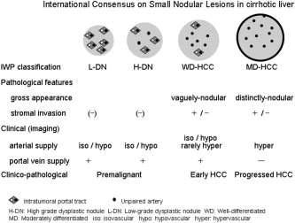
10 Multi-step hepatocarcinogenesis Improved imaging and development of surveillance protocols highlighted spectrum of nodules in cirrhotic livers Defined histologically but emerging molecular signatures International Working Party of World Congress of Gastroenterology 1995 classification Large regenerative nodule Low grade dysplastic nodule High grade dysplastic nodule Small hepatocellular carcinoma
11 Accumulation of genetic and epigenetic insults with clonal expansion of liver cells and progenitors in context of chronic liver disease Early over-expression of TGF α and IGF-2 Gene activation by promoter methylation Additional effects in HBV with genomic instability Telomerase activation with decreased apoptosis
12 Chromosomal amplifications LOH Global DNA hypomethylation Promoter hypermethylation Wnt/β catenin pathway commonly disrupted either by mutation or epigenetic silencing p53 and Rb1 pathways also altered PI3K/Akt/mTor pathway disrupted by activation of tyrosine kinase receptors or PTEN loss of function


13 Hypercellular with expansile growth pattern: may have peripheral scar Evidence of clonality May be large cell change No pseudoglands or nodule in nodule May have siderosis or CAP Normally contain portal tracts: most blood from PV rare aberrant arteries On imaging appear of normal vascularity or hypovascular? Distinction from LRN


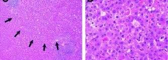
14 Vaguely nodular Frequently show diffuse or focal small cell change Steatosis Clear cell change MD bodies/p62 inclusions Thickened liver cell plates Acinar structures Nodule-in-nodule: worrying feature: most are HCC evolution


15 Early HCC: vaguely nodular appearance; well differentiated Progressed HCC: distinctly nodular appearance; moderately differentiated; may be microvascular invasion Of prognostic significance: with ehcc 5YS = 89% cf. phcc = 48% Takayama et al (1998)


16 Difficult to recognise macro and microscopically Recognised by Japanese for almost 25 years Now some (?) international consensus as to distinction from HGDN Small lesions (<2 cm diameter) Many of the cytological features of HGDN seen: merge with adjacent liver Stromal invasion required for diagnosis Absence of ductular reaction (cf. pseudoinvasion)


17 Not all small HCCs represent early HCCs Classical HCC if trabecular architecture; pseudoacinar change; ++ aberrant arteries; presence of capsule Vascular supply differs between early HCC and classical HCC Portal tracts seen in former Reflected in imaging where early HCC may have normal vascularity but arterialised classical HCC is hypervascular
18 34 pathologists from 13 countries Several meetings cases All resected specimens with lesions < 2 cm: no transplants or biopsies Restricted to Japanese or Korean patients All HBV or HCV associated Highlighted East-West divide! Importance of stromal invasion for diagnosis of ehcc Kappa values of 0.49 (after tuning the violins!)

19 LGDN HGDN Early hepatocellular carcinoma

 Nakano et")
20 Tumour cell invasion into portal tracts or fibrous septa in vaguely nodular lesions Kondo et al (1994) Nakano et al (1997)
21
")
22 Park et al (2007)
")
23 Park et al (2007)
24 HSP 70 CAP2 Glypican 3 Glutamine synthetase Epigenetic markers mirnas Llovet & Bruix (2008) Sakamoto et al (2008) Suriawinata & Thung (2010)
25 Rarely positive with serum markers eg. afp HSP70 upregulated: seen in majority of early HCC but rare in HGDN Glypican 3: expression higher in early HCC than HGDN: reported sensitivity of 77% and specificity of 96% Increased staining and abnormal distribution of glutamine synthetase also a feature (normal - < 10% hepatocytes Can these be used to make a diagnosis of early HCC in the absence of stromal invasion?
26 HSP 70 Anti-apoptotic Most abundantly over-expressed gene in early HCC Nuclear and cytoplasmic: may be focal Glypican 3 Cell surface heparan sulphate proteoglycan Predominantly cytoplasmic (+/- membranous and canalicular) May be focal and expressed in regeneration Glutamine synthetase Ammonia metabolism: glutamine a source of energy for HCC Normally expressed in PV hepatocytes Diffuse in 50% of HCCs

")
27 Di Tommaso et al (2009)

")
28 Di Tommaso et al (2009)
29 88% hepatocellular cacinomas positive for GPC3 Only 3% non-hepatic epithelial tumours GPC3 positive All adenomas and FNH: GPC3 negative Distinctive pattern with CD34 in HCC Incomplete vascular pattern in adenoma and FNH Coston et al (2008)
30 T Primary tumour TX Primary tumour cannot be assessed T0 No evidence of primary tumour T1 Solitary tumour without vascular invasion T2 Solitary tumour with vascular invasion or multiple tumours, none more than 5 cm in greatest dimension T3 Multiple tumours any more than 5 cm or tumour involving a major branch of the portal or hepatic vein(s) T3a Multiple tumours any more than 5 cm T3b Tumour involving a major branch of the portal or hepatic vein(s) T4 Tumour(s) with direct invasion of adjacent organs other than the gall bladder or with perforation of visceral peritoneum T3b Tumour involving a major branch of the portal or Stage I T1 N0 M0 Stage II T2 N0 M0 Stage IIIA T3a N0 M0 Stage IIIB T3b N0 M0 N Regional lymph nodes NX Regional lymph nodes cannot be assessed Stage IIIC T4 N0 M0 N0 No regional lymph-node metastasis Stage IVA Any T N1 M0 N1 Regional lymph-node metastasis Stage IVB Any T Any N M1 M Distant metastasis M0 No distant metastasis M1 Distant metastasis
31 Stage A: very early/early HCC Stage B: asymptomatic mutinodular HCC Stage C: invasive/extrhepatic HCC Stage D: terminal HCC Linked to treatment pathways Endorsed by EASL and AASLD Does not take into account histology: not to be mistaken with histological concept of early HCC
32 Benign Hepatocellular adenoma 8170/0 Focal nodular hyperplasia Malignancy-associated and premalignant lesions Large cell change (formerly dysplasia ) Small cell change (formerly dysplasia ) Dysplastic nodules Low grade High grade Malignant Hepatocellular carcinoma 8170/3 Hepatocellular carcinoma, fibrolamellar variant 8171/3 Hepatoblastoma, epithelial variants 8970/3 Undifferentiated carcinoma 8020/3 Calcifying nested epithelial stromal tumour 8975/1* Carcinosarcoma 8980/3 Combined hepatocellularcholangiocarcinoma 8180/3 Hepatoblastoma, mixed epithelial-mesenchymal 8970/3 Malignant rhabdoid tumour 8963/3
33 Cytological changes Stromal invasion Vascular changes LGDN HGDN Early HCC HCC How important clinically? How well can we identify this in real life?


34
PATHOLOGY OF LIVER TUMORS
 PATHOLOGY OF LIVER TUMORS Pathobasic, 31.05.2016 WHO Classification Approach to a Liver Mass Lesion in a patient with chronic liver disease? Lesion in a patient without chronic liver disease? Malignant
Pathological Classification of Hepatocellular Carcinoma
 3 rd APASL Single Topic Conference: HCC in 3D Pathological Classification of Hepatocellular Carcinoma Glenda Lyn Y. Pua, M.D. HCC Primary liver cancer is the 2 nd most common cancer in Asia HCC is the
Pitfalls in the diagnosis of well-differentiated hepatocellular lesions
 2013 Colorado Society of Pathology Pitfalls in the diagnosis of well-differentiated hepatocellular lesions Sanjay Kakar, MD University of California, San Francisco Outline Hepatocellular adenoma: new WHO
Dysplastic Nodules. Department of Pathology, Chonbuk National University Medical School. Woo Sung Moon. Introduction
 Dysplastic Nodules Department of Pathology, Chonbuk National University Medical School Woo Sung Moon 만성간질환에발생하는간의결절병변에는재생결절, 형성이상결절및간세포암종이있다. 여러인자에의해손상받은간세포는괴사와재생과정을반복하며재생결절, 형성이상결절을형성하는동안유전자의변이와 epigenetic
Pathological Analysis of Small Hepatocellular Carcinoma with Poor Prognosis
 Pathological Analysis of Small Hepatocellular Carcinoma with Poor Prognosis Haeryoung Kim, M.D., Ph.D. Department of Pathology Seoul National University Bundang Hospital Small HCC Definition: HCC < 2cm
O Farrell Legacy UPDATE ON WHO NOMENCLATURE. World Health Organization, 2010 DISCLOSURES WITH EMPHASIS ON PROBLEM HEPATOCELLULAR TUMORS
 O Farrell Legacy UPDATE ON WHO NOMENCLATURE WITH EMPHASIS ON PROBLEM HEPATOCELLULAR TUMORS Linda Ferrell, MD University of California San Francisco Vice Chair, Director of Surgical Pathology World Health
Invited Re vie W. Analytical histopathological diagnosis of small hepatocellular nodules in chronic liver diseases
 Histol Histopathol (1 998) 13: 1077-1 087 http://www.ehu.es/histoi-histopathol Histology and Histopathology Invited Re vie W Analytical histopathological diagnosis of small hepatocellular nodules in chronic
Detection and Characterization of Hepatocellular Carcinoma by Imaging
 CLINICAL GASTROENTEROLOGY AND HEPATOLOGY 2005;3:S136 S140 Detection and Characterization of Hepatocellular Carcinoma by Imaging OSAMU MATSUI Department of Imaging Diagnosis and Interventional Radiology,
Liver Specialty Evening Conference. Matthew M. Yeh, MD, PhD Professor of Pathology Adjunct Professor of Medicine University of Washington, Seattle
 Liver Specialty Evening Conference Matthew M. Yeh, MD, PhD Professor of Pathology Adjunct Professor of Medicine University of Washington, Seattle Case History A 65 year-old man presents with abdominal
Outline. Hepatocellular Carcinoma Histologic variants. HCC: Histologic variants
 2018 Park City AP Update Hepatocellular Carcinoma Histologic variants Sanjay Kakar, MD University of California, San Francisco Outline Histologic variants of HCC Morphologic and Immunohistochemical pitfalls
Liver Tumors. Prof. Dr. Ahmed El - Samongy
 Liver Tumors Prof. Dr. Ahmed El - Samongy Objective 1. Identify the most important features of common benign liver tumors 2. Know the risk factors, diagnosis, and management of hepatocellular carcinoma
Liver Cancer. Su Jong Yu, M.D. Department of Internal Medicine, Liver Research Institute, Seoul National University College of Medicine
 Liver Cancer Su Jong Yu, M.D. Department of Internal Medicine, Liver Research Institute, Seoul National University College of Medicine Primary Liver Cancer Hepatocellular carcinoma (HCC) : > 80% Derived
HEPATOCYTE SPECIFIC CONTRAST MEDIA: WHERE DO WE STAND?
 HEPATOCYTE SPECIFIC CONTRAST MEDIA: WHERE DO WE STAND? Andrew T. Trout, MD @AndrewTroutMD Disclosures No relevant disclosures Outline Review of hepatocyte specific contrast media Review of hepatocellular
Neoplasms of the Canine, Feline and Lemur Liver:
 Neoplasms of the Canine, Feline and Lemur Liver: Classification and Prognosis Annual Seminar of the French Society of Veterinary Pathology John M. Cullen VMD PhD DACVP North Carolina State University Primary
Review Article Assessment of Stromal Invasion for Correct Histological Diagnosis of Early Hepatocellular Carcinoma
 SAGE-Hindawi Access to Research International Hepatology Volume 2011, Article ID 241652, 7 pages doi:10.4061/2011/241652 Review Article Assessment of Stromal Invasion for Correct Histological Diagnosis
Histopathologic Features of Hepatocellular Carcinoma
 REVIEW REVIEW Histopathologic Features of Hepatocellular Carcinoma Elizabeth M. Brunt, M.D. Paradoxically, with the recognized increase in hepatocellular carcinoma, liver biopsy is used less frequently
Focus on Dysplastic Nodules and Early Hepatocellular Carcinoma: An Eastern Point of View. Masamichi Kojiro
 Focus on Dysplastic Nodules and Early Hepatocellular Carcinoma: An Eastern Point of View Masamichi Kojiro Although increasing numbers of equivocal nodular lesions have been detected in patients with liver
Multistep hepatocarcinogenesis is characterized by the following
 Early hepatocellular carcinoma with high-grade atypia in small vaguely nodular lesions Hidenori Ojima, 1 Yohei Masugi, 1 Hanako Tsujikawa, 1 Katsura Emoto, 1 Yoko Fujii-Nishimura, 1 Mami Hatano, 1 Miho
New insights into fatty liver disease. Rob Goldin Centre for Pathology, Imperial College
 New insights into fatty liver disease Rob Goldin Centre for Pathology, Imperial College r.goldin@imperial.ac.uk Prevalence of NASH Global prevalence of NAFLD is 25% with highest prevalence in the Middle
Evaluation of Liver Mass Lesions. American College of Gastroenterology 2013 Regional Postgraduate Course
 Evaluation of Liver Mass Lesions American College of Gastroenterology 2013 Regional Postgraduate Course Lewis R. Roberts, MB ChB, PhD Division of Gastroenterology and Hepatology Mayo Clinic College of
ACRIN 6690 PATHOLOGY MANUAL
 ACRIN 6690 PATHOLOGY MANUAL ACRIN 6690 Prepared by the American College of Radiology Imaging Network Original: December 15, 2010 Version Date: May 3, 2012 American College of Radiology Imaging Network
Microarray Analysis and Liver Diseases
 Microarray Analysis and Liver Diseases Snorri S. Thorgeirsson M.D., Ph.D. Laboratory of Experimental Carcinogenesis Center for Cancer Research, NCI, NIH Application of Microarrays to Cancer Research Identifying
Intrahepatic cholangiocarcinoma Histologic spectrum, novel markers and molecular assays
 2018 Current Issues in Surgical Pathology Summary (not actual lecture) Intrahepatic cholangiocarcinoma Histologic spectrum, novel markers and molecular assays Sanjay Kakar, MD University of California,
LIVER IMAGING TIPS IN VARIOUS MODALITIES. M.Vlychou, MD, PhD Assoc. Professor of Radiology University of Thessaly
 LIVER IMAGING TIPS IN VARIOUS MODALITIES M.Vlychou, MD, PhD Assoc. Professor of Radiology University of Thessaly Hepatocellular carcinoma is a common malignancy for which prevention, screening, diagnosis,
Hepatocellular Carcinoma HCC Updated November 2015 by: Dr. Mohammed Alghamdi (Medical Oncology Fellow, University of Calgary)
") Hepatocellular Carcinoma HCC Updated November 2015 by: Dr. Mohammed Alghamdi (Medical Oncology Fellow, University of Calgary) Staff Reviewers: Dr. Yoo Joung Ko (Medical Oncologist, Sunnybrook Odette Cancer
Hepatocellular Carcinoma HCC Updated November 2015 by: Dr. Mohammed Alghamdi (Medical Oncology Fellow, University of Calgary) Staff Reviewers: Dr. Yoo Joung Ko (Medical Oncologist, Sunnybrook Odette Cancer
Jesse Civan, M.D. Medical Director, Jefferson Liver Tumor Center
 Liver Tumors Jesse Civan, M.D. Medical Director, Jefferson Liver Tumor Center Differential Diagnosis Malignant Metastatic from non-hepatic primary Hepatocellular carcinoma Cholangiocarcinoma Biliary cystcarcinoma
Atypical Well differentiated Hepatocellular Neoplasms Cruising through the maze of criteria, terminology and risk assessment
 2016 Hans Popper Companion Meeting Atypical Well differentiated Hepatocellular Neoplasms Cruising through the maze of criteria, terminology and risk assessment Disclosure Dr. Kakar has nothing to Disclose
C ORPUS UTERI C ARCINOMA STAGING FORM (Carcinosarcomas should be staged as carcinomas)
") CLINICAL C ORPUS UTERI C ARCINOMA STAGING FORM PATHOLOGIC Extent of disease before S TAGE C ATEGORY D EFINITIONS Extent of disease through any treatment completion of definitive surgery y clinical staging
CLINICAL C ORPUS UTERI C ARCINOMA STAGING FORM PATHOLOGIC Extent of disease before S TAGE C ATEGORY D EFINITIONS Extent of disease through any treatment completion of definitive surgery y clinical staging
Raga Ramachandran, MD, PhD Assistant Professor and Director of Medical Education, UCSF Pathology
 Variants of Hepatocellular Carcinoma: Practical Issues Raga Ramachandran, MD, PhD Assistant Professor and Director of Medical Education, UCSF Pathology raga.ramachandran@ucsf.edu A full copy of the presentation
Kidney Case 1 SURGICAL PATHOLOGY REPORT
 Kidney Case 1 Surgical Pathology Report February 9, 2007 Clinical History: This 45 year old woman was found to have a left renal mass. CT urography with reconstruction revealed a 2 cm medial mass which
Surveillance for Hepatocellular Carcinoma
 Surveillance for Hepatocellular Carcinoma Marion G. Peters, MD John V. Carbone, MD, Endowed Chair Professor of Medicine Chief of Hepatology Research University of California San Francisco Recorded on April
Colorectal Cancer Structured Pathology Reporting Proforma DD MM YYYY
 Colorectal Cancer Structured Pathology Reporting Proforma Mandatory questions (i.e. protocol standards) are in bold (e.g. S1.03). Family name Given name(s) Date of birth DD MM YYYY S1.02 Clinical details
Non Small Cell Lung Cancer Histopathology ד"ר יהודית זנדבנק
 Non Small Cell Lung Cancer Histopathology ד"ר יהודית זנדבנק 26.06.09 Lecture outlines WHO histological classification Macro/Micro assessment Early diagnosis Minimal pathology Main subtypes SCC, AdCa, LCLC
Innovations in HCC Imaging: MDCT/MRI
 Innovations in HCC Imaging: MDCT/MRI Anthony E. Cheng, M.D. Cardinal MRI Center Cardinal Santos Medical Center, Wilson Street, San Juan Innovations in HCC Imaging: Goals/Objectives MDCT/MRI Learn the diagnostic
Hepatocelluar nodules in liver cirrhosis: hemodynamic evaluation (angiographyassisted CT) with special reference to multi-step hepatocarcinogenesis
 with special reference to multi-step hepatocarcinogenesis") Abdominal Imaging ª The Author(s) 2011. This article is published with open access at Springerlink.com Published online: 26 January 2011 Abdom Imaging (2011) 36:264 272 DOI: 10.1007/s00261-011-9685-1 INVITED
Abdominal Imaging ª The Author(s) 2011. This article is published with open access at Springerlink.com Published online: 26 January 2011 Abdom Imaging (2011) 36:264 272 DOI: 10.1007/s00261-011-9685-1 INVITED
Workup of a Solid Liver Lesion
 Workup of a Solid Liver Lesion Joseph B. Cofer MD FACS Chief Quality Officer Erlanger Health System Affiliate Professor of Surgery UTHSC-Chattanooga I have no financial or other relationships with any
Disclosures. Parathyroid Pathology. Objectives. The normal parathyroid 11/10/2012
 Disclosures Parathyroid Pathology I have nothing to disclose Annemieke van Zante MD/PhD Assistant Professor of Clinical Pathology Associate Chief of Cytopathology Objectives 1. Review the pathologic features
Evangelos Chartampilas Bioclinic Hospital Thessaloniki, Greece
 Evangelos Chartampilas Bioclinic Hospital Thessaloniki, Greece Hepatospecificcontrast agents Gadobenate dimeglumine (Multihance) Gadoxeticacid (Primovist) 3-5% liver uptake 50% liver uptake Hepatobiliary
Clinical Staging for Hepatocellular Carcinoma: Eastern Perspectives. Osamu Yokosuka, M.D. Graduate School of Medicine, Chiba University, Chiba, Japan
 Clinical Staging for Hepatocellular Carcinoma: Eastern Perspectives Osamu Yokosuka, M.D. Graduate School of Medicine, Chiba University, Chiba, Japan Why is staging system important? Cancer stage can be
Video Microscopy Tutorial 8
 Video Microscopy Tutorial 8 Common and Uncommon Lesions of the Liver Gladwyn Leiman, MD There are no disclosures necessary. Common and Uncommon Lesions in Liver FNA Gladwyn Leiman University of Vermont
HEPATO-BILIARY IMAGING
 HEPATO-BILIARY IMAGING BY MAMDOUH MAHFOUZ MD PROF.OF RADIOLOGY CAIRO UNIVERSITY mamdouh.m5@gmail.com www.ssregypt.com CT ABDOMEN Indications Patient preparation Patient position Scanogram Fasting 4-6 hours
Benign liver tumors : Diagnosis and management
 4th International Hepatology Conference 2016 HEPATOLOGY SOCIETY, DHAKA, BANGLADESH Benign liver tumors : Diagnosis and management Pr Laurence Chiche Hepato biliary surgery and transplantation Bordeaux,
LIVER PATHOLOGY(3) Prof. Andras Kiss. M.D., Ph.D., D.Sc. Semmelweis University 2 nd Department of Pathology Budapest February 26.
 Prof. Andras Kiss. M.D., Ph.D., D.Sc. Semmelweis University 2 nd Department of Pathology Budapest February 26.") LIVER PATHOLOGY(3) Prof. Andras Kiss. M.D., Ph.D., D.Sc. Semmelweis University 2 nd Department of Pathology Budapest February 26. 2018 Vascular disorders Inflow A.hepatica thromb., embolia infarctus V.portae
LIVER PATHOLOGY(3) Prof. Andras Kiss. M.D., Ph.D., D.Sc. Semmelweis University 2 nd Department of Pathology Budapest February 26. 2018 Vascular disorders Inflow A.hepatica thromb., embolia infarctus V.portae
Hepatocellular neoplasia - Recent developments
 22 November 2018 Hepatocellular neoplasia - Recent developments Dina G. Tiniakos Institute of Cellular Medicine, Faculty of Medical Sciences, Newcastle University, UK Dept of Pathology, Aretaieion Hospital,
Interesting Cases from Liver Tumor Board. Jeffrey C. Weinreb, M.D.,FACR Yale University School of Medicine
 Interesting Cases from Liver Tumor Board Jeffrey C. Weinreb, M.D.,FACR Yale University School of Medicine jeffrey.weinreb@yale.edu Common Liver Diseases Hemangioma Cyst FNH Focal Fat/Sparing THID Non-Cirrhotic
XIII. Tumours of the liver and biliary system
 XIII. Tumours of the liver and biliary system V. PONOMARKOV 1 & L. J. MACKEY 2 In this histological classification of liver and gall bladder tumours the tumour types largely correspond to those found in
Papillary Lesions of the Breast A Practical Approach to Diagnosis. (Arch Pathol Lab Med. 2016;140: ; doi: /arpa.
 Papillary Lesions of the Breast A Practical Approach to Diagnosis (Arch Pathol Lab Med. 2016;140:1052 1059; doi: 10.5858/arpa.2016-0219-RA) Papillary lesions of the breast Span the spectrum of benign,
Malignant Focal Liver Lesions
 Malignant Focal Liver Lesions Other Than HCC Pablo R. Ros, MD, MPH, PhD Departments of Radiology and Pathology University Hospitals Cleveland Medical Center Case Western Reserve University Pablo.Ros@UHhospitals.org
Clonal evolution of human cancers
 Clonal evolution of human cancers -Pathology-based microdissection and genetic analysis precisely demonstrates molecular evolution of neoplastic clones- Hiroaki Fujii, MD Ageo Medical Laboratories, Yashio
With the widespread use of hepatic imaging, liver masses
 2B: Liver Assessment of the Liver Mass: What Do You Need to Know? With the widespread use of hepatic imaging, liver masses are detected either unexpectedly or in the course of screening for liver cancer
Surveillance for HCC Who, how Diagnosis of HCC Surveillance for HCC in Practice
 Surveillance for Hepatocellular Carcinoma Hashem B. El-Serag, MD, MPH Dan L. Duncan Professor of Medicine Chief, Gastroenterology and Hepatology Houston VA & Baylor College of Medicine Houston, TX Outline
Case presentation. Eran Zittan. MD Mount Sinai Hospital, Toronto, Canada. Emek Medical Center, Afula, Israel. March, 2016
 Case presentation Eran Zittan. MD Mount Sinai Hospital, Toronto, Canada. Emek Medical Center, Afula, Israel. March, 2016 60 y/o man with long standing UC+PSC. Last 10 years on clinical and endoscopic remission.
Basic patterns of liver damage what information can a liver biopsy provide and what clinical information does the pathologist need?
 Basic patterns of liver damage what information can a liver biopsy provide and what clinical information does the pathologist need? Rob Goldin r.goldin@imperial.ac.uk Fatty liver disease Is there fatty
MRI for HCC surveillance and reporting: LI-RADS. Donald G. Mitchell, M.D. Thomas Jefferson University Philadelphia, PA
 MRI for HCC surveillance and reporting: LI-RADS Donald G. Mitchell, M.D. Thomas Jefferson University Philadelphia, PA Cirrhotic Nodules Regenerative Nodule Atypical Nodule Hyperplastic Nodule Dysplastic
British Liver Transplant Group Pathology meeting September Leeds cases
 British Liver Transplant Group Pathology meeting September 2014 Leeds cases Leeds Case 1 Male 61 years Liver transplant for HCV cirrhosis with HCC in January 2014. Now raised ALT and bilirubin,? acute
B. Environmental Factors. a. The major risk factor to papillary thyroid cancer is exposure to ionizing radiation, during the first 2 decades of life.
 B. Environmental Factors. a. The major risk factor to papillary thyroid cancer is exposure to ionizing radiation, during the first 2 decades of life. b. Deficiency of dietary iodine: - Is linked with a
HCC and mass effect. Hepatocellular cancer: what if the AFP is rising but no lesion seen on imaging? What you need to know about AFP.
 Hepatocellular cancer: what if the AFP is rising but no lesion seen on imaging? Arun J Sanyal M.B.B.S., M.D. Charles Caravati Professor of Medicine Virginia Commonwealth University Imaging features used
Neoplasia 2018 Lecture 2. Dr Heyam Awad MD, FRCPath
 Neoplasia 2018 Lecture 2 Dr Heyam Awad MD, FRCPath ILOS 1. List the differences between benign and malignant tumors. 2. Recognize the histological features of malignancy. 3. Define dysplasia and understand
Carcinogenesis in IBD
 Oxford Inflammatory Bowel Disease MasterClass Carcinogenesis in IBD Dr Simon Leedham, Oxford, UK Oxford Inflammatory Bowel Disease MasterClass Carcinogenesis in Inflammatory Bowel Disease Dr Simon Leedham
Hepatocellular Carcinoma: A major global health problem. David L. Wood, MD Interventional Radiology Banner Good Samaritan Medical Center
 Hepatocellular Carcinoma: A major global health problem David L. Wood, MD Interventional Radiology Banner Good Samaritan Medical Center Hepatocellular Carcinoma WORLDWIDE The #2 Cancer Killer Overall cancer
Definition of Synoptic Reporting
 Definition of Synoptic Reporting The CAP has developed this list of specific features that define synoptic reporting formatting: 1. All required cancer data from an applicable cancer protocol that are
STAGE CATEGORY DEFINITIONS
 CLINICAL Extent of disease before any treatment y clinical staging completed after neoadjuvant therapy but before subsequent surgery TX Tis Tis (DCIS) Tis (LCIS) Tis (Paget s) T1 T1mi T1a T1b T1c a b c
Molecular signature for management of hepatocellular carcinoma
 Molecular signature for management of hepatocellular carcinoma Yujin Hoshida, MD, PhD Liver Cancer Program, Tisch Cancer Institute Division of Liver Diseases, Department of Medicine Icahn School of Medicine
G3.02 The malignant potential of the neoplasm should be recorded. CG3.02a
 G3.02 The malignant potential of the neoplasm should be recorded. CG3.02a Conventional adrenocortical neoplasm. Each of the below parameters is scored 0 when absent and 1 when present. 3 or more of these
Papillary Lesions of the breast
 Papillary Lesions of the breast Emad Rakha Professor of Breast Pathology The University of Nottingham Papillary lesions of the breast are a heterogeneous group of disease, which are characterised by neoplastic
Dynamic Risk Stratification:
 Dynamic Risk Stratification: Using Risk Estimates to Guide Initial Management R Michael Tuttle, MD Clinical Director, Endocrinology Service Memorial Sloan Kettering Cancer Center Professor of Medicine
Pancreatic intraepithelial
 Pancreatic intraepithelial neoplasia (PanIN) Markéta Hermanová St. Anne s University Hospital Brno Faculty of Medicine, Masaryk University Precursor lesions of invasive pancreatic cancer Pancreatic intraepithelial
Hepatocellular Carcinoma: Diagnosis and Management
 Hepatocellular Carcinoma: Diagnosis and Management Nizar A. Mukhtar, MD Co-director, SMC Liver Tumor Board April 30, 2016 1 Objectives Review screening/surveillance guidelines Discuss diagnostic algorithm
NEOPLASMS AND TUMOR-LIKE CONDITIONS OF LIVER
 NEOPLASMS AND TUMOR-LIKE CONDITIONS OF LIVER Epithelial Tumors Focal nodular hyperplasia Focal nodular hyperplasia is a localized hyperplasic overgrowth of hepatocytes around a vascular anomaly, particularly
GUIDELINES ON NON-MUSCLE- INVASIVE BLADDER CANCER
 GUIDELINES ON NON-MUSCLE- INVASIVE BLADDER CANCER (Limited text update December 21) M. Babjuk, W. Oosterlinck, R. Sylvester, E. Kaasinen, A. Böhle, J. Palou, M. Rouprêt Eur Urol 211 Apr;59(4):584-94 Introduction
Hepatocellular Carcinoma. Markus Heim Basel
 Hepatocellular Carcinoma Markus Heim Basel Outline 1. Epidemiology 2. Surveillance 3. (Diagnosis) 4. Staging 5. Treatment Epidemiology of HCC Worldwide, liver cancer is the sixth most common cancer (749
Tumor incidence varies significantly, depending on geographical location.
 Hepatocellular carcinoma is the 5 th most common malignancy worldwide with male-to-female ratio 5:1 in Asia 2:1 in the United States Tumor incidence varies significantly, depending on geographical location.
Differential diagnosis of HCC
 Hepatocellular Carcinoma Quest for an Ideal Immunohistochemical Panel Sanjay Kakar, MD UCSF Differential diagnosis of HCC Hepatocellular lesions Adenoma, FNH, HG dysplasia Adenocarcinoma CholangioCA, metastasis
A) PUBLIC HEALTH B) PRESENTATION & DIAGNOSIS
 PUBLIC HEALTH B) PRESENTATION & DIAGNOSIS") Hepatocellular Carcinoma HCC Updated November 2015 by: Dr. Mohammed Alghamdi (Medical Oncology Fellow, University of Calgary), April 2017 by Dr. Jenny Ko (Medical Oncologist, Abbotsford Centre, BC Cancer
Hepatocellular Carcinoma HCC Updated November 2015 by: Dr. Mohammed Alghamdi (Medical Oncology Fellow, University of Calgary), April 2017 by Dr. Jenny Ko (Medical Oncologist, Abbotsford Centre, BC Cancer
Update on staging colorectal carcinoma, the 8 th edition AJCC. General overview of staging. When is staging required? 11/1/2017
 Update on staging colorectal carcinoma, the 8 th edition AJCC Dale C. Snover, MD November 3, 2017 General overview of staging Reason for uniform staging Requirements to use AJCC manual and/or CAP protocols
HEPATOCELLULAR CARCINOMA: SCREENING, DIAGNOSIS, AND TREATMENT
 HEPATOCELLULAR CARCINOMA: SCREENING, DIAGNOSIS, AND TREATMENT INTRODUCTION: Hepatocellular carcinoma (HCC): Fifth most common cancer worldwide Third most common cause of cancer mortality In Egypt: 2.3%
Hepatocellular Carcinoma (HCC): Burden of Disease
: Burden of Disease") Hepatocellular Carcinoma (HCC): Burden of Disease Blaire E Burman, MD VM Hepatology Hepatocellular Carcinoma (HCC) Primary HCCs most often arise in the setting of chronic inflammation, liver damage, and
Hepatocellular Carcinoma (HCC): Burden of Disease Blaire E Burman, MD VM Hepatology Hepatocellular Carcinoma (HCC) Primary HCCs most often arise in the setting of chronic inflammation, liver damage, and
The role of Hepatitis C Virus in hepatocarcinogenesis
 The role of Hepatitis C Virus in hepatocarcinogenesis Laura Beretta Fred Hutchinson Cancer Research Center l8 Incidence and mortality of the five most common cancers worldwide, 2000 Incidence Lung Breast
NAACCR Webinar Series 1
 Collecting Cancer Data: Liver 2013 2014 NAACCR Webinar Series June 5, 2014 Q&A Please submit all questions concerning webinar content through the Q&A panel. Reminder: If you have participants watching
Updates in Urologic Pathology WHO Made Those Changes?! Peyman Tavassoli Pathology Department BC Cancer Agency
 Updates in Urologic Pathology WHO Made Those Changes?! Peyman Tavassoli Pathology Department BC Cancer Agency World Health Organization Available in Feb 2016 Frame work for reporting Major contributing
04/10/2018. Intraductal Papillary Neoplasms Of Breast INTRADUCTAL PAPILLOMA
 Intraductal Papillary Neoplasms Of Breast Savitri Krishnamurthy MD Professor of Pathology Deputy Division Head The University of Texas MD Anderson Cancer Center 25 th Annual Seminar in Pathology Pittsburgh,
Dr Rodney Itaki Lecturer Anatomical Pathology Discipline. University of Papua New Guinea School of Medicine & Health Sciences Division of Pathology
 Neoplasia Dr Rodney Itaki Lecturer Anatomical Pathology Discipline University of Papua New Guinea School of Medicine & Health Sciences Division of Pathology General Considerations Overview: Neoplasia uncontrolled,
!! 2 to 3% of All New Visceral Cancers.!! Peak Incidence is 6th Decade!! M:F = 2:1
 !! Kathleen M. O Toole, M.D.!! 2 to 3% of All New Visceral Cancers!! Peak Incidence is 6th Decade!! M:F = 2:1!! Grossly is a Bright Yellow, Necrotic Mass with a Pseudocapsule 1 !!Conventional RCC! Clear
Cholangiocarcinoma. Judy Wyatt Dundee November 2010
 Cholangiocarcinoma Judy Wyatt Dundee November 2010 Making sense of cholangiocarcinoma Difficulties with diagnostic criteria How many entities within cholangiocarcinoma? Rapidly evolving Intrahepatic cholangiocarcinoma
21/02/2014. Disclosures. HCC: predicting recurrence. Outline. Liver transplant: Beyond Milan?
 Disclosures HCC: predicting recurrence Peter Ghali, MD, FRCPC, MSc (epid) None relevant to this talk other than off-label use of sirolimus Toronto, February 2014 Outline Recurrence after what? Locoregional
CT & MRI of Benign Liver Neoplasms Srinivasa R Prasad
 CT & MRI of Benign Liver Neoplasms Srinivasa R Prasad No financial disclosures Acknowledgements Many thanks to Drs. Heiken, Narra & Menias (MIR) Dr. Sahani (MGH) for sharing images Benign Liver Tumors:
CELL BIOLOGY - CLUTCH CH CANCER.
 !! www.clutchprep.com CONCEPT: OVERVIEW OF CANCER Cancer is a disease which is primarily caused from misregulated cell division, which form There are two types of tumors - Benign tumors remain confined
The impact of the treatment of HCV in developing Hepatocellular Carcinoma
 The impact of the treatment of HCV in developing Hepatocellular Carcinoma Paul Y Kwo, MD Professor of Medicine Medical Director, Liver Transplantation Gastroenterology/Hepatology Division Indiana University
Hepatocellular Carcinoma: Transplantation, Resection or Ablation?
 Hepatocellular Carcinoma: Transplantation, Resection or Ablation? Roberto Gedaly MD Chief, Abdominal Transplantation Transplant Service Line University of Kentucky Nothing to disclose Disclosure Objective
Hyperplastische Polyps Innocent bystanders?
 Hyperplastische Polyps Innocent bystanders?? K. Geboes P th l i h O tl dk d Pathologische Ontleedkunde, KULeuven Content Historical Classification Relation Hyperplastic polyps carcinoma The concept cept
2 to 3% of All New Visceral Cancers Peak Incidence is 6th Decade M:F = 2:1 Grossly is a Bright Yellow, Necrotic Mass with a Pseudocapsule
 GENITOURINARY PATHOLOGY Kathleen M. O Toole, M.D. Renal Cell Carcinoma 2 to 3% of All New Visceral Cancers Peak Incidence is 6th Decade M:F = 2:1 Grossly is a Bright Yellow Necrotic Mass Grossly is a Bright
Malignant neoplasms of the gastrointestinal (GI) tract,
 tract,") Special Section First Chinese American Pathologists Association Diagnostic Pathology Course, Part II Practical Immunohistochemistry in Neoplastic Pathology of the Gastrointestinal Tract, Liver, Biliary
Special Section First Chinese American Pathologists Association Diagnostic Pathology Course, Part II Practical Immunohistochemistry in Neoplastic Pathology of the Gastrointestinal Tract, Liver, Biliary
C ORPUS UTERI C ARCINOMA STAGING FORM (Carcinosarcomas should be staged as carcinomas)
") C ORPUS UTERI C ARCINOMA STAGING FORM CLINICAL Extent of disease before any treatment y clinical staging completed after neoadjuvant therapy but before subsequent surgery Tis * T1 I T1a IA NX N0 N1 N2
C ORPUS UTERI C ARCINOMA STAGING FORM CLINICAL Extent of disease before any treatment y clinical staging completed after neoadjuvant therapy but before subsequent surgery Tis * T1 I T1a IA NX N0 N1 N2
Neoplasia part I. Dr. Mohsen Dashti. Clinical Medicine & Pathology nd Lecture
 Neoplasia part I By Dr. Mohsen Dashti Clinical Medicine & Pathology 316 2 nd Lecture Lecture outline Review of structure & function. Basic definitions. Classification of neoplasms. Morphologic features.
This form may provide more data elements than required for collection by standard setters such as NCI SEER, CDC NPCR, and CoC NCDB.
 1 Terms of Use The cancer staging form is a specific document in the patient record; it is not a substitute for documentation of history, physical examination, and staging evaluation, or for documenting
Normal thyroid tissue
 Thyroid Pathology Overview Normal thyroid tissue Normal thyroid tissue with follicles filled with colloid. Thyroid cells form follicles, spheres of epithelial cells (always single layered in health, usually
LOINC. Clinical information. RCPA code. Record if different to report header Operating surgeon name and contact details. Absent.
 Complete as narrative or use the structured format below 55752-0 17.02.28593 Clinical information 22027-7 17.02.30001 Record if different to report header Operating surgeon name and contact details 52101004
Breast pathology. 2nd Department of Pathology Semmelweis University
 Breast pathology 2nd Department of Pathology Semmelweis University Breast pathology - Summary - Benign lesions - Acute mastitis - Plasma cell mastitis / duct ectasia - Fat necrosis - Fibrocystic change/
CTA/MRA of Pediatric Hepatic Masses Radiology-Pathology Correlation
 Acta Radiológica Portuguesa, Vol.XVIII, nº70, pág. 41-50, Abr.-Jun., 2006 CTA/MRA of Pediatric Hepatic Masses Radiology-Pathology Correlation Marilyn J. Siegel Mallinckrodt Institute of Radiology, Washington
Gastric Cancer Histopathology Reporting Proforma
 Gastric Cancer Histopathology Reporting Proforma Mandatory questions (i.e. protocol standards) are in bold (e.g. S1.01). S1.01 Identification Family name Given name(s) Date of birth Sex Male Female Intersex/indeterminate