Endoscopic Assisted resection for congenital Midline Nasal Mass
|
|
|
- Reynard Booker
- 5 years ago
- Views:
Transcription
1 Endoscopic Assisted resection for congenital Midline Nasal Mass Ahmed Aly Ibrahim A.prof ORL Department Alexandria University Emad. A Magdy prof ORL Department Alexandria University Haytham Morsi,MD Mohammad Fawzy,MD Embryology During formation of skull base and nose, mesenchymal structures are formed from several centers which will eventually fuse and ossify. Before their fusion, there are recognized spaces which are important in the development of congenital midline nasal masses Fonticulus frontalis Prenasal space Foramen cecum 1

 Congenital Midline")
2 Embyrology Fonticulus frontalis space between the frontal and nasal bones Eventually fuses with foramen cecum to create a separation between intracranial and extracranial structures Prenasal space is between the nasal bones and the nasal capsule (precursor of the septum and nasal cartilages) Congenital Midline Nasal Masses 2
3 Encephalocele Extracranial herniations of cranial contents through a defect in the skull. May include meninges only (meningocele), or both brain and meninges (meningoencephalocele). 40% have other associated anomalies. Encephalocele Divided into three categories: Occipital 75% Sincipital 15% Basal 10% Sincipital encephaloceles: A bony defect between the frontal and ethmoid bones anterior to the crista galli Nasofrontal, nasoethmoidal, nasoorbital. Basal encephaloceles: A bony defect between the cribriform plate and the superior orbital or posterior clinoid fissure, presenting as an intranasal mass. Trans-ethmoidal, Spheno-ethmoidal, Trans-sphenoidal, sphenoorbital. 3
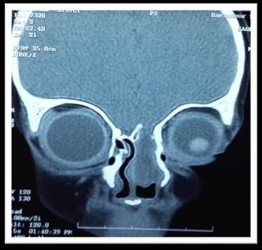
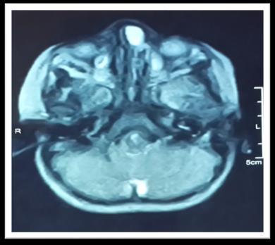
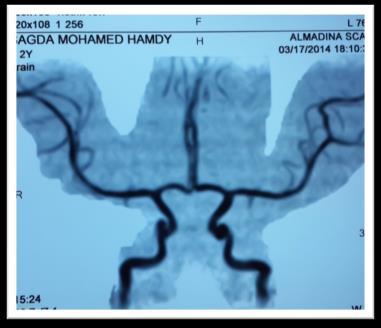
4 Encephalocele Imaging The diagnosis is radiologically confirmed by CT and/or MRI CT evaluation should include high-resolution which help delineate the infant s cartilaginous skull base. MRI provides complementary information regarding the fluid and soft tissue characteristics of the mass and is valuable in identifying an intracranial connection. It is also useful in helping to differentiate a meningocele from a meningoencephalocele. Encephalocele Treatment Early intervention in the first few months of life: minimize the risk of meningitis and cosmetic deformities. Small lesions with minimal skull base defects may be managed endoscopically. Larger lesions require craniotomy / combined approach. 4

5 5
6 Glioma Gliomas are unencapsulated collections of glial cells situated outside the CNS. Possible theories of development include: (1) sequestration of glial tissue of the olfactory bulb entrapped during cribriform plate fusion (2) ectopic neural tissue cells (3) pinched encephalocele (4) inappropriate closure of the anterior neuropore (fonticulus frontalis), with failure of mesoderm to enter the region, resulting in inadequate bone formation % of nasal gliomas have a fibrous stalk connection to the intracranial space. Glioma Types: extranasal (60%) intranasal (30%) combined (10%) Firm, noncompressible purple or gray mass. Site: Extranasal: glabella or nasomaxillary suture Intranasal: lateral nasal wall near MT or nasal septum sometimes difficult to differentiate between gliomas and encephaloceles; however, the presence of ependymal tissue is consistent with an encephalocele. 6


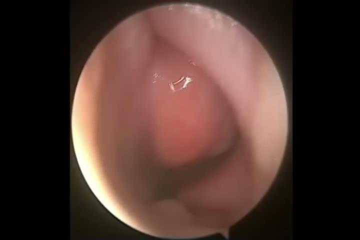
7 Glioma Nasal gliomas appear hypo- or isointense on T1-weighted images and hyperintense on T2- weighted images. Management of nasal glioma consists of surgical excision. Intranasal gliomas can be excised with endoscopic techniques 7
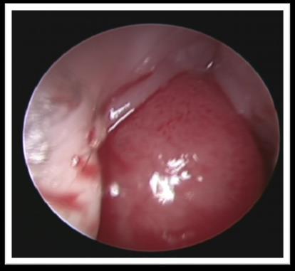
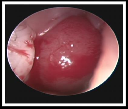
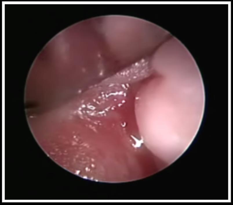
8 Intranasal Glioma 8
9 Nasal Dermoid Most common congenital nasal abnormality. 1 3% of all dermoids and 10 12% of head and neck dermoids. Congenital dermoids contain only ectodermal and mesodermal embryonic elements. Mesodermal elements, which include hair follicles, sebaceous glands, and sweat glands are found in the wall of the cyst and thus differentiate these masses from simple epidermoid cysts Teratomas, contain all three embryonal germ layers. Nasal Dermoid Aetiology of these lesions is controversial. Prenasal space theory, is based on the abnormal development of the fonticulus frontalis. The retracting dura may drag the surface epithelium inward, causing formation of a sinus tract. In some patients, the sinus tract extends into the intracranial cavity or prenasal space. the dermal sinus or cyst may persist anywhere from the foramen cecum to the nasal tip. 9


10 Nasal Dermoids Manifest as a simple cyst, a cyst with a sinus tract, or a sinus tract alone. may be intermittent discharge or infection. Protruding hair is seen in a minority of patients, but is pathognomic. Firm, lobulated, noncompressible midline mass over the nasal dorsum +/- sinus opening. negative Furstenberg test and do not transilluminate. Intracranial extension 4 45%. Nasal Dermoids CT and MRI provide complementary information. Common findings include a bifid crista galli and an enlarged foramen cecum. A normal crista galli and foramen cecum may be used to exclude intracranial extension. The neonatal crista galli does not contain marrow fat and hence a high intensity signal on T1-weighted images is suggestive of an intracranial dermoid. 10


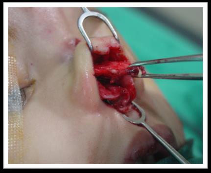
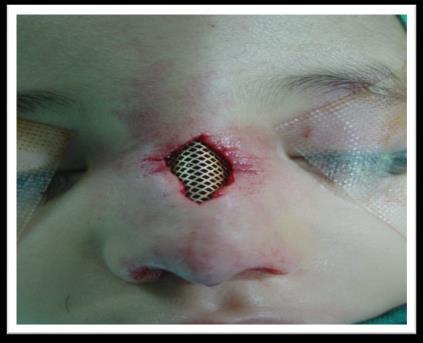
11 Nasal Dermoid Approaches to nasal dermoid should fulfill four criteria: 1. Provide excellent access to the midline 2. Allow access to the base of the skull 3. Provide adequate exposure for reconstruction of the nasal dorsum 4. Result in an acceptable scar Several extracranial approaches have been described: lateral rhinotomy,external rhinoplasty, midline vertical incisions, medial paracanthal incisions The external rhinoplasty incision best cosmetic result approach gives access to the skull base and allows for exposure of the nasal dorsum limited access to lesions in the glabellar region. Dermoid 11
12 12


13 Gliomas Encephaloceles Dermoids Contents Glial cells Meninges+/- brain tissue Ectoderm/mesoderm derivatives Characteristics Reddish, firm, nonpulsatile Bluish, soft, pulsatile Nonpulsatile cyst, sinus, or fistula Furstenberg test -ve +ve -ve Intracranial extension ~15% 100% ~30% Take Home Message A Multi-modality team should be considered in managing these type of congenital lesions Approaches to midline congenital nasal mass should fulfill four criteria: 1. Provide excellent access to the midline 2. Allow access to the base of the skull 3. Provide adequate exposure for reconstruction of the nasal dorsum 4. Result in an acceptable scar Endoscopic and Several extracranial approaches have been described: lateral rhinotomy,external rhinoplasty, midline vertical incisions, medial paracanthal incisions and bicronal external approches. 13
14 Thank You for your Attention 14
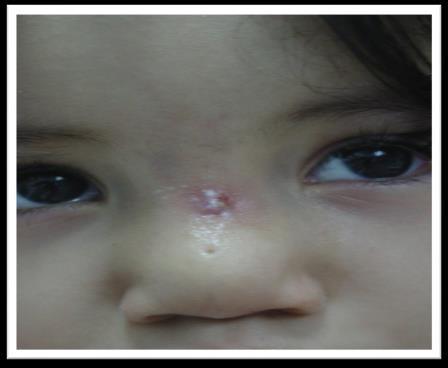
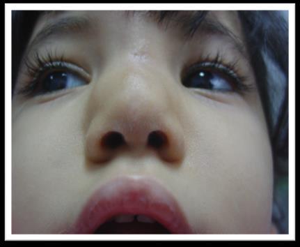
Management of Nasal Dermoid Sinus Cyst by Modified
 AIJCR CASE REPORT Management of Nasal Dermoid Sinus Cyst by Modified Bipedicle 10.5005/jp-journals-10013-1247 Advancement Flap Technique Management of Nasal Dermoid Sinus Cyst by Modified Bipedicle Advancement
Biological Early Detection of Nasal Dermoid Cyst Using CT/ MRI Scan - A Review
 Biological Early Detection of Nasal Dermoid Cyst Using CT/ MRI Scan - A Review Neeraj Singla 1, Sugandha Sharma 2 M.Tech Student, Dept. of CSE, CGC, Gharuan, Punjab, India 1 Assistant Professor, Dept.
CASE REPORT-NASO-ETHMOIDAL ENCEPHALOCELE Shrishail Patil 1, Tanvi Choubey 2
 -NASO-ETHMOIDAL ENCEPHALOCELE Shrishail Patil 1, Tanvi Choubey 2 HOW TO CITE THIS ARTICLE: Shrishail Patil, Tanvi Choubey. Case Report-_Naso-ethmoidal Encephalocele. Journal of Evolution of Medical and
Congenital Nasal Neuroglial Heterotopia and Encephaloceles: An Update on Current Evaluation and Management
 The Laryngoscope VC 2016 The American Laryngological, Rhinological and Otological Society, Inc. Congenital Nasal Neuroglial Heterotopia and Encephaloceles: An Update on Current Evaluation and Management
Remember from the first year embryology Trilaminar disc has 3 layers: ectoderm, mesoderm, and endoderm
 Development of face Remember from the first year embryology Trilaminar disc has 3 layers: ectoderm, mesoderm, and endoderm The ectoderm forms the neural groove, then tube The neural tube lies in the mesoderm
A Case of Naso-Ethmoidal Meningoencephalocele
 A Case of Naso-Ethmoidal Meningoencephalocele Divyanshu Dubey, Sonjjay Pande, Pradeep Dubey, Anshudha Sawhney Vol. 3 No. 8 (August 2011) International Journal of Collaborative Research on Internal Medicine
Nasal Glioma: Prenatal Diagnosis and Multidisciplinary Surgical Approach
 Nasal Glioma: Prenatal Diagnosis and Multidisciplinary Surgical Approach The Harvard community has made this article openly available. Please share how this access benefits you. Your story matters Citation
An important indication for imaging the anterior skull base
 ORIGINAL RESEARCH D.C. Hughes M.J. Kaduthodil D.J.A. Connolly P.D. Griffiths Dimensions and Ossification of the Normal Anterior Cranial Fossa in Children BACKGROUND AND PURPOSE: Interpretation of CT of
Rare Combination of Frontonasal and Bilateral Naso-orbital Encephaloceles
 Rare Combination of Frontonasal and Bilateral Naso-orbital Encephaloceles Alan A. Alexander 1*, Megan R. Saettele 2, Daniel L'Heureux 1, Paras A. Shah 3, Kristin A. Fickenscher 4 1. Department of Radiology,
SKULL AS A WHOLE + ANTERIOR CRANIAL FOSSA
 SKULL AS A WHOLE + ANTERIOR CRANIAL FOSSA LEARNING OBJECTIVES At the end of this lecture, the student should be able to know: Parts of skeleton (axial and appendicular) Parts of skull Sutures of skull
Case Report A Case of Nasal Glial Heterotopia in an Adult
 Case Reports in Otolaryngology, Article ID 354672, 4 pages http://dx.doi.org/10.1155/2014/354672 Case Report A Case of Nasal Glial Heterotopia in an Adult Akira Hagiwara, 1,2 Noriko Nagai, 1 Yasuo Ogawa,
RECURRENT INTRA NASAL GLIOMA: A CASE REPORT D. Ranganath Swamy 1, K. Nagaraju 2, E. Yugandhar 3, B. Kathyayani 4, T. Shankar 5
 RECURRENT INTRA NASAL GLIOMA: A D. Ranganath Swamy 1, K. Nagaraju 2, E. Yugandhar 3, B. Kathyayani 4, T. Shankar 5 HOW TO CITE THIS ARTICLE: D. Ranganath Swamy, K. Nagaraju, E. Yugandhar, B. Kathyayani,
From the Mount Vernon Centre for Plastic Surgery, Northwood
 THE FRONTO-NASAL GLIOMA By R. L. G. DAWSON, M.B., F.R.C.S., and I. F. K. MUIR, M.B.E., M.B., F.R.C.S. From the Mount Vernon Centre for Plastic Surgery, Northwood CHILDREN quite commonly present with swellings
OBSTRUCTIVE NASAL AND NASOPHARYNGEAL AIRWAY MASSES IN CHILDREN AND ADOLESCENTS
 OBSTRUCTIVE NASAL AND NASOPHARYNGEAL AIRWAY MASSES IN CHILDREN AND ADOLESCENTS Michael J. Cunningham MD Department of Otolaryngology and Communication Enhancement Children s Hospital Boston Nasal Airway
Skull-2. Norma Basalis Interna. Dr. Heba Kalbouneh Assistant Professor of Anatomy and Histology
 Skull-2 Norma Basalis Interna Dr. Heba Kalbouneh Assistant Professor of Anatomy and Histology Norma basalis interna Base of the skull- superior view The interior of the base of the skull is divided into
Malformations of the nose, Clefts & Tumors of the nose. Summer School David Holzmann
 Malformations of the nose, Clefts & Tumors of the nose Summer School 2018 David Holzmann Department of Otorhinolaryngology Head & Neck Surgery University Hospital Zurich University Zurich Switzerland Content
The management of midline transcranial nasal dermoid sinus cysts *
 British Journal of Plastic Surgery (2005) 58, 1043 1050 The management of midline transcranial nasal dermoid sinus cysts * M. Hanikeri*, N. Waterhouse, N. Kirkpatrick, D. Peterson, I. Macleod Department
NEUROCRANIUM VISCEROCRANIUM VISCEROCRANIUM VISCEROCRANIUM
 LECTURE 4 SKULL NEUROCRANIUM VISCEROCRANIUM VISCEROCRANIUM VISCEROCRANIUM CRANIUM NEUROCRANIUM (protective case around brain) VISCEROCRANIUM (skeleton of face) NASOMAXILLARY COMPLEX MANDIBLE (DESMOCRANIUM)
The cribriform plate. ethmoid bone. Ethmoid bone consists from: 1) A horizontal cribriform plate. 2) A perpendicular plate. 3) Two lateral labyrinths.
 A horizontal cribriform plate. 2) A perpendicular plate. 3) Two lateral labyrinths.") ethmoid bone Ethmoid bone consists from: 1) A horizontal cribriform plate. 2) A perpendicular plate. 3) Two lateral labyrinths. The cribriform plate 1) Connect the two labyrinths to the perpendicular plate.
ethmoid bone Ethmoid bone consists from: 1) A horizontal cribriform plate. 2) A perpendicular plate. 3) Two lateral labyrinths. The cribriform plate 1) Connect the two labyrinths to the perpendicular plate.
Skeletal System. Prof. Dr. Malak A. Al-yawer Department of Anatomy/Embryology Section
 Skeletal System Prof. Dr. Malak A. Al-yawer Department of Anatomy/Embryology Section Learning objectives At the end of this lecture, the medical student will be able to: State the embryonic origin of skeletal
Biology 218 Human Anatomy. Adapted from Martini Human Anatomy 7th ed. Chapter 6 The Skeletal System: Axial Division
 Adapted from Martini Human Anatomy 7th ed. Chapter 6 The Skeletal System: Axial Division Introduction The axial skeleton: Composed of bones along the central axis of the body Divided into three regions:
Chapter 7: Head & Neck
 Chapter 7: Head & Neck Osteology I. Overview A. Skull The cranium is composed of irregularly shaped bones that are fused together at unique joints called sutures The skull provides durable protection from
Skull-2. Norma Basalis Interna Norma Basalis Externa. Dr. Heba Kalbouneh Associate Professor of Anatomy and Histology
 Skull-2 Norma Basalis Interna Norma Basalis Externa Dr. Heba Kalbouneh Associate Professor of Anatomy and Histology Norma basalis interna Base of the skull- superior view The interior of the base of the
Neuroanatomy. Assistant Professor of Anatomy Faculty of Medicine The University of Jordan Dr Maha ELBeltagy
 Neuroanatomy Dr. Maha ELBeltagy Assistant Professor of Anatomy Faculty of Medicine The University of Jordan 2018 Development of the Central Nervous System Development of the nervous system Development
Chapter 7. Skeletal System
 Chapter 7 Skeletal System 1 Skull A. The skull is made up of 22 bones: 8 cranial bones, 13 facial bones, and the mandible. B. The Cranium encloses and protects the brain, provides attachments for muscles,
A case of posterior fossa dermoid
 University Journal of Surgery and Surgical Specialties ISSN 2455-2860 Volume 2 Issue 4 2016 A case of posterior fossa dermoid SURESH BABU THANGAVEL Department of Neuro Surgery, MADRAS MEDICAL COLLEGE AND
Skeletal System: Skull.
 Skeletal System: Skull www.fisiokinesiterapia.biz Bones of the Skull SPLANCHNOCRANIUM Nasal (2) Maxilla (2) Lacrimal (2) Zygomatic (2) Palatine (2) Inferior concha (2) Vomer Mandible NEUROCRANIUM Frontal
Introduction to Local Anesthesia and Review of Anatomy
 5-Sep Introduction and Anatomy Review 12-Sep Neurophysiology and Pain 19-Sep Physiology and Pharmacology part 1 26-Sep Physiology and Pharmacology part 2 Introduction to Local Anesthesia and Review of
Boundaries Septum Turbinates & Meati Lamellae Drainage Pathways Variants
 The Fastest 20 Minutes in Michelle A. Michel, MD Professor of Radiology and Otolaryngology Medical College of Wisconsin, Milwaukee Overview Nasal cavity Anterior skull base Ostiomeatal complex Frontal
Superior View of the Skull (Norma Verticalis) Anteriorly the frontal bone articulates with the two parietal bones AT THE CORONAL SUTURE
 Anteriorly the frontal bone articulates with the two parietal bones AT THE CORONAL SUTURE") Superior View of the Skull (Norma Verticalis) Anteriorly the frontal bone articulates with the two parietal bones AT THE CORONAL SUTURE 1 The two parietal bones articulate in the midline AT THE SAGITTAL
Superior View of the Skull (Norma Verticalis) Anteriorly the frontal bone articulates with the two parietal bones AT THE CORONAL SUTURE 1 The two parietal bones articulate in the midline AT THE SAGITTAL
LUMPS, TUFTS AND DIMPLES IT S THE PITS!! Session Information. Faculty Disclosure Information
 Session Information Session Title: Lumps, Tufts and Dimples Session Number: F3056, F2130 Faculty Name: Mark S. Dias, MD, FAAP Faculty Institution: Penn State Children s Hospital, Penn State University
Bones of the skull & face
 Bones of the skull & face Cranium= brain case or helmet Copyright The McGraw-Hill Companies, Inc. Permission required for reproduction or display. The cranium is composed of eight bones : frontal Occipital
Dr. Sami Zaqout, IUG Medical School
 The skull The skull is composed of several separate bones united at immobile joints called sutures. Exceptions? Frontal bone Occipital bone Vault Cranium Sphenoid bone Zygomatic bones Base Ethmoid bone
Juvenile Angiofibroma
 Juvenile Angiofibroma Disclaimer The pictures used in this presentation have been obtained from a number of sources. Their use is purely for academic and teaching purposes. The contents of this presentation
Congenital Midline Nasal Masses: Diagnosis and Management
 Congenital Midline Nasal Masses: Diagnosis and Management CHARLES D. KENNARD, M.D. JAMES E. RASMUSSEN, M.D. HONORABLE IMENTION Abstract. Congenital midline nasal masses (CMNMs) are rare lesions most commonly
Sinonasal Tumors. Objectives. Objectives. Incidence of Paranasal Sinus Tumors. Demographics of Paranasal Sinus Tumors. Paranasal Sinus Tumors
 Sinonasal Tumors Objectives Incidence and demographics of sinonasal tumors Separating tumors from inflammatory changes Common and notable histologic types of sinonasal tumors Staging of sinonasal tumors
Chapter 7 Part A The Skeleton
 Chapter 7 Part A The Skeleton Why This Matters Understanding the anatomy of the skeleton enables you to anticipate problems such as pelvic dimensions that may affect labor and delivery The Skeleton The
Sectional Anatomy Head Practice Problems
 1. Which of the following is illustrated by #3? (Fig. 5-42) A) maxillary sinus B) vomer C) septal cartilage D) perpendicular plate of ethmoid bone 2. What number illustrates the cornea? (Fig. 5-42) A)
SPLIT NOTOCHORD SYNDROME ASSOCIATION. DR. Hasan Nugud Consultant Paediatric Surgeon
 SPLIT NOTOCHORD SYNDROME ASSOCIATION DR. Hasan Nugud Consultant Paediatric Surgeon CASE PRESENTATION :- New born baby, boy, referred to the paediatric surgical team at the age of 14 hours. Birth History
Congenital dermoid cysts (CDCs) develop from. Considerations in the management of congenital cranial dermoid cysts
 develop from. Considerations in the management of congenital cranial dermoid cysts") CLINICAL ARTICLE J Neurosurg Pediatr 20:30 34, 2017 Considerations in the management of congenital cranial dermoid cysts Syed Khalid, BS, 1,2 and John Ruge, MD 1 1 Division of Pediatric Neurosurgery, Advocate
CLINICAL ARTICLE J Neurosurg Pediatr 20:30 34, 2017 Considerations in the management of congenital cranial dermoid cysts Syed Khalid, BS, 1,2 and John Ruge, MD 1 1 Division of Pediatric Neurosurgery, Advocate
Nasal Encephalocele: Endoscopic excision with anesthetic consideration
 Nasal Encephalocele: Endoscopic excision with anesthetic consideration Introduction An encephalocele is a herniation of cranial contents through a defect in the skull. It may include meninges only, termed
Case Studies in Sella/Parasellar Region. Child thirsty, increased urination. Imaging. Suprasellar Germ Cell Tumor (Germinoma) No Disclosures
 No Disclosures") Case Studies in Sella/Parasellar Region No Disclosures 2018 Head and Neck Imaging Conference Child thirsty, increased urination Suprasellar Germ Cell Tumor (Germinoma) Midline Pineal >> Suprasellar > Other
Case Studies in Sella/Parasellar Region No Disclosures 2018 Head and Neck Imaging Conference Child thirsty, increased urination Suprasellar Germ Cell Tumor (Germinoma) Midline Pineal >> Suprasellar > Other
Fetal Medicine. Case Presentations. Dr Ermos Nicolaou Fetal Medicine Unit Chris Hani Baragwanath Hospital. October 2003
 Case Presentations Dr Ermos Nicolaou Fetal Medicine Unit Chris Hani Baragwanath Hospital October 2003 Case 1 Ms A M 22year old P0 G1 Referred from Sebokeng Hospital at 36w for polyhydramnios On Ultrasound:
in compact bone, large vertical canals carrying blood vessels and nerves. in compact bone, large horizontal canals carrying blood vessels and nerves.
 Carl Christensen, PhD Skeletal System (Bones`) Bio. 2304 Human Anatomy 1. Identify a term for each of the following: shaft of a long bone ends of a long bone ossified remnant of the "growth plate" connective
Skull basic structures. Neurocranium
 Assoc. Prof. Květuše Lovásová, M.V.D., PhD. Skull basic structures Skull consists of two groups of bones: neurocranium (bones forming the brain box) splanchnocranium (bones forming the facial skeleton)
Giant high occipital encephalocele
 122 Agrawal et al Giant high occipital encephalocele Giant high occipital encephalocele Amit Agrawal 1, Umamaheshwara Reddy V. 2, Kishor V. Hegde 2, Suneetha P. 2, Divya Siddharth Kolikipudi 2 Narayana
External Acoustic Meatus. Mastoid Process. Zygomatic Process. Temporal Bone
 Bone lab review 1. Frontal Bone 2. Supra-Orbital Foramen 3. Orbit (Orbital Cavity) 4. Superior Orbital Fissure 5. Inferior Orbital Fissure 6. Zygomatic Bone 7. Infra-Orbital Foramen 8. Maxilla 9. Mandible
Coding For Craniosynostosis. Peggy Feeley RHIA, CCS, CCS-P, COC AHIMA Approved ICD-10-CM/PCS Trainer
 Coding For Craniosynostosis Peggy Feeley RHIA, CCS, CCS-P, COC AHIMA Approved ICD-10-CM/PCS Trainer Cranial sagittal Synostosis Cranium job is to protect the brain The top portion of the skull, which protects
REVIEW OF HEAD AND NECK CRANIAL NERVES AND EVERYTHING ELSE
 REVIEW OF HEAD AND NECK CRANIAL NERVES AND EVERYTHING ELSE OLFACTORY NERVE CN I ANTERIOR CRANIAL FOSSA CRISTA GALLI OF ETHMOID OLFACTORY FORAMINA IN CRIBIFORM PLATE OF ETHMOID BONE CN I OLFACTORY NERVE
Dr. Sami Zaqout Faculty of Medicine IUG
 The Nose External Nose Nasal Cavity External Nose Blood and Nerve Supplies of the External Nose Blood Supply of the External Nose The skin of the external nose Branches of the ophthalmic and the maxillary
Cranium Facial bones. Sternum Rib
 Figure 7.1 The human skeleton. Skull Thoracic cage (ribs and sternum) Cranium Facial bones Sternum Rib Bones of pectoral girdle Vertebral column Sacrum Vertebra Bones of pelvic girdle (a) Anterior view
Encephaloceles and menignoencephaloceles of the base of the skull: imaging, differential diagnosis and pictorial essay.
 Encephaloceles and menignoencephaloceles of the base of the skull: imaging, differential diagnosis and pictorial essay. Poster No.: C-0060 Congress: ECR 2013 Type: Educational Exhibit Authors: S. Pollice,
Skeletal System -Axial System. Chapter 7 Part A
 Skeletal System -Axial System Chapter 7 Part A Skeleton Learn: Names of the s. Identify specific landmarks that allow: Bones to fit into each other, Organs to fit into the cavities, Muscles to attach,
Bisection of Head & Nasal Cavity 頭部對切以及鼻腔. 解剖學科馮琮涵副教授 分機
 Bisection of Head & Nasal Cavity 頭部對切以及鼻腔 解剖學科馮琮涵副教授 分機 3250 E-mail: thfong@tmu.edu.tw Outline: The structure of nose The concha and meatus in nasal cavity The openings of paranasal sinuses Canals, foramens
Encephaloceles and menignoencephaloceles of the base of the skull: imaging, differential diagnosis and pictorial essay.
 Encephaloceles and menignoencephaloceles of the base of the skull: imaging, differential diagnosis and pictorial essay. Poster No.: C-0060 Congress: ECR 2013 Type: Educational Exhibit Authors: S. Pollice,
Anatomy Made Easy MSS
 Anatomy Made Easy MSS part #1 هذا الملف يشمل تفريغ المحاضرة الثانية لعون بدءا من الصفحة 11 وحتى األخير Done By :MohamedA. Diabat Edited by Awn Academic team The Axial Skeleton The axial skeleton consist
Endoscopic Approach a Rare Adult Nasal Glioma and Review of the Literature
 International Archives of Medical Research Volume5, No. 2, pp.15-21, 2013. CASE REPORT Endoscopic Approach a Rare Adult Nasal Glioma and Review of the Literature Mehmet Akdag 1, Ulaş Alabalik 2, Abdurrahim
Small lesions involving scalp and skull in pediatric age.
 Small lesions involving scalp and skull in pediatric age. Poster No.: C-1149 Congress: ECR 2013 Type: Educational Exhibit Authors: M. J. Yi, J. H. Yoo; Seoul/KR Keywords: Education and training, Education,
Essentials in Head and Neck Embryology. Part 3 Development of the head, face, and oral cavity
 Essentials in Head and Neck Embryology Part 3 Development of the head, face, and oral cavity Outline General overview of prenatal development Embryonic period phase 1 Formation of bilaminar disk Formation
APPENDICULAR SKELETON 126 AXIAL SKELETON SKELETAL SYSTEM. Cranium. Skull. Face. Skull and associated bones. Auditory ossicles. Associated bones.
 SKELETAL SYSTEM 206 AXIAL SKELETON 80 APPENDICULAR SKELETON 26 Skull Skull and associated s 29 Cranium Face Auditory ossicles 8 4 6 Associated s Hyoid Thoracic cage 25 Sternum Ribs 24 Vertebrae 24 column
Small lesions involving scalp and skull in pediatric age.
 Small lesions involving scalp and skull in pediatric age. Poster No.: C-1149 Congress: ECR 2013 Type: Educational Exhibit Authors: M. J. Yi, J. H. Yoo; Seoul/ Keywords: Education and training, Education,
Mucocele of paranasal sinuses
 From the SelectedWorks of Balasubramanian Thiagarajan March 7, 2012 Mucocele of paranasal sinuses Balasubramanian Thiagarajan Available at: https://works.bepress.com/drtbalu/57/ Mucoceles of paranasal
ANATOMY & PHYSIOLOGY I Laboratory Version B Name Section. REVIEW SHEET Exercise 10 Axial Skeleton
 ANATOMY & PHYSIOLOGY I Laboratory Version B Name Section REVIEW SHEET Exercise 10 Axial Skeleton 1 POINT EACH. THE SKULL MULTIPLE CHOICE 1. The major components of the axial skeleton include the 7. The
Case Studies in the Skull Base
 Case Studies in the Skull Base Amy C Tsai, MD Neuroradiology Fellow Department of Radiology and Imaging Sciences University of Utah Health Sciences Center Salt Lake City, Utah, USA No disclosures related
SINUS ANATOMY AND FUNCTION
 EMBRYOLOGY AND DEVELOPMENT SINUS ANATOMY AND FUNCTION -4 th week gestation: -frontonasal process identified, arises over developing forebrain -ectodermal -contributes to nasal capsule -9 th and 10 th week
MANAGEMENT OF CSF. Steven D. Schaefer, MD, FACS. Department of Otolaryngology New York Eye and Ear Infirmary
 MANAGEMENT OF CSF RHINORRHEA, MENIGIOCELES, Steven D. Schaefer, MD, FACS Professor and Chair Department of Otolaryngology New York Eye and Ear Infirmary New York Medical College Anatomy and Physiology
Anatomic Relations Summary. Done by: Sohayyla Yasin Dababseh
 Anatomic Relations Summary Done by: Sohayyla Yasin Dababseh Anatomic Relations Lecture 1 Part-1 - The medial wall of the nose is the septum. - The vestibule lies directly inside the nostrils (Nares). -
Unit 18: Cranial Cavity and Contents
 Unit 18: Cranial Cavity and Contents Dissection Instructions: The calvaria is to be removed without damage to the dura mater which is attached to the inner surface of the calvaria. Cut through the outer
Head & Neck Clinical Sub Group. Network Agreed Imaging Guidelines for UAT and Thyroid Cancer. Measure Nos: 11-1C-105i & 11-1C-106i
 Greater Manchester, Lancashire & South Cumbria Strategic Clinical Network & Senate Head & Neck Clinical Sub Group Network Agreed Imaging Guidelines for UAT and Thyroid Cancer Measure Nos: 11-1C-105i &
Bony orbit Roof The orbital plate of the frontal bone Lateral wall: the zygomatic bone and the greater wing of the sphenoid
 Bony orbit Roof: Formed by: The orbital plate of the frontal bone, which separates the orbital cavity from the anterior cranial fossa and the frontal lobe of the cerebral hemisphere Lateral wall: Formed
Skull. Sphenoid and Ethmoid bones
 Skull. Sphenoid and Ethmoid bones PhD., Dr. David Lendvai Department of Anatomy, Histology and Embriology Semmelweis University, Faculty of Medicine 2018. Skeletal system Structure of the skull Border
 Drawings illustrating the human pharyngeal apparatus. Drawings illustrating the human pharyngeal apparatus. Drawings illustrating the human pharyngeal apparatus. Drawings illustrating the human pharyngeal
04 Development of the Face and Neck. Development of the Face Development of the neck
 04 Development of the Face and Neck Development of the Face Development of the neck Development of the face Overview of facial development The fourth week ~ the twelfth week of prenatal development Between
REVIEW OF CLINICAL EMBRYOLOGY OF HEAD AND NECK
 REVIEW OF CLINICAL EMBRYOLOGY OF HEAD AND NECK OUTLINE - EMBRYOLOGY UNDERLYING CLINICAL CONDITIONS I. EARLY DEVELOPMENT OF FACE: CLEFT LIP, CLEFT PALATE, OBSTRUCTED NASOLACRIMAL DUCT II. BRANCHIAL ARCHES
Bones of the Skull Lateral View
 Bones of the Skull Lateral View Frontal Bone Parietal Bone Occipital Bone Temporal Bone Sphenoid Bone Pterion Sutures of the Skull Lateral View Coronal Suture Lambdoid Suture Squamous Suture Sutures of
RADIOLOGY TEACHING CONFERENCE
 RADIOLOGY TEACHING CONFERENCE John Athas, MD Monica Tadros, MD Columbia University, College of Physicians & Surgeons Department of Otolaryngology- Head & Neck Surgery September 27, 2007 CT SCAN IMAGING
Musculoskeletal System (Part A-1) Module 7 -Chapter 10 Overview. Functions
 Module 7 -Chapter 10 Overview. Functions") Musculoskeletal System (Part A-1) Module 7 -Chapter 10 Overview Susie Turner, M.D. 1/8/13 Muscles Attachments Bones Bone types Surface features of bones Divisions of the skeletal system Joints or Articulations
Musculoskeletal System (Part A-1) Module 7 -Chapter 10 Overview Susie Turner, M.D. 1/8/13 Muscles Attachments Bones Bone types Surface features of bones Divisions of the skeletal system Joints or Articulations
Review Article INTRODUCTION AND BACKGROUND. Abstract
 Review Article Identification of varied Sudanese infants ages (at birth, day 4, and 8 weeks) by determination of ossification pattern in the anterior skull base using computed tomography scan (A qualitative
SKULL / CRANIUM BONES OF THE NEUROCRANIUM (7) Occipital bone (1) Sphenoid bone (1) Temporal bone (2) Frontal bone (1) Parietal bone (2)
 Occipital bone (1) Sphenoid bone (1) Temporal bone (2) Frontal bone (1) Parietal bone (2)") Important! 1. Memorizing these pages only does not guarantee the succesfull passing of the midterm test or the semifinal exam. 2. The handout has not been supervised, and I can not guarantee, that these
Important! 1. Memorizing these pages only does not guarantee the succesfull passing of the midterm test or the semifinal exam. 2. The handout has not been supervised, and I can not guarantee, that these
Bones Ethmoid bone Inferior nasal concha Lacrimal bone Maxilla Nasal bone Palatine bone Vomer Zygomatic bone Mandible
 splanchnocranium - Consists of part of skull that is derived from branchial arches - The facial bones are the bones of the anterior and lower human skull Bones Ethmoid bone Inferior nasal concha Lacrimal
Radiological anatomy of frontal sinus By drtbalu
 2009 Radiological anatomy of frontal sinus By drtbalu Anatomy of frontal sinus is highly variable. Precise understanding of these variables will help a surgeon to avoid unnecessary complications during
The orbit-1. Dr. Heba Kalbouneh Assistant Professor of Anatomy and Histology
 The orbit-1 Dr. Heba Kalbouneh Assistant Professor of Anatomy and Histology Orbital plate of frontal bone Orbital plate of ethmoid bone Lesser wing of sphenoid Greater wing of sphenoid Lacrimal bone Orbital
TRANSVERSE SECTION PLANE Scalp 2. Cranium. 13. Superior sagittal sinus
 TRANSVERSE SECTION PLANE 1 1. Scalp 2. Cranium 3. Superior sagittal sinus 4. Dura mater 5. Falx cerebri 6. Frontal lobes of the cerebrum 7. Middle meningeal artery 8. Cortex, grey matter 9. Cerebral vessels
Development of the Pharyngeal Arches
 Development of the Pharyngeal Arches Thomas A. Marino, Ph.D. Temple University School of Medicine Competencies: Upon completion of this section of the course, the student must be able to: 1. Recall the
Principles Arteries & Veins of the CNS LO14
 Principles Arteries & Veins of the CNS LO14 14. Identify (on cadaver specimens, models and diagrams) and name the principal arteries and veins of the CNS: Why is it important to understand blood supply
Persistent Terminal Ventricle
 Persistent Terminal Ventricle Ventriculus Terminalis Incomplete regression of TV of 2 neurulation, continuity with central canal small cavity PTV vs terminal myelocystocele (?severe manifestation from
THIEME. Scalp and Superficial Temporal Region
 CHAPTER 2 Scalp and Superficial Temporal Region Scalp Learning Objectives At the end of the dissection of the scalp, you should be able to identify, understand and correlate the clinical aspects: Layers
A TEN YEAR RETROSPECTIVE STUDY ON ENCEPHALOCELES AS SEEN AND MANAGED AT KENYATTA NATIONAL HOSPITAL (JANUARY 1992-DECEMBER 2001)
") A TEN YEAR RETROSPECTIVE STUDY ON ENCEPHALOCELES AS SEEN AND MANAGED AT KENYATTA NATIONAL HOSPITAL (JANUARY 1992-DECEMBER 2001) BY DR. NYAGAH ROBERT WAMAE (MB.ChB) A DISSERTATION SUBMITTED IN PART FULFILLMENT
A TEN YEAR RETROSPECTIVE STUDY ON ENCEPHALOCELES AS SEEN AND MANAGED AT KENYATTA NATIONAL HOSPITAL (JANUARY 1992-DECEMBER 2001) BY DR. NYAGAH ROBERT WAMAE (MB.ChB) A DISSERTATION SUBMITTED IN PART FULFILLMENT
Skeletal system. Prof. Abdulameer Al-Nuaimi. E. mail:
 Skeletal system Prof. Abdulameer Al-Nuaimi E-mail: a.al-nuaimi@sheffield.ac.uk E. mail: abdulameerh@yahoo.com Functions of Bone and The Skeletal System Support: The skeleton serves as the structural framework
An Introduction to the Axial Skeleton. Copyright 2009 Pearson Education, Inc., publishing as Pearson Benjamin Cummings
 An Introduction to the Axial Skeleton Copyright 2009 Pearson Education, Inc., publishing as Pearson Benjamin Cummings Terms: Structures of Bones Articulations: Contacts with other bones Landmarks (Bone
Midgut. Over its entire length the midgut is supplied by the superior mesenteric artery
 Gi Embryology 3 Midgut the midgut is suspended from the dorsal abdominal wall by a short mesentery and communicates with the yolk sac by way of the vitelline duct or yolk stalk Over its entire length the
The sebaceous glands (glands of Zeis) open directly into the eyelash follicles, ciliary glands (glands of Moll) are modified sweat glands that open
 open directly into the eyelash follicles, ciliary glands (glands of Moll) are modified sweat glands that open") The Orbital Region The orbits are a pair of bony cavities that contain the eyeballs; their associated muscles, nerves, vessels, and fat; and most of the lacrimal apparatus upper eyelid is larger and more
The Orbital Region The orbits are a pair of bony cavities that contain the eyeballs; their associated muscles, nerves, vessels, and fat; and most of the lacrimal apparatus upper eyelid is larger and more
Skullbase Lesions. Skullbase Surgery Open vs endoscopic. Choice Of Surgical Approaches 12/28/2015. Skullbase Surgery: Evolution
 Skullbase Lesions Skullbase Surgery Open vs endoscopic Prof Asim Mahmood,FRCS,FACS,FICS,FAANS, Professor of Neurosurgery Henry Ford Hospital Detroit, MI, USA Anterior Cranial Fossa Subfrontal meningioma
View of a Skull, 1489 by Leonardo Da Vinci. Kaan Yücel M.D., Ph.D Tuesday
 View of a Skull, 1489 by Leonardo Da Vinci Kaan Yücel M.D., Ph.D. 26.11.2013 Tuesday 1.SKULL skeleton of the head cranium 22 bones excluding ossicles of the ear 1.SKULL Mandible Lower jaw bone Neurocranium
DISCLOSURES LEARNING OBJECTIVES WE WILL NOT DISCUSS. CSB: Birdseye View MESSAGE NAVIGATING THE SELLA AND CENTRAL SKULL BASE
 NAVIGATING THE SELLA AND CENTRAL SKULL BASE Christopher P. Hess, M.D., Ph.D. DISCLOSURES Research Support, General Electric SLIDES: http://www.radiology.ucsf.edu/research/meetings/rsna LEARNING OBJECTIVES
AXIAL SKELETON SKULL
 AXIAL SKELETON SKULL CRANIAL BONES (8 total flat bones w/ 2 paired) 1. Frontal forms forehead & upper portion of eyesocket (orbital) 2. Parietal paired bones; form superior & lateral walls of cranium 3.
Imaging Orbit/Periorbital Injury
 Imaging Orbit/Periorbital Injury 9 th Nordic Trauma Radiology Course 2016 Stuart E. Mirvis, M.D., FACR Department of Radiology University of Maryland School of Medicine Fireworks Topics to Cover Struts
LESSON ASSIGNMENT. Positioning for Exams of the Cranium, Sinuses, and Mandible. After completing this lesson, you should be able to:
 LESSON ASSIGNMENT LESSON 5 Positioning for Exams of the Cranium, Sinuses, and Mandible. LESSON ASSIGNMENT Paragraphs 5-1 through 5-9. LESSON OBJECTIVES After completing this lesson, you should be able