monotonous, stippled, round, smoothcontoured nuclei and scanty acidophilic or
|
|
|
- Moses Morris
- 5 years ago
- Views:
Transcription
1
2


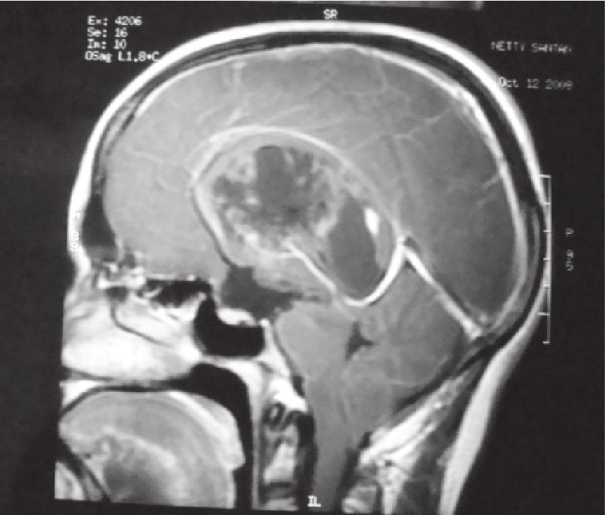
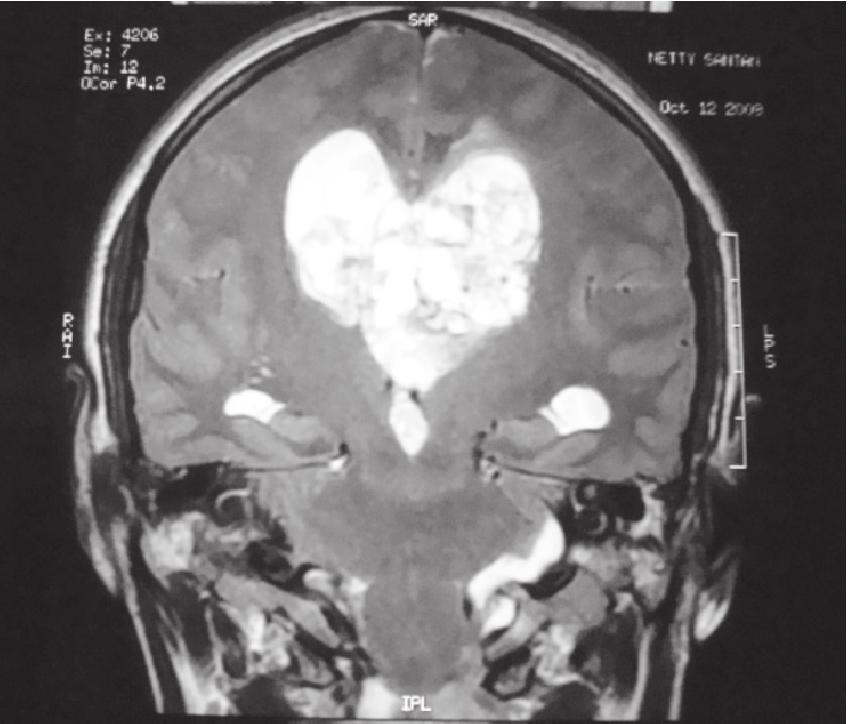
3 monotonous, stippled, round, smoothcontoured nuclei and scanty acidophilic or vacuolated cytoplasm. The cells are surrounded by a loose fibrillary stroma that is traversed by delicate capillaries. Ill formed perivascular rosettes are appreciated. Mitotic activity is inconspicuous. Many cells contain granular blackish brown pigment. There is no vascular endothelial proliferation or necrosis. central neurocytoma have been described in the literature to date. The first account of a pigmented central neurocytoma identified cytoplasmic lipofuscin granules.13 In a similar subsequent case an intimate association of lipofuscin and Fig. 1 Im m u n o h is to c h e m is tr y s h o we d cytoplasmic staining for synaptophysin but not for glial fibrillary associated protein(gfap), epithelial membrane antigen (EMA), neuron specific enolase (NSE), chromogranin, or neurofilament.ki 67 proliferation index was less than 2%. Prussian blue staining was negative. Periodic acid-schiff (PAS) staining was positive. Diagnosis Central neurocytoma (pigmented), septum pellucidum. (a) Discussion As such pigment is an infrequent feature of neuroepithelial neoplasms. The normally melanotic neoplasms like meningeal melanocytoma and intracranial malignant melanoma are indeed rare. Exceptional cases of pigmented lesions have been described for tumour types that normally do not contain the pigment for example, choroid plexus papilloma, ependymoma and carcinoma were found to have both lipofuscin and neuromelanin.9,10,11,12 Other examples are medulloblastoma-medulloepithelioma, schwannoma and meningioma. The accumulation of pigment in neuroepithelial tumours has not been associated with a change in biological behaviour.9,10 Only rare cases of pigmented Bombay Hospital Journal, Vol. 53, No. 2, 2011 (b) H and E stained sections showing (a) monotonous population of small cells having round nuclei and pale acidophilic cytoplasm. (b) In the right half many pigment containing cells are seen. H and E x
 Figure shows PAS positivity in tumour cells indicating the presence of neuromelanin and lipofuscin X380 (b) (a) Synaptophysin immunohistochemistry showing diffuse cytoplasmic staining.")

 such a case has not been previously described in the literature.")
4 Fig. 2 Fig. 3 (a) Figure shows PAS positivity in tumour cells indicating the presence of neuromelanin and lipofuscin X380 (b) (a) Synaptophysin immunohistochemistry showing diffuse cytoplasmic staining. (b) This view shows marked decrease in the amount of melanin pigment which has been bleached with potassium permanganate. X 380 neuromelanin was noted.14 The authors suggested autocatalytic peroxidation of lipofuscin as a possible mechanism for neuromelanin formation. It was also thought to be a manifestation of the low turnover rate of tumour cells, compatible with the usually slowly growing nature of 240 central neurocytoma. The current case is unique because it contained all the three pigments (neuromelanin, melanin and lipofuscin) such a case has not been previously described in the literature.1,2 Marked reduction in the amount of pigment on bleaching (potassium permanganate) indicates melanin (Fig. 2b) and positive staining for periodic acidschiff indicates the presence of neuromelanin and lipofuscin. (Fig. 3) The present case is reported to highlight this rare morphological feature of central neurocytoma. References 1. Figarella-Branger D, Soylemezoglu F, Kleihues P, Hassoun J, Central neurocytoma. In : Kleihues P, Cavenee WK (eds) World Health Organization classification of tumors : pathology and genetics of tumours of the nervous system. IARC Press, Lyon, pp 2000; Hassoun J, Gambarelli D, Grisoli F, Pellet W, Salamon G, Pellissier JF, Toga M, Central neurocytoma. An electronmicroscopic study of two cases. Acta Neuropathol (Berl) 1982;56: Stephan CL, Kepes JJ, Arnold P, Green KD, Chamberlin F Neurocytoma of the cauda equina. Case report. J Neurosurg 1999; 90: Bombay Hospital Journal, Vol. 53, No. 2, 2011
5
CNS pathology Third year medical students. Dr Heyam Awad 2018 Lecture 12: CNS tumours 2/3
 CNS pathology Third year medical students Dr Heyam Awad 2018 Lecture 12: CNS tumours 2/3 Pilocytic astrocytoma Relatively benign ( WHO grade 1) Occurs in children and young adults Mostly: in the cerebellum
ISSN X (Print) Case Report
 Case Report") Scholars Journal of Applied Medical Sciences (SJAMS) Sch. J. App. Med. Sci., 2014; 2(1A):128-132 Scholars Academic and Scientific Publisher (An International Publisher for Academic and Scientific Resources)
Scholars Journal of Applied Medical Sciences (SJAMS) Sch. J. App. Med. Sci., 2014; 2(1A):128-132 Scholars Academic and Scientific Publisher (An International Publisher for Academic and Scientific Resources)
Pathologic features and clinical outcome of central neurocytoma: analysis of 15 cases
 Original Article Pathologic features and clinical outcome of central neurocytoma: analysis of 15 cases Yu Li, Xiu-Feng Ye, Guo Qian, Yu Yin, Qian-Guan Pan Institute of Neuroscience, Department of Pathology,
General: Brain tumors are lesions that have mass effect distorting the normal tissue and often result in increased intracranial pressure.
 1 Lecture Objectives Know the histologic features of the most common tumors of the CNS. Know the differences in behavior of the different tumor types. Be aware of the treatment modalities in the various
CNS TUMORS. D r. Ali Eltayb ( U. of Omdurman. I ). M. Path (U. of Alexandria)
. M. Path (U. of Alexandria)") CNS TUMORS D r. Ali Eltayb ( U. of Omdurman. I ). M. Path (U. of Alexandria) CNS TUMORS The annual incidence of intracranial tumors of the CNS ISmore than intraspinal tumors May be Primary or Secondary
CNS TUMORS D r. Ali Eltayb ( U. of Omdurman. I ). M. Path (U. of Alexandria) CNS TUMORS The annual incidence of intracranial tumors of the CNS ISmore than intraspinal tumors May be Primary or Secondary
Financial disclosures
 Mesenchymal Neoplasms with Melanocytic Differentiation By Konstantinos Linos MD, FCAP, FASDP Bone, Soft Tissue and Dermatopathology Assistant Professor of Pathology Dartmouth-Hitchcock Medical Center Geisel
MR Imaging of Pial Melanosis Secondary to a Posterior Fossa Melanotic Ependymoma
 AJNR Am J Neuroradiol 26:804 808, April 2005 Case Report MR Imaging of Pial Melanosis Secondary to a Posterior Fossa Melanotic Ependymoma Sait Albayram, Efsun Urger, Buge Oz, Ali Kafadar, Civan Islak,
Anna Maria Buccoliero Department of Biomedicine, Careggi Hospital Florence
 PEDIATRIC RHABDOID MENINGIOMA Anna Maria Buccoliero Department of Biomedicine, Careggi Hospital Florence CLINICAL HISTORY A 3-year-old boy, with a recent history of seizures, was admitted to the Neurosurgery
Sporadic Hemangioblastoma of the Kidney: a rare renal tumor
 Liu et al. Diagnostic Pathology 2012, 7:49 CASE REPORT Open Access Sporadic Hemangioblastoma of the Kidney: a rare renal tumor Yang Liu 1,2, Xue-shan Qiu 1,2* and En-Hua Wang 1,2 Abstract: Hemangioblastoma
Tumors of the Nervous System
 Tumors of the Nervous System Peter Canoll MD. PhD. What I want to cover What are the most common types of brain tumors? Who gets them? How do they present? What do they look like? How do they behave? 1
Classification of spontaneous brain tumors in rats
 Classification of spontaneous brain tumors in rats Central Nervous System Neoplasms in the Rat (Solleveld HA, et al., 1991) 1. Tumors of Neuroepithelial Tissue A. Astrocytic and oligodendroglial tumors
Pigmented astrocytoma with suprasellar location: case report and literature review
 Acta Neuropathol (2004) 108: 461 466 DOI 10.1007/s00401-004-0903-6 CASE REPORT Ba rd Kronen Krossnes Æ Olav Mella Æ Knut Wester Sverre Jarl Mørk Pigmented astrocytoma with suprasellar location: case report
Peter Canoll MD. PhD.
 Tumors of the Nervous System Peter Canoll MD. PhD. What I want to cover What are the most common types of brain tumors? Who gets them? How do they ypresent? What do they look like? How do they behave?
Tumors of the Central Nervous System
 Tumors of the Central Nervous System 1 Financial Disclosures I have NO SIGNIFICANT FINANCIAL, GENERAL, OR OBLIGATION INTERESTS TO REPORT Introduction General: Brain tumors are lesions that have mass effect
Histopathological Study and Categorisation of Brain Tumors
 Histopathological Study and Categorisation of Brain Tumors Ruchira Wadhwa 1*, Purvi Patel 2, Hansa Goswami 3 1 Third Year Resident, 2 Assistant Professor, 3 Professor and Head, Department of Pathology,
OMICS PUBLISHING GROUP/Clinical Meningioma with remarkable multiple rosette formation: A diagnostically difficult case
 OMICS PUBLISHING GROUP/Clinical Meningioma with remarkable multiple rosette formation: A diagnostically difficult case --Manuscript Draft-- Manuscript Number: Full Title: Short Title: Article Type: Section/Category:
Brain Tumors. Medulloblastoma. Pilocytic astrocytoma: Ahmed Koriesh, MD. Pathological finding
 NeuroPathology Page 8 Brain Tumors Pathological finding Pseudorosette Rosenthal fibers Rosettes Wet Keratin Psammoma bodies Fried egg Tumor Ependymoma, SEGA Pilocytic astrocytoma Medulloblastoma Craniopharyngioma
Anaplastic Pilocytic Astrocytoma: The fusion of good and bad
 Anaplastic Pilocytic Astrocytoma: The fusion of good and bad Alexandrina Nikova 1, Charalampos-Chrysovalantis Chytoudis-Peroudis 2, Penelope Korkolopoulou 3 and Dimitrios Kanakis 4 Abstract 5 Pilocytic
Intramedullary Clear Cell Ependymoma in the Thoracic Spinal Cord: A Case with Its Crush Smear and Ultrastructural Findings
 J Korean Med Sci 2007; 22 (Suppl): S149-53 ISSN 1011-8934 Copyright The Korean Academy of Medical Sciences Intramedullary Clear Cell Ependymoma in the Thoracic Spinal Cord: A Case with Its Crush Smear
1/10/2018. Soft Tissue Tumors Showing Melanocytic Differentiation. Overview. Desmoplastic/ Spindle Cell Melanoma
 2016 MFMER slide-1 2016 MFMER slide-2 2016 MFMER slide-3 Soft Tissue Tumors Showing Melanocytic Differentiation Andrew L. Folpe, M.D. Professor of Laboratory Medicine and Pathology Mayo Clinic, Rochester,
A case of multinodular high-grade neuroepithelial tumor
 Brain Tumor Pathol (2011) 28:253 257 DOI 10.1007/s10014-011-0032-6 CASE REPORT A case of multinodular high-grade neuroepithelial tumor with ependymal differentiation Kensaku Kamada Yuko Tanaka Takayuki
Neurocytoma a Rare Intraventricular Tumor
 Neurocytoma a Rare Intraventricular Tumor J. A. Mallick,S. A. Ali ( Department of Oncology, Liaquat National Postgraduate Medical Centre, Karachi. ) Introduction Central neurocytoma was first recognized
Hemosiderin pigmentation of tumour cells in cerebellar pilocytic astrocytoma associated with post-traumatic hemorrhage in adults
 Case report Hemosiderin pigmentation of tumour cells in cerebellar pilocytic astrocytoma associated with post-traumatic hemorrhage in adults Anna Taraszewska 1, Zbigniew Czernicki 2, Jaros³aw Andrychowski
Astroblastoma : A Case Report
 J Korean Med Sci 2004; 19: 772-6 ISSN 1011-8934 Copyright The Korean Academy of Medical Sciences Astroblastoma : A Case Report Astroblastoma is one of the very unusual type of tumors, whose histogenesis
Intramedullary melanocytoma of thoracic spine: A rare case report
 1 di 12 16/07/2014 11.16 Asian J Neurosurg. 2014 Jan-Mar; 9(1): 36 39. doi: 10.4103/1793-5482.131068 PMCID: PMC4038865 Intramedullary melanocytoma of thoracic spine: A rare case report 1 Pranav Dorwal,
Immunohistochemical Staining for Claudin-1 Can Help Distinguish Meningiomas From Histologic Mimics
 Anatomic Pathology / CLAUDIN-1 IN MENINGIOMAS Immunohistochemical Staining for Claudin-1 Can Help Distinguish Meningiomas From Histologic Mimics Hejin P. Hahn, MD, PhD, Elizabeth A. Bundock, MD, PhD, and
Supplementary Information
 Rise in Glioblastoma Multiforme incidence in England 1995 2015 suggests an adverse environmental or lifestyle factor Alasdair Philips, Denis L Henshaw, Graham Lamburn, Michael J O Carroll Supplementary
Methoden / Methods inc. ICCC-3 105
 Methoden / Methods inc. ICCC-3 105 Internationale Klassifikation der Krebserkrankungen bei Kindern (ICCC-3) Zuordnung von ICD-O-3-Codes für Morphologie und Topographie zu diagnostischen Kategorien International
Patients Treated with Leksell Gamma Knife
 Patients Treated with Leksell Gamma Knife 1968-2016 TREATMENTS REPORTED 2016 BY REGION AND INDICATION INDICATION Asia excl. Europe Latin Middle East & Africa North Grand Total Benign Tumors 12283 9778
Pathologic Analysis of CNS Surgical Specimens
 2015 Kenneth M. Earle Memorial Neuropathology Review Pathologic Analysis of CNS Surgical Specimens Peter C. Burger, MD Interdisciplinary Quality Control Familiarity with entities Use of diagnostic algorithm
Cerebral Parenchymal Lesions: I. Metastatic Neoplasms
 Chapter 4 Cerebral Parenchymal Lesions: I. Metastatic Neoplasms After one has reasonably ruled out the possibility of a nonneoplastic diagnosis (see Chap. 3), one is left with considering a diagnosis of
AMERICAN ASSOCIATION OF NEUROPATHOLOGISTS COMPANION SOCIETY MEETING at the 106 th ANNUAL MEETING OF THE USCAP San Antonio, March 4, 2017
 AMERICAN ASSOCIATION OF NEUROPATHOLOGISTS COMPANION SOCIETY MEETING at the 106 th ANNUAL MEETING OF THE USCAP San Antonio, March 4, 2017 SYLLABUS Papillary Tumor of the Pineal Region and the Differential
Non-hematogenous endogenous pigments
 Non-hematogenous endogenous pigments 0 This group contains the following : 1. Melanins. 2. Lipofuscins. 3. Chromaffin. 4. Pseudomelanosis. 5. Dubin-Johnson pigments. 6. Ceroid-type lipofuscins. 7. Hamazaki-Weisenberg
Journal of American Science 2014;10(3)
") Central Neurocytoma: Experience at King Abdulaziz University Hospital Jeddah Saudi Arabia Shabnum Sultana, Awatif A. Jamal., Dahlia S. Mirdad, Fahad A. Alghamdi Department of Pathology, Faculty of Medicine,
Central Neurocytoma: Experience at King Abdulaziz University Hospital Jeddah Saudi Arabia Shabnum Sultana, Awatif A. Jamal., Dahlia S. Mirdad, Fahad A. Alghamdi Department of Pathology, Faculty of Medicine,
Rhabdoid Meningioma In A Background Of Atypical Meningioma With Lipomatous Metaplasia: Case Report And Review Of Literature
 ISPUB.COM The Internet Journal of Neurosurgery Volume 5 Number 1 Rhabdoid Meningioma In A Background Of Atypical Meningioma With Lipomatous Metaplasia: Case Report And Review Of Literature S Shuja, T Lucey,
Case Report Intracranial Capillary Hemangioma in the Posterior Fossa of an Adult Male
 Case Reports in Radiology Volume 2016, Article ID 6434623, 4 pages http://dx.doi.org/10.1155/2016/6434623 Case Report Intracranial Capillary Hemangioma in the Posterior Fossa of an Adult Male Jordan Nepute,
Pineal region tumors
 Case Series Pineal region tumors Meena Patil, Manjiri Karandikar Abstract The pineal gland is located near the center of the brain, between two hemispheres in between the two thalamic bodies. It is activated
Evaluation of the Squash Smear Technique in the Rapid Diagnosis of Central Nervous System Tumors: A Cytomorphological Study
 ISPUB.COM The Internet Journal of Pathology Volume 11 Number 1 Evaluation of the Squash Smear Technique in the Rapid Diagnosis of Central Nervous System Tumors: A N Pawar, K Deshpande, S Surase, G D costa,
Site Specific Coding Rules MALIGNANT CENTRAL NERVOUS SYSTEM TUMORS
 Multiple Primary and Histology Site Specific Coding Rules MALIGNANT CENTRAL NERVOUS SYSTEM TUMORS 1 Prerequisites 2 Completion of Multiple Primary and Histology General Coding Rules 3 There are many ways
Intramedullary central neurocytoma of the thoracic spinal cord: A case report and literature review
 MOLECULAR AND CLINICAL ONCOLOGY 8: 539-543, 2018 Intramedullary central neurocytoma of the thoracic spinal cord: A case report and literature review ZHIMIN LI, JUN GAO, TIANYU WANG, XIANGYI KONG, JIAN
J of Evolution of Med and Dent Sci/ eissn , pissn / Vol. 3/ Issue 30/July 28, 2014 Page 8403
 BENIGN TUMOR OF NOSE AND PARANASAL SINUS IN A CHILD: A CASE REPORT Mujtaba Khan 1, S. Muneeruddin Ahmed 2, A. Sesha Prasad 3, M. Mahendra Kumar 4, G. Shahul Hameed 5 HOW TO CITE THIS ARTICLE: Mujtaba Khan,
Paraganglioma of cauda equina - case report and literature review
 22 H. Oo et al. / Brunei Darussalam Journal of Health Volume 4, 2009/2010 Paraganglioma of cauda equina - case report and literature review Hla Oo 1, Pemasiri Upali Telisinghe 1, Ghazala Kafeel 1, Murugaiyan
2. Subependymal giant cell astrocytoma:
 I. Astrocytomas: A. Diffusely infiltrating ( astrocytoma, anaplastic astrocytoma, GBM) B. Localised (pilocytic astrocytoma, pleomorphic xanthoastrocytoma, SGCA) *Grading: Diffuse: 1. Astrocytoma WHO grade
1 NORMAL ANATOMY AND HISTOLOGY
 1 NORMAL ANATOMY AND HISTOLOGY OF THE CNS Anatomy 1 Histology 4 ANATOMY Knowledge of nervous system anatomy is essential for success in surgical neuropathology. Familiarity with native cellular elements
Benign and malignant epithelial lesions: Seborrheic keratosis: A common benign pigmented epidermal tumor occur in middle-aged or older persons more
 Benign and malignant epithelial lesions: Seborrheic keratosis: A common benign pigmented epidermal tumor occur in middle-aged or older persons more common on the trunk; but extremities, head and neck are
Pathological Pigmentation
 Pathological Pigmentation By Dr. Hemn Hassan Othman PhD, Pathology, Fall 2018 10/20/2018 1 Pathological Pigmentation: Pigments: Pigments are colored substances accumulate abnormally within the tissue and
Ocular Neoplasia CL Davis 9/08. Richard R Dubielzig
 Ocular Neoplasia CL Davis 9/08 Richard R Dubielzig 2135/5722 Canine Melanocytic Tumors Outside the Globe: 264 Conjunctival: 159 Eye Lid: 72 Skin: 33 Affecting the Globe: 1871 Anterior Uveal Melanocytoma:
External Neoplasms in Goats: A Clinicopathological Study on Five Types. Abu-Seida, A.M and Kawkab, A. Ahmed
 External Neoplasms in Goats: A Clinicopathological Study on Five Types By Abu-Seida, A.M and Kawkab, A. Ahmed Introduction Introduction Neoplasia is occasionally diagnosed in goats. A survey of 800000
Case 1. Maysa Al-Hussaini MD FRCPath
 Case 1 Maysa Al-Hussaini MD FRCPath MAYSA King AL-HUSSAINI Hussein Cancer MD Center MRCPATH KING HUSSEIN Amman CANCER Jordan CENTER Clinical history 4 year old boy History of frontal headache, sleepiness.
Decreased Vision and Junctional Scotoma from Pituicytoma
 Decreased Vision and Junctional Scotoma from Pituicytoma The Harvard community has made this article openly available. Please share how this access benefits you. Your story matters. Citation Published
SPECIAL SLIDE SEMINAR CASE 3
 SPECIAL SLIDE SEMINAR CASE 3 Tihana Džombeta, MD Leo Pažanin, MD, PhD Department of Pathology, School of Medicine, University of Zagreb Department of Pathology, Clinical Hospital Centre Sestre milosrdnice
Case Report Xanthomatous meningioma: a case report with review of the literature
 Int J Clin Exp Pathol 2013;6(10):2242-2246 www.ijcep.com /ISSN:1936-2625/IJCEP1308033 Case Report Xanthomatous meningioma: a case report with review of the literature Mitsuaki Ishida 1, Tadateru Fukami
Symtomatic Subependymoma Of The Lateral Ventricle: A Rare Entity A Case report and review of literature
 ISPUB.COM The Internet Journal of Neurosurgery Volume 7 Number 1 Symtomatic Subependymoma Of The Lateral Ventricle: A Rare Entity A Case report and review of M Sharma, V Velho, P Ghodgaonkar, D Palande
Special Techniques for the Study of Cutaneous Neural Tumors
 Special Techniques for the Study of Cutaneous Neural Tumors 2 Keywords A variety of histochemical and immunohistochemical methods, if employed in the appropriate context and in a suitable combination,
hemangioblastoma of the retroperitoneum
 Int J Clin Exp Pathol 2014;7(4):1777-1781 www.ijcep.com /ISSN:1936-2625/IJCEP1401042 Case Report Yong Huang 1, Xiang-Chun Han 2, Guo-Shi Lv 3 1 Department of Pathology, 251 Hospital of PLA, Zhangjiakou,
Rosette-forming glioneuronal tumor in the pineal gland and the third ventricle: a case with radiological and clinical implications
 Case Report Rosette-forming glioneuronal tumor in the pineal gland and the third ventricle: a case with radiological and clinical implications Junqing Xu 1, Yong Yang 1, Ying Liu 1, Mengqi Wei 1, Jing
Histological pattern of central nervous system neoplasms
 ecommons@aku Department of Pathology and Laboratory Medicine Medical College, Pakistan April 2001 Histological pattern of central nervous system neoplasms Z. Ahmed Aga Khan University S. Muzaffar Aga Khan
Embryonal tumor with multilayered rosettes, C19MC-altered: Report of an extremely rare malignant pediatric central nervous system neoplasm
 745208SCO0010.1177/2050313X17745208SAGE Open Medical Case ReportsTariq et al. case-report2017 Case Report SAGE Open Medical Case Reports Embryonal tumor with multilayered rosettes, C19MC-altered: Report
Alveolar Soft Part Sarcoma of the Uterine Cervix: A Case Report and Review of the Literature
 The Korean Journal of Pathology 2014; 48: 361-365 CASE STUDY Alveolar Soft Part Sarcoma of the Uterine Cervix: A Case Report and Review of the Literature Hyun Ju Lee Department of Pathology, Soonchunhyang
Five Most Common Problems in Surgical Neuropathology
 Five Most Common Problems in Surgical Neuropathology If the brain were so simple that we could understand it, we would be so simple that we couldn t Emerson Pugh What is your greatest difficulty in neuropathology?
Site Specific Coding Rules Benign and Borderline Intracranial and CNS Tumors
 Multiple Primary and Histology Site Specific Coding Rules Benign and Borderline Intracranial and CNS Tumors 1 Prerequisites 2 Completion of Multiple Primary and Histology General Coding Rules 3 There are
Epidemiology of Primary Brain and Central Nervous System Tumors in Korea
 www.jkns.or.kr 1.334/jkns.21.48.2.145 J Korean Neurosurg Soc 48 : 145-152, 21 Print ISSN 25-3711 On-line ISSN 1598-7876 Copyright 21 The Korean Neurosurgical Society Clinical Article Epidemiology of Primary
Case #3. USCAP Neuropathology Evening Seminar/Companion Meeting
 Case #3 USCAP Neuropathology Evening Seminar/Companion Meeting Clinical History A 71-year year-old man presented with a 4-4 week history of word finding difficulty. An initial screening head CT followed
Pancreatitis: A Potential Pitfall in Endoscopic Ultrasound Guided Pancreatic FNA
 Pancreatitis: A Potential Pitfall in Endoscopic Ultrasound Guided Pancreatic FNA Jack Yang, MD Department of Pathology, Medical University of South Carolina Objectives Understand the indication of EUS
Nasal Cavity and Paranasal Sinuses
 Chapter 2 Nasal Cavity and Paranasal Sinuses Introduction Included in this chapter are nasal cavities, frontal sinus, ethmoid complex, sphenoid sinus, and maxillary sinuses. These cavities and sinuses
Diagnostic accuracy of central nervous system tumors by squash cytology
 Original Research Article Diagnostic accuracy of central nervous system tumors by squash cytology Ramana PV 1, Vijaya Nirmala B 2*, Srujana Shyamala 3 1 Assistant Professor, Department of Pathology, Gandhi
Special slide seminar
 Special slide seminar Tomáš Rozkoš The Fingerland Department of Pathology Charles University Medical Faculty and Faculty Hospital in Hradec Králové Czech Republic Case history, 33 years old resistance
Charles Halsey, DVM, PhD, DACVP Pfizer, Inc. IHC Resources
 Charles Halsey, DVM, PhD, DACVP Pfizer, Inc. IHC Resources 1 IHC Identification Targets Specimens Controls 2 Tissue controls Trouble Spots 3 The Key to Description IHC Description 4 Intermediate Filaments
Morphological features and genetic alterations
 Morphological features and genetic alterations Tutor : Audrey Rousseau Caget Lise: Université d Angers Iorio Vittoria: Seconda Università degli studi di Napoli Manaila Roxana: Iuliu Hatieganu University
Unknown Case 6. Ann T. Moriarty, MD
 Unknown Case 6 Ann T. Moriarty, MD Unknown Case 6 61 year old male with an enlarged cervical lymph node. He has a history of lung carcinoma, renal cell carcinoma and lymphoma. Case 6 Image 1: Fine needle
The recently updated World Health Organization (WHO)
") REVIEW ARTICLE The Expanding Family of Glioneuronal Tumors Daniela S. Allende, MD and Richard A. Prayson, MD Abstract: Three new entities have been recently added to the group of glioneuronal tumors in
REVIEW ARTICLE The Expanding Family of Glioneuronal Tumors Daniela S. Allende, MD and Richard A. Prayson, MD Abstract: Three new entities have been recently added to the group of glioneuronal tumors in
Histology of the CNS
 Histology of the CNS Lecture Objectives Describe the histology of the cerebral cortex layers. Describe the histological features of the cerebellum; layers and cells of cerebellar cortex. Describe the elements
C.L. Davis Foundation Descriptive Veterinary Pathology Course
 C.L. Davis Foundation 2015 Descriptive Veterinary Pathology Course IHC Resources IHC Identification Targets Antibodies Antibodies 1 Antibodies Specimens Antigen Retrieval Unmasks antigen epitopes Methods
IAP XXVI International Congress Slide Seminar 07 (SS07)
") IAP XXVI International Congress Slide Seminar 07 (SS07) Pitfalls in Surgical Neuropathology Case 6 Richard A. Prayson, M.D. Cleveland Clinic Foundation Clinical History 64M S/P resection of pituitary adenoma
IAP XXVI International Congress Slide Seminar 07 (SS07) Pitfalls in Surgical Neuropathology Case 6 Richard A. Prayson, M.D. Cleveland Clinic Foundation Clinical History 64M S/P resection of pituitary adenoma
Diseases of the breast (1 of 2)
") Diseases of the breast (1 of 2) Introduction A histology introduction Normal ducts and lobules of the breast are lined by two layers of cells a layer of luminal cells overlying a second layer of myoepithelial
Diseases of the breast (1 of 2) Introduction A histology introduction Normal ducts and lobules of the breast are lined by two layers of cells a layer of luminal cells overlying a second layer of myoepithelial
Case Report Hemangioblastoma in the Lung: Metastatic or Primary Lesions?
 Hindawi Publishing Corporation Volume 2014, Article ID 468671, 5 pages http://dx.doi.org/10.1155/2014/468671 Case Report Hemangioblastoma in the Lung: Metastatic or Primary Lesions? Li Lu, Peter A. Drew,
From Morphology to Molecular Pathology: A Practical Approach for Cytopathologists Part 1-Cytomorphology. Songlin Zhang, MD, PhD LSUHSC-Shreveport
 From Morphology to Molecular Pathology: A Practical Approach for Cytopathologists Part 1-Cytomorphology Songlin Zhang, MD, PhD LSUHSC-Shreveport I have no Conflict of Interest. FNA on Lymphoproliferative
Neuroendocrine Lung Tumors Myers
 Diagnosis and Classification of Neuroendocrine Lung Tumors Jeffrey L. Myers, M.D. A. James French Professor Director, Anatomic Pathology & MLabs University of Michigan, Ann Arbor, MI myerjeff@umich.edu
Spinal Meningeal Melanocytoma with Benign Histology Showing Leptomeningeal Spread: Case Report
 Case Report Musculoskeletal Imaging A 37-year-old previously healthy man presented with a 2 months history of increasing posterior neck pain and radiating pain to the right upper extremity. Neurological
Diagnosis of a granular cell tumour at the abdominal wall using fine needle aspiration cytology and histology: Case report
 Case Report Diagnosis of a granular cell tumour at the abdominal wall using fine needle aspiration cytology and histology: Case report Journal of International Medical Research 2015, Vol. 43(4) 592 596!
H Haloes cautions, 57 neurocytomas, perinuclear, 56 Headache blue cell tumors, 147 cautions, 135, 147, 152 clinical history, 132, 144, 148
 Index A ADC. See Apparent diffusion coefficient Adult. See also Supratentorial mass, adult cerebral tumor, 1 headache and ataxia cysts, mural nodules, 118 sporadic tumors, 118 headaches and visual changes,
Genetic differences between neurocytoma and dysembryoplastic neuroepithelial tumor and oligodendroglial tumors
 J Neurosurg 97:1350 1355, 2002 Genetic differences between neurocytoma and dysembryoplastic neuroepithelial tumor and oligodendroglial tumors HIRONORI FUJISAWA, M.D., KOHEI MARUKAWA, M.D., MITSUHIRO HASEGAWA,
Gangliogliomas: A Report of Five Cases
 Case Report Gangliogliomas: A Report of Five Cases Nair V, Suri VS, Tatke M, Saran RK, Malhotra V, Singh D* Departments of Pathology and *Neurosurgery, G. B. Pant Hospital, New Delhi, India. Correspondence
Ó Journal of Krishna Institute of Medical Sciences University 104
 ISSN 2231-4261 CASE REPORT Unusual Alveolar Pattern in Node Based Diffuse Large B-cell Lymphoma 1* 1 1 1 Archana C. Buch, Jay Y. Sheth, Sunita A Bamanikar, Aditi A. Pandey 1 Department of Pathology, Padmashri
DISCUSSION: PLGA accounts for about 2% of all salivary gland tumours and occurs almost exclusively in the minor salivary glands.
 SWELLING ON THE HARD PALATE PRESENTING AS POLYMORPHOUS LOW GRADE ADENOCARCINOMA: A AND REVIEW OF LITERATURE Swapnil D. Chandekar 1, Sunita S. Dantkale 2, Rahul R. Narkhede 3, Snehal V. Chavhan 4, Khushboo
PATHOLOGY Intracellular Degeneration LAB 1
 PATHOLOGY Intracellular Degeneration LAB 1 Cellular swelling Liver Organ :- Liver Lesion :- 1. Narrowing of hepatic sinusoids due to the swelling of hepatocyte. 2. The cytoplasm of affected hepatocyte
2008 Gross Ocular Pathology. Gross Pathology 2
 2008 Gross Ocular Pathology Gross Pathology 2 08rd1281 Feline T-Cell Lymphoma 08rd1300 Canine Iridociliary Adenoma Foam Cell Variant 08rd1331 Feline Feline Iridociliary Adenoma 08rd1340 Canine Retinal
Participants Identification No. % Evaluation. Mitotic figure Educational Erythrocyte precursor, abnormal/
 Cell Identification BMD-09 Participants Identification No. % Evaluation Mitotic figure 233 96.7 Educational Erythrocyte precursor, abnormal/ 4 1.7 Educational dysplastic nuclear features Erythrocyte precursor
Small (and large) Blue Cell Tumors of the Skull Base
 Blue Cell Tumors of the Skull Base") Small (and large) Blue Cell Tumors of the Skull Base Jennifer L. Hunt, MD, MEd Aubrey J. Hough Jr, MD, Endowed Professor of Pathology Chair of Pathology and Laboratory Medicine University of Arkansas for
Small (and large) Blue Cell Tumors of the Skull Base Jennifer L. Hunt, MD, MEd Aubrey J. Hough Jr, MD, Endowed Professor of Pathology Chair of Pathology and Laboratory Medicine University of Arkansas for
Pleomorphic adenoma of breast - a case report and distinction with metaplastic carcinoma D Gupta, S Agrawal, N Trivedi, A Tewari
 of breast - a case report and distinction with metaplastic carcinoma D Gupta, S Agrawal, N Trivedi, A Tewari Introduction, also known as mixed tumour, is a benign tumour which typically presents as a painless,
TUMORS of nervous system
 TUMORS of nervous system By: Shifaa Alqa qa Done By : Ola Hijjawi CNS tumors : The annual incidence of CNS tumors ranges from 10 to 17 per 100,000 persons for intracranial tumors and 1 to 2 per 100,000
CASE REPORT Benign epithelioid peripheral nerve sheath tumour resembling schwannoma
 Malaysian J Pathol 2014; 36(3) : 217 221 CASE REPORT Benign epithelioid peripheral nerve sheath tumour resembling schwannoma Thejasvi KRISHNAMURTHY MD and SR NIVEDITHA MD, DNB Department of Pathology,
Case 2. Dr. Sathima Natarajan M.D. Kaiser Permanente Medical Center Sunset
 Case 2 Dr. Sathima Natarajan M.D. Kaiser Permanente Medical Center Sunset History 24 year old male presented with a 3 day history of right flank pain, sharp in nature Denies fever, chills, hematuria or
Immunohistochemical Evaluation Of Small Round Cell Tumors Of Childhood
 Immunohistochemical Evaluation Of Small Round Cell Tumors Of Childhood Pages with reference to book, From 87 To 89 Sajid H. Shah,Irshad N. Soomro,M. Shahid Siddiqui,Shahid Pervez,Sheema H. Hassan ( Department
Case Report Disseminated Cerebrospinal Embryonal Tumor in the Adult
 Case Reports in Pathology Volume 2016, Article ID 6785459, 5 pages http://dx.doi.org/10.1155/2016/6785459 Case Report Disseminated Cerebrospinal Embryonal Tumor in the Adult Alessandro Caporlingua, 1 Daniele
59 yo male with past medical history of prostate carcinoma, presented with upper abdominal pain
 December 2016 59 yo male with past medical history of prostate carcinoma, presented with upper abdominal pain Contributed by: Divya Sharma, MD. Fellow, Gastrointestinal Pathology, Department of Pathology
Experimental Neoplastic Formation in Embryonic Chick Brains
 Experimental Neoplastic Formation in Embryonic Chick Brains by BENGT KALLEN 1 From the Tornblad Institute of Comparative Embryology, Lund WITH TWO PLATES IN mammalian teratology, a malformation consisting
Pleomorphic Xanthoastrocytoma
 Pleomorphic Xanthoastrocytoma Christine E. Fuller Keywords Pleomorphic xanthoastrocytoma; Pleomorphic xanthoastrocytoma with anaplastic features 2.1 OVERVIEW Pleomorphic xanthoastrocytoma (PXA) is an uncommon
University of Zurich. Histology and Immunophenotype of Invasive Lobular Breast Cancer. Daily Practice and Pitfalls. Zurich Open Repository and Archive
 University of Zurich Zurich Open Repository and Archive Winterthurerstr. 190 CH-8057 Zurich http://www.zora.uzh.ch Year: 2009 Histology and Immunophenotype of Invasive Lobular Breast Cancer. Daily Practice