PITUITARY PARASELLAR LESIONS. Kim Learned, MD
|
|
|
- Peter Powers
- 6 years ago
- Views:
Transcription

1 PITUITARY PARASELLAR LESIONS Kim Learned, MD

2 DIFFERENTIALS Pituitary Sella Clivus, Sphenoid Sinus Suprasellar Optic chiasm, Hypothalamus, Circle of Willis Parasellar Cavernous Sinus


3 Case 1 17 YEAR-OLD FEMALE BITEMPORAL SUPERIOR QUADRANTOPSIA Diffuse gland enlargement
4 Adenoma Rathke s cleft cyst Apoplexy SELLAR-PITUITARY PATHOLOGIES Pituitary Infectious/Inflammatory: lymphocytic hypophysitis, Sarcoid, Abscess Metastasis: 1-3% sellar mass (breast, lung, GI, typically involved stalk/hypothalamus)


5 PITUITARY ADENOMA Microadenoma < 10 mm Local mass effect Deviation of normal pituitary Stalk and Gland


6 PITUITARY ADENOMA Hemorrhagic Cystic changes Cavernous sinus Protrusion (ICA abutment < 180) Invasion (ICA abutment >270)


7 RATHKE S CLEFT CYST Intrasellar or Upper margin of gland anterior to stalk Variable CT and T1-T2 intensity: CSF Mucoid/Hemorrhage. No Ca+ Intracystic nodule on T2: 75% cholesterol/mucopolysaccharide-hemorrhage Imperceptible-thin enhancing wall


8 PITUITARY APOPLEXY Infarction pituitary Headache, visual symptoms to shock Post-partum Sheehan syndrome Adenoma acute bleed with expansion results in clinical symptoms Bromocriptine predisposes to tumor hemorrhage Pituitary hemorrhage on MRI Clinical Apoplexy
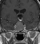
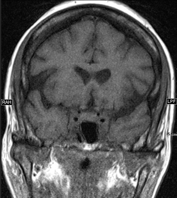
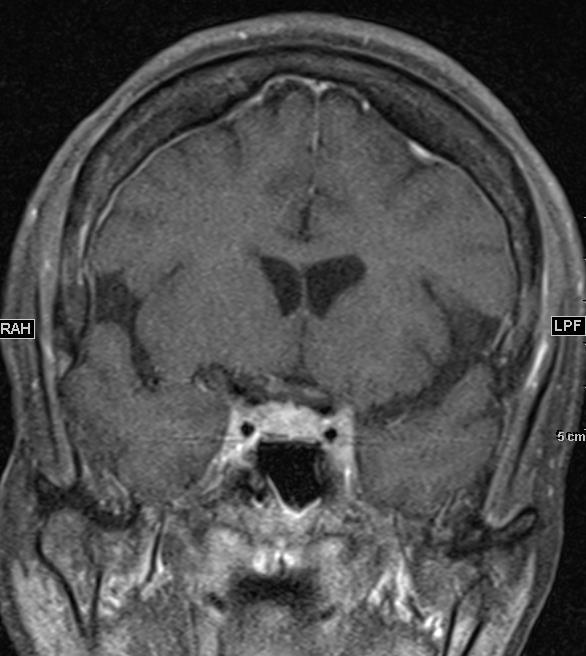
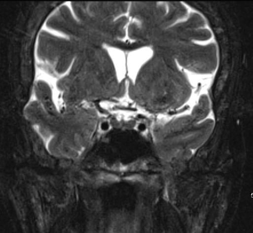
9 Case 1 LYMPHOCYTIC HYPOPHYSITIS Diffuse infiltration of the gland with convex contour, elevation-thickening of the stalk
10 DIFFUSE PITUITARY GLAND PROCESS Hyperplasia Macroadenoma Infiltrative processes lymphocytic hypophysitis, sarcoid, granulomatous disease


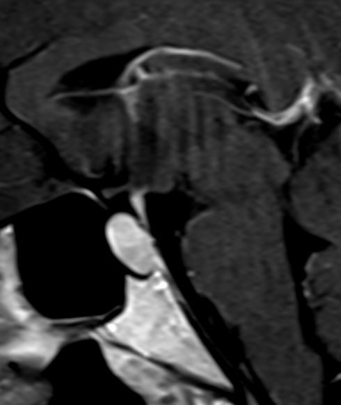
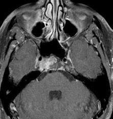
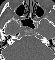
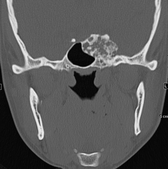
11 Case 3 INCIDENTAL FINDINGS A. Pituitary adenoma B. Sinonasal tumor C. Chordoma D. All above
12 Case 3 PITUITARY MACROADENOMA Adenoma from inferior pituitary eroded sellar floor, protruded into sphenoid sinus Normal fatty marrow clivus


13 GIANT MACROADENOMA Most common Sellar-Parasellar lesions in Adults Arise from Pituitary gland Burrow into bone Intermediate T2 signal, Solid Enhancement


14 CHORDOMA Bone Erosion Arc-Ring matrix in Chondrosarcoma T2 Hyperintensity Variable Degree of Enhancement
15 CHONDROSARCOMA

16 OSTEOSARCOMA 23 year-old female well-differentiated low-grade type

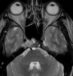
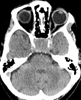
17 Case 4 VISUAL CHANGE A. Macroadenoma B. Meningioma C. Metastasis D. Chordoma
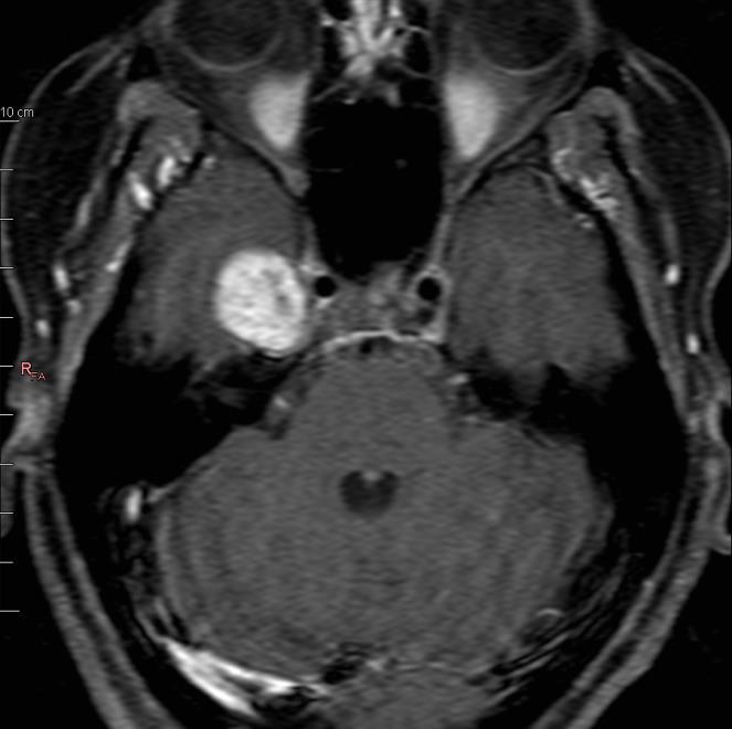
18 Case 4 MENINGIOMA 2 nd common Parasellar mass in Adults Dural-based Hyperostosis, Enhancing Dural Tail Similar to gray matter, Avid enhancement
19 SELLAR PARASELLAR ENHANCING LESIONS Pituitary origin Adenoma >> Infectious/inflammatory, craniopharyngioma, metastasis Dura origin Meningioma >> Metastasis Clivus origin Chordoma, Chondrosarcoma Metastasis, Plasmacytoma Aneurysm Cavernous ICA, Acom
20 Case 5 HEADACHE
21 Case 5 SELLAR-SUPRASELLAR CYSTIC LESIONS A. Hemorrhagic/Cystic Adenoma B. Rathke s Cleft Cyst C. Epidermoid D. Craniopharyngioma E. Arachnoid cyst


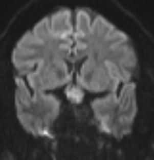
22 CSF + DWI DDx: Dermoid -- Fat EPIDERMOID

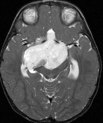
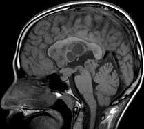
23 CRANIOPHARYNGIOMA Bimodal: ½ Childhood Adolescence ½ Adulthood Adamantinoma Children/Adults Wet karatin mass surrounded by inflammatory infiltrate 90 % mixed solid cystic, T1, Ca + Squamous-Papillary Adults Pappilary finger-like projection of squamous epithelium Solid
24 Case 5 CRANIOPHARYNGIOMA Supra-Sellar 75% Sellar-Suprasellar 20% Cystic, Rim-Nodular Ca+80% Multilobulated into multiple anatomic locations Encase vessels, optic nerve Recurrence
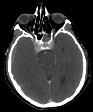
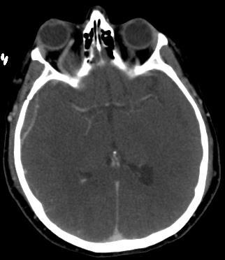
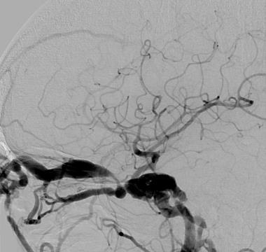
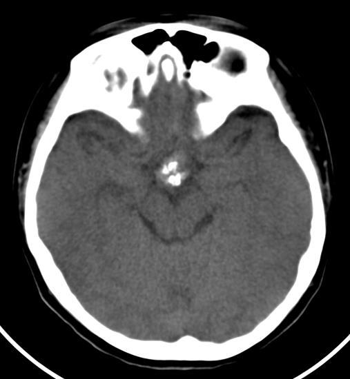
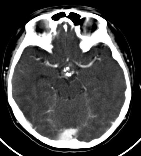
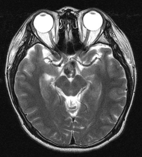
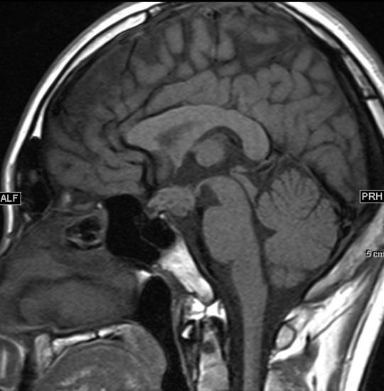
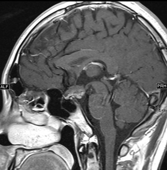
25 Case 6 VISUAL CHANGE A. Macroadenoma B. Craniopharyngioma C. Aneurysm D. Germinoma
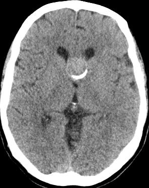
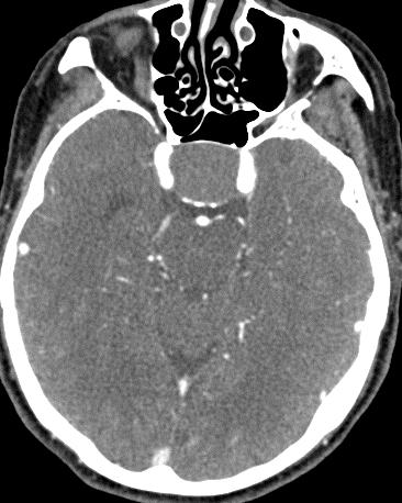
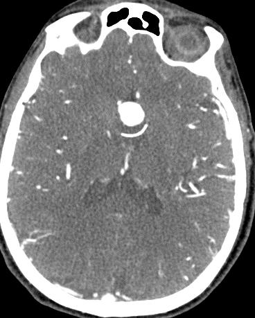


26 Case 6 MACROADENOMA ACOMM ANEURYSM


27 ACOM ANEURYSM Partial thrombosed Aneurysm: Mixed signal of thrombus
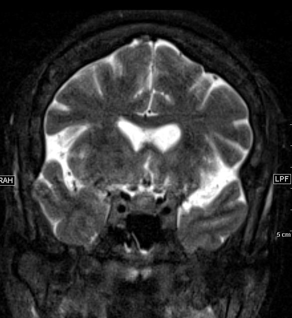
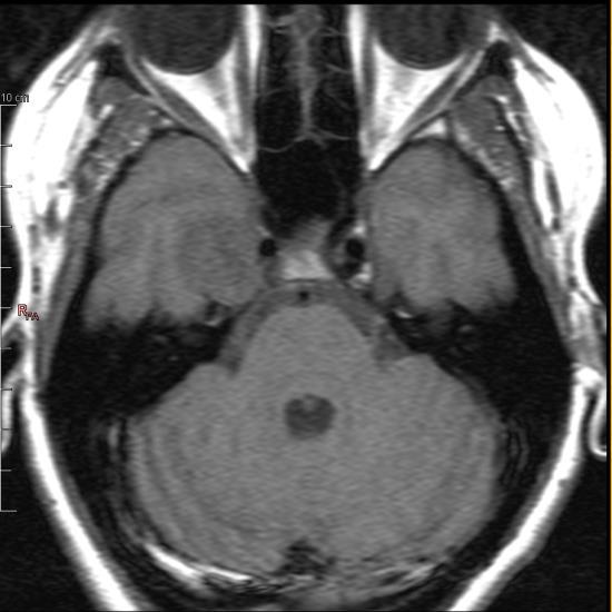
28 Case 1 SEIZURE A. Astrocytoma B. Aneurysm C. Hamartoma D. Epidermoid

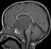
29 Case 1 TUBER CINEREUM HAMARTOMA Congenital heterotopia of Gray matter Gelastic seizure Precocious puberty: LHRH secretion

30 Case 7 VISUAL CHANGE HEADACHE A. Craniopharyngioma B. Astrocytoma C. Giant Aneurysm D. Germinoma


31 Case 7 HYPOTHALAMIC-OPTIC CHIASM ASTROCYTOMA 10 year old JPA 60 year old Anaplastic Astrocytoma Children: Low grade, Juvenile Pilocytic Astrocytoma (JPA) Adults: High grade
32 GERMINOMA Suprasellar Pineal
33
34 SUPRASELLAR SOLID LESIONS Aneurysm: Acom, flow artifact, Mixed signal of thrombosis Optic chiasm/hypothalamic Astrocytoma: infiltrative, T2, variable enhancement
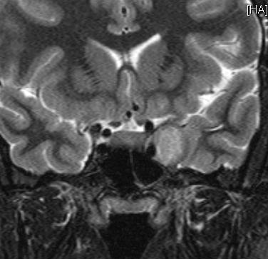
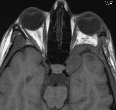
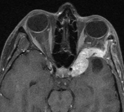
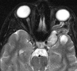
35 Case 8 DIPLOPIA CAVERNOUS SINUS LESIONS A. Aneurysm B. Schwannoma C. Lymphoma/Perin eural Spread of Tumor D. Meningioma
36 CAVERNOUS ICA ANEURYSM

37 CAVERNOUS SINUS MENINGIOMA


38 LYMPHOMA SELLAR- CAVERNOUS SINUS Diffuse marrow replacement DDx: Perineural spread of tumor
39 EPIDERMOID CAVERNOUS SINUS
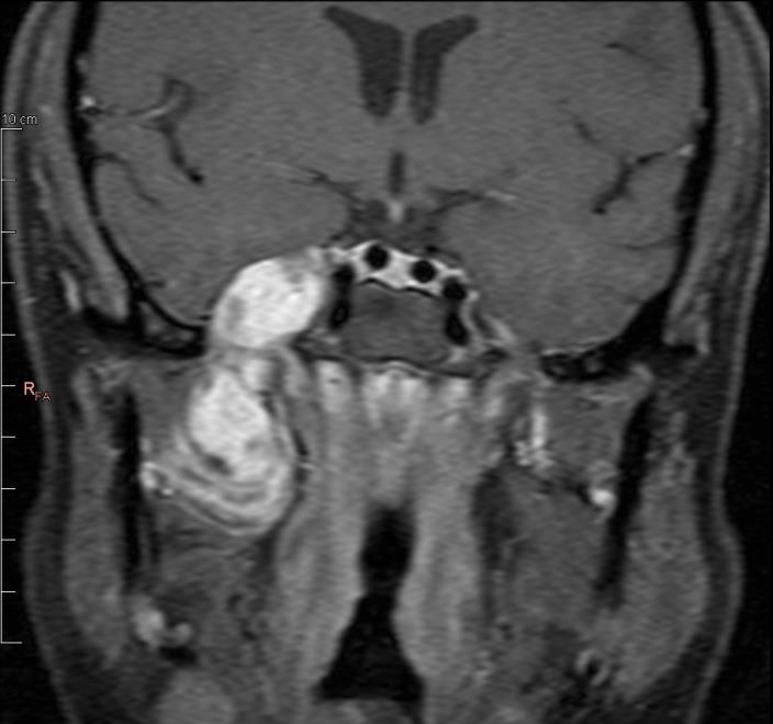
40 Case 8 V1 SCHWANNOMA

41 CAROTID CAVERNOUS FISTULA


42 CAVERNOUS HEMANGIOMA T2 hyperintense T1 Isointense No narrowing ICA Avid nhancement Gradual filling
43 Pituitary Sella Clivus, Sinus Suprasellar Hypothalamus, Optic chiasm, Circle of Willis Parasellar Cavernous Sinus DIFFERENTIALS LOCATIONS CLINICAL


44 MENINGIOMA Narrowing of cavernous ICA Dural tail Petroclival meningioma DDx: Pituitary adenomas, Bone lesion, Lymphoma, Perineural spread of neoplasm, Tolosa-Hunt 45

45 OPHTHALMOPLEGIA 46 2 wks after steroid and chemo Cavernous sinus thrombosis Lymphoma Metastasis Tolosa-Hunt

46 ESRD on HD What Next?
47
48 CAVERNOUS ICA ANEURYSM THROMBUS Concentric ring of thrombus

49 RIGHT FACIAL PAIN Neurilemomas/Schwannoma Intracavernous aneurysms Cavernous hemangiomas 50


50 CAVERNOUS HEMANGIOMA T2 hyper intense Lobular Fill-in
51 MANDIBLE METASTASIS

52 16 YO BOY VISUAL CHANGE
53 HEADACHE
54 CRANIOPHARYNGI OMA Supra-Sellar 75% Sellar-Suprasellar 20% Cystic, Rim-Nodular Ca+80% Multilobulated into multiple anatomic locations Encase vessels, optic nerve Recurrence


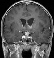
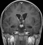
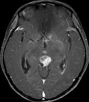
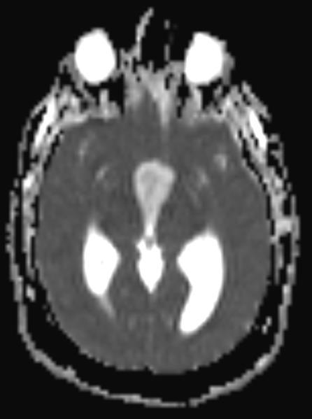
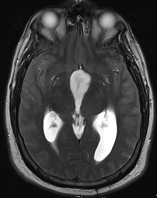
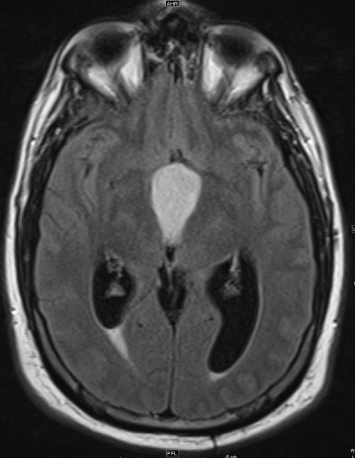
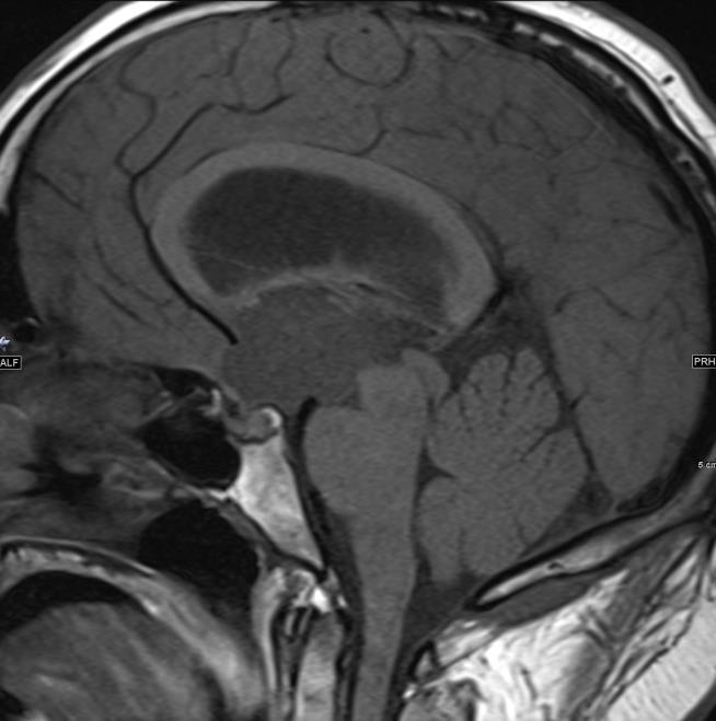
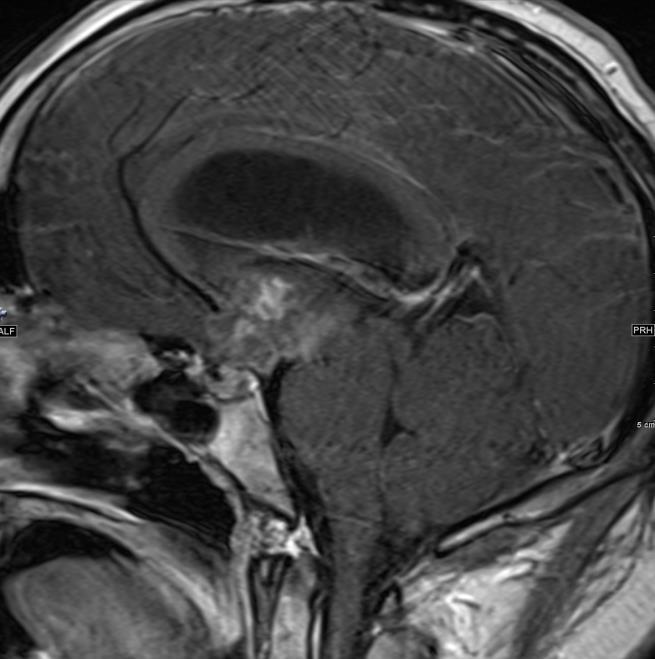
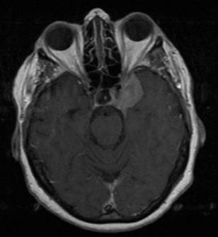
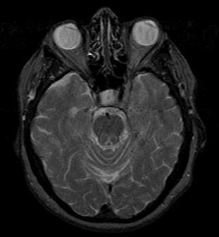
55 Thick enhancing pituitary stalk DDx: Germ cell tumor, Sarcoid, Lymphoma
Metastasis. 57 year old with progressive Headache and Right Sided Visual Loss
 Metastasis 1% of sellar/parasellar masses Usually occurs with known primary Can involve third ventricle, hypothalamus, infundibular stalk May be both supra-, intrasellar 57 year old with progressive Headache
Laurie A. Loevner, MD
 Laurie A. Loevner, MD Chief, Division of Neuroradiology UPHS Professor of Radiology, Otorhinolaryngology: Head & Neck Surgery, Neurosurgery, and Ophthalmology University of Pennsylvania Health System Disclosures
EXPERT DIFFERENTIAL DIAGNOSIS:
 EXPERT DIFFERENTIAL DIAGNOSIS: Sellar Region Anne G. Osborn, M.D. DISCLOSURE: Published RSNA 2008 SELLA, PITUITARY: Normal Gross, 3T Anatomy SELLA, PITUITARY: Anatomically-Based Differential Diagnoses
Imaging The Turkish Saddle. Russell Goodman, HMS III Dr. Gillian Lieberman
 Imaging The Turkish Saddle Russell Goodman, HMS III Dr. Gillian Lieberman Learning Objectives Review the anatomy of the sellar region Discuss the differential diagnosis of sellar masses Discuss typical
Where Has My Vision Gone? Evaluation of Sellar Lesions. Caleb Stowell,, HMS III Gillian Lieberman, MD November 2008
 Where Has My Vision Gone? Evaluation of Sellar Lesions Caleb Stowell,, HMS III Gillian Lieberman, MD November 2008 Objectives Present a case highlighting the clinical presentation and evaluation of a sellar
DISCLOSURES LEARNING OBJECTIVES WE WILL NOT DISCUSS. CSB: Birdseye View MESSAGE NAVIGATING THE SELLA AND CENTRAL SKULL BASE
 NAVIGATING THE SELLA AND CENTRAL SKULL BASE Christopher P. Hess, M.D., Ph.D. DISCLOSURES Research Support, General Electric SLIDES: http://www.radiology.ucsf.edu/research/meetings/rsna LEARNING OBJECTIVES
Case Studies in Sella/Parasellar Region. Child thirsty, increased urination. Imaging. Suprasellar Germ Cell Tumor (Germinoma) No Disclosures
 No Disclosures") Case Studies in Sella/Parasellar Region No Disclosures 2018 Head and Neck Imaging Conference Child thirsty, increased urination Suprasellar Germ Cell Tumor (Germinoma) Midline Pineal >> Suprasellar > Other
Case Studies in Sella/Parasellar Region No Disclosures 2018 Head and Neck Imaging Conference Child thirsty, increased urination Suprasellar Germ Cell Tumor (Germinoma) Midline Pineal >> Suprasellar > Other
Neuro - imaging. Sella. ssregypt.com
 Neuro - imaging Sella ssregypt.com Bony Sella AP diameter Depth Contents 16mm 14mm Pituitary gland, part of infundibular stalk, CSF CT Technique 5 mm slices Axial and coronal Contrast injection Bone and
Part II - Revising the sellar and parasellar region: differential diagnosis of a sellar region mass
 Part II - Revising the sellar and parasellar region: differential diagnosis of a sellar region mass Poster No.: C-1390 Congress: ECR 2015 Type: Educational Exhibit Authors: I. Candelaria, C. Figueira,
Imaging pituitary gland tumors
 November 2005 Imaging pituitary gland tumors Neel Varshney,, Harvard Medical School Year IV Two categories of presenting signs of a pituitary mass Functional tumors present with symptoms due to excess
TABLES. Imaging Modalities Evidence Tables Table 1 Computed Tomography (CT) Imaging. Conclusions. Author (Year) Classification Process/Evid ence Class
 Imaging. Conclusions. Author (Year) Classification Process/Evid ence Class") TABLES Imaging Modalities Evidence Tables Table 1 Computed Tomography (CT) Imaging Author Clark (1986) 9 Reformatted sagittal images in the differential diagnosis meningiomas and adenomas with suprasellar
TABLES Imaging Modalities Evidence Tables Table 1 Computed Tomography (CT) Imaging Author Clark (1986) 9 Reformatted sagittal images in the differential diagnosis meningiomas and adenomas with suprasellar
RADIOANATOMY OF SELLA TURCICA
 RADIOANATOMY OF SELLA TURCICA O.BAKKACHA, H.MALAJATI, M.RHISSASSI, H. BENCHAABOUNE, N.CHAKIR, My R. EL HASSANI,M.JIDDANE Department of Neuroradiology specialties Hospital. Rabat Objective: New imaging
Brain Tumors. Medulloblastoma. Pilocytic astrocytoma: Ahmed Koriesh, MD. Pathological finding
 NeuroPathology Page 8 Brain Tumors Pathological finding Pseudorosette Rosenthal fibers Rosettes Wet Keratin Psammoma bodies Fried egg Tumor Ependymoma, SEGA Pilocytic astrocytoma Medulloblastoma Craniopharyngioma
Traditional Approach. Pathways for Skull Base Pathology. Special Pathways Approach. 1. Traditional Approach. Central Skull Base. Anterior Skull Base
 Traditional Approach Pathways for Skull Base Pathology Anatomy Local Pathology Wade Wong DO FACR Professor of Radiology University of California, San Diego Special Pathways Approach Perineural Perivascular
Case Studies in the Skull Base
 Case Studies in the Skull Base Amy C Tsai, MD Neuroradiology Fellow Department of Radiology and Imaging Sciences University of Utah Health Sciences Center Salt Lake City, Utah, USA No disclosures related
panhypopituitarism Pattawan Wongwijitsook Maharat Nakhon Ratchasima hospital 17 Nov 2013
 panhypopituitarism Pattawan Wongwijitsook Maharat Nakhon Ratchasima hospital 17 Nov 2013 PITUITARY GLAND (HYPOPHYSIS CEREBRI) The master of endocrine glands master of endocrine glands It is a small oval
Visual pathways in the chiasm
 Visual pathways in the chiasm Intracranial relationships of the optic nerve Fixation of the chiasm Chiasmatic pathologies The function of the optic chiasm may be altered by the presence of : 4) Artero
Radiology of hypothalamic lesions: A pictorial essay depicting characteristic hypothalamic pathologies
 Radiology of hypothalamic lesions: A pictorial essay depicting characteristic hypothalamic pathologies Poster No.: C-2713 Congress: ECR 2010 Type: Scientific Exhibit Topic: Neuro Authors: A. J. B. Baxi,
MRI of the Pituitary Gland
 MRI of the Pituitary Gland Jean- François Bonneville Fabrice Bonneville Françoise Cattin Sonia Nagi MRI of the Pituitary Gland With a Foreword by A. Beckers Jean-François Bonneville, MD Department of
Sellar and Parasellar Lesions: over and above adenomas.
 Sellar and Parasellar Lesions: over and above adenomas. Poster No.: C-2052 Congress: ECR 2013 Type: Educational Exhibit Authors: S. Paz Maya, P. Lemercier, I. lópez blasco, D. Soriano Mena, J. P. Ruiz
Intrasphenoidal Rathke's Cleft Cyst: Case presentation and review of the literature
 Romanian Neurosurgery Volume XXX Number 4 2016 October - December Article Intrasphenoidal Rathke's Cleft Cyst: Case presentation and review of the literature Umit Kocaman, Muhammet Bahadir Yilmaz, Hakan
Imaging of Hearing Loss
 Contemporary Imaging of Sensorineural Hearing Loss Imaging of Hearing Loss Discussion Outline (SNHL) Imaging Approaches Anatomic Relationships Lesions: SNHL KL Salzman, MD University of Utah School of
DIFFERENTIAL DIAGNOSIS OF SELLAR MASSES
 ~~ ~~ ~ ADVANCES IN PITUITARY TUMOR THERAPY 0889-8529/99 $8.00 +.OO DIFFERENTIAL DIAGNOSIS OF SELLAR MASSES Pamela U. Freda, MD, and Kalmon D. Post, MD Pituitary adenomas are the most common cause of a
Transplanum Approach for Suprasellar pathology
 Transplanum Approach for Suprasellar pathology Omar A. El-Banhawy Prof. of otorhinolaryngology El Menoufyia University, Egypt Why Endoscopic Approach For Suprasellar Pathology Constant improvements in
Pediatric CNS Tumors. Disclosures. Acknowledgements. Introduction. Introduction. Posterior Fossa Tumors. Whitney Finke, MD
 Pediatric CNS Tumors Disclosures Whitney Finke, MD Neuroradiology Fellow PGY-6 University of Utah Health Sciences Center Salt Lake City, Utah None Acknowledgements Introduction Nicholas A. Koontz, MD Luke
ANATOMY AND IMAGING APPEARANCES OF COMMON PATHOLOGIES OF THE PITUITARY REGION: A PICTORIAL REVIEW
 ANATOMY AND IMAGING APPEARANCES OF COMMON PATHOLOGIES OF THE PITUITARY REGION: A PICTORIAL REVIEW Sitheeque F 1, Udupihille JJKH 2, Amarasinghe VGPS 1 1 Department of Radiology and Medical Imaging, Teaching
Cross sectional imaging of Intracranial cystic lesions Abdel Razek A
 Cross sectional imaging of Intracranial cystic lesions Abdel Razek A Department of Radiology. Mansoura Faculty of Medicine, Mansoura. Egypt. arazek@mans.edu.eg Introduction Intracranial cystic lesions
Patients Treated with Leksell Gamma Knife
 Patients Treated with Leksell Gamma Knife 1968-2016 TREATMENTS REPORTED 2016 BY REGION AND INDICATION INDICATION Asia excl. Europe Latin Middle East & Africa North Grand Total Benign Tumors 12283 9778
Supra- and infratentorial brain tumors from childhood to maternity
 Supra- and infratentorial brain tumors from childhood to maternity What to expect? I am going to show you the characteristic imaging findings of following tumors: Thierry A.G.M. Huisman, MD, FICIS, EQNR
Sellar and Parasellar pathologies: a comprehensive review on MRI
 Sellar and Parasellar pathologies: a comprehensive review on MRI Poster No.: C-1854 Congress: ECR 2016 Type: Educational Exhibit Authors: S. Sahni, K. Saggar, K. GUPTA, C. Kakkar, A. Banerjee ; 1 1 2 2
Pituitary gland diseases
 Pituitary gland diseases Pituitary Gland Weight 600 mg Is located within the sella turcica Anatomically and functionally distinct anterior and posterior lobes Pituitary Development The pituitary originate
Imaging of Petrous Apex: Anatomy and Pathology
 University of Utah Head and Neck Conference 2018 Petrous apex Imaging of Petrous Apex: Anatomy and Pathology Philip Chapman MD University of Alabama, Birmingham Good News PAs tend to be symmetric A quick
Cavernous sinus síndrome. Diferencial diagnosis.
 Cavernous sinus síndrome. Diferencial diagnosis. Poster No.: C-1766 Congress: ECR 2014 Type: Educational Exhibit Authors: V. M. Vilela, H. C. Marques, L. L. Macedo, R. V. Leite, L. C. 1 1 1 1 1 2 2 2 Campos,
Making Sense of Sellar Region Pathology: Image-Based Diagnostic Algorithm
 Volume 38 Number 22 October 31, 2015 Making Sense of Sellar Region Pathology: Image-Based Diagnostic Algorithm Ammar A. Chaudhry, MD, Rajesh Gupta, MD, Luboslav Woroch, DO, Alexander Filatov, MD, Robert
Spinal Neoplasms. First Things First!! Localize the Lesion!! Ependymomas. Common Intramedullary Lesions
 Acta Radiológica Portuguesa, Vol.XXIII, nº 90, pág. 101-114, Abr.-Jun., 2011 Spinal Neoplasms Bruno A Policeni University of Iowa Hospitals and Clinics Assistant Professor of Radiology Disclosure of Commercial
No Financial Interest
 Pituitary Apoplexy Michael Vaphiades, D.O. Professor Department of Ophthalmology, Neurology, Neurosurgery University of Alabama at Birmingham, Birmingham, AL No Financial Interest N E U R O L O G I C
Update on Sellar & Orbital Imaging
 Update on Sellar & Orbital Imaging Gabriella Szatmáry, MD, PhD Director of Neuro-Ophthalmology Neuroimager Hattiesburg Clinic, PA American Society of Neuroimaging 36 th Annual Meeting 01/19/2013 2 Intra
Pituitary Macroadenoma Joseph Junewick, MD FACR
 Pituitary Macroadenoma Joseph Junewick, MD FACR 08/13/2010 History 12 year old female with headache and visual disturbance. Diagnosis Pituitary Macroadenoma Additional Clinical Markedly elevated growth
Adult Brain Tumours: an approach based on imaging findings
 Adult Brain Tumours: an approach based on imaging findings Robert J Sevick, MD, FRCPC, FACR Professor, Radiology and Clinical Neurosciences Cumming School of Medicine University of Calgary Learning objectives:
Characterization of sellar and parasellar
 NEURORADIOLOGY REVIEW SERIES Sellar and Parasellar Imaging Carlos Zamora, MD, PhD Mauricio Castillo, MD Department of Radiology, Division of Neuroradiology, University of North Carolina School of Medicine,
Imaging the Spinal Cord & Intradural Disease
 Department of Radiology University of California San Diego Imaging the Spinal Cord & Intradural Disease John R. Hesselink, M.D. Spinal Cord Diseases Tumors Syringohydromyelia Trauma Ischemia / Infarction
Contents. Basic Ultrasound Principles and Terminology. Ultrasound Nodule Characteristics
 Contents Basic Ultrasound Principles and Terminology Basic Ultrasound Principles... 1 Ultrasound System... 2 Linear Transducer for Superficial Images and Ultrasound-Guided FNA... 3 Scanning Planes... 4
NANOS Patient Brochure
 NANOS Patient Brochure Pituitary Tumor Copyright 2015. North American Neuro-Ophthalmology Society. All rights reserved. These brochures are produced and made available as is without warranty and for informational
MR Imaging of the ellar and Juxtasellar
 .... S MR Imaging of the ellar and Juxtasellar. 1,: Regions DavidE.Jobnsen, MD. William W. Woodruff MD Ira S. Allen, MD PeterJ. Cera, MD George R. Funkbouser, MD Linda L. Coleman, MD. Multiplanar capability
RADIOLOGY TEACHING CONFERENCE
 RADIOLOGY TEACHING CONFERENCE John Athas, MD Monica Tadros, MD Columbia University, College of Physicians & Surgeons Department of Otolaryngology- Head & Neck Surgery September 27, 2007 CT SCAN IMAGING
Case Report. Michael H. Goldman, MD; Alison T. Gruber; Marc A. Herman, MD ABSTRACT
 Case Report CONCURRENT PANHYPOPITUITARISM AND HYPERPROLACTINEMIA DUE TO A GIANT INTERNAL CAROTID ANEURYSM REVEALED BY THYROID HORMONE WITHDRAWAL DURING FOLLOW-UP MANAGEMENT OF THYROID CANCER Michael H.
Original Research Paper
 Original Research Paper Radiology Dr. Abhishek J. Arora Dr. Richa Arora Dr. Jyotsna Yarlagadda Magnetic Resonance Evaluation of Non Vascular and Non Cystic Sellar, Suprasellar and Parasellar lesions Assistant
Pituitary Apoplexy: Early Detection with Diffusion-Weighted MR Imaging
 AJNR Am J Neuroradiol 23:1240 1245, August 2002 Case Report Pituitary Apoplexy: Early Detection with Diffusion-Weighted MR Imaging Jeffrey M. Rogg, Glenn A. Tung, Gordon Anderson, and Selina Cortez Summary:
Diseases of pituitary gland
 Diseases of pituitary gland A brief introduction Anterior lobe = adenohypophysis Posterior lobe = neurohypophysis The production of most pituitary hormones is controlled in large part by positively and
10/23/2010. Excludes Single Surgeon Pituitary (N=~140) Skull Base Volume 12 Month UC SF. Patients. Anterior/Midline. Pituitary CSF Leak.
 Skull Base Volume 12 Month UC SF. Patients. Anterior/Midline. Pituitary CSF Leak.") Advances in Pituitary Surgery Ivan El-Sayed MD, FACS Director- Otolaryngology Minimally Invasive Skull Base Surgery Program Otolaryngology-Head and Neck Surgery University of California-San Francisco Minimally
Advances in Pituitary Surgery Ivan El-Sayed MD, FACS Director- Otolaryngology Minimally Invasive Skull Base Surgery Program Otolaryngology-Head and Neck Surgery University of California-San Francisco Minimally
Skullbase Lesions. Skullbase Surgery Open vs endoscopic. Choice Of Surgical Approaches 12/28/2015. Skullbase Surgery: Evolution
 Skullbase Lesions Skullbase Surgery Open vs endoscopic Prof Asim Mahmood,FRCS,FACS,FICS,FAANS, Professor of Neurosurgery Henry Ford Hospital Detroit, MI, USA Anterior Cranial Fossa Subfrontal meningioma
RINGS N THINGS: Imaging Patterns in Differential Diagnosis. Anne G. Osborn, M.D.
 RINGS N THINGS: Imaging Patterns in Differential Diagnosis Anne G. Osborn, M.D. ExpDDxs: Intra-axial (Parenchymal) Lesions Ring-enhancing lesions, solitary 1 Ring-enhancing lesion crossing corpus callosum
Craniopharyngioma. Michael Gottschalk, MD,PhD University of California San Diego Rady Children s Hospital
 Craniopharyngioma Michael Gottschalk, MD,PhD University of California San Diego Rady Children s Hospital Objectives Incidence Clinical Presentation Treatment Options Perioperative concerns Long-term endocrine
Disclosures. Posterior Fossa Masses. I m from the Government. and I here to help! Differential Diagnosis
 Posterior Fossa Masses Differential Diagnosis James G. Smirniotopoulos, M.D. Radiology, Neurology, Biomedical Informatics Uniformed Services University Bethesda, Maryland http://rad.usuhs.edu http://medpix.usuhs.edu
CNS TUMORS. D r. Ali Eltayb ( U. of Omdurman. I ). M. Path (U. of Alexandria)
. M. Path (U. of Alexandria)") CNS TUMORS D r. Ali Eltayb ( U. of Omdurman. I ). M. Path (U. of Alexandria) CNS TUMORS The annual incidence of intracranial tumors of the CNS ISmore than intraspinal tumors May be Primary or Secondary
CNS TUMORS D r. Ali Eltayb ( U. of Omdurman. I ). M. Path (U. of Alexandria) CNS TUMORS The annual incidence of intracranial tumors of the CNS ISmore than intraspinal tumors May be Primary or Secondary
INDEX INTRODUCTION & PATHOLOGY NORMAL ANATOMY PITUITARY MICROADENOMA PITUITARY MACROADENOMA
 INDEX www.yassermetwally.com INTRODUCTION & PATHOLOGY NORMAL ANATOMY PITUITARY MICROADENOMA PITUITARY MACROADENOMA CONTRAST ISSUES IN PITUITARY ADENOMAS PITUITARY APOPLEXY EMPTY SELLA SYNDROME INTRODUCTION
Peter Canoll MD. PhD.
 Tumors of the Nervous System Peter Canoll MD. PhD. What I want to cover What are the most common types of brain tumors? Who gets them? How do they ypresent? What do they look like? How do they behave?
25/06/2010. Scaricato da 1
 Approcci chirurgici al Clivus DIPARTIMENTO DI NEUROCHIRURGIA SECONDA UNIVERSITÀ DI NAPOLI Prof. Aldo Moraci Surgical Anatomy of the Clivus Scaricato da www.sunhope.it 1 Midsagittal Section of the Skull
Pure Intracavernous Sinus Epidermoid Cyst: Diffusion-Weighted (DW) and Constructive Interference in Steady State (CISS) Images 1
 and Constructive Interference in Steady State (CISS) Images 1") Pure Intracavernous Sinus Epidermoid Cyst: Diffusion-Weighted (DW) and Constructive Interference in Steady State (CISS) Images 1 Suk Jin Park, M.D., In Kyu Yu, M.D., Min Sun Kim, M.D., Hyeon Mi Yoo, M.D.,
Pure Intracavernous Sinus Epidermoid Cyst: Diffusion-Weighted (DW) and Constructive Interference in Steady State (CISS) Images 1 Suk Jin Park, M.D., In Kyu Yu, M.D., Min Sun Kim, M.D., Hyeon Mi Yoo, M.D.,
What we will cover. Evaluation of the Child with Suspected Pituitary Disease. ituitary
 Evaluation of the Child with Suspected Pituitary Disease Craig Alter, MD University of Pennsylvania Children s Hospital of Philadelphia What we will cover * What laboratory tests to order * MRI: common
Case Report Successful Pregnancy in a Female with a Large Prolactinoma after Pituitary Tumor Apoplexy
 Case Reports in Obstetrics and Gynecology Volume 2013, Article ID 817603, 4 pages http://dx.doi.org/10.1155/2013/817603 Case Report Successful Pregnancy in a Female with a Large Prolactinoma after Pituitary
Introduction to Neurosurgical Subspecialties:
 Introduction to Neurosurgical Subspecialties: Tumor and Skull Base Neurosurgery Brian L. Hoh, MD 1 and Gregory J. Zipfel, MD 2 1 University of Florida, 2 Washington University Tumor / Skull Base Neurosurgery
Intracranial Lesions: MRI Signs for Localization
 Intracranial Lesions: MRI Signs for Localization Poster No.: C-1574 Congress: ECR 2017 Type: Educational Exhibit Authors: M. Cucos, A. Puiu, S. Manole ; Cluj-Napoca/RO, Cluj napoca/ RO Keywords: Cerebrospinal
Non-Functioning Tumours and Pituitary Hormone Testing. Miguel Debono Consultant in Endocrinology
 Non-Functioning Tumours and Pituitary Hormone Testing Miguel Debono Consultant in Endocrinology Agenda Pituitary masses Non functioning pituitary adenomas Testing pituitary function Pituitary Hormone Replacement
Case Studies in CPA/IAC
 Outline Case Studies in CPA/IAC Atul K Mallik MD PhD Department of Radiology and Imaging Sciences University of Utah Health Sciences Center Salt Lake City, Utah, USA Case based review of cerebellopontine
MRI of the Pituitary Gland. Jean-François Bonneville Fabrice Bonneville Françoise Cattin Sonia Nagi
 MRI of the Pituitary Gland Jean-François Bonneville Fabrice Bonneville Françoise Cattin Sonia Nagi 123 MRI of the Pituitary Gland Jean- François Bonneville Fabrice Bonneville Françoise Cattin Sonia Nagi
The central nervous system
 Sectc.qxd 29/06/99 09:42 Page 81 Section C The central nervous system CNS haemorrhage Subarachnoid haemorrhage Cerebral infarction Brain atrophy Ring enhancing lesions MRI of the pituitary Multiple sclerosis
Meningioma tumor. Meningiomas are named according to their location (Fig. 1) and cause various symptoms: > 1
 and cause various symptoms: > 1") Meningioma tumor Overview A meningioma is a type of tumor that grows from the protective membranes, called meninges, which surround the brain and spinal cord. Most meningiomas are benign (not cancer) and
Meningioma tumor Overview A meningioma is a type of tumor that grows from the protective membranes, called meninges, which surround the brain and spinal cord. Most meningiomas are benign (not cancer) and
Cholesteatoma and Non-cholesteatomatous Inflammatory Disease. Cholesteatoma. Disclosures. Overview EAC. Cholesteatoma. None
 Disclosures Cholesteatoma and Non-cholesteatomatous Inflammatory Disease None Amy F Juliano, MD Staff Radiologist, Massachusetts Eye and Ear Infirmary Assistant Professor of Radiology, Harvard Medical
Pituitary Macroadenoma with Superior Orbital Fissure Syndrome
 1 CASE REPORT OPEN ACCESS Pituitary Macroadenoma with Superior Orbital Fissure Syndrome Tapan Nagpal, Ankit Singhania ABSTRACT Introduction: Pituitary adenomas are benign tumours which arise within the
Neuroimaging Core Curriculum
 Neuroimaging Core Curriculum Program Content The purpose of the training program is to prepare the physician for the independent practice of neuroimaging. Neuroimaging is the subspecialty of Neurology
CSF Rhinorrhoea after Transsphenoidal Surgery
 ISPUB.COM The Internet Journal of Neurosurgery Volume 5 Number 1 CSF Rhinorrhoea after Transsphenoidal Surgery E Elgamal Citation E Elgamal. CSF Rhinorrhoea after Transsphenoidal Surgery. The Internet
HISTORY. CRANIOPHARYNGIOMA Mclean in 1930, Frazier and Alpes in 1931 Cushing in 1932
 CRANIOPHARYNGIOMA HISTORY CRANIOPHARYNGIOMA Mclean in 1930, Frazier and Alpes in 1931 Cushing in 1932 CUSHING MOST FORBIDDING OF THE INTRACRANIAL TUMORS RUTKA THERE IS PERHAPS NO OTHER BRAIN TUMOR THAT
Accuracy of intra-operative rapid diagnosis by Squash smear in CNS lesions An early institutional experience. KK Bansal,
 Accuracy of intra-operative rapid diagnosis by Squash smear in CNS lesions An early institutional experience. KK Bansal, Monika Bansal, Sanjeev Kishore, Anuradha K, Meena H, Dushyant G. Department of Neurosurgery
MRI findings in childhood neurohypophyseal germinomas
 MRI findings in childhood neurohypophyseal germinomas Poster No.: C-1587 Congress: ECR 2015 Type: Scientific Exhibit Authors: C. Laganâ, S. I. Sirvent, M. A. Lopez-Pino, G. Albi, I. Solis Muniz, E. García
M. PIEMONTE SOC O.R.L. Az. Ospedaliero-Universitaria S.M.M., Udine
 M. PIEMONTE SOC O.R.L. Az. Ospedaliero-Universitaria S.M.M., Udine LIMITS OF ENDOSCOPIC RESECTIONS IN ANTERIOR SKULL BASE TUMORS Limiti delle resezioni endoscopiche nei tumori della rinobase anteriore
Pediatric Spine Tumors (and other masses)
") Pediatric Spine Tumors (and other masses) Francisco A Perez, MD, PhD Assistant Professor Neuroradiology and Pediatric Radiology Seattle Children s Hospital University of Washington, Seattle Commercial
Pediatric Spine Tumors (and other masses) Francisco A Perez, MD, PhD Assistant Professor Neuroradiology and Pediatric Radiology Seattle Children s Hospital University of Washington, Seattle Commercial
Neuroradiology MR Protocols
 Neuroradiology MR Protocols Brain protocols N 1: Brain MRI without contrast N 2: Pre- and post-contrast brain MRI N 3 is deleted N 4: Brain MRI without or pre-/post-contrast (seizure protocol) N 5: Pre-
David Rand, MD April 5, 2012
 David Rand, MD April 5, 2012 History 46-yr-old woman presented to SIUH ophthalmology clinic 1/20/12 for initial visit, referred by endocrinology for routine visual field testing for pituitary adenoma No
Clinical significance of thickened sphenoid sinus mucosa in Rathke s cleft cyst
 SNI: General Neurosurgery OPEN ACCESS For entire Editorial Board visit : http://www.surgicalneurologyint.com Editor: C. David Hunt, M.D. Marquette General Neurosurgery, Brooklyn, NY, USA Original Article
Unknown Cases from the Participants
 Unknown Cases from the Participants Case 1: 1 Case 1: Case 1: DDX? Answer on next slide Case 1: MS V5 Neuropathy Case 2: Case 2: 76 year old woman Ultrasound for multinodular goiter finds suspicious nodule
The View through the Nose: ENT considerations for Pituitary/Skull Base Surgery
 The View through the Nose: ENT considerations for Pituitary/Skull Base Surgery Edsel Kim, M.D. Otolaryngology-Head and Neck Surgery The Oregon Clinic Providence Brain and Spine Institute Pituitary, Thyroid
Role of Diffusion weighted Imaging in the Evaluation of Intracranial Tumors
 IOSR Journal of Dental and Medical Sciences (IOSR-JDMS) e-issn: 2279-0853, p-issn: 2279-0861.Volume 15, Issue 12 Ver. IX (December. 2016), PP 99-104 www.iosrjournals.org Role of Diffusion weighted Imaging
Unknown Cases. Financial Disclosures & Disclaimers. Unknown Cases Case #2. Unknown Cases Case #1. Differentials in Pediatric Brain Imaging
 Financial Disclosures & Disclaimers Differentials in Pediatric Brain Imaging William T. O Brien, Sr., D.O. Program Director, Diagnostic Radiology Residency, DGMC Associate Clinical Professor of Radiology
Treatment of Post Traumatic Internal Carotid Artery Pseudo Aneurysm with Intravascular Coil Embolization
 American Journal of Oral and Maxillofacial Surgery Page 1 of 5 http://ivyunion.org/index.php/maxillofacial Pandey S et al. American Journal of Oral and Maxillofacial Surgery 2016, 3:23-27 Case Report Treatment
Posterior fossa tumors: clues to differential diagnosis with case-based review
 Posterior fossa tumors: clues to differential diagnosis with case-based review Poster No.: C-0323 Congress: ECR 2017 Type: Educational Exhibit Authors: H. A. Aboughalia, M. Abdelhady; Doha/QA Keywords:
Imaging: When to get MRI, CT or PET-CT?
 Imaging: When to get MRI, CT or PET-CT? Alina Uzelac, D.O. Assistant Clinical Professor Neuroradiology UCSF Department of Radiology and Biomedical Imaging San Francisco General Hospital Overview CT MRI
CT & MRI Evaluation of Brain Tumour & Tumour like Conditions
 CT & MRI Evaluation of Brain Tumour & Tumour like Conditions Dr. Anjana Trivedi 1, Dr. Jay Thakkar 2, Dr. Maulik Jethva 3, Dr. Ishita Virda 4 1 M.D. Radiology, Professor and Head, P.D.U. Medical College
182 Ligia Tataranu et al Endoscopic endonasal transsphenoidal approach
 182 Ligia Tataranu et al Endoscopic endonasal transsphenoidal approach Endoscopic endonasal transsphenoidal approach in the management of sellar and parasellar lesions: alternative surgical techniques,
Cavernous sinus pathology: CT and MRI findings
 Cavernous sinus pathology: CT and MRI findings Poster No.: C-1788 Congress: ECR 2011 Type: Educational Exhibit Authors: F. imbriano, S. Ballester, A. E. Buzzi, J. P. OPRANDI, E. 1 2 1 2 3 2 2 3 salum ;
Brain tumors: tumor types
 Brain tumors: tumor types Tumor types There are more than 120 types of brain tumors. Today, most medical institutions use the World Health Organization (WHO) classification system to identify brain tumors.
Coincidental aneurysms with tumours of pituitary origin
 Journal ofneurology, Neurosurgery, and Psychiatry, 1978, 41, 972-979 Coincidental aneurysms with tumours of pituitary origin JAN JAKUBOWSKI AND BRIAN KENDALL From the Gough Cooper Department of Neurological
A Journey Down The Canal
 A Journey Down The Canal Radiological Assessment of Spinal Cord Masses John Berry-Candelario HMS III Gillian Lieberman, MD BIDMC Objectives Patient review Anatomy of the spine Imaging techniques Classification
Pituitary Region Tumours: Not Always An Adenoma
 Pituitary Region Tumours: Not Always An Adenoma Poster No: C-0944 Congress: ECR 2014 Type: Educational Exhibit Authors: C W Oh, F Gaillard, Y D Weerakkody ; Melbourne/AU, 1 2 1 2 1 Perth/AU Keywords: CNS,
Benign brain lesions
 Benign brain lesions Diagnostic and Interventional Radiology Hung-Wen Kao Department of Radiology, Tri-Service General Hospital, National Defense Medical Center Computed tomography Hounsfield unit (HU)
Sinonasal Tumors. Objectives. Objectives. Incidence of Paranasal Sinus Tumors. Demographics of Paranasal Sinus Tumors. Paranasal Sinus Tumors
 Sinonasal Tumors Objectives Incidence and demographics of sinonasal tumors Separating tumors from inflammatory changes Common and notable histologic types of sinonasal tumors Staging of sinonasal tumors
Pituitary Apoplexy: A Pictorial Review.
 Pituitary Apoplexy: A Pictorial Review. Poster No.: C-1655 Congress: ECR 2015 Type: Educational Exhibit Authors: D. Palominos Pose 1, J. J. Sanchez Fernandez 2, P. Puyalto 2, Keywords: DOI: D. Rodriguez
(3) Pituitary tumours
 Pituitary tumours") Hypopituitarism Diabetes Insipidus Pituitary tumours (2) Dr T Kemp - Endocrinology and Metabolism Unit - Steve Biko Academic Hospital (3) Pituitary tumours Pituitary microadenoma - intrasellar adenoma
Hypopituitarism Diabetes Insipidus Pituitary tumours (2) Dr T Kemp - Endocrinology and Metabolism Unit - Steve Biko Academic Hospital (3) Pituitary tumours Pituitary microadenoma - intrasellar adenoma
Dosimetry, see MAGIC; Polymer gel dosimetry. Fiducial tracking, see CyberKnife radiosurgery
 Subject Index Acoustic neuroma, neurofibromatosis type 2 complications 103, 105 hearing outcomes 103, 105 outcome measures 101 patient selection 105 study design 101 tumor control 101 105 treatment options
Non-Traumatic Neuro Emergencies
 Department of Radiology University of California San Diego Non-Traumatic Neuro Emergencies John R. Hesselink, M.D. Nontraumatic Neuroemergencies 1. Acute focal neurological deficit 2. Worst headache of