INTRACRANIAL ARACHNOID CYSTS: CLASSIFICATION AND MANAGEMENT. G. Tamburrini, Rome
|
|
|
- Kathlyn Pitts
- 5 years ago
- Views:
Transcription
1 INTRACRANIAL ARACHNOID CYSTS: CLASSIFICATION AND MANAGEMENT G. Tamburrini, Rome
2 Incidence 2% of occasional neuroradiological findings From clinical studies (1960 s): 0.4-1% of intracranial space occupying lesions (bias: asymptomatic cases) From autopsies: 0.1% (bias: tearing of the lining when brain is removed)
3 Classification: SUPRATENTORIAL Syivian fissure Sellar region Interhemispheric / Parasagittal


")


4 INFRATENTORIAL Dandy-Walker Malformation (DWM) Mega Cisterna Magna (MCM) Dandy-Walker Variant (DWV) Persisting Blake s Pouch Arachnoid Cysts (AC) - Retrocerebellar - Laterocerebellar
5 INTRA/PARAVENTRICULAR Quadrigeminal region / Tentorial notch Subependymal/ paraventricular cysts Choroid plexus cysts
 (50%) Sellar region Interhemispheric /")
6 Classification: SUPRATENTORIAL Syivian fissure (SAC) (50%) Sellar region Interhemispheric / Parasagittal

7 Sylvian fissure cysts: E Galassi et al. Surg Neurol 17: ,1982 CT Scan and Metrizamide CT Cisternography in Arachnoid Cysts of the Middle Cranial Fossa: Classification and Pathophysiological aspects
8 PROBLEMS 1.NATURAL HISTORY Quiescient throughout life Dormant for years before showing clinical manifestations Occasionally disappearing spontaneously
9 NATURAL HISTORY Progressively enlarging, compressing and dislocating surrounding structures, and interferring with CSF circulation
10 NATURAL HISTORY Pathogenesis of enlargement: Ball-valve mechanism (microsurgical and endoscopic observations) Fluid production by the cyst wall cells (similar to the subdural neuroepithelium) Osmotic gradient
11 Are all related SYMPTOMS? Headache? Macrocrania, focal skull enlargement Intracranial hypertension Focal neurological deficits Seizure disorder? Psychomotor retardation?
12 ..a survey Tamburrini et al, Child s Nerv Syst, 2008 INTERNATIONAL SURVEY ON NEUROSURGICAL ATTITUDE RECOMMENDATIONS..the case proposed HEADACHE Indication for surgery: 13.3% Prophylactic surgery: 28.8% Prolonged clinical follow-up and seriated MR: 42%
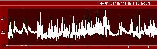
13 HEADACHE Tamburrini et al, Child s Nerv Syst, 2008 Presenting sign: up to 70% of symptomatic cases Chronic, unspecific, unrelated to cyst size, compression,distortion of surrounding cerebrovascular structures Unrelated to intracystic pressure BENEFIT OF ICP RECORDING CONTROVERSIAL
14 SEIZURES Tamburrini et al, Child s Nerv Syst, 2008 INTERNATIONAL SURVEY ON NEUROSURGICAL ATTITUDE RECOMMENDATIONS Indication for surgery: 37% Prophylactic surgery: 26.6% Prolonged clinical follow-up and seriated MR: 15.5%
15 SEIZURES Tamburrini et al, Child s Nerv Syst, 2008 Extensive documentation in literature on the uncertain correlation between surgical excision of the cyst and epilepsy control One fourth of the patients may have developmental cortical anomalies far from the cyst Rare concordance between cyst location and seizures semeiology Controlateral EEG anomalies common
16 PSYCHOMOTOR RETARDATION Tamburrini et al, Child s Nerv Syst, 2008 INTERNATIONAL SURVEY ON NEUROSURGICAL ATTITUDE RECOMMENDATIONS Indication for surgery: 31.1% Prophylactic surgery: 11.1% Prolonged clinical follow-up and seriated MR: 24.3%
17 PSYCHOMOTOR RETARDATION Tamburrini et al, Child s Nerv Syst, 2008 Limited amount of information concerning psychomotor evaluation in literature
18 PSYCHOMOTOR RETARDATION Tamburrini et al, Child s Nerv Syst, 2008 Most of the data indicating postoperative cognitive improvement from adults and from one single research group ( few selected neuropsychological tests it remains uncertain whether these laboratory test results reflect true clinical problem for the patient ) Anomalies associated to the reduced volume of the temporal lobe rather than to the volume of the cyst Language dominance preserved on the side of the cyst


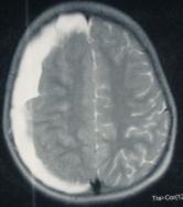
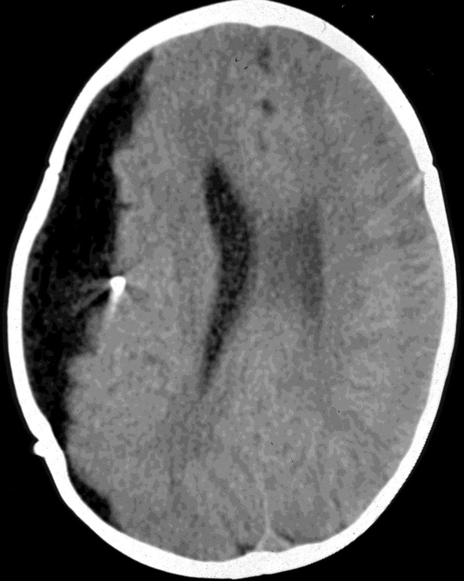
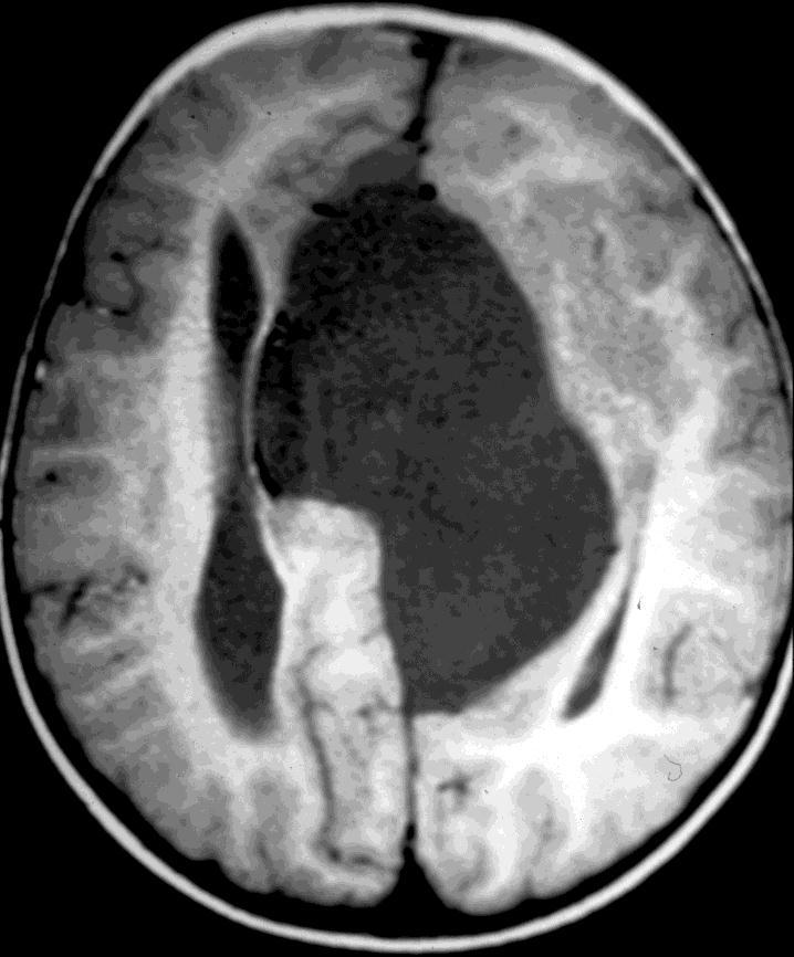
19 Is there a role for prophylactic surgery? 9 y old boy. Type II Cyst Prophylactic cyst excision PRE Immediate POST


")
 Fewel")

20 CAN IT AVOID/REDUCE THE RATE OF SUBDURAL HYGROMAS/ HEMATOMAS? Prophylactic surgical treatment of Sylvian arachnoid cysts is weighted by a twofold risk of subdural hygromas (5-7%) than the natural history of the condition (2-3%) Fewel et al., 1996, Parsch et al., 1997, Donaldson et al., 2000, Gelabert-Gonzalez et al, 2002, Tamburrini et al., 2003


21 Sylvian fissure cysts: SURGICAL INDICATION Hypometabolism Abnormally high ICP SPECT
22 Sylvian fissure cysts: Surgical options: Cystoperitoneal shunt In favor: 1. Easy and 2. Effective operation Against: 1.Shunt dependency 2.Plugging of the shunt by the cyst lining
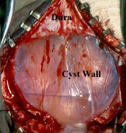
23 Sylvian fissure cysts: Surgical options: Craniotomy and cyst lining excision / marsupialization Against: 1.Severity of the operation 2.Frequent failures 3.Interference with CSF circulation

24 Sylvian fissure cysts:

25 Sylvian fissure cysts:
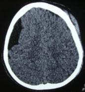
26 COMPLICATIONS Ext drain Preop Postop Shunt Post ext drain

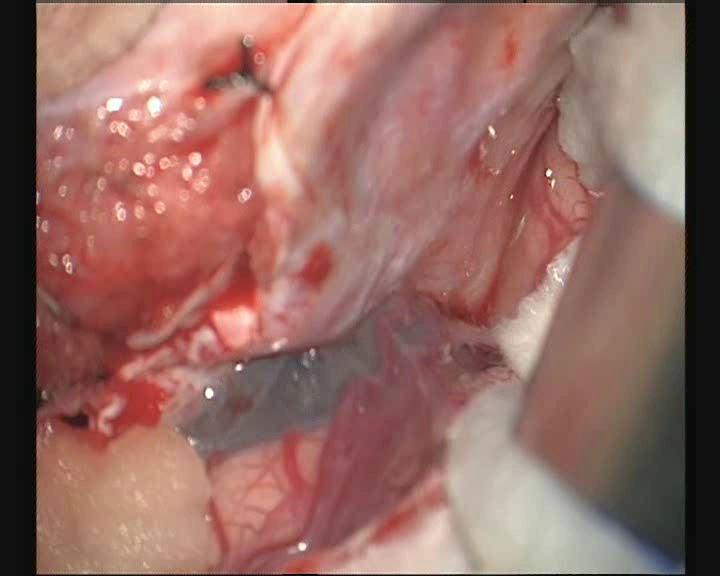
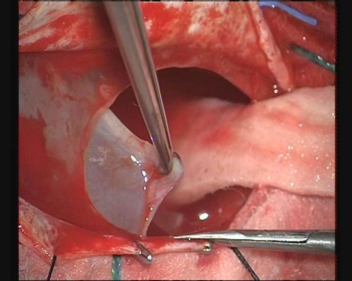
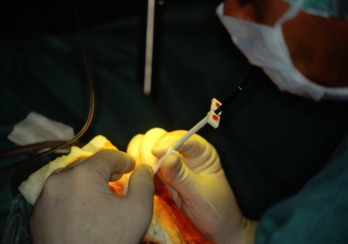
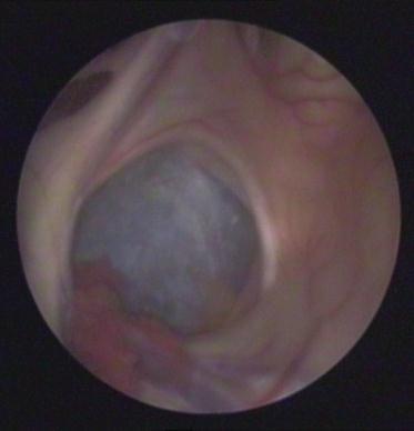
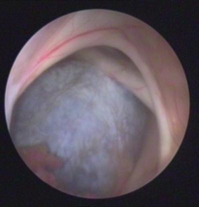
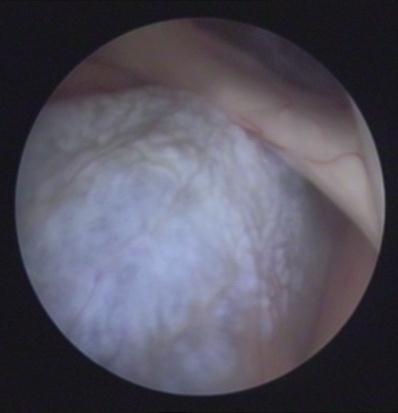
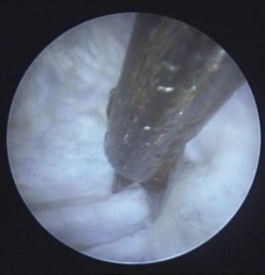
27 Sylvian fissure cysts: Endoscopic cyst fenestration


28 Sylvian fissure cysts: Endoscopic cyst fenestration Minicraniotomy and limited cyst membrane excision

29 Sylvian fissure cysts:

30 Sylvian fissure cysts:

31 Sylvian fissure cysts:

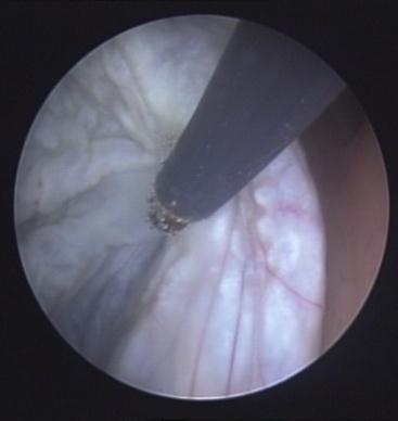
32 Sylvian fissure cysts: Post-endoscopic opening Pre

33 Sylvian fissure cysts:
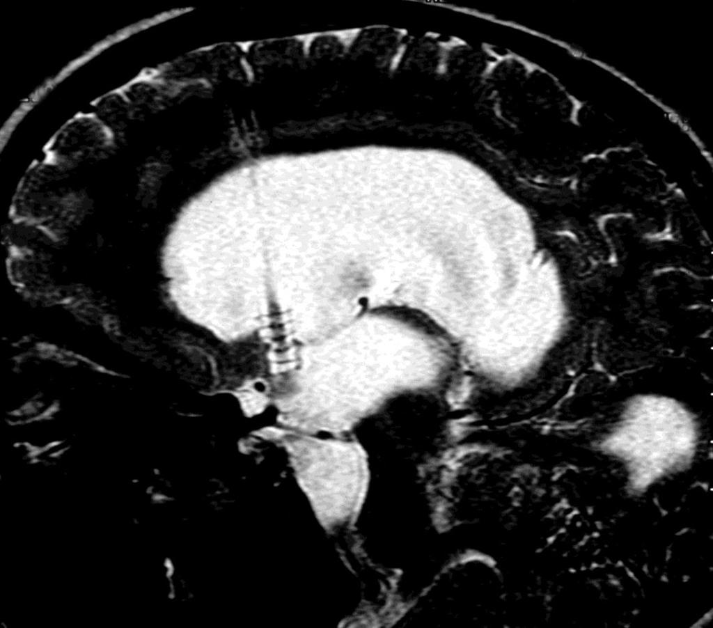
34 Sellar region cysts: Varieties: Intrasellar: typically in adults (mean age:42 yrs) Suprasellar: typically in children (78% < 1y.o in our series) Expand in all directions; Hydrocephalus common (obstructive or from CSF absorption)
")
35 Sellar region cysts: RISK VISUAL IMPAIRMENT ENDOCRINE DYSFUNCTION (Growth retardation, isosexual precocity)

36 Sellar region cysts:

37 Sellar region cysts:

38 Sellar region cysts:


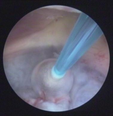
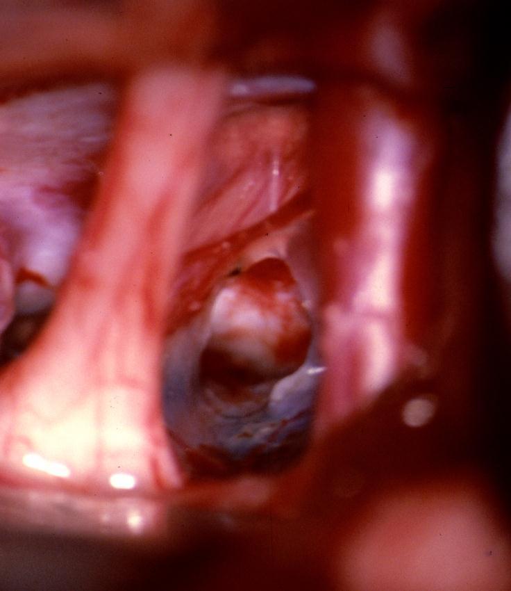
39 Sellar region cysts: ENDOSCOPIC APPROACH: cyst opening into the III ventricle and basal cisterns


40 Sellar region cysts: ENDOSCOPIC APPROACH: cyst opening into the III ventricle and basal cisterns
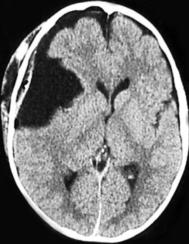
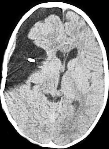
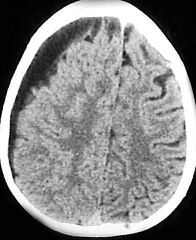
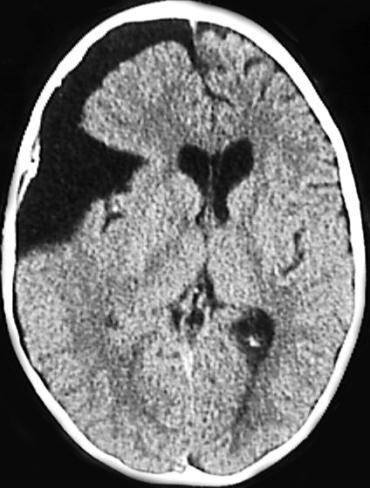
41 Cerebral convexity cysts: CHILDREN Localized skull bulging Cranial asymmetry without neurological deficits ADULTS Increased intracranial pressure Epilepsy Neurological deficits

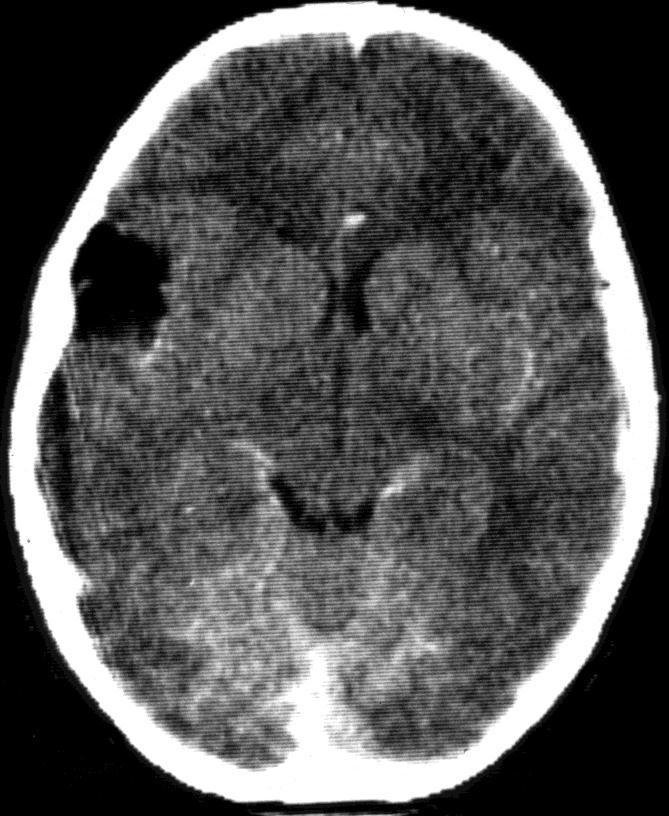
42 PRE PRE POST POST


43 Cerebral convexity cysts: Varieties Focal Treatment: Craniotomy and cyst wall excision Hemispheric Treatment: Shunting (?)

44 Cerebral convexity cysts:
45 PRE POST
46 Interhemispheric fissure cysts: Varieties Interhemispheric Parasagittal RISK Hydrocephalus Motor deficits TREATMENT Cyst membrane fenestration (endoscopy, craniotomy) Shunting
47 Interhemispheric fissure cysts:

48 Interhemispheric fissure cysts: Treatment: Endoscopy!


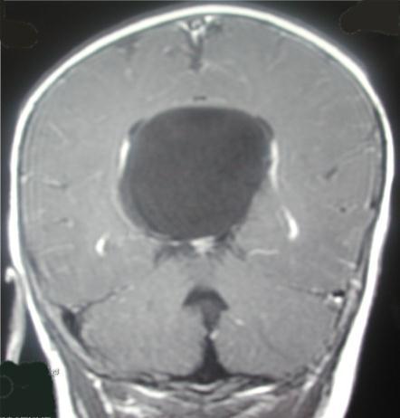
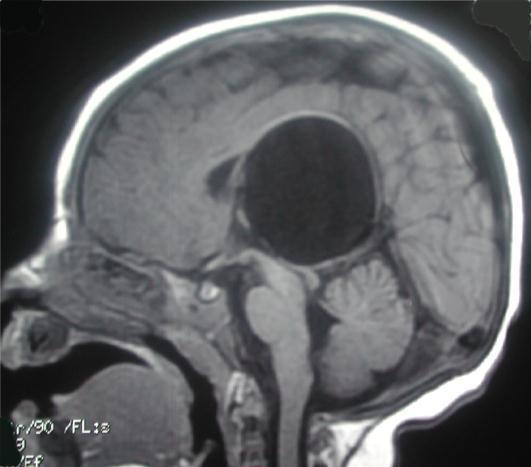
49 INTRA/PARAVENTRICULAR Quadrigeminal region / Tentorial notch Subependymal/ paraventricular cysts Choroid plexus cysts
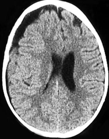
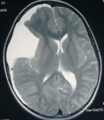
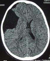
50 INTRA/PARAVENTRICULAR Clinical manifestations Symptoms of increased ICP due to obstructive hydrocephalus in most cases Focal signs (Parinaud, motor deficits) less frequent
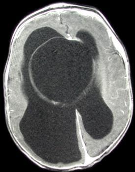
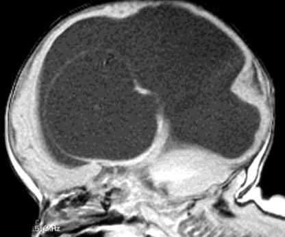

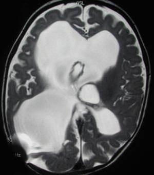
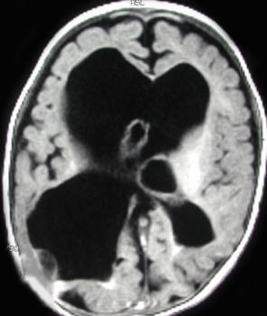
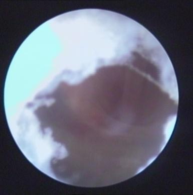
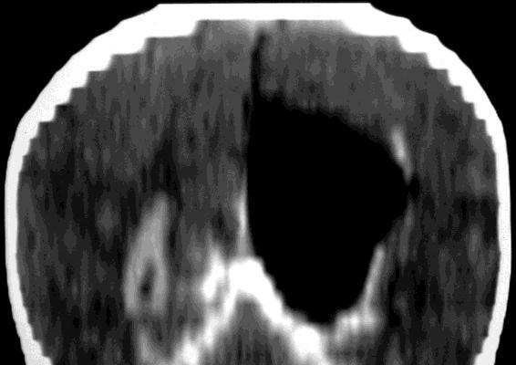
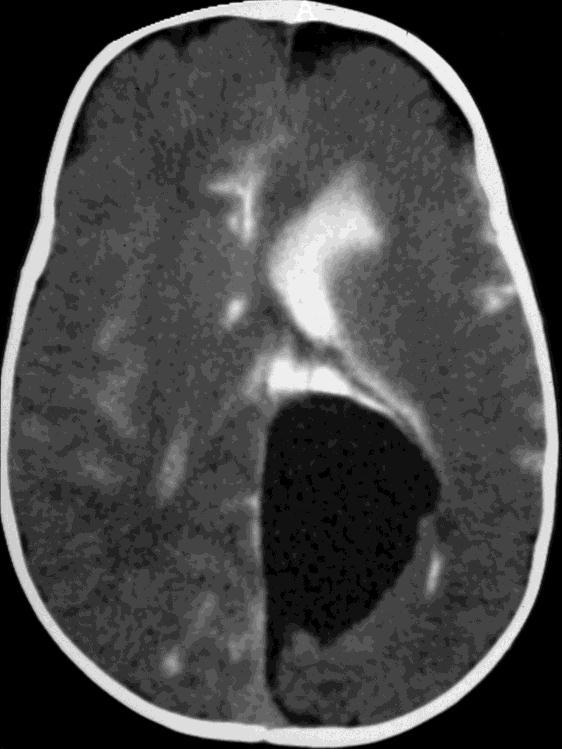
51 Intraventricular cysts treatment: endoscopy Postop
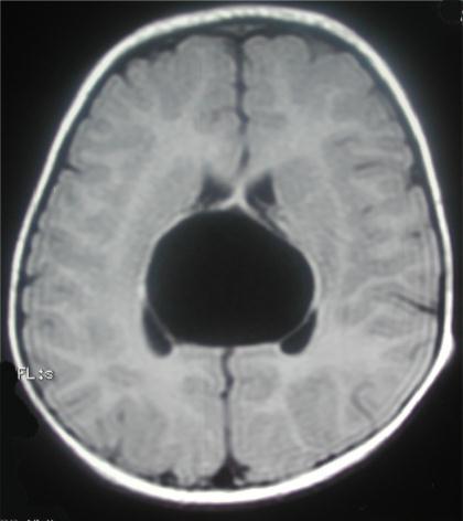
52 LIMITED VENTRICULAR ACCESS DOES NOT REPRESENT A CONTROINDICATION

53 LIMITED VENTRICULAR ACCESS DOES NOT REPRESENT A CONTROINDICATION
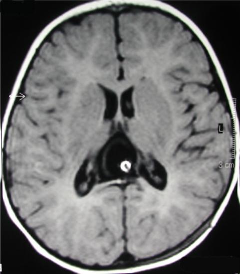

54 LIMITED VENTRICULAR ACCESS DOES NOT REPRESENT A CONTROINDICATION

55 INFRATENTORIAL Fastigium present Vermis normal Retrocerebellar: DD from DWC Scalloping of occipital bone
56 INFRATENTORIAL Laterocerebellar

57 INFRATENTORIAL Clinical symptoms Retrocerebellar Laterocerebellar Related to secondary Hy Cerebellar ataxia Cranial nerves Related to secondary Hy

58 INFRATENTORIAL Management options Retrocerebellar Laterocerebellar Endoscopic cysto-ventriculostomy whenever possible


59 INFRATENTORIAL Management options Retrocerebellar Cyst excision Laterocerebellar (open or endoscopic/endoscopic assisted)

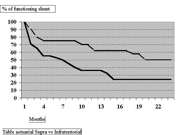
60 Avoid CP shunt Supratentorial Infratentorial
Intracranial arachnoid cysts: radiological study of the incidental, the symptomatic and the complicated.
 Intracranial arachnoid cysts: radiological study of the incidental, the symptomatic and the complicated. Poster No.: C-1092 Congress: ECR 2015 Type: Educational Exhibit Authors: C. Ospina Moreno, I. Montejo
Complex Hydrocephalus
 2012 Hydrocephalus Association Conference Washington, DC - June 27-July1, 2012 Complex Hydrocephalus Marion L. Walker, MD Professor of Neurosurgery & Pediatrics Primary Children s Medical Center University
Intracranial arachnoid cyst: an institutional experience
 136 Sharma et al Intracranial arachnoid cyst Intracranial arachnoid cyst: an institutional experience Mukesh Sharma, R.S. Mittal, Rajeev Bansal, Achal Sharma Department of Neurosurgery,S.M.S. Medical College
Suprasellar Arachnoid Cysts. Wan Tew SEOW FRACS Singapore
 Suprasellar Arachnoid Cysts Wan Tew SEOW FRACS Singapore Distribution Intracranial Arachnoid Cysts Sylvian fissure 49% CPA 11% Quadrigeminal 10% Vermian 9% Sellar and suprasellar 9% Interhemispheric 5%
Neuropathology Specialty Conference
 Neuropathology Specialty Conference March 22, 2010 Case 2 Rebecca Folkerth, MD Brigham and Women s Hospital Children s Hospital Harvard Medical School Clinical History 18-gestational-week fetus found on
Han-Sung Kwon M.D. Department of Obstetrics and Gynecology Konkuk University School of Medicine Seoul, Korea
 Han-Sung Kwon M.D. Department of Obstetrics and Gynecology Konkuk University School of Medicine Seoul, Korea Embryologic features of the developing hindbrain Embryologic features of the developing hindbrain
Surgery of posterior fossa arachnoid cyst causing hydrocephalus unmasking the cause of epilepsy
 CASE REPORTS ALBANIAN MEDICAL JOURNAL Surgery of posterior fossa arachnoid cyst causing hydrocephalus unmasking the cause of epilepsy Florian Dashi 1, Arben Rroji 1, Eugen Enesi 1, Suzana Gjeci 1, Ridvan
Head CT Scan Interpretation: A Five-Step Approach to Seeing Inside the Head Lawrence B. Stack, MD
 Head CT Scan Interpretation: A Five-Step Approach to Seeing Inside the Head Lawrence B. Stack, MD Five Step Approach 1. Adequate study 2. Bone windows 3. Ventricles 4. Quadrigeminal cistern 5. Parenchyma
CNS Embryology 5th Menstrual Week (Dorsal View)
") Imaging of the Fetal Brain; Normal & Abnormal Alfred Abuhamad, M.D. Eastern Virginia Medical School CNS Embryology 5th Menstrual Week (Dorsal View) Day 20 from fertilization Neural plate formed in ectoderm
Imaging of the Fetal Brain; Normal & Abnormal Alfred Abuhamad, M.D. Eastern Virginia Medical School CNS Embryology 5th Menstrual Week (Dorsal View) Day 20 from fertilization Neural plate formed in ectoderm
Update on Pediatric Brain Tumors
 Update on Pediatric Brain Tumors David I. Sandberg, M.D. Director of Pediatric Neurosurgery & Associate Professor Dr. Marnie Rose Professorship in Pediatric Neurosurgery Pre-talk Questions for Audience
Symposium: OB/GY US (Room B) CNS Anomalies
 CNS Anomalies") 82 Symposium: OB/GY US (Room B) 11 : 50 1 2 : 10 CNS Anomalies Brain area Midline structure S u p r a t e n t o r i a l ventricular system Cerebral hemisphere Posterior fossa Head size and shape Image
82 Symposium: OB/GY US (Room B) 11 : 50 1 2 : 10 CNS Anomalies Brain area Midline structure S u p r a t e n t o r i a l ventricular system Cerebral hemisphere Posterior fossa Head size and shape Image
Surgical management of the fourth ventricle arachnoid cysts
 Annals of Medical Research DOI: 10.5455/annalsmedres.2018.09.199 2019;26(1):42-6 Original Article Surgical management of the fourth ventricle arachnoid cysts Sukru Oral 1, Hanifi Bayarogullari 2, Atilla
For Emergency Doctors. Dr Suzanne Smallbane November 2011
 For Emergency Doctors Dr Suzanne Smallbane November 2011 A: Orbit B: Sphenoid Sinus C: Temporal Lobe D: EAC E: Mastoid air cells F: Cerebellar hemisphere A: Frontal lobe B: Frontal bone C: Dorsum sellae
The clinical spectrum of Blake s pouch cyst: report of six illustrative cases
 Childs Nerv Syst (2010) 26:1057 1064 DOI 10.1007/s00381-010-1085-2 ORIGINAL PAPER The clinical spectrum of Blake s pouch cyst: report of six illustrative cases Erwin M. J. Cornips & Geke M. Overvliet &
NEURO IMAGING 2. Dr. Said Huwaijah Chairman of radiology Dep, Damascus Univercity
 NEURO IMAGING 2 Dr. Said Huwaijah Chairman of radiology Dep, Damascus Univercity I. EPIDURAL HEMATOMA (EDH) LOCATION Seventy to seventy-five percent occur in temporoparietal region. CAUSE Most likely caused
Original Article CT grouping and microsurgical treatment strategies of hypertensive cerebellar hemorrhage
 Int J Clin Exp Med 2016;9(8):15921-15927 www.ijcem.com /ISSN:1940-5901/IJCEM0022273 Original Article CT grouping and microsurgical treatment strategies of hypertensive cerebellar hemorrhage Xielin Tang
intracranial anomalies
 Chapter 5: Fetal Central Nervous System 84 intracranial anomalies Hydrocephaly Dilatation of ventricular system secondary to an increase in the amount of CSF. Effects of hydrocephalus include flattening
Posterior fossa malformations
 ANDREA ROSSI, MD Head, Department of Pediatric Neuroradiology G. Gaslini Children s Research Hospital Genoa Italy andrearossi@ospedale-gaslini.ge.it Posterior fossa malformations Cerebellar ataxia Hypotonia
PHYSIOLOGY OF CSF AND PATHOPHYSIOLOGY OF HYDROCEPHALUS
 PHYSIOLOGY OF CSF AND PATHOPHYSIOLOGY OF HYDROCEPHALUS Introduction Dynamic component of CNS Invaluable tool to diagnosis Physiological reservoir of human proteome Reflects the physiologic state of CNS
Arachnoid cysts account for 1% of all intracranial. Endoscopic treatment of intraparenchymal arachnoid cysts in children.
 J Neurosurg Pediatrics 14:501 507, 2014 AANS, 2014 Endoscopic treatment of intraparenchymal arachnoid cysts in children Clinical article Nasser M. F. El-Ghandour, M.D. Department of Neurosurgery, Faculty
The "Keyhole": A Sign of
 473 The "Keyhole": A Sign of Herniation of a Trapped Fourth Ventricle and Other Posterior Fossa Cysts Barbara J. Wolfson' Eric N. Faerber' Raymond C. Truex, Jr. 2 When a cystic structure in the posterior
Neurosonography: State of the art
 Neurosonography: State of the art Lisa H Lowe, MD, FAAP Professor and Academic Chair, University MO-Kansas City Pediatric Radiologist, Children s Mercy Hospitals and Clinics Learning objectives After this
SURGICAL MANAGEMENT OF BRAIN TUMORS
 SURGICAL MANAGEMENT OF BRAIN TUMORS LIGIA TATARANU, MD, Ph D NEUROSURGICAL CLINIC, BAGDASAR ARSENI CLINICAL HOSPITAL BUCHAREST, ROMANIA SURGICAL INDICATIONS CONFIRMING HISTOLOGIC DIAGNOSIS REDUCING TUMOR
Communicating Hydrocephalus Accompanied by Arachnoid Cyst in Aneurismal Subarachnoid Hemorrhage
 Journal of Cerebrovascular and Endovascular Neurosurgery ISSN 2234-8565, EISSN 2287-3139, http://dx.doi.org/10.7461/jcen.2013.15.4.311 Case Report Communicating Hydrocephalus Accompanied by Arachnoid Cyst
EANS Training Course Moscow, 5 th 8 th May 2019 Tumours
 EANS Training Course Moscow, 5 th 8 th May 2019 Tumours SUNDAY, MAY 5 th, 2019 09:00 09:10 Welcome 09:15 10:30 Tumours Chair: Meling 09:15 09:30 Molecular biology of brain tumours Reinert 09:30 09:45 Imaging
Chapter 3. Neonatal cranial ultrasonography: how to optimize its performance
 Chapter 3 Neonatal cranial ultrasonography: how to optimize its performance Sylke J. Steggerda Lara M. Leijser Frans J. Walther Gerda van Wezel-Meijler Early Human Development 2009; 85(2): 93-99 Chapter
Spontaneous Intracystic Haemorrhage of an Arachnoid Cyst Associated with a Subacute Subdural Haematoma: A Case Report and Literature Review
 DOI: 10.5137/1019-5149.JTN.20885-17.2 Received: 20.06.2017 / Accepted: 20.09.2017 Published Online: 31.10.2017 Case Report Spontaneous Intracystic Haemorrhage of an Arachnoid Cyst Associated with a Subacute
Brain Meninges, Ventricles and CSF
 Brain Meninges, Ventricles and CSF Lecture Objectives Describe the arrangement of the meninges and their relationship to brain and spinal cord. Explain the occurrence of epidural, subdural and subarachnoid
Dandy-Walker syndrome: different modalities of treatment and outcome in 42 cases
 Child s Nerv Syst (2001) 17:348 352 DOI 10.1007/s003810000425 ORIGINAL PAPER Raj Kumar Manoj Kumar Jain Devendra Kumar Chhabra Dandy-Walker syndrome: different modalities of treatment and outcome in 42
Prenatal Prediction of The Neurologically Impaired Neonate By Ultrasound
 Prenatal Prediction of The Neurologically Impaired Neonate By Ultrasound Robert H. Debbs, D.O.,F.A.C.O.O.G. Professor of OB-GYN Perelman School of Medicine, University of Pennsylvania Director, Pennsylvania
Cavum velum interpositum cyst causing symptomatic trapped ventricle: A case report
 Cavum velum interpositum cyst causing symptomatic trapped ventricle: A case report Poster No: R-0286 Congress: 2014 CSM Type: Scientific Exhibit Authors: T Singh, S Dupre; NAMBOUR/AU Keywords: CNS, Neuroradiology
Neurosurgery. Neurosurgery
 Neurosurgery Neurosurgery Neurosurgery Telephone Numbers: Appointment: 202-476-3020 Fax: 202-476-3091 Administration: 202-476-3020 Evenings and Weekends: 202-476-5000 Robert Keating, MD, Chief The Division
A STUDY OF POSTERIOR FOSSA MALFORMATIONS: MR IMAGING Ravi Ningappa 1, Vaishali D. M 2, Vijayaraghavachari T. V 3, Manjappa B. H 4
 A STUDY OF POSTERIOR FOSSA MALFORMATIONS: MR IMAGING Ravi Ningappa 1, Vaishali D. M 2, Vijayaraghavachari T. V 3, Manjappa B. H 4 HOW TO CITE THIS ARTICLE: Ravi Ningappa, Vaishali D. M, Vijayaraghavachari
Applicable Neuroradiology
 For the Clinical Neurology Clerkship LSU Medical School New Orleans Amy W Voigt, MD Clerkship Director Introduction The field of Radiology first developed following the discovery of X-Rays by Wilhelm Roentgen
Original Article Analysis on clinical characteristics of intracranial Arachnoid Cysts in 488 pediatric cases
 Int J Clin Exp Med 2015;8(10):18343-18350 www.ijcem.com /ISSN:1940-5901/IJCEM0012149 Original Article Analysis on clinical characteristics of intracranial Arachnoid Cysts in 488 pediatric cases Jian-Huang
Index. aneurysm, 92 carotid occlusion, 94 ICA stenosis, 95 intracranial, 92 MCA, 94
 A ADC. See Apparent diffusion coefficient (ADC) Aneurysm cerebral artery aneurysm, 93 CT scan, 93 gadolinium, 93 Angiography, 13 Anoxic brain injury, 25 Apparent diffusion coefficient (ADC), 7 Arachnoid
SOP: Cerebral Ultrasound
 SOP: Cerebral Ultrasound Version Author(s) Date Changes Approved by 1.0 Cornelia Hagmann Manon Benders 29.5.2012 Initial Version Gorm Greisen 1.1 Cornelia Hagmann 18.6.2012 Minor changes Gorm Greisen 1.2
Supra- and infratentorial brain tumors from childhood to maternity
 Supra- and infratentorial brain tumors from childhood to maternity What to expect? I am going to show you the characteristic imaging findings of following tumors: Thierry A.G.M. Huisman, MD, FICIS, EQNR
GEORGE E. PERRET, M.D., AND CARL J. GRAF, M.D.
 J Neurosurg 47:590-595, 1977 Subgaleal shunt for temporary ventricle decompression and subdural drainage GEORGE E. PERRET, M.D., AND CARL J. GRAF, M.D. Division of Neurological Surgery, University of Iowa
Differential diagnosis of intracranial cystic lesions.
 Differential diagnosis of intracranial cystic lesions. Poster No.: C-0215 Congress: ECR 2015 Type: Educational Exhibit Authors: S. P. G. Alandete, M. A. Meseguer, E. De la Via, D. Uceda, C. Poyatos; Valencia/ES
Measurements of the Posterior Fossa in Normal Fetus MRI
 Measurements of the Posterior Fossa in Normal Fetus MRI Ber Roee, 3 rd year medical student, Sackler School of Medicine, Tel Aviv University Supervised by: Dr. Katorza Eldad, Antenatal Diagnostic Unit,The
Meninges and Ventricles
 Meninges and Ventricles Irene Yu, class of 2019 LEARNING OBJECTIVES Describe the meningeal layers, the dural infolds, and the spaces they create. Name the contents of the subarachnoid space. Describe the
Intraventricular neoplasms are uncommon, Intraventricular Tumor: An. Analysis of 18 Cases Dhaka, Bangladesh
 Original Article Nepal Journal of Neuroscience 13:23-29, 2016 Shamsul Alam, MS Intraventricular Tumor: An Analysis of 18 Cases Abu N W Uddin, MS Mashiur R Majumder, MS Comilla Medical College Comilla,
Marc Norman, Ph.D. - Do Not Use without Permission 1. Cerebrovascular Accidents. Marc Norman, Ph.D. Department of Psychiatry
 Cerebrovascular Accidents Marc Norman, Ph.D. Department of Psychiatry Neuropsychiatry and Behavioral Medicine Neuropsychology Clinical Training Seminar 1 5 http://www.nlm.nih.gov/medlineplus/ency/images/ency/fullsize/18009.jpg
Enhancement of Cranial US: Utility of Supplementary Acoustic Windows and Doppler Harriet J. Paltiel, MD
 Enhancement of Cranial US: Utility of Supplementary Acoustic Windows and Doppler Harriet J. Paltiel, MD Boston Children s Hospital Harvard Medical School None Disclosures Conventional US Anterior fontanelle
Fetal Medicine. Case Presentations. Dr Ermos Nicolaou Fetal Medicine Unit Chris Hani Baragwanath Hospital. October 2003
 Case Presentations Dr Ermos Nicolaou Fetal Medicine Unit Chris Hani Baragwanath Hospital October 2003 Case 1 Ms A M 22year old P0 G1 Referred from Sebokeng Hospital at 36w for polyhydramnios On Ultrasound:
Computed Tomographic Evaluation of Posterior Fossa Lesions
 ORIGINAL ARTICLE Computed Tomographic Evaluation of Posterior Fossa Lesions Gupta MK 1, Rauniyar RK 1, Bhatta N 2, Raja S 3, Ahmad K 1 1 Department of Radiodiagnosis & Imaging, 2 Department of Internal
 www.yassermetwally.com MANAGEMENT OF CEREBRAL HAEMORRHAGE (ICH): A QUICK GUIDE Overview 10% of strokes is caused by ICH. Main Causes: Less than 40 years old: vascular malformations and illicit drug use.
Surgical decompression of arachnoid cysts: a study on 44 pediatric patients
 264 Tascu, Florea Surgical decompression of arachnoid cysts Surgical decompression of arachnoid cysts: a study on 44 pediatric patients A. Tascu 1, Simona Mihaela Florea 2 1 Assist. Prof. at the Universtity
UPSTATE Comprehensive Stroke Center. Neurosurgical Interventions Satish Krishnamurthy MD, MCh
 UPSTATE Comprehensive Stroke Center Neurosurgical Interventions Satish Krishnamurthy MD, MCh Regional cerebral blood flow is important Some essential facts Neurons are obligatory glucose users Under anerobic
Intracranian arahnoid cysts in children (ACs)
") 68 A.V. Ciurea et al Intracranian arahnoid cysts in children (ACs) Intracranian arahnoid cysts in children (ACs) A.V. Ciurea 1, A. Tascu 1, A. Iliescu 1, C. Mihalache 2, F. Brehar 1, C. Palade 1, A. Spatariu
68 A.V. Ciurea et al Intracranian arahnoid cysts in children (ACs) Intracranian arahnoid cysts in children (ACs) A.V. Ciurea 1, A. Tascu 1, A. Iliescu 1, C. Mihalache 2, F. Brehar 1, C. Palade 1, A. Spatariu
Residence of Discipline of Neurosurgery of Hospital da Santa Casa de Misericórdia of Sao Paulo Sao Paulo, Brazil
 Cronicon OPEN ACCESS NEUROLOGY Research Article Efficacy of the Lamina Terminalis Fenestration Associated With the Liliequist Membrane Fenestration in Reducing Shunt-Dependent Hydrocephalus Following Aneurysm
S Alandete, M Meseguer, CR Poyatos, D Uceda, E de la Via, J Sales, J Vilar. H.U. Dr Peset, Valencia (Spain)
") S Alandete, M Meseguer, CR Poyatos, D Uceda, E de la Via, J Sales, J Vilar. H.U. Dr Peset, Valencia (Spain) Introduction Cystic lesions are usually a common finding in clinical practice and you can find
S Alandete, M Meseguer, CR Poyatos, D Uceda, E de la Via, J Sales, J Vilar. H.U. Dr Peset, Valencia (Spain) Introduction Cystic lesions are usually a common finding in clinical practice and you can find
Ventricles, CSF & Meninges. Steven McLoon Department of Neuroscience University of Minnesota
 Ventricles, CSF & Meninges Steven McLoon Department of Neuroscience University of Minnesota 1 Coffee Hour Thursday (Sept 14) 8:30-9:30am Surdyk s Café in Northrop Auditorium Stop by for a minute or an
Vascular Malformations of the Brain: A Review of Imaging Features and Risks
 Vascular Malformations of the Brain: A Review of Imaging Features and Risks Comprehensive Neuroradiology: Best Practices October 27-30, 2016 Sudhakar R. Satti, MD Associate Director Neurointerventional
Quick practical guide to Cranial Ultrasound in the newborn
 Quick practical guide to Cranial Ultrasound in the newborn Introduction A standard set of views is taken to assist with consistent visualisation of structures and in the interpretation of possible abnormalities.
CISTERNOGRAPHY (CEREBRO SPINAL FLUID IMAGING): A VERSATILE DIAGNOSTIC PROCE DURE
: A VERSATILE DIAGNOSTIC PROCE DURE") VOL. 115, No. i E D I T 0 R I A L CISTERNOGRAPHY (CEREBRO SPINAL FLUID IMAGING): A VERSATILE DIAGNOSTIC PROCE DURE C ISTERNOGRAPHY (CSF imaging) is a diagnostic study based on the premise that certain
VOL. 115, No. i E D I T 0 R I A L CISTERNOGRAPHY (CEREBRO SPINAL FLUID IMAGING): A VERSATILE DIAGNOSTIC PROCE DURE C ISTERNOGRAPHY (CSF imaging) is a diagnostic study based on the premise that certain
What Are We Going to Do? Fourth Year Meds Clinical Neuroanatomy. Hydrocephalus and Effects of Interruption of CSF Flow. Tube Blockage Doctrine
 Fourth Year Meds Clinical Neuroanatomy Ventricles, CSF, Brain Swelling etc. David A. Ramsay, Neuropathologist, LHSC What Are We Going to Do? Hydrocephalus and some effects of the interruption of CSF flow
Case Report Multiple Intracranial Meningiomas: A Review of the Literature and a Case Report
 Case Reports in Surgery Volume 2013, Article ID 131962, 4 pages http://dx.doi.org/10.1155/2013/131962 Case Report Multiple Intracranial Meningiomas: A Review of the Literature and a Case Report F. Koech,
Ventriculostomy and Risk of Upward Herniation in Patients with Obstructive Hydrocephalus from Posterior Fossa Mass Lesions
 https://doi.org/10.1007/s12028-017-0487-3 ORIGINAL ARTICLE Ventriculostomy and Risk of Upward Herniation in Patients with Obstructive Hydrocephalus from Posterior Fossa Mass Lesions Sherri A. Braksick
Rare case of multiple meningiomas in nonneurofibromatosis
 Romanian Neurosurgery Volume XXXI Number 2 2017 April-June Article Rare case of multiple meningiomas in nonneurofibromatosis patient at unusual locations Vikrant Setia, Deepashu Sachdeva, Shrinivas Odugoudar,
Cerebro-vascular stroke
 Cerebro-vascular stroke CT Terminology Hypodense lesion = lesion of lower density than the normal brain tissue Hyperdense lesion = lesion of higher density than normal brain tissue Isodense lesion = lesion
Transfontanelar Ultrasound Technique, Normal Anatomy, Anatomic Variants and Classification Review
 Transfontanelar Ultrasound Technique, Normal Anatomy, Anatomic Variants and Classification Review Poster No.: C-2615 Congress: ECR 2013 Type: Educational Exhibit Authors: S. E. Vazquez, R. E. Ochoa Albíztegui
Aria Fallah MD, MSc, FRCSC
 Aria Fallah MD, MSc, FRCSC Department of Neurosurgery David Geffen School of Medicine at UCLA Pineal Region Tumors Brain Tumor Symposium August 22, 2015 Disclosures None Pineal Gland Arises from an invagination
2/20/2019 BRAIN DISSECTION CODING AND DOCUMENTATION OBJECTIVES INTRODUCTION
 BRAIN DISSECTION CODING AND DOCUMENTATION Diana R. Phelps, CPC, CPC-I, CEMC OBJECTIVES Identify general structure of the human brain Describe how the different parts work Recognized the two hemispheres
CNS pathology Third year medical students. Dr Heyam Awad 2018 Lecture 5: disturbed fluid balance and increased intracranial pressure
 CNS pathology Third year medical students Dr Heyam Awad 2018 Lecture 5: disturbed fluid balance and increased intracranial pressure ILOs Understand causes and symptoms of increased intracranial pressure.
SWISS SOCIETY OF NEONATOLOGY. Severe apnea and bradycardia in a term infant
 SWISS SOCIETY OF NEONATOLOGY Severe apnea and bradycardia in a term infant October 2014 2 Walker JH, Arlettaz Mieth R, Däster C, Division of Neonatology, University Hospital Zurich, Switzerland Swiss Society
Recurrent Subdural Hematomas in Benign Macrocrania of Infancy
 Case Report imedpub Journals http://www.imedpub.com Medical & Clinical DOI: 10.21767/2471-299X.100032 Recurrent Subdural Hematomas in Benign Macrocrania of Infancy Ademar Lucas Junior São Camilo Diagnostic
REVIEW ARTICLE Egypt. J. Hum. Genet. Vol. 8, No. 2, Nov Dandy-Walker Malformation
 REVIEW ARTICLE Egypt. J. Hum. Genet. Vol. 8, No. 2, Nov. 2007 Medical Genetics Center, Ain Shams University INTRODUCTION Dandy-Walker malformation is a rare congenital malformation and involves the cerebellum
Intracranial air on computerized tomography ANNE G. OSBORN, M.D., JONATHAN H. DAINES, M.D., S. DOUGLAS WING, M.D., AND ROBERT E. ANDERSON, M.D.
 J Neurosurg 48:355-359, 1978 Intracranial air on computerized tomography ANNE G. OSBORN, M.D., JONATHAN H. DAINES, M.D., S. DOUGLAS WING, M.D., AND ROBERT E. ANDERSON, M.D. Department of Radiology, University
NEURORADIOLOGY DIL part 3
 NEURORADIOLOGY DIL part 3 Bleeds and hemorrhages K. Agyem MD, G. Hall MD, D. Palathinkal MD, Alexandre Menard March/April 2015 OVERVIEW Introduction to Neuroimaging - DIL part 1 Basic Brain Anatomy - DIL
Department of Neurosurgery, First Hospital of Jilin University, Changchun , China 2
 Case Reports in Medicine Volume 2010, Article ID 634839, 7 pages doi:10.1155/2010/634839 Case Report Reoperation as a Result of Raised Intracranial Pressure Associated with Cyst Formation in Tumor Cavity
Intra-Fourth Ventricular Schwannoma With Obstructive Hydrocephalus A Rare Case Report
 ISPUB.COM The Internet Journal of Neurosurgery Volume 7 Number 1 Intra-Fourth Ventricular Schwannoma With Obstructive Hydrocephalus A Rare Case Report A Babbu, R Katheerayson Citation A Babbu, R Katheerayson..
Stroke - Intracranial hemorrhage. Dr. Amitesh Aggarwal Associate Professor Department of Medicine
 Stroke - Intracranial hemorrhage Dr. Amitesh Aggarwal Associate Professor Department of Medicine Etiology and pathogenesis ICH accounts for ~10% of all strokes 30 day mortality - 35 45% Incidence rates
Neuroendoscopic management of interhemispheric cysts in children
 J Neurosurg (3 Suppl Pediatrics) 105:194 202, 2006 Neuroendoscopic management of interhemispheric cysts in children GIUSEPPE CINALLI, M.D., PAOLA PERETTA, M.D., PIETRO SPENNATO, M.D., LUCIANO SAVARESE,
Introduction to Neurosurgical Subspecialties:
 Introduction to Neurosurgical Subspecialties: Pediatric Neurosurgery Brian L. Hoh, MD 1 and Gregory J. Zipfel, MD 2 1 University of Florida, 2 Washington University Pediatric Neurosurgery Pediatric neurosurgeons
5/22/2009. Pediatric Neurosurgery Pediatric Neurology Neuroradiology Neurophysiology Neuropathology Neuropsychology
 Current Surgical Treatment Strategies for the Management of Pediatric Epilepsy University of California, San Francisco Department of Neurological Surgery San Francisco, California Kurtis Ian Auguste, M.D.
Brain Imaging. Bearbeitet von Klaus Sartor, Stefan Hähnel, Bodo Kress
 Brain Imaging Bearbeitet von Klaus Sartor, Stefan Hähnel, Bodo Kress 1. Auflage 2007. Taschenbuch. 312 S. Paperback ISBN 978 3 13 143961 1 Format (B x L): 12,5 x 19 cm Weitere Fachgebiete > Medizin > Sonstige
Anatomy, Terminology and Treatment in Pediatric Neurosurgery Part I
 Anatomy, Terminology and Treatment in Pediatric Neurosurgery Part I John Ragheb, MD, FACS, FAAP Professor of Neurosurgery and Pediatrics, Affiliated Faculty of University of Miami, Miller School of Medicine
Multicompartmental congenital intracranial immature teratoma
 Neurology Asia 2013; 18(1) : 117 121 Multicompartmental congenital intracranial immature teratoma 1 Dharmendra Ganesan MS FRCS(SN), 1 Sheau Fung Sia MS MRCS, 1 Vairavan Narayanan MS, 2 Gnana Kumar FRCR,
The arrest of treated hydrocephalus in children
 J Neurosurg 61:752-756, 1984 The arrest of treated hydrocephalus in children A radionuclide study IAN H. JOHNSTON, F.R.C.S., ROBERT HOWMAN-GILES, F.R.A.C.P., AND IAN R. WHITTLE, M.B., B.S. T. Y. Nelson
CNS pathology Third year medical students,2019. Dr Heyam Awad Lecture 2: Disturbed fluid balance and increased intracranial pressure
 CNS pathology Third year medical students,2019 Dr Heyam Awad Lecture 2: Disturbed fluid balance and increased intracranial pressure ILOs Understand causes and symptoms of increased intracranial pressure.
Basic Training. ISUOG Basic Training Distinguishing Between Normal & Abnormal Appearances of the Skull & Brain
 ISUOG Distinguishing Between Normal & Abnormal Appearances of the Skull & Brain Learning objectives At the end of the lecture you will be able to: Describe how to obtain the 3 planes required to assess,
Dural Arteriovenous Malformations and Fistulae (DAVM S DAVF S)
") Jorge Guedes Campos NEUROIMAGING DEPARTMENT HOSPITAL SANTA MARIA UNIVERSITY OF LISBON PORTUGAL DEFINITION region of arteriovenous shunting confined to a leaflet of packymeninges often adjacent to a major
Jorge Guedes Campos NEUROIMAGING DEPARTMENT HOSPITAL SANTA MARIA UNIVERSITY OF LISBON PORTUGAL DEFINITION region of arteriovenous shunting confined to a leaflet of packymeninges often adjacent to a major
PRENATAL DIAGNOSIS OF ARACHNOID CYSTS
 REVIEW ARTICLE PRENATAL DIAGNOSIS OF ARACHNOID CYSTS Chih-Ping Chen* Department of Obstetrics and Gynecology, Mackay Memorial Hospital, Taipei, and Department of Biotechnology, Asia University, Taichung,
Acute stroke. Ischaemic stroke. Characteristics. Temporal classification. Clinical features. Interpretation of Emergency Head CT
 Ischaemic stroke Characteristics Stroke is the third most common cause of death in the UK, and the leading cause of disability. 80% of strokes are ischaemic Large vessel occlusive atheromatous disease
EEG IN FOCAL ENCEPHALOPATHIES: CEREBROVASCULAR DISEASE, NEOPLASMS, AND INFECTIONS
 246 Figure 8.7: FIRDA. The patient has a history of nonspecific cognitive decline and multiple small WM changes on imaging. oligodendrocytic tumors of the cerebral hemispheres (11,12). Electroencephalogram
ECMUS The Safety Committee of EFSUMB : Tutorial
 Neonatal cranial ultrasound Safety Aspects (2013) Prepared for ECMUS by B.J. van der Knoop, M.D. 1, J.I.P. de Vries, M.D., PhD 1, I.A. Zonnenberg, M.D. 2, J.I.M.L. Verbeke, M.D. 3 R.J. Vermeulen, M.D.,
CSF Leaks. Abnormal communication between the subarachnoid space and the tympanomastoid space or nasal cavity. Presenting symptoms:
 CSF Leaks Steven Wright, M.D. Faculty Advisor: Matthew Ryan, M.D. The University of Texas Medical Branch Department of Otolaryngology Grand Rounds Presentation January 5, 2005 CSF Leaks Abnormal communication
Temporal Lobe Cystic Collection and Associated Oedema: A Rare Complication of Translabyrinthine Resection of Vestibular Schwannoma
 Open Access Case Report DOI: 10.7759/cureus.2217 Temporal Lobe Cystic Collection and Associated Oedema: A Rare Complication of Translabyrinthine Resection of Vestibular Schwannoma Abdurrahman Raeiq 1 1.
The optimal treatment for arachnoid cysts is controversial. Cyst-ventricle stent as primary or salvage treatment for posterior fossa arachnoid cysts
 J Neurosurg Pediatrics 7:000 000, 7:549 556, 2011 Cyst-ventricle stent as primary or salvage treatment for posterior fossa arachnoid cysts Clinical article Daniel H. Fulkerson, M.D., 1 Todd D. Vogel, M.D.,
Small and Big Operations: New Tools of the Trade for Brain Tumors. Disclosure. Incidence of Childhood Cancer
 Small and Big Operations: New Tools of the Trade for Brain Tumors Nalin Gupta MD PhD Chief, Division of Pediatric Neurosurgery Departments of Neurosurgery and Pediatrics University of California San Francisco
HYDROCEPHALUS OF THE INFANT (ABOUT 86 CASES)
") HYDROCEPHALUS OF THE INFANT (ABOUT 86 CASES) K.EL KHOU;R.ANDALOUSSI;L.OUZIDANE Pediatric radiology department-chu Ibn Rochd Casablanca-Morroco Morroco. Introduction Hydrocephalus of infant is a progressive
HYDROCEPHALUS OF THE INFANT (ABOUT 86 CASES) K.EL KHOU;R.ANDALOUSSI;L.OUZIDANE Pediatric radiology department-chu Ibn Rochd Casablanca-Morroco Morroco. Introduction Hydrocephalus of infant is a progressive
Neuroendoscopy is widely accepted as a safe and effective
 » This article has been updated from its originally published version to correct the omission of an author. See the corresponding erratum notice in this issue, p 1128. «clinical article J Neurosurg 124:1047
ISUOG Basic Training. Distinguishing Between Normal & Abnormal Appearances of the Skull & Brain. Seshadri Suresh, India
 ISUOG Basic Training Distinguishing Between Normal & Abnormal Appearances of the Skull & Brain Seshadri Suresh, India Learning objectives 4 & 5 At the end of the lecture you will be able to: Describe how
Traumatic brain Injury- An open eye approach
 Traumatic brain Injury- An open eye approach Dr. Sunit Dr Sunit, Apollo children's hospital Blah blah Lots of head injury Lot of ill children Various methods of injury Various mechanisms of brain damage
Hemimegalencephaly without seizures: report of a case and review of literature
 Romanian Neurosurgery Volume XXXI Number 3 2017 July-September Article Hemimegalencephaly without seizures: report of a case and review of literature Agrawal Atul, Dutta Gautam, Singh Daljit, Sachdeva
Appendix 3.5 Case Inclusion Guidance for Potentially Zika-related Birth Defects
 Appendix 3.5 Case Inclusion Guidance for Potentially Zika-related Birth Defects Appendix 3.5 A3.5-1 Case Definition Appendix 3.5 Case Inclusion Guidance for Potentially Zika-related Birth Defects Contents
ventriculography compared, those with mass lesions involving the cerebral hemispheres were excluded.
 Journal of Neurology, Neurosurgery, and Psychiatry, 1976, 39, 203-211 Computerized tomography (the EMAI Scanner): a comparison with pneumoencephalography and ventriculography J. GAWLER, G. H. DU BOULAY,