The Brain, Cranial Nerves, and Sensory and Motor Pathways
|
|
|
- Matthew Henry
- 5 years ago
- Views:
Transcription

1 13 The Brain, Cranial Nerves, and Sensory and Motor Pathways Lecture Presentation by Lori Garrett
2 Note to the Instructor: For the third edition of Visual Anatomy & Physiology, we have updated our PowerPoints to fully integrate text and art. The pedagogy now more closely matches that of the textbook. The goal of this revised formatting is to help your students learn from the art more effectively. However, you will notice that the labels on the embedded PowerPoint art are not editable. You can easily import editable art by doing the following: Copying slides from one slide set into another You can easily copy the Label Edit art into the Lecture Presentations by using either the PowerPoint Slide Finder dialog box or Slide Sorter view. Using the Slide Finder dialog box allows you to explicitly retain the source formatting of the slides you insert. Using the Slide Finder dialog box in PowerPoint: 1. Open the original slide set in PowerPoint. 2. On the Slides tab in Normal view, click the slide thumbnail that you want the copied slides to follow. 3. On the toolbar at the top of the window, click the drop down arrow on the New Slide tab. Select Reuse Slides. 4. Click Browse to look for the file; in the Browse dialog box, select the file, and then click Open. 5. If you want the new slides to keep their current formatting, in the Slide Finder dialog box, select the Keep source formatting checkbox. When this checkbox is cleared, the copied slides assume the formatting of the slide they are inserted after. 6. To insert selected slides: Click the slides you want to insert. Slides will place immediately after the slide you have selected in the Slides tab in Normal view.
3 Section 1: Functional Anatomy of the Brain and Cranial Nerves Learning Outcomes 13.1 Describe the origins of the different regions of the brain from the embryonic neural tube Name the four major regions of the brain, and describe their functions Explain how the brain is protected and supported and how cerebrospinal fluid forms and circulates List the main components of the medulla oblongata, and specify their functions.
4 Section 1: Functional Anatomy of the Brain and Cranial Nerves Learning Outcomes (continued) 13.5 List the main components of the pons, and specify their functions List the main components of the cerebellum, and specify the functions of each List the main components of the midbrain, and specify the functions of each List the main components of the diencephalon, and specify the functions of each.
5 Section 1: Functional Anatomy of the Brain and Cranial Nerves Learning Outcomes (continued) 13.9 Identify the main components of the limbic system, and specify the locations and functions of each Describe the structure and function of the basal nuclei of the cerebrum Identify the major superficial landmarks of the cerebrum, and cite the location of each.
6 Section 1: Functional Anatomy of the Brain and Cranial Nerves Learning Outcomes (continued) Identify the locations of the motor, sensory, and association areas of the cerebral cortex, and discuss the functions of each Discuss the significance of the white matter of the cerebral hemispheres Clinical Module: Discuss the origin and significance of the major categories of brain waves seen in an electroencephalogram (EEG) Identify the cranial nerves by name and number, and cite the functions of each.
7 Module 13.1: The brain develops from a hollow neural tube Brain anatomy Typical brain volume 1200 ml, but size varies On average, male brain ~ 10% larger than female s because of body size difference No correlation between brain size and intelligence
8 Module 13.1: Brain development Brain development Central nervous system begins as hollow cylinder neural tube Visible by fourth week of development

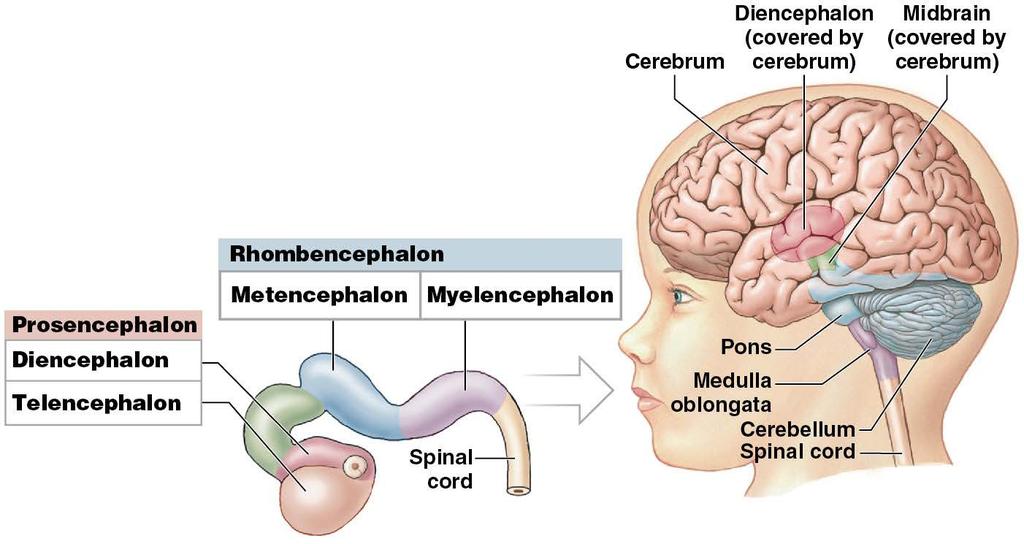
9 Module 13.1: Brain development Primary brain vesicles = three swellings at cephalic end of tube Prosencephalon = forebrain; anterior tip of neural tube Mesencephalon = midbrain; caudal to prosencephalon Rhombencephalon = hindbrain; most caudal part; continuous with spinal cord
10 Module 13.1: Brain development Secondary brain vesicles By week 5 of development: Primary brain vesicles subdivide; form secondary brain vesicles
11 Module 13.1: Brain development Secondary brain vesicles (continued) Prosencephalon subdivides into: Diencephalon becomes major relay/processing center for information headed to/from cerebrum Telencephalon expands rapidly; forms cerebrum Cerebrum continues enlarging to become largest part of brain and cover other regions
 Rhombencephalon subdivides into: Metencephalon caudal to")
12 Module 13.1: Brain development Secondary brain vesicles (continued) Rhombencephalon subdivides into: Metencephalon caudal to midbrain (mesencephalon) Forms cerebellum and pons of adult brain Myelencephalon becomes medulla oblongata
13 Brain development
14 Module 13.1: Review A. Name the three primary brain vesicles. B. Which structures form the secondary brain vesicles? C. Which embryonic brain vesicle develops into the largest region of the adult brain? Learning Outcome: Describe the origins of the different regions of the brain from the embryonic neural tube.
15 Module 13.2: Each region of the brain has distinct structural and functional characteristics Four major brain regions 1. Cerebrum 2. Cerebellum 3. Diencephalon 4. Brainstem
16 Module 13.2: Major brain regions Cerebrum Left and right cerebral hemispheres; divided by deep fissures Surface Gyri (folds) and sulci (grooves) increase surface area
17 Module 13.2: Major brain regions Cerebral cortex Superficial layer of gray matter Thin mm thick Major cortical functions Conscious thought Memory storage and processing Sensory processing Control of skeletal muscles

18 Module 13.2: Major brain regions Cerebellum Partially hidden by cerebral hemispheres Second largest brain structure Makes up 10 percent of brain s volume, but > 50 percent of its neurons Major cerebellar functions Coordinate/modulate motor commands from cerebral cortex

19 Module 13.2: Major brain regions Diencephalon Structural/functional link between cerebral hemispheres and rest of CNS Two parts 1. Thalamus Sensory relay/ processing 2. Hypothalamus has centers involved with: Emotions Autonomic function Hormone production
20 Module 13.2: Major brain regions Brainstem Three parts midbrain, pons, and medulla oblongata Midbrain contains nuclei that: Process visual and auditory information Control reflexes triggered by them Helps maintain consciousness Pons connects cerebellum/ brainstem Tracts and relay centers Somatic and visceral motor control
 Three parts (continued) Medulla oblongata Relays")
21 Module 13.2: Major brain regions Brainstem (continued) Three parts (continued) Medulla oblongata Relays sensory information through brainstem and to the thalamus Regulates many autonomic functions
22 Module 13.2: Major brain regions
23 Module 13.2: Major brain regions Ventricles of the brain Chambers formed by neural tube expansion during development Filled with cerebrospinal fluid; lined by ependymal cells
24 Module 13.2: Major brain regions Ventricular system components Lateral ventricles two, one in each cerebral hemisphere Interventricular foramen connect lateral ventricles to third ventricle Third ventricle located in diencephalon Cerebral aqueduct slender canal within midbrain; connects third ventricle to fourth ventricle Fourth ventricle extends from metencephalon upper part of medulla oblongata Narrows to become the central canal of spinal cord

25 Module 13.2: Major brain regions

26 Module 13.2: Major brain regions Interconnections between ventricles Corpus callosum thick tract of white matter that interconnects the cerebral hemispheres Septum pellucidum thin partition separating lateral ventricles
27 Module 13.2: Review A. Describe the role of the medulla oblongata. B. Which region of the brain contains two ventricles? C. Compare the corpus callosum with the septum pellucidum. D. Which region of the brain is enclosed or hidden by the cerebrum? Learning Outcome: Name the four major regions of the brain, and describe their functions.
28 Module 13.3: The cranial meninges and cerebrospinal fluid protect and support the brain Protection of the brain Cranial bones Cerebrospinal fluid Blood brain barrier Cranial meninges
29 Module 13.3: The cranial meninges and cerebrospinal fluid protect and support the brain Cranial meninges Continuous with spinal meninges Three layers 1. Dura mater 2. Arachnoid mater 3. Pia mater
 fused to periosteum of cranial bones No epidural")
30 Module 13.3: Protection of the brain Dura mater Two fibrous layers; mostly fused Periosteal layer (outer layer) fused to periosteum of cranial bones No epidural space Meningeal layer (inner layer) In some areas, including dural venous sinuses, layers are separated by gap containing fluid and blood vessels
31 Module 13.3: Protection of the brain Arachnoid mater Arachnoid membrane closest to dura mater Arachnoid trabeculae fibrous strands through subarachnoid space; connect to the pia mater Subarachnoid space between arachnoid membrane/pia mater
32 Module 13.3: Protection of the brain Pia mater Bound to surface of brain by astrocyte processes (sticks to the brain) Extends into every fold Accompanies branches of cerebral blood vessels as they penetrate the surface into the brain
33 Module 13.3: Protection of the brain Dural folds Inward extensions of inner dural layer Stabilize/support the brain Three large dural folds 1. Falx cerebri 2. Tentorium cerebelli 3. Falx cerebelli
34 Module 13.3: Protection of the brain Dural venous sinuses Large collecting veins located within dural folds Superior sagittal sinus is the largest
35 Module 13.3: Protection of the brain Falx cerebri Dural fold between cerebral hemispheres Runs from crista galli of ethmoid bone (anteriorly) to internal occipital crest of occipital bone (posteriorly) Contains the superior and inferior sagittal sinuses

36 Module 13.3: Protection of the brain Tentorium cerebelli Separates cerebral hemispheres from cerebellum Falx cerebelli Separates cerebellar hemispheres along midsagittal line Inferior to tentorium cerebelli
37 Module 13.3: Protection of the brain Cerebrospinal fluid (CSF) Three functions 1. Support weight of brain 2. Cushion brain/spinal cord from physical trauma 3. Transport nutrients, chemical messengers, wastes Produced/maintained by choroid plexus (ependymal cells with tight junctions, and capillaries) A choroid plexus is in each ventricle
 CSF circulates")
38 Module 13.3: Protection of the brain Cerebrospinal fluid (continued) CSF circulates from choroid plexus through ventricles into central canal of spinal cord
")
39 Module 13.3: Protection of the brain Cerebrospinal fluid (continued) Materials diffuse between CSF and interstitial fluid of CNS
40 Module 13.3: Protection of the brain Cerebrospinal fluid (continued) CSF is absorbed into venous circulation at arachnoid granulations Fingerlike extensions of arachnoid membrane Penetrate meningeal layer of dura mater Extend into superior sagittal sinus

41 Formation, circulation, and functions of CSF
42 Module 13.3 Review A. From superficial to deep, name the layers that make up the cranial meninges. B. What brain tissues have tight junctions? C. How would decreased diffusion across the arachnoid granulations affect the volume of cerebrospinal fluid in the ventricles? Learning Outcome: Explain how the brain is protected and supported and how cerebrospinal fluid forms and circulates.
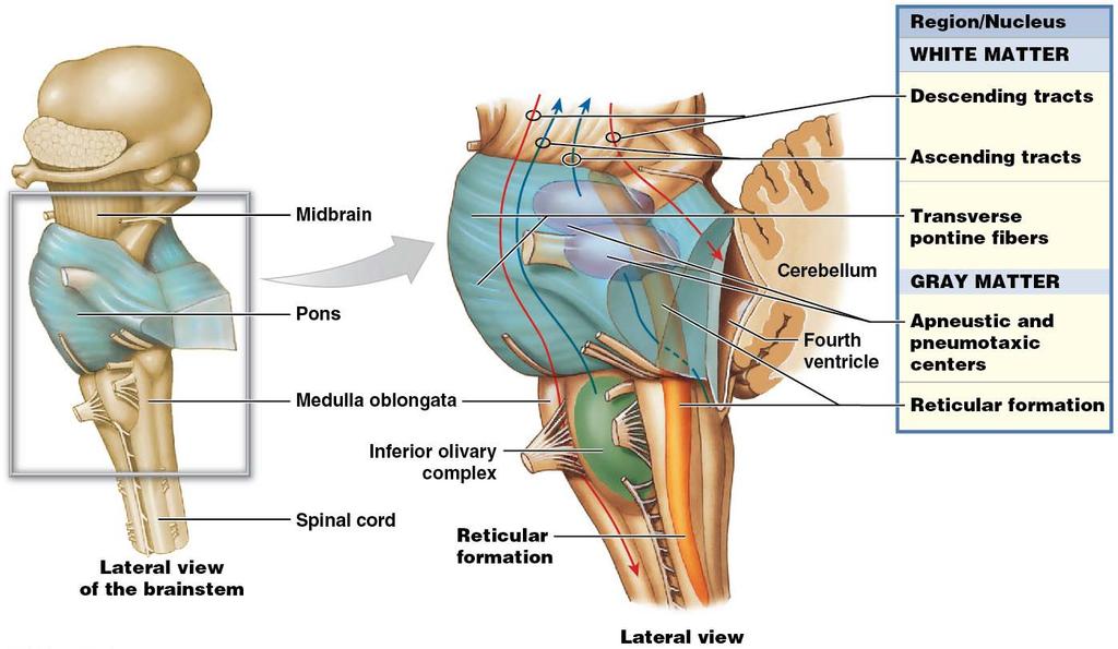
43 Module 13.4: The medulla oblongata contains autonomic reflex centers, relay stations, and ascending and descending tracts Medulla oblongata All communication between brain and spinal cord travels in ascending and descending tracts through medulla oblongata Coordinates complex autonomic reflexes and visceral functions
44 Module 13.4: Medulla oblongata Medulla oblongata contains: Autonomic centers for vital functions Relay stations along sensory and motor pathways Nuclei associated with five cranial nerves Structures Olive olive-shaped bulge on anterolateral surface Pyramids contain tracts of motor fibers from cerebral cortex Some fibers cross to opposite side of medulla (decussation)

45 Module 13.4: Medulla oblongata

46 Organization of the medulla oblongata

47 Module 13.4: Medulla oblongata
48 Module 13.4: Review A. Describe the pyramids of the medulla oblongata and a decussation. B. Which medulla oblongata components relay somatic sensory information to the thalamus? C. What is the function of the ascending and descending tracts in the medulla oblongata? Learning Outcome: List the main components of the medulla oblongata, and specify their functions.
49 Module 13.5: The pons links the cerebellum to the brain and spinal cord and has vital autonomic reflex centers Pons links cerebellum with midbrain, diencephalon, cerebrum, medulla oblongata, spinal cord Four groups of structures 1. Ascending, descending, transverse tracts 2. Nuclei controlling respiration 3. Sensory and motor nuclei of cranial nerves 4. Nuclei/tracts that process/relay information sent to/from cerebellum
50 Module 13.5: Pons Reticular formation contains nuclei/centers regulating vital autonomic functions; spans from medulla oblongata to midbrain
51 Organization of the Pons

52 Module 13.5: Pons
53 Module 13.5: Review A. List the four groups of pontine structures. Learning Outcome: List the main components of the pons, and specify their functions.

54 Module 13.6: The cerebellum coordinates learned and reflexive patterns of muscular activity at the subconscious level Cerebellum Automatic processing center Monitors proprioceptive, visual, tactile, balance, and auditory sensations
55 Module 13.6: The cerebellum Cerebellum (continued) Two primary functions 1. Adjusting postural muscles Modifies activities of brainstem centers 2. Programming/fine-tuning movements controlled at conscious/subconscious levels Refines learned movement patterns Compares motor commands with proprioceptive information, makes adjustments as needed

56 Module 13.6: The cerebellum Cerebellum anatomy Anterior and posterior lobes, separated by primary fissure Two hemispheres Surface covered by thin layer of gray matter = cerebellar cortex Separated by narrow band of cortex called the vermis (worm) Folia = folds of the surface; less prominent than cerebral gyri

57 Module 13.6: The cerebellum Cerebellar cortex Outer molecular layer Inner granular layer Intermediate Purkinje layer with stacked Purkinje cells Highly branched cells (extensive dendrites) Receive input from up to 200,000 synapses Relay motor commands The only axons leaving cerebellar cortex are from Purkinje cells
58 Module 13.6: The cerebellum Internal organization and peduncles Arbor vitae: tree of life = branching cerebellar white matter Cerebellar peduncles Tracts linking cerebellum with brain stem, cerebrum, and spinal cord Three on each side 1. Superior peduncle carries most cerebellar output 2. Middle and inferior carry most cerebellar input Ataxia results from cerebellum damage/impairment Inability to coordinate muscle movement If severe, person cannot sit or stand without assistance

59 Organization of the cerebellum

60 Module 13.6: The cerebellum
61 Module 13.6: Review A. Identify the components of the cerebellar gray matter. B. Describe the arbor vitae, including its makeup, location, and function. C. Describe ataxia. Learning Outcome: List the main components of the cerebellum, and specify the functions of each.

62 Module 13.7: The midbrain regulates auditory and visual reflexes and controls alertness Midbrain Most complex/integrative part of brainstem Can direct complex motor patterns at subconscious level Influences level of activity in entire nervous system
63 Module 13.7: The midbrain Corpora quadrigemina two pairs of sensory nuclei 1. Superior colliculus (colliculus, hill) Receives visual input from thalamus Controls reflex movements of eyes, head, and neck in response to visual stimuli 2. Inferior colliculus Receives auditory input from medulla oblongata and pons Controls reflex movements of head, neck, and trunk in response to auditory inputs
64 Module 13.7: The midbrain Reticular activating system (RAS) Specialized part of the reticular formation Stimulation increases alertness and attentiveness Damage to the RAS produces unconsciousness Red nucleus Red from rich blood supply Receives information from cerebrum and cerebellum Sends subconscious motor commands for upper limb position and muscle tone
65 Module 13.7: The midbrain Substantia nigra (nigra, black) Contains darkly pigmented cells Inhibits activity in cerebral basal nuclei Cerebral peduncles Nerve fiber bundles with descending fibers Go to cerebellum, via pons Carry voluntary motor commands from cerebral cortex

66 Organization of the midbrain

67 Module 13.7: The midbrain Internal midbrain structures Tectum Roof of the midbrain Region posterior to the cerebral aqueduct Tegmentum Region anterior to the cerebral aqueduct

68 Origins of cranial nerves from the brainstem

69 Module 13.7: The midbrain
70 Module 13.7: Review A. Identify the sensory nuclei contained within the corpora quadrigemina. B. Give the functions of the superior colliculi and inferior colliculi. C. Which cranial nerves arise from the brainstem? D. What part of the midbrain influences the activity of the entire nervous system? Learning Outcome: List the main components of the midbrain, and specify the functions of each.
71 Module 13.8: The diencephalon consists of the epithalamus, thalamus, and hypothalamus Three parts 1. Epithalamus 2. Thalamus 3. Hypothalamus
72 Module 13.8: Diencephalon Anterior commissure Connects cerebral hemispheres Optic chiasm Where optic nerves (II) cross; enter brain Interthalamic adhesion Bumplike extension of gray matter into third ventricle from sides of thalamus; no fibers cross Missing in about 20 percent of human brains
73 Location of the diencephalon
74 Module 13.8: Diencephalon Epithalamus Roof of diencephalon; superior to third ventricle Contains extensive choroid plexus that extends through interventricular foramina
75 Module 13.8: Diencephalon Epithalamus (continued) Pineal gland Endocrine structure secretes melatonin Located in posterior epithalamus Melatonin regulates day night cycles and reproductive functions
76 Module 13.8: Diencephalon Thalamus Superior to midbrain Filters sensory information, passing on only small portion Final relay for ascending sensory information being projected to cerebral cortex Each thalamic region connected to specific areas in cortex

77 Major relay areas of the thalamus
 Lateral geniculate body Receives visual information from optic tract Sends signals to midbrain and")
78 Module 13.8: Diencephalon Thalamus (continued) Lateral geniculate body Receives visual information from optic tract Sends signals to midbrain and occipital lobe Medial geniculate nucleus Relays auditory information from receptors in inner ear to appropriate areas in cerebral cortex

79 Module 13.8: Diencephalon

80 Module 13.8: Diencephalon Hypothalamus Contains important control and integrative centers May be stimulated by: 1. Sensory information from cerebrum, brainstem, spinal cord 2. Changes in composition of CSF and interstitial fluid 3. Chemicals in circulating blood (lacks blood brain barrier) Infundibulum connects it to pituitary gland (endocrine)

81 Organization of the hypothalamus

82 Module 13.8: Diencephalon
83 Module 13.8: Review A. Damage to the lateral geniculate bodies of the thalamus would interfere with what particular function? B. Which component of the diencephalon is stimulated by changes in body temperature? Learning Outcome: List the main components of the diencephalon, and specify the functions of each.
84 Module 13.9: The limbic system is a functional group of tracts and nuclei located in the cerebrum and diencephalon Limbic system Functional grouping of tracts and nuclei along border of cerebrum and diencephalon Functions include: 1. Establishing emotional states 2. Linking conscious, intellectual functions of cerebral cortex with unconscious and autonomic functions of brainstem 3. Facilitating memory storage/retrieval 4. Affecting motivation
85 Module 13.9: Limbic system Major components of limbic system Diencephalic components Thalamus anterior thalamic nuclei from mammillary body (in hypothalamus) to cingulate gyrus Hypothalamus hypothalamic nuclei, mammillary body Emotions (rage, fear, pain, sexual arousal, pleasure) Produce general alertness/excitement or lethargy/sleep via stimulation from reticular formation
86 Module 13.9: Limbic system Major components of limbic system (continued) Cerebral components Limbic lobe cortical areas (three cerebral gyri) Cingulate gyrus Dentate gyrus Parahippocampal gyrus Tracts Fornix = tract of white matter connecting hippocampus and hypothalamus
87 Module 13.9: Limbic system Major components of limbic system (continued) Cerebral components (continued) Nuclei Amygdaloid body interface between limbic system, sensory systems, and cerebrum; role in regulating heart rate, fight-or-flight response, and linking emotions and memories Hippocampus learning, especially storage/retrieval of long-term memories

88 Organization of the limbic system

89 The functional components of the limbic system
90 Module 13.9: Review A. List the primary functions of the limbic system. B. What are some functions of the amygdaloid body? C. Which region of the limbic system is particularly important for the storage and retrieval of longterm memories? Learning Outcome: Identify the main components of the limbic system, and specify the locations and functions of each.

91 Module 13.10: The basal nuclei of the cerebrum adjust and refine ongoing voluntary movements Basal nuclei of the cerebrum Masses of gray matter within each cerebral hemisphere, deep to floor of lateral ventricles Subconscious control of skeletal muscle tone Help coordinate learned movement patterns Do not initiate movements; provide general pattern/rhythm

92 Module 13.10: Basal nuclei The basal nuclei consist of: Caudate nucleus Lentiform nucleus Medial globus pallidus Lateral putamen Internal capsule Bundles of axons linking cerebral cortex to diencephalon and brainstem; pass between and around the basal nuclei

93 Locations of the basal nuclei
94 13.10: Basal nuclei Roles of the basal nuclei in modifying ongoing movements Direct control over movement Stimulate motor nuclei (the red nucleus, superior and inferior colliculi, reticular formation) in the brainstem Example: control cycles of arm/thigh movements while walking Indirect modification of movement Send output to cerebral cortex after synapsing in thalamus Example: subconsciously adjust muscle tone as you begin a voluntary movement

95 Roles of the basal nuclei in movement
96 Module 13.10: Review A. Define basal nuclei. B. Describe the function of the caudate nucleus. C. What signs and symptoms would be present in a person who has basal nuclei damage? Learning Outcome: Describe the structure and function of the basal nuclei of the cerebrum.

97 Module 13.11: Superficial landmarks divide the cerebral hemispheres into lobes Overview of cerebral lobes Each cerebral hemisphere divided into regions lobes Named after overlying bones of the skull (except for the insula) Lobes of the cerebrum 1. Frontal lobe 2. Parietal lobe 3. Temporal lobe 4. Occipital lobe 5. Insula (insula, island)
98 Module 13.11: Lobes of the cerebrum Superficial landmarks Each person has unique pattern of gyri and sulci Lateral sulcus separates frontal lobe from temporal lobe Parieto-occipital sulcus separates parietal lobe from occipital lobe
99 Module 13.11: Lobes of the cerebrum Superficial landmarks (continued) Central sulcus separates frontal lobe from parietal lobe Precentral gyrus ridge anterior to central sulcus Contains primary motor cortex Controls voluntary movements Postcentral gyrus Posterior to central sulcus Contains primary somatosensory cortex Conscious awareness of somatosensory information

100 Lobes and superficial anatomy of the cerebrum
101 Module 13.11: Lobes of the cerebrum

102 Module 13.11: Lobes of the cerebrum
103 Module 13.11: Lobes of the cerebrum General facts about the cerebral hemispheres Each hemisphere receives sensory information from and sends motor commands to opposite side of body Crossing over occurs in brainstem and spinal cord Has no known functional significance Hemispheres may look identical and have similar functions, but still have important differences Imprecise mapping of specific functions and regions Boundaries are indistinct and overlap Some functions (such as consciousness) use multiple regions Normal individuals use ALL portions of the brain
104 Module 13.11: Review A. Identify the lobes of the cerebrum, and indicate the basis for their names. B. Describe the insula. C. What effect would damage to the left postcentral gyrus produce? Learning Outcome: Identify the major superficial landmarks of the cerebrum, and cite the location of each.
105 Module 13.12: The lobes of the cerebral cortex have regions with specific functions Cerebral cortex divided into six functional categories 1. Motor cortex sends voluntary commands to skeletal muscles 2. Sensory cortex receives general somatic sensory information 3. Visual cortex (vision) 4. Auditory cortex (hearing) 5. Olfactory cortex (smell) 6. Gustatory cortex (taste) Each sensory and motor region connected to nearby association area = region that interprets incoming data or coordinates motor response

106 Module 13.12: Functional regions of cerebral cortex 1. Motor cortex Primary motor cortex directs voluntary movement by controlling motor neurons in brainstem/spinal cord Premotor cortex (somatic motor association area) coordinates learned movements

107 Module 13.12: Functional regions of cerebral cortex 2. Sensory cortex Primary somatosensory cortex receives sensory information from receptors for touch, pressure, pain, vibration, or temperature Somatosensory association area monitors activity in primary somatosensory cortex; recognizes different sensations
 In insula receives information from taste receptors 4.")
108 Module 13.12: Functional regions of cerebral cortex 3. Gustatory cortex (taste) In insula receives information from taste receptors 4. Olfactory cortex (smell) Receives sensory information from olfactory receptors
 Primary auditory cortex monitors auditory information Auditory association area activity")
109 Module 13.12: Functional regions of cerebral cortex 5. Auditory cortex (hearing) Primary auditory cortex monitors auditory information Auditory association area activity in auditory cortex; recognizes different sounds (such as speech)

110 Module 13.12: Functional regions of cerebral cortex 6. Visual cortex Primary visual cortex receives visual information from lateral geniculate bodies Visual association area monitors activity and patterns in visual cortex; interprets the information Example: Primary visual cortex sees symbols c, a, t; visual association area interprets as cat

111 Module 13.12: Functional regions of cerebral cortex Integrative centers Receive information from association areas Direct motor activities Perform analytical functions In lobes/cortical areas of both cerebral hemispheres Language areas typically associated with left hemisphere
112 Module 13.12: Functional regions of cerebral cortex Integrative centers (continued) Broca s area (motor speech area) speech production Regulates breathing/vocalization patterns for normal speech If damaged, can make sounds but not form words Prefrontal cortex integrates information from sensory association areas; performs intellectual functions
113 Module 13.12: Functional regions of cerebral cortex Integrative centers (continued) Frontal eye field controls learned eye movements Example: scanning lines of text Wernicke s area associated with language comprehension Receives information from somatosensory association areas Plays essential role in personality by linking sensory information to complex visual and auditory memories
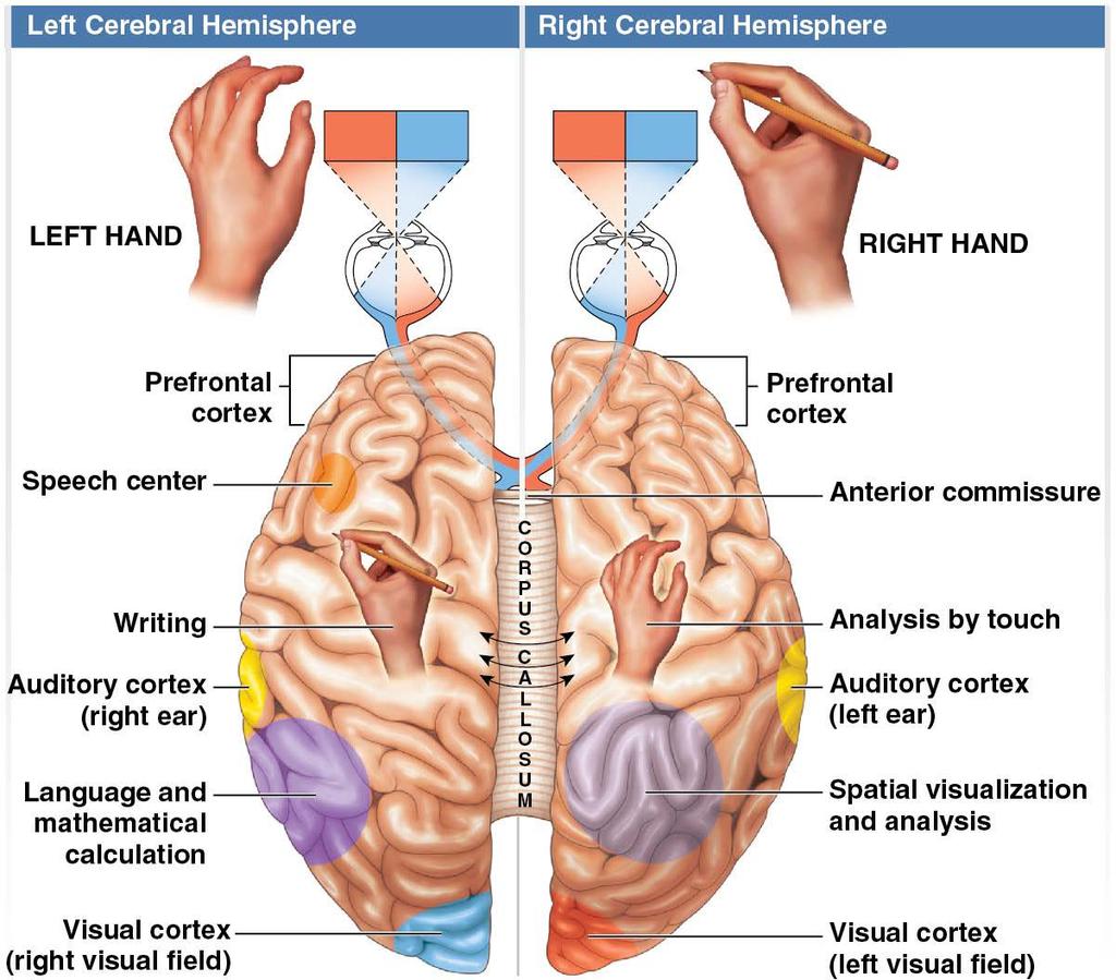
114 Module 13.12: Functional regions of cerebral cortex Hemispheric lateralization Regional specialization of each hemisphere Left cerebral hemisphere Specialized language areas Language-based skills, such as reading, writing, speaking Premotor cortex for hand movements is larger on left side in right-handed people than in left-handed people Analytical tasks, such as math and logic
115 Module 13.12: Functional regions of cerebral cortex Hemispheric lateralization (continued) Right cerebral hemisphere Analyzes sensory information; relates body to it Interpretive centers for identifying familiar objects by touch, smell, sight, taste, or feel Facial recognition Understanding three-dimensional relationships Analyzes emotional context/tone of conversation
116 Hemispheric lateralization
117 Module 13.12: Functional regions of cerebral cortex Approximately 9 percent of the population is lefthanded Primary motor cortex of right hemisphere controls motor function for left hand, but left hemisphere usually still controls speech and analytical functions Seen in an unusually high percentage of musicians/artists
118 Module 13.12: Review A. Where is the primary motor cortex located? B. Which senses are affected by damage to the temporal lobes? C. Which brain region has been affected in a stroke victim who is unable to speak? Learning Outcome: Identify the locations of the motor, sensory, and association areas of the cerebral cortex, and discuss the functions of each.
119 Module 13.13: White matter connects the cerebral hemispheres and the lobes of each hemisphere and links the cerebrum to the rest of the brain Functional grouping of white matter Interior of cerebral hemispheres mostly consists of white matter Organized into groups sharing common function

120 Module 13.13: Cerebral white matter Association fibers connect areas in same cerebral hemisphere Arcuate fibers shortest; curve in an arc to connect gyri Longest are organized in bundles, or fasciculi Longitudinal fasciculi connect frontal lobe to other lobes in same cerebral hemisphere

121 Module 13.13: Cerebral white matter Commissural fibers connect the two cerebral hemispheres Corpus callosum most important band of commissural fibers Allows communication/coordination between the two hemispheres > 200 million axons carrying about 4 billion impulses per second
122 Module 13.13: Cerebral white matter Commissural fibers (continued) Anterior commissure smaller tract also connecting the two hemispheres; becomes more important if corpus callosum injured
123 Module 13.13: Cerebral white matter Projection fibers link cerebral cortex to diencephalon, brainstem, cerebellum, and spinal cord All projection fibers pass through diencephalon Internal capsule = collection of ascending (sensory) and descending (motor) fibers passing through diencephalon
124 Module 13.13: Review A. What is the function of the longitudinal fasciculi? B. What special names are given to axons in the white matter of the cerebral hemispheres? Learning Outcome: Discuss the significance of the white matter of the cerebral hemispheres.
125 Module 13.14: Brain activity can be monitored using external electrodes; the record is called an electroencephalogram, or EEG Electrical activity of the brain Neural function depends on electrical events Electrical activity of all the neurons in the brain generates an electrical field, measurable by electrodes placed on scalp Printed report of that activity = electroencephalogram (EEG) Electrical patterns observed are called brain waves
126 Module 13.14: EEG Brain waves Alpha waves Occur in healthy, awake adults while resting with eyes closed Disappear during sleep Disappear with concentration on a specific task Beta waves Higher frequency than alpha waves Appear in people who are concentrating on a task, under stress, or in state of psychological tension
127 Module 13.14: EEG Brain waves (continued) Theta waves Appear transiently during sleep in normal adults Usually observed in children and intensely frustrated adults Presence under other circumstances may indicate brain disorder
128 Module 13.14: EEG Brain waves (continued) Delta waves Large-amplitude, low-frequency waves Normally seen during deep sleep in all ages Also seen in brains of infants (incomplete cortical development) Seen in awake adults when tumor, vascular block, or inflammation has damaged parts of the brain
129 Electroencephalogram (EEG) showing the brain waves
130 Module 13.14: EEG Abnormal brain activity Electrical activity in cerebral hemispheres usually synchronized by pacemaker that involves the thalamus Asynchrony may indicate localized damage or cerebral abnormality Example: tumor or injury in one hemisphere changes its pattern, losing alignment between hemispheres
131 Module 13.14: EEG Seizure = temporary cerebral disorder accompanied by unusual neural activity (movements, sensations, behaviors) Marked by change in pattern of electrical activity Epilepsies, or seizure disorders = clinical conditions characterized by seizures
132 Module 13.14: Review A. Name and describe the four wave types associated with an EEG. B. Differentiate between a seizure and epilepsy. Learning Outcome: Discuss the origin and significance of the major categories of brain waves seen in an electroencephalogram (EEG).

133 Module 13.15: The twelve pairs of cranial nerves are classified as sensory, special sensory, motor, or mixed nerves Twelve pairs of cranial nerves Numbered using Roman numerals (I XII) preceded by CN

134 Cranial nerve distribution

135 Module 13.15: Cranial nerves

136 Module 13.15: Cranial nerves
137 Module 13.15: Review A. Which cranial nerves are involved with the eye? B. Which cranial nerves have motor functions only? Learning Outcome: Identify the cranial nerves by name and number, and cite the functions of each.
138 Section 2: Sensory and Motor Pathways Learning Outcomes Describe the basic events that occur along a sensory pathway Explain the ways in which receptors can be classified List the types of tactile receptors, and specify the functions of each Identify and describe the major sensory pathways Describe the components, processes, and functions of the somatic motor pathways.
139 Section 2: Sensory and Motor Pathways Learning Outcomes (continued) Describe the levels of information processing involved in motor control Clinical Module: Describe the roles of the nervous system in referred pain, Parkinson s disease, rabies, cerebral palsy, amyotrophic lateral sclerosis, Alzheimer s disease, and multiple sclerosis.
140 Module 13.16: Sensations carried by sensory pathways to the CNS begin with transduction at a sensory receptor Sensory terminology Sensory receptors = specialized cells or cell processes that alert CNS about conditions in/out of your body General senses temperature, pain, touch, pressure, vibration, proprioception (position); receptors distributed throughout body Sensory pathways begin at peripheral receptors, end within CNS
 Sensation = information carried by a sensory pathway Perception = conscious awareness")
141 Module 13.16: Sensory pathways Sensory terminology (continued) Sensation = information carried by a sensory pathway Perception = conscious awareness of a sensation Receptive field = area monitored by single receptor cell Larger the receptor field, less able to localize stimulus

142 Module 13.16: Sensory pathways Basic events along sensory and motor pathways Depolarization of receptor Physical/chemical stimulus results in graded change in receptor cell (transduction)
143 Module 13.16: Sensory pathways Action potential generation If receptor cell depolarizes to threshold, triggers action potential Greater depolarization = greater frequency of action potentials
144 Module 13.16: Sensory pathways Propagation over labeled line Labeled line = axons carrying information about one type of stimulus (touch, pressure, temperature) CNS interprets stimulus based on which line carries it
145 Module 13.16: Sensory pathways CNS processing Occurs at every synapse along labeled line Line may branch repeatedly, distributing sensory information to multiple nuclei/centers in spinal cord and brain

146 Module 13.16: Sensory pathways CNS processing can trigger motor response Involuntary motor pathways immediate involuntary response even before sensations reach cerebral cortex Example: reflex response

147 Module 13.16: Sensory pathways Voluntary motor pathways Perception only ~ 1 percent of arriving sensations are relayed to primary sensory cortex, where perception occurs Can initiate voluntary response not immediate; can moderate, enhance, supplement simple reflexive response
148 Module Review A. Define the term general senses. B. Relate receptive field size to stimulus localization. Learning Outcome: Describe the basic events that occur along a sensory pathway.

149 Module 13.17: Receptors are classified by function or response to the stimulus Free nerve endings Tips of branching dendrites of sensory neurons Simplest receptors Respond to many different stimuli, so not much receptor specificity Example: pain receptors stimulated by chemicals, pressure, temperature, or trauma
150 Module 13.17: Receptor classification Nociceptors = pain receptors Free nerve endings with large receptive fields and broad sensitivity Do not adapt quickly Two axon types carry pain information 1. Myelinated type A fibers 2. Unmyelinated type C fibers
151 Module 13.17: Receptor classification Nociceptors (continued) Two axon types carry pain signals 1. Myelinated type A fibers fast pain (prickling pain) Sensations quickly reach primary somatosensory cortex for conscious awareness; often trigger somatic reflexes Can usually localize the stimulus
152 Module 13.17: Receptor classification Nociceptors (continued) 2. Unmyelinated type C fibers slow pain Burning/aching General activation of reticular formation and thalamus Aware of pain but just general idea what area is affected

153 Module 13.17: Receptor classification Thermoreceptors = temperature receptors Free nerve endings in dermis, skeletal muscles, liver, hypothalamus Cold receptors three to four times more numerous than warm receptors No structural differences between warm and cold thermoreceptors

154 Module 13.17: Receptor classification Chemoreceptors respond to water-soluble and lipid-soluble substances dissolved in body fluids (interstitial fluid, blood, CSF)
155 Module 13.17: Receptor classification Mechanoreceptors respond to physical distortion of their plasma membranes Membranes have mechanically gated ion channels that open/close in response to stretching, compression, twisting, etc. Three types of mechanoreceptors 1. Proprioceptors 2. Baroreceptors 3. Tactile receptors
156 Module 13.17: Receptor classification Mechanoreceptors (continued) Three types (continued) 1. Proprioceptors monitor position of joints/muscles most complex of general sensory receptors Example: muscle spindle 2. Baroreceptors detect pressure changes in blood vessels and in digestive, respiratory, and urinary tracts 3. Tactile receptors provide sensations of touch (shape/texture), pressure, and vibration Fine touch and pressure receptors give detailed information; extremely sensitive Crude touch and pressure receptors give little information; poor localization

157 Module 13.17: Receptor classification

158 Module 13.17: Receptor classification Receptors categorized by nature of their response to stimulation Tonic receptors Slow-adapting; always active Action potential frequency reflects level of stimulation Increased stimulus = increased action potential frequency

159 Module 13.17: Receptor classification Phasic receptors Fast-adapting; normally inactive Become active for short time when a change occurs in what they are monitoring
160 Module 13.17: Receptor classification Adaptation = reduction in sensitivity with constant stimulus Two types 1. Peripheral adaptation occurs when level of receptor activity changes; receptor first responds strongly, then activity declines Especially characteristic of phasic receptors Example: temperature not noticed much unless it changes
161 Module 13.17: Receptor classification 2. Central adaptation occurs along sensory pathways in CNS; involves inhibition of nuclei along a sensory pathway Example: new smell once it is initially detected, awareness almost stops even though neurons are still sending signals
162 Module 13.17: Review A. List the four types of general sensory receptors based on function, and identify the type of stimulus that excites each type. B. Describe the three classes of mechanoreceptors. C. Explain adaptation, and differentiate between peripheral adaptation and central adaptation. Learning Outcome: Explain the ways in which receptors can be classified.
163 Module 13.18: Tactile receptors have a simple structure and are abundant in the skin Tactile sensory receptors = mechanoreceptors responding to touch, pressure, vibration Greatest diversity in the skin Six types of tactile receptors in the skin 1. Free nerve endings 2. Root hair plexuses 3. Tactile discs 4. Tactile corpuscles 5. Lamellar corpuscles 6. Bulbous corpuscles

164 Module 13.18: Tactile receptors Six types of tactile receptors in skin (continued) 1. Free nerve endings Branching dendrite tips of sensory neurons Nonspecific respond to touch, pressure, pain, temperature Most common receptors in skin

165 Module 13.18: Tactile receptors Six types of tactile receptors in skin (continued) 2. Root hair plexus Monitor distortion/movement of hair follicle Displacement of hair distorts sensory dendrites; generates action potentials Adapt rapidly

166 Module 13.18: Tactile receptors Six types of tactile receptors in skin (continued) 3. Tactile discs Fine touch and pressure Extremely sensitive tonic receptors Very small receptive fields A tactile disc is composed of a Merkel cell (large epithelial cell in stratum basale) and dendritic processes from single myelinated afferent fiber that are in contact with it
 Fine touch, pressure, low-frequency vibration Adapt within a second Abundant in eyelids, lips, fingertips, nipples, external genitalia")
167 Module 13.18: Tactile receptors Six types of tactile receptors in skin (continued) 4. Tactile corpuscles (Meissner s corpuscles) Fine touch, pressure, low-frequency vibration Adapt within a second Abundant in eyelids, lips, fingertips, nipples, external genitalia Tactile corpuscles composed of dendrites surrounded by Schwann cells, all wrapped in fibrous capsule
 Sensitive to deep pressure Fast-adapting most sensitive to pulsing or high-frequency vibration Large receptors up to 4 mm long and 1 mm in diameter Abundant")
168 Module 13.18: Tactile receptors Six types of tactile receptors in skin (continued) 5. Lamellar corpuscles (Pacinian corpuscles) Sensitive to deep pressure Fast-adapting most sensitive to pulsing or high-frequency vibration Large receptors up to 4 mm long and 1 mm in diameter Abundant in fingers, mammary glands, external genitalia, fasciae, joint capsules, some viscera (mesenteries, pancreas, urethra, and bladder) Lamellar corpuscles composed of single dendrite within concentric layers of collagen fibers and fibroblasts
 Sensitive to pressure and distortion of deep dermis (skin stretched) Tonic receptors; little adaptation Ruffini corpuscles are composed of capsule around core")
169 Module 13.18: Tactile receptors Six types of tactile receptors in skin (continued) 6. Bulbous corpuscles (Ruffini corpuscles) Sensitive to pressure and distortion of deep dermis (skin stretched) Tonic receptors; little adaptation Ruffini corpuscles are composed of capsule around core of collagen fibers continuous with those of adjacent dermis, and inner network of dendrites wrapped around collagen fibers Tension/distortion of dermis stimulates dendrite

170 Module 13.18: Tactile receptors
171 Module 13.18: Review A. Which types of tactile receptors are located only in the dermis? B. Which is likely to be more sensitive to continuous deep pressure: a lamellar corpuscle or a bulbous corpuscle? Learning Outcome: List the types of tactile receptors, and specify the functions of each.
172 Module 13.19: Three major somatic sensory pathways carry information from the skin and muscles to the CNS Somatotopy = functional map of primary somatosensory cortex Sensory homunculus ( little human ) = somatotope showing the relative size of cortex devoted to any specific body area

173 Module 13.19: Somatic sensory pathways Three major somatic sensory pathways 1. Spinothalamic pathway 2. Posterior column pathway 3. Spinocerebellar pathway
174 Module 13.19: Somatic sensory pathways Spinothalamic pathway Anterior spinothalamic tracts Crude touch and pressure sensations from body Lateral spinothalamic tracts Pain and temperature sensations from body First-order neuron from receptor to spinal cord; synapses with second-order neuron in posterior gray horns Second-order neuron from posterior gray horn; crosses spinal cord; ascends to thalamus Third-order neuron from thalamus to primary sensory cortex

175 Module 13.19: Somatic sensory pathways
176 Module 13.19: Somatic sensory pathways Posterior column pathway Highly localized ( fine ) touch, pressure, vibration, proprioception From peripheral receptor to primary somatosensory cortex Sensory axons ascend in medial gracile fasciculus and lateral cuneate fasciculus Medial lemniscus tract leading from gracile nucleus and cuneate nucleus to the thalamus

177 Module 13.19: Somatic sensory pathways
178 Module 13.19: Somatic sensory pathways Spinocerebellar pathway Proprioceptive information about position of skeletal muscles, joints, and tendons; goes to cerebellum Axons of posterior spinocerebellar tracts do not cross to other side pass through inferior cerebellar peduncle on same side Anterior spinocerebellar tract axons do cross to opposite side of spinal cord

179 Module 13.19: Somatic sensory pathways
180 Module 13.19: Review A. Define somatotopy. B. Which spinal tracts carry action potentials generated by nociceptors? C. Which cerebral hemisphere receives impulses conducted by the right gracile fasciculus? Learning Outcome: Identify and describe the major sensory pathways.
181 Module 13.20: The somatic nervous system controls skeletal muscles through upper and lower motor neurons Somatic motor pathways Always involve at least two motor neurons 1. Upper motor neuron Cell body in a CNS processing center 2. Lower motor neuron Cell body in a nucleus of brainstem or spinal cord Upper motor neuron synapses on lower motor neuron, which then innervates a single motor unit of a skeletal muscle
182 Module 13.20: The somatic nervous system controls skeletal muscles through upper and lower motor neurons Motor homunculus = functional map of primary motor cortex Proportions reflect number of motor units innervated and degree of fine motor control in corresponding body region
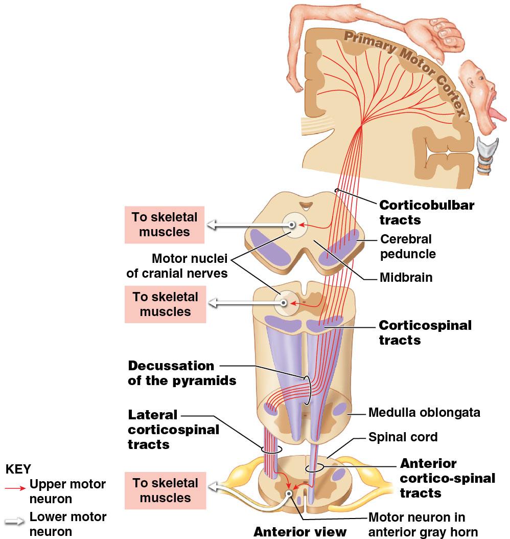
183 Module 13.20: Motor pathways Corticospinal pathway Voluntary control of skeletal muscles Sometimes called the pyramidal system upper motor neurons start at pyramidal cells in primary motor cortex Upper motor neuron axons descend into brainstem and spinal cord Synapse with lower motor neurons that control skeletal muscles
184 Module 13.20: Motor pathways Tracts of the corticospinal pathway 1. Corticobulbar (bulbar, brainstem) tracts conscious control of skeletal muscles for eye, jaw, face, some muscles of neck/pharynx Synapse on lower motor neurons in cranial nerve motor nuclei 2. Corticospinal tracts conscious control of skeletal muscles Visible along ventral surface of medulla oblongata as pair of thick bands (pyramids) ~15 percent descend uncrossed as anterior corticospinal tracts; cross over in anterior white commissure before synapsing on lower motor neurons in anterior gray horns
185 Corticospinal pathways
186 Module 13.20: Motor pathways Two main pathways for subconscious motor commands Commands issued from cerebrum, diencephalon, brainstem 1. Lateral pathway muscle tone/precise movements of distal limb muscles Upper motor neurons start in red nucleus Axons cross to opposite side of brain; descend through rubrospinal (ruber, red) tracts
187 Module 13.20: Motor pathways 2. Medial pathway gross movements of trunk, proximal limb Upper motor neurons located in vestibular nuclei, superior and inferior colliculi, and reticular formation Three major sets of tracts in medial pathway 1. Reticulospinal tracts 2. Vestibulospinal tracts 3. Tectospinal tracts
188 Module 13.20: Motor pathways Medial pathway (continued) Reticulospinal tracts Axons of upper motor neurons in reticular formation Reticular formation receives input from almost every ascending/descending pathway and has extensive connections with cerebrum, cerebellum, and brainstem Vestibulospinal tracts Start at vestibular nuclei of CN VIII (vestibulocochlear nerve.) Get sensory information from internal ear about head position/movement; send motor commands to alter muscle tone and position of neck, head, eyes, limbs
189 Module 13.20: Motor pathways Medial pathway (continued) Tectospinal tracts Upper motor neurons in superior/inferior colliculi (midbrain) Reflex changes in position of head, neck, upper limb in response to bright light, sudden movement, loud noises

190 The Medial Pathway

191 Cross-section of spinal cord showing positions of the various motor tracts
192 Module 13.20: Review A. Describe the role of the corticobulbar tracts. B. Define corticospinal tracts. C. What effect would increased stimulation of the motor neurons of the red nucleus have on muscle tone? Learning Outcome: Describe the components, processes, and functions of the somatic motor pathways.
193 Module 13.21: There are multiple levels of somatic motor control Many nuclei in spinal cord and brain are involved in controlling skeletal muscle contractions Generally, the closer the motor center is to the cerebral cortex, the more complex the motor activity Cerebellum coordinates motor activities at multiple levels
194 Module 13.21: Levels of somatic motor control Levels of somatic motor control (least to most complex): 1. Brainstem/spinal cord 2. Pons/medulla oblongata 3. Hypothalamus 4. Thalamus/midbrain 5. Basal nuclei 6. Cerebellum 7. Cerebral cortex

195 Levels of motor control

196 Module 13.21: Levels of somatic motor control Preparing for movement Decision to move begins relay of information from: Frontal lobes premotor cortex basal nuclei/cerebellum

197 Module 13.21: Levels of somatic motor control As movement begins: Information is sent from premotor cortex to primary motor cortex Commands are modified by feedback from cerebellum/basal nuclei
198 Module 13.21: Levels of somatic motor control Effects of primary motor cortex damage Person loses fine motor control over skeletal muscles Some voluntary movements will still be controlled by basal nuclei with input from prefrontal cortex Cerebellum cannot fine-tune movements because corticospinal pathway is inoperative An individual is able to stand, balance, and walk, but movements are hesitant, awkward, poorly controlled
199 Module 13.21: Review A. Which brain regions control reflexes in response to visual and auditory stimuli that are experienced while viewing a movie? B. The basic reflex motor patterns related to eating and drinking are controlled by which region of the brain? C. During a tennis match, you decide how and where to hit the ball. Explain how the premotor cortex is involved in your decisions. Learning Outcome: Describe the levels of information processing involved in motor control.

200 Module 13.22: Nervous system disorders may result from problems with neurons, pathways, or a combination of the two Referred pain Sensation of pain in a part of the body other than its actual source Example: pain of a heart attack felt in left arm Strong visceral pain sensations arriving at a spinal cord segment can stimulate interneurons in spinothalamic pathway Stimulates primary sensory cortex Pain perceived pain as coming from corresponding part of body surface (see sensory homunculus)
 Every part of a movement must be voluntarily controlled through intense effort and")
201 13.22: Nervous system disorders Parkinson s disease Substantia nigra neurons damaged or secrete less dopamine Basal nuclei become more active, increasing muscle tone and producing stiffness and rigidity Starting movements is difficult because antagonistic muscle groups do not relax (must be overpowered) Every part of a movement must be voluntarily controlled through intense effort and concentration

202 13.22: Nervous system disorders Rabies Bite from rabid animal injects rabies virus into peripheral tissues; virus enters axon terminals Virus carried by axons back into CNS through retrograde flow Potentially fatal results Many toxins, pathogenic bacteria, and other viruses also bypass CNS defenses through axoplasmic transport
 Genetic defect disrupting motor pathway")
203 13.22: Nervous system disorders Cerebral palsy (CP) Number of disorders that affect voluntary movement Motor dysfunction is nonprogressive Appears during infancy/childhood; persists throughout life Cause may be: Trauma from premature/stressful childbirth Maternal exposure to drugs (including alcohol) Genetic defect disrupting motor pathway development
; Stephen Hawking, noted physicist, also afflicted with")
204 13.22: Nervous system disorders Amyotrophic lateral sclerosis (ALS) Progressive, degenerative disorder affecting motor neurons in spinal cord, brainstem, cerebrum Destroys upper/lower motor neurons Associated skeletal muscles atrophy Likely defect in axonal transport Also called Lou Gehrig s disease (famous Yankees player who died from it); Stephen Hawking, noted physicist, also afflicted with ALS
205 13.22: Nervous system disorders Alzheimer s disease (AD) Progressive disorder causing loss of higher-order cerebral functions AD patients have intracellular and extracellular abnormalities in hippocampus (affects memory processing) Most common cause of senile dementia, or senility
; ~100,000")
206 13.22: Nervous system disorders Alzheimer s disease (AD) (continued) Symptoms may appear at age or later; also affects younger people Estimated 2 million people affected in United States (~15 percent of those over 65, ~50 percent of those over 85); ~100,000 deaths/year
 Time between incidents and degree of recovery vary In about one-third of patients,")
207 13.22: Nervous system disorders Multiple sclerosis (sklerosis, hardness) (MS) CNS disease characterized by recurrent incidents of demyelination in axons of optic nerve, brain, spinal cord Common signs/symptoms include impairment of vision, speech, balance, general motor coordination (including urinary/bowel) Time between incidents and degree of recovery vary In about one-third of patients, disease progresses more functional impairment occurs with each incident Onset often occurs at age years; 1.5 times more common in women
BRAIN PART I (A & B): VENTRICLES & MENINGES
: VENTRICLES & MENINGES") BRAIN PART I (A & B): VENTRICLES & MENINGES Cranial Meninges Cranial meninges are continuous with spinal meninges Dura mater: inner layer (meningeal layer) outer layer (endosteal layer) fused to periosteum
BRAIN PART I (A & B): VENTRICLES & MENINGES Cranial Meninges Cranial meninges are continuous with spinal meninges Dura mater: inner layer (meningeal layer) outer layer (endosteal layer) fused to periosteum
Anatomy and Physiology (Bio 220) The Brain Chapter 14 and select portions of Chapter 16
 The Brain Chapter 14 and select portions of Chapter 16") Anatomy and Physiology (Bio 220) The Brain Chapter 14 and select portions of Chapter 16 I. Introduction A. Appearance 1. physical 2. weight 3. relative weight B. Major parts of the brain 1. cerebrum 2.
Anatomy and Physiology (Bio 220) The Brain Chapter 14 and select portions of Chapter 16 I. Introduction A. Appearance 1. physical 2. weight 3. relative weight B. Major parts of the brain 1. cerebrum 2.
b. The groove between the two crests is called 2. The neural folds move toward each other & the fuse to create a
 Chapter 13: Brain and Cranial Nerves I. Development of the CNS A. The CNS begins as a flat plate called the B. The process proceeds as: 1. The lateral sides of the become elevated as waves called a. The
The Nervous System: Sensory and Motor Tracts of the Spinal Cord
 15 The Nervous System: Sensory and Motor Tracts of the Spinal Cord PowerPoint Lecture Presentations prepared by Steven Bassett Southeast Community College Lincoln, Nebraska Introduction Millions of sensory
Blood supply to the brain Blood brain barrier isolates neural tissue from general circulation
 The Brain and Cranial Nerves Objectives Name the major regions of the brain and describe their functions. Discuss the formation, circulation, and functions of the CSF. List the main components of the medulla
Neural Integration I: Sensory Pathways and the Somatic Nervous System
 15 Neural Integration I: Sensory Pathways and the Somatic Nervous System PowerPoint Lecture Presentations prepared by Jason LaPres Lone Star College North Harris An Introduction to Sensory Pathways and
Chapter 3. Structure and Function of the Nervous System. Copyright (c) Allyn and Bacon 2004
 Allyn and Bacon 2004") Chapter 3 Structure and Function of the Nervous System 1 Basic Features of the Nervous System Neuraxis: An imaginary line drawn through the center of the length of the central nervous system, from the
Chapter 3 Structure and Function of the Nervous System 1 Basic Features of the Nervous System Neuraxis: An imaginary line drawn through the center of the length of the central nervous system, from the
The Nervous System PART B
 7 The Nervous System PART B PowerPoint Lecture Slide Presentation by Jerry L. Cook, Sam Houston University ESSENTIALS OF HUMAN ANATOMY & PHYSIOLOGY EIGHTH EDITION ELAINE N. MARIEB The Reflex Arc Reflex
Chapter 18: The Brain & Cranial Nerves. Origin of the Brain
 Chapter 18: The Brain & Cranial Nerves BIO 218 Fall 2015 Origin of the Brain The brain originates from a structure called the neural tube, which arises during a developmental stage called neurulation.
Chapter 14: The Brain and Cranial Nerves. Copyright 2009, John Wiley & Sons, Inc.
 Chapter 14: The Brain and Cranial Nerves Development of the Brain Three to four-week embryo: prosencephalon, mesencephalon and rhombencephalon. Five-week embryo: telencephalon (cerebrum), diencephalon
Neural Integration I: Sensory Pathways and the Somatic Nervous System
 C h a p t e r 15 Neural Integration I: Sensory Pathways and the Somatic Nervous System PowerPoint Lecture Slides prepared by Jason LaPres Lone Star College - North Harris Copyright 2009 Pearson Education,
Chapter 13 Brain and Cranial Nerves
 Chapter 13 Brain and Cranial Nerves 13-1 Brain and Cranial Nerves Brain Part of CNS contained in cranial cavity Control center for many of body s functions Much like a complex computer but more Parts of
I. Anatomy of the Brain A. Cranial Meninges and Ventricles of the Brain 1. Meninges a. Dura mater 1) Endosteal/Periosteal Layer - Outer 2) Meningeal
 Endosteal/Periosteal Layer - Outer 2) Meningeal") I. Anatomy of the Brain A. Cranial Meninges and Ventricles of the Brain 1. Meninges a. Dura mater 1) Endosteal/Periosteal Layer - Outer 2) Meningeal Layer - Inner 3) Falx cerebri a) Superior sagittal sinus
I. Anatomy of the Brain A. Cranial Meninges and Ventricles of the Brain 1. Meninges a. Dura mater 1) Endosteal/Periosteal Layer - Outer 2) Meningeal Layer - Inner 3) Falx cerebri a) Superior sagittal sinus
The neurvous system senses, interprets, and responds to changes in the environment. Two types of cells makes this possible:
 NERVOUS SYSTEM The neurvous system senses, interprets, and responds to changes in the environment. Two types of cells makes this possible: the neuron and the supporting cells ("glial cells"). Neuron Neurons
Chapter 16: Sensory, Motor, and Integrative Systems. Copyright 2009, John Wiley & Sons, Inc.
 Chapter 16: Sensory, Motor, and Integrative Systems Sensation n Conscious and subconscious awareness of changes in the external or internal environment. n Components of sensation: Stimulation of the sensory
The Nervous System 7PART B. PowerPoint Lecture Slide Presentation by Patty Bostwick-Taylor, Florence-Darlington Technical College
 PowerPoint Lecture Slide Presentation by Patty Bostwick-Taylor, Florence-Darlington Technical College The Nervous System 7PART B What is a reflex? What is a reflex? What is meant by the statement that
Unit Three. The brain includes: cerebrum, diencephalon, brain stem, & cerebellum. The brain lies within the cranial cavity of the skull.
 Human Anatomy & Physiology 11 Divisions of the Nervous System Karen W. Smith, Instructor Unit Three BRAIN & SPINAL CORD Refer to the following URLs. Be sure to study these along with your book. http://www.sirinet.net/~jgjohnso/nervous.html
Human Anatomy. Brain and Cranial Nerves
 Human Anatomy Brain and Cranial Nerves 1 Brain and Cranial Nerves An adult brain weighs between 1.35 and 1.4 kilograms (kg) (around 3 pounds) and has a volume of about 1200 cubic centimeters (cc). Brain
Principles of Anatomy and Physiology
 Principles of Anatomy and Physiology 14 th Edition CHAPTER 14 The Brain and Cranial Nerves Introduction The purpose of the chapter is to: 1. Understand how the brain is organized, protected, and supplied
Ch 13: Central Nervous System Part 1: The Brain p 374
 Ch 13: Central Nervous System Part 1: The Brain p 374 Discuss the organization of the brain, including the major structures and how they relate to one another! Review the meninges of the spinal cord and
The Central Nervous System I. Chapter 12
 The Central Nervous System I Chapter 12 The Central Nervous System The Brain and Spinal Cord Contained within the Axial Skeleton Brain Regions and Organization Medical Scheme (4 regions) 1. Cerebral Hemispheres
ACTIVITY 7: NERVOUS SYSTEM HISTOLOGY, BRAIN, CRANIAL NERVES
 ACTIVITY 7: NERVOUS SYSTEM HISTOLOGY, BRAIN, CRANIAL NERVES LABORATORY OBJECTIVES: 1. Histology: Identify structures indicated on three different slides or images of nervous system tissue. These images
The Nervous System PART B
 7 The Nervous System PART B PowerPoint Lecture Slide Presentation by Jerry L. Cook, Sam Houston University ESSENTIALS OF HUMAN ANATOMY & PHYSIOLOGY EIGHTH EDITION ELAINE N. MARIEB Central Nervous System
Biological Bases of Behavior. 3: Structure of the Nervous System
 Biological Bases of Behavior 3: Structure of the Nervous System Neuroanatomy Terms The neuraxis is an imaginary line drawn through the spinal cord up to the front of the brain Anatomical directions are
Biology 218 Human Anatomy
 Chapter 21 Adapted form Tortora 10 th ed. LECTURE OUTLINE A. Overview of Sensations (p. 652) 1. Sensation is the conscious or subconscious awareness of external or internal stimuli. 2. For a sensation
The Brain. Brain. Spinal Cord. Cauda Equina
 The Brain Brain Spinal Cord Cauda Equina The Brain Ventricles- cavities in the brain filled with cerebrospinal fluid connected to the subarachnoid space- fluid filled space surrounding the brain Brain
Chapter 13 Lecture Outline *
 Anatomy and Physiology, Seventh Edition Rod R. Seeley Idaho State University Trent D. Stephens Idaho State University Philip Tate Phoenix College Chapter 13 Lecture Outline * *See PowerPoint Image Slides
Chapter 14: Integration of Nervous System Functions I. Sensation.
 Chapter 14: Integration of Nervous System Functions I. Sensation A. General Organization 1. General senses have receptors a. The somatic senses provide information about & 1. Somatic senses include: a.
Brain and Cranial Nerves (Ch. 15) Human Anatomy lecture. caudal = toward the spinal cord)
 Human Anatomy lecture. caudal = toward the spinal cord)") Insight: Some cranial nerve disorders Brain and Cranial Nerves (Ch. 15) Human Anatomy lecture I. Overview (Directional terms: rostral = toward the forehead caudal = toward the spinal cord) A. 3 Major parts
Insight: Some cranial nerve disorders Brain and Cranial Nerves (Ch. 15) Human Anatomy lecture I. Overview (Directional terms: rostral = toward the forehead caudal = toward the spinal cord) A. 3 Major parts
Lecture - Chapter 13: Central Nervous System
 Lecture - Chapter 13: Central Nervous System 1. Describe the following structures of the brain, what is the general function of each: a. Cerebrum b. Diencephalon c. Brain Stem d. Cerebellum 2. What structures
Anatomy Lecture Notes Chapter 13
 I. embryonic development of the CNS A. neurulation is the formation of the CNS in the embryo invagination of dorsal ectoderm (outer layer of embryo cells) this process is induced (caused) by the notochord
Homework Week 2. PreLab 2 HW #2 Synapses (Page 1 in the HW Section)
") Homework Week 2 Due in Lab PreLab 2 HW #2 Synapses (Page 1 in the HW Section) Reminders No class next Monday Quiz 1 is @ 5:30pm on Tuesday, 1/22/13 Study guide posted under Study Aids section of website
Homework Week 2 Due in Lab PreLab 2 HW #2 Synapses (Page 1 in the HW Section) Reminders No class next Monday Quiz 1 is @ 5:30pm on Tuesday, 1/22/13 Study guide posted under Study Aids section of website
Unit 12a: The Nervous System The Brain. MDL231 Principle of Anatomy
 Unit 12a: The Nervous System The Brain MDL231 Principle of Anatomy The Brain - Overview Cerebrum T PP H midbrain Cerebellum pons m.o. Brain stem medulla oblongata (M.O.) pons midbrain (mesencephalon) Diencephalon
stored information, making decisions, and taking action. 1. It is also the center for intellect, emotions, behavior, and memory.
 Chapter 14 - Outline I. INTRODUCTION A. The brain is the center for registering sensations, correlating them with one another and with stored information, making decisions, and taking action. 1. It is
Embryonic Brain Development
 Chapter 14 The Brain and Cranial Nerves Largest organ in the body? Brain functions in sensations, memory, emotions, decision making, behavior 19-1 19-2 Embryonic Brain Development Principal Parts of the
14 - Central Nervous System. The Brain Taft College Human Physiology
 14 - Central Nervous System The Brain Taft College Human Physiology Development of the Brain The brain begins as a simple tube, a neural tube. The tube or chamber (ventricle) is filled with cerebrospinal
Parts of the Brain. Hindbrain. Controls autonomic functions Breathing, Heartbeat, Blood pressure, Swallowing, Vomiting, etc. Upper part of hindbrain
 Parts of the Brain The human brain is made up of three main parts: 1) Hindbrain (or brainstem) Which is made up of: Myelencephalon Metencephalon 2) Midbrain Which is made up of: Mesencephalon 3) Forebrain
Sheep Brain Dissection
 Sheep Brain Dissection Mammalian brains have many features in common. Human brains may not be available, so sheep brains often are dissected as an aid to understanding the mammalian brain since he general
Brainstem. By Dr. Bhushan R. Kavimandan
 Brainstem By Dr. Bhushan R. Kavimandan Development Ventricles in brainstem Mesencephalon cerebral aqueduct Metencephalon 4 th ventricle Mylencephalon 4 th ventricle Corpus callosum Posterior commissure
M555 Medical Neuroscience Lab 1: Gross Anatomy of Brain, Crainal Nerves and Cerebral Blood Vessels
 M555 Medical Neuroscience Lab 1: Gross Anatomy of Brain, Crainal Nerves and Cerebral Blood Vessels Anatomical Directions Terms like dorsal, ventral, and posterior provide a means of locating structures
Neurology study of the nervous system. nervous & endocrine systems work together to maintain homeostasis
 Nervous System Neurology study of the nervous system nervous & endocrine systems work together to maintain homeostasis Nervous System works very fast Uses electrical signals called nerve impulses Short-lived
CENTRAL NERVOUS SYSTEM
 Student Name CHAPTER 13 CENTRAL NERVOUS SYSTEM Approximately one hundred billion neurons make up the brain. Everything we are and everything we hope to become are centered in this structure, which is about
Organization of The Nervous System PROF. MOUSAED ALFAYEZ & DR. SANAA ALSHAARAWY
 Organization of The Nervous System PROF. MOUSAED ALFAYEZ & DR. SANAA ALSHAARAWY Objectives At the end of the lecture, the students should be able to: List the parts of the nervous system. List the function
Brain ميهاربا لض اف دمح ا د The Meninges 1- Dura Mater of the Brain endosteal layer does not extend meningeal layer falx cerebri tentorium cerebelli
 .احمد د فاضل ابراهيم Lecture 15 Brain The Meninges Three protective membranes or meninges surround the brain in the skull: the dura mater, the arachnoid mater, and the pia mater 1- Dura Mater of the Brain
Good Morning! Take out your notes and vocab 1-10! Copyright 2003 Pearson Education, Inc. publishing as Benjamin Cummings
 Good Morning! Take out your notes and vocab 1-10! Functions of the Nervous System 1. Sensory input gathering information To monitor changes occurring inside and outside the body (changes = stimuli) 2.
meninges Outermost layer of the meninge dura mater arachnoid mater pia mater membranes located between bone and soft tissue of the nervous system
 membranes located between bone and soft tissue of the nervous system meninges Outermost layer of the meninge dura mater middle layer of the meninges, contains no blood vessels arachnoid mater Innermost
Organization of The Nervous System PROF. SAEED ABUEL MAKAREM
 Organization of The Nervous System PROF. SAEED ABUEL MAKAREM Objectives By the end of the lecture, you should be able to: List the parts of the nervous system. List the function of the nervous system.
Chapter 12 The Central Nervous System Chapter Outline
 Chapter 12 The Central Nervous System Chapter Outline Module 12.1 Overview of the Central Nervous System (Figures 12.1, 12.2, 12.3) A. The central nervous system (CNS) includes the and, and is involved
Divisions of the Nervous System
 Marieb s Human Anatomy and Physiology Marieb Hoehn Chapter 12 The Central Nervous System Lecture 19 1 Divisions of the Nervous System You are here CNS PNS 3 Brain Embryology & Overview Table & Figure From:
Central Nervous System (CNS) -> brain and spinal cord. Major Divisions of the nervous system:
 -> brain and spinal cord. Major Divisions of the nervous system:") Central Nervous System (CNS) -> brain and spinal cord Major Divisions of the nervous system: Afferent (sensory input) -> cell bodies outside of the central nervous system (CNS), carry info into the CNS
Central Nervous System (CNS) -> brain and spinal cord Major Divisions of the nervous system: Afferent (sensory input) -> cell bodies outside of the central nervous system (CNS), carry info into the CNS
Somatosensory System. Steven McLoon Department of Neuroscience University of Minnesota
 Somatosensory System Steven McLoon Department of Neuroscience University of Minnesota 1 Course News Dr. Riedl s review session this week: Tuesday (Oct 10) 4-5pm in MCB 3-146B 2 Sensory Systems Sensory
Note to Self. The Brain and Cranial Nerves. Organization of the Brain
 Note to Self Despite what you think you can get through this chapter in 1 class period, or one with only a few slides left over The Brain and Cranial Nerves Chapter 14 Organization of the Brain The brain
Chapter 12b. Overview
 Chapter 12b Spinal Cord Overview Spinal cord gross anatomy Spinal meninges Sectional anatomy Sensory pathways Motor pathways Spinal cord pathologies 1 The Adult Spinal Cord About 18 inches (45 cm) long
The Brain and Cranial Nerves Student Objectives
 The Brain and Cranial Nerves Student Objectives Chapter 14 Textbook and Laboratory Manual Name the major regions of the brain and describe their functions Name the ventricles of the brain and describe
Introduction to the Central Nervous System: Internal Structure
 Introduction to the Central Nervous System: Internal Structure Objective To understand, in general terms, the internal organization of the brain and spinal cord. To understand the 3-dimensional organization
BIO 210 CHAPTER 13. The Central Nervous System SUPPLEMENT 2. PowerPoint by John McGill Supplemental Notes by Beth Wyatt CEREBELLUM
 BIO 210 CHAPTER 13 The Central Nervous System SUPPLEMENT 2 PowerPoint by John McGill Supplemental Notes by Beth Wyatt CEREBELLUM Second Largest Division of the Brain Lies Below the Posterior Portion of
The Nervous System. PowerPoint Lecture Slides C H A P T E R 7. Prepared by Patty Bostwick-Taylor, Florence-Darlington Technical College
 PowerPoint Lecture Slides Prepared by Patty Bostwick-Taylor, Florence-Darlington Technical College C H A P T E R 7 The Nervous System NERVOUS SYSTEM OVERVIEW Essential Question: What are the primary functions
action potential afferent neuron Weblike; specifically, the weblike middle layer of the three meninges. arachnoid astrocytes autonomic nervous system
 action potential A large transient depolarization event, including polarity reversal, that is conducted along the membrane of a muscle cell or a nerve fiber. afferent neuron Nerve cell that carries impulses
If I Only Had a Brain
 If I Only Had a Brain A Heart. (The Nerve!) Regions of the Brain Cerebral hemisphere Diencephalon Cerebellum (b) Adult brain Brain stem Regions of the Brain: Cerebrum Precentral gyrus Frontal lobe Central
Nervous System: Part IV The Central Nervous System The Brain
 Nervous System: Part IV The Central Nervous System The Brain Can you survive when part of your brain is destroyed? 2 Essential Knowledge 3.D.2 2. Cells communicate with each other through direct contact
CNS consists of brain and spinal cord Cephalization Evolutionary development of rostral (anterior) portion of CNS Increased number of neurons in head
 portion of CNS Increased number of neurons in head") CNS consists of brain and spinal cord Cephalization Evolutionary development of rostral (anterior) portion of CNS Increased number of neurons in head Highest level reached in human brain 1 Mostly to orient
CNS consists of brain and spinal cord Cephalization Evolutionary development of rostral (anterior) portion of CNS Increased number of neurons in head Highest level reached in human brain 1 Mostly to orient
Nervous System - PNS and CNS. Bio 105
 Nervous System - PNS and CNS Bio 105 Outline I. Central Nervous System vs Peripheral Nervous System II. Peripheral Nervous System A. Autonomic Nervous Systems B. Somatic Nervous Systems III. Autonomic
3/15/17. Outline. Nervous System - PNS and CNS. Two Parts of the Nervous System
 Nervous System - PNS and CNS Bio 105 Outline I. Central Nervous System vs Peripheral Nervous System II. Peripheral Nervous System A. Autonomic Nervous Systems B. Somatic Nervous Systems III. Autonomic
Functional Organization of the Central Nervous System
 Functional Organization of the Central Nervous System Hierarchical orgnization CNS consists of the brain and the spinal cord The brain analyzes and interprets the information Response messages are
Lesson 14. The Nervous System. Introduction to Life Processes - SCI 102 1
 Lesson 14 The Nervous System Introduction to Life Processes - SCI 102 1 Structures and Functions of Nerve Cells The nervous system has two principal cell types: Neurons (nerve cells) Glia The functions
Introduction to the Organization of the Brain 98% of the body s neural tissue is in the brain
 Introduction to the Organization of the Brain 98% of the body s neural tissue is in the brain An avg. human brain weighs 1.4 kg (3lbs.) and has a volume of 1200 cc brains average about 10% bigger than
The Nervous System: Central Nervous System
 The Nervous System: Central Nervous System I. Anatomy of the nervous system A. The CNS & the body by: 1. monitoring of the body 2. & information between parts of the body 3. acting as a to gather, store,
I: To describe the pyramidal and extrapyramidal tracts. II: To discuss the functions of the descending tracts.
 Descending Tracts I: To describe the pyramidal and extrapyramidal tracts. II: To discuss the functions of the descending tracts. III: To define the upper and the lower motor neurons. 1. The corticonuclear
The Brain and Cranial Nerves Pg Three Main Regions of the Brain. Forebrain
 The Brain and Cranial Nerves Pg. 129 Three Main Regions of the Brain Forebrain Cerbral hemispheres Diencephalon Midbrain Brain stem Hindbrain Pons Cerebellum Medulla oblongata Interprets sensory inputs
NOTES CHAPTER 9 (Brief) The Nervous System LECTURE NOTES
 The Nervous System LECTURE NOTES") NOTES CHAPTER 9 (Brief) The Nervous System LECTURE NOTES I. Divisions of the Nervous System two major divisions A. Central Nervous System (CNS) 1. brain 2. spinal cord B. Peripheral Nervous System (PNS)
NOTES CHAPTER 9 (Brief) The Nervous System LECTURE NOTES I. Divisions of the Nervous System two major divisions A. Central Nervous System (CNS) 1. brain 2. spinal cord B. Peripheral Nervous System (PNS)
The Brain and Cranial Nerves Pg. 129
 The Brain and Cranial Nerves Pg. 129 Three Main Regions of the Brain Forebrain Cerbral hemispheres Diencephalon Midbrain Brain stem Hindbrain Pons Cerebellum Medulla oblongata Forebrain Interprets sensory
The Brain Worksheet Sections 5-7
 The Brain Worksheet Sections 5-7 1. neuroglia 2. autonomic nervous system 3. sensory neurons 4. oligodendrocytes 5. ascending tracts 6. descending tracts 7. saltatory propagation 8. continuous propagation
SOME BASIC TERMINOLOGY CNS: Central Nervous System: Brain + Spinal Cord
 SOME BASIC TERMINOLOGY CNS: Central Nervous System: Brain + Spinal Cord CEREBROSPINAL FLUID (CSF): The fluid filling the ventricles, cerebral aqueduct, central canal, and subarachnoid space. It is a filtrate
Collin County Community College. BIOL 2401 : Anatomy/ Physiology PNS
 Collin County Community College BIOL 2401 : Anatomy/ Physiology PNS Peripheral Nervous System (PNS) PNS all neural structures outside the brain and spinal cord Includes sensory receptors, peripheral nerves,
Lecture 4 The BRAINSTEM Medulla Oblongata
 Lecture 4 The BRAINSTEM Medulla Oblongata Introduction to brainstem 1- Medulla oblongata 2- Pons 3- Midbrain - - - occupies the posterior cranial fossa of the skull. connects the narrow spinal cord
Chapter 15! Chapter 15 Sensory Pathways, Somatic Nervous System! Neural Integration I: Sensory Pathways and the Somatic Nervous System!
 Chapter 15! Neural Integration I: Sensory Pathways and the SECTION 15-2! Sensory receptors connect our internal and external environments with the nervous system! 2 Sensation and Receptors! Transduction!
Somatic Nervous Systems. III. Autonomic Nervous System. Parasympathetic Nervous System. Sympathetic Nervous Systems
 7/21/2014 Outline Nervous System - PNS and CNS I. II. Two Parts of the Nervous System Central Nervous System vs Peripheral Nervous System Peripheral Nervous System A. B. Brain and Spinal Cord III. Autonomic
ACTIVITY 7: NERVOUS SYSTEM HISTOLOGY, BRAIN, CRANIAL NERVES NERVOUS SYSTEM TISSUES: HISTOLOGY SLIDES
 ACTIVITY 7: NERVOUS SYSTEM HISTOLOGY, BRAIN, CRANIAL NERVES OBJECTIVES: 1) How to get ready: Read Chapter 14 & 15 McKinley et al., Human Anatomy, 4e. All text references are for this textbook. Read dissection
Located below tentorium cerebelli within posterior cranial fossa. Formed of 2 hemispheres connected by the vermis in midline.
 The Cerebellum Cerebellum Located below tentorium cerebelli within posterior cranial fossa. Formed of 2 hemispheres connected by the vermis in midline. Gray matter is external. White matter is internal,
Nervous system, integration: Overview, and peripheral nervous system:
 Nervous system, integration: Overview, and peripheral nervous system: Some review & misc. parts [Fig. 28.11B, p. 573]: - white matter --> looks white due to the myelinated sheaths, which are quite fatty.
Department of Cognitive Science UCSD
 Department of Cognitive Science UCSD Verse 1: Neocortex, frontal lobe, Brain stem, brain stem, Hippocampus, neural node, Right hemisphere, Pons and cortex visual, Brain stem, brain stem, Sylvian fissure,
Nervous System. 1. What N.S. division controls skeletal muscles? 3. What kind of neuroglia myelinates axons in the PNS?
 . What N.S. division controls skeletal muscles? Nervous System SRS Review %. Central nervous system %. Peripheral nervous system %. Afferent division %. Somatic division %. Autonomic division %. Sympathetic
CEREBRUM Dr. Jamila Elmedany Dr. Essam Eldin Salama
 CEREBRUM Dr. Jamila Elmedany Dr. Essam Eldin Salama Objectives At the end of the lecture, the student should be able to: List the parts of the cerebral hemisphere (cortex, medulla, basal nuclei, lateral
CHAPTER 48: NERVOUS SYSTEMS
 CHAPTER 48: NERVOUS SYSTEMS Name I. AN OVERVIEW OF NERVOUS SYSTEMS A. Nervous systems perform the three overlapping functions of sensory input, integration, and motor output B. Networks of neurons with
Telencephalon (Cerebral Hemisphere)
") Telencephalon (Cerebral Hemisphere) OUTLINE The Cortex - Lobes, Sulci & Gyri - Functional Subdivisions - Limbic Lobe & Limbic System The Subcortex - Basal Ganglia - White Matter (Internal Capsule) - Relations
Telencephalon (Cerebral Hemisphere) OUTLINE The Cortex - Lobes, Sulci & Gyri - Functional Subdivisions - Limbic Lobe & Limbic System The Subcortex - Basal Ganglia - White Matter (Internal Capsule) - Relations
49a A&P: Nervous System -! Synaptic Transmission and Central Nervous System
 49a A&P: Nervous System -! Synaptic Transmission and Central Nervous System 49a A&P: Nervous System -! Synaptic Transmission and Central Nervous System! Class Outline" 5 minutes" "Attendance, Breath of
CEREBRUM & CEREBRAL CORTEX
 CEREBRUM & CEREBRAL CORTEX Seonghan Kim Dept. of Anatomy Inje University, College of Medicine THE BRAIN ANATOMICAL REGIONS A. Cerebrum B. Diencephalon Thalamus Hypothalamus C. Brain Stem Midbrain Pons
Anatomy & Physiology Central Nervous System Worksheet
 1. What are the two parts of the CNS? 2. What are the four functions of the CNS Anatomy & Physiology Central Nervous System Worksheet 3. What are the four functions of the meninges? (p430) 4. Starting
CHAPTER 16 LECTURE OUTLINE
 CHAPTER 16 LECTURE OUTLINE I. INTRODUCTION A. The components of the brain interact to receive sensory input, integrate and store the information, and transmit motor responses. B. To accomplish the primary
2/22/2012. Cerebrum CNS
 Chapter 8 outline CNS: Consists of???? Structural organization of the brain Cerebrum Diencephalon Midbrain and hindbrain Spinal cord tracts Cranial and spinal nerves Receives input from???? neurons Directs
Gross Morphology of the Brain
 Gross Morphology of the Brain Done by : Marah Marahleh & Razan Krishan *slides in bold Principal Parts of the Brain Cerebrum : largest part of the brain Diencephalon Thalamus & hypothalamus Cerebellum
The CNS Part II pg
 The CNS Part II pg. 455-474 Protection of the Brain Objectives Describe how the meninges, cerebrospinal fluid, and the blood brain barrier protect the CNS. Explain how Cerebrospinal fluid is formed, and
The Nervous System. Functions of the Nervous System input gathering To monitor occurring inside and outside the body Changes =
 The Nervous System Functions of the Nervous System input gathering To monitor occurring inside and outside the body Changes = To process and sensory input and decide if is needed output A response to integrated
Dissection of the Sheep Brain
 Dissection of the Sheep Brain Laboratory Objectives After completing this lab, you should be able to: 1. Identify the main structures in the sheep brain and to compare them with those of the human brain.
SHORT ANSWER. Write the word or phrase that best completes each statement or answers the question.
 Exam Name 1) A change in the conditions in the synaptic terminal can influence the soma as a result of axoplasmic transport. 2) The nervous system is composed of the brain and spinal cord. A) efferent
Chapter 9. Nervous System
 Chapter 9 Nervous System Central Nervous System (CNS) vs. Peripheral Nervous System(PNS) CNS Brain Spinal cord PNS Peripheral nerves connecting CNS to the body Cranial nerves Spinal nerves Neurons transmit
The Central Nervous System
 The Central Nervous System 1 The Central Nervous System Brain Spinal Cord Components 2 Protection of the Brain The brain is protected from injury by The skull enough said! You already know this. What else
A recap of the Brain- Bio 230
 A recap of the Brain- Bio 230 This recap of the brain is to help you make sense of that 3 pound tofu blob that you carry around everyday. My hope is that if you get these basics, you can build and add
PROPERTY OF ELSEVIER SAMPLE CONTENT - NOT FINAL. Gross Anatomy and General Organization of the Central Nervous System
 3 Gross Anatomy and General Organization of the Central Nervous System C h a p t e r O u t l i n e The Long Axis of the CNS Bends at the Cephalic Flexure Hemisecting a Brain Reveals Parts of the Diencephalon,
Histology of the CNS
 Histology of the CNS Lecture Objectives Describe the histology of the cerebral cortex layers. Describe the histological features of the cerebellum; layers and cells of cerebellar cortex. Describe the elements