Supra- and infratentorial brain tumors from childhood to maternity
|
|
|
- Benedict Anthony
- 5 years ago
- Views:
Transcription

 Hemangioblastoma Glioblastoma multiforme (GBM) Choroid plexus carcinoma Pineoblastoma Choroid Plexus papilloma Subependymal Giant cell astrocytoma")

1 Supra- and infratentorial brain tumors from childhood to maternity What to expect? I am going to show you the characteristic imaging findings of following tumors: Thierry A.G.M. Huisman, MD, FICIS, EQNR Professor of Radiology, Pediatrics and Neurology Director of Pediatric Radiology and Pediatric Neuroradiology Johns Hopkins Hospital Pilomyxoid astrocytoma Juvenile pilocytic astrocytoma Astroblastoma Primitive Neuroectodermal Tumor (PNET) Atypical Teratoid-Rhabdoid Tumor (ATRT) Ganglioglioma Desmoplastic infantile ganglioglioma Oligodendroglioma Ependymoma Ependymoblastoma Pleomorphic xanthoastrocytoma (PXA) Hemangioblastoma Glioblastoma multiforme (GBM) Choroid plexus carcinoma Pineoblastoma Choroid Plexus papilloma Subependymal Giant cell astrocytoma What to expect? Please listen carefully and do not distract or interrupt me, otherwise I cannot get through my 344 characteristic slides in the coming 25 min We will only discuss the most frequent tumors the most characteristic tumors The really important questions: Is there a tumor? Is the lesion benign or malignant? Narrow differential diagnosis Additional important questions: Localize and characterize the lesion Identify threatened structures Identify life threatening complications (e.g. herniation) Look for additional lesions outside of primary field of view Give clinician the most reliable information for their decision making 1


2 Children are not small adults What is different from adults Clinical symptoms Type and incidence of tumor Imaging characteristica Treatment Prognosis Neuronal/functional plasticity,... Type and incidence of tumor Supratentorial 30% Astrocytoma 15% Craniopharyngeoma 15% Optic pathway glioma 60% 30% of all supratentorial tumors in children M=F, all age groups are affected, peak at 7-8Y Symptoms depend on location Clinics: Seizures, focal neurological defect, signs related to ICP Most are low grade, GBM does occur (J-)PA are less frequent supratentorial than infratentorial Spontaneous malignant degeneration may occur Variable appearance on imaging Solid, solid with necrosis, cystic with mural nodule May occur at any location Somewhat more frequent in deep location: thalamus, basal ganglia Often large at initial presentation Symptoms depend on location -hyperintense, minimal mass effect, infiltration along fiber tracts 2

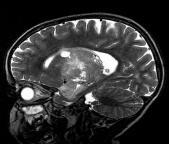
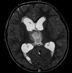
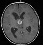
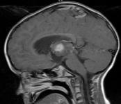
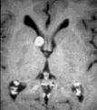
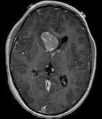
3 Solid, cystic, minimal enhancement and mass effect Partial, nodular enhancement Giant cell astrocytoma Glioblastoma multiforme (grade IV) Similar to adults Rare in children May mimic abscess + 22m Near foramen of Monro Benign lesion 5-15% of TSC patients M=F, any age, peak: 5-10Y Clinics: Hydrocephalus Rarely malignant degeneration Arise from subependymal hamartoma? Giant cell astrocytoma Differential diagnosis Many more tumors are known: Ependymoma PNET ATRT Choroid plexus papilloma May look similar to high grade astrocytomas, biopsy may be necessary 3


 M, 7Y, failure to")
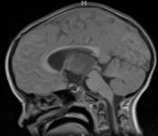
4 Sellar and suprasellar tumors Optic pathway glioma/astrocytomas Optic Nerve gliomas Craniopharyngeoma Hypothalamic hamartoma Langerhans Cell Histiocytosis Pituitary tumors Suprasellar germ cell tumors Optic pathway glioma Optic nerve glioma Optic nerve glioma: Starts in intraorbital segment of optic nerve; slow growth; JPA-like histology Hypothalamic/chiasmatic tumor: Starts in hypothalamus; more aggressive/invasive, histology similar to hemispheric astrocytomas Clinics: Diminished vision, pituitary dysfunction, hydrocephalus, diencephalic syndrome 20-50% have NF1 Optic nerve glioma Optic nerve glioma Frequently stable on follow up Look for other (NF1) lesions (UBO, JPA, ) uuhsc.utah.edu Optic nerve glioma Optic pathway glioma Subarachnoid optic nerve sheath surrounds glioma Differentiation from optic nerve sheath meningeoma (NF2, MISME) M, 7Y, failure to thrive, diencephalic syndrome, pilomyxoid type juvenile pilocytic astrocytoma 4


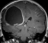
5 Always look beyond the most obvious findings Always look beyond the most obvious findings Glioma of the optic chiasm, UBO s, MCA infarction Glioma of the optic chiasm, UBO s, MCA infarction, Moya Moya, post ECA-MCA anastomosis in NF1 patient Craniopharyngeoma Craniopharyngeoma Along hypothalamic-pituitary axis 15% of all supratentorial tumors M>F, peak between years Originate from remnants of pluripotents cells Clinics: Visual field defects, pituitary or hypothalamic dysfunction, hydrocephalus Imaging: Solid w/wo cysts, calcifications, vary greatly in size May infiltrate adjacent brain Germinoma Pituitary macroadenoma Vision loss, 11Y Girl, confirmed by biopsy 11y female endocrine disorder 5


 Usually in older children More solid, small")

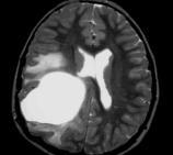
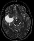
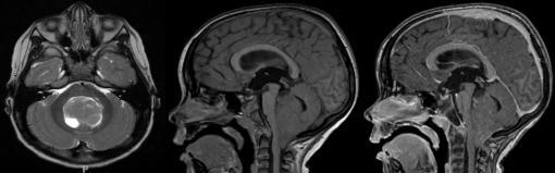
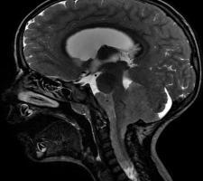
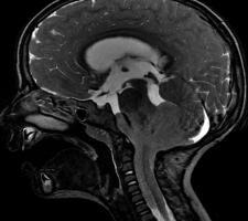
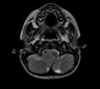
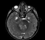
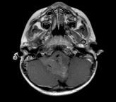
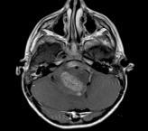
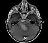
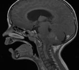
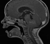
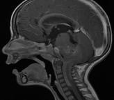
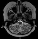
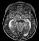
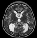
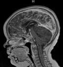
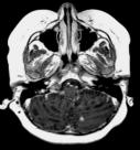
6 Hypothalamic hamartoma Tuber cinereum hamartoma Rare congenital malformations Normal neuronal tissue In region of mamillary bodies/tuber cinereum Precocious puberty, gelastic seizures Infratentorial tumors in children Take advantage of statistics Cerebellar astrocytoma 30-35% 20-25% Brainstem glioma 20-25% Ependymoma 10-15% Total 80-90% Meningiomas, schwannomas, metastasis are rare in children!!! Prognosticators precocious puberty Cerebellar astrocytoma Most frequently encountered posterior fossa tumor (30-35%) Peak incidence 5-13 years 1:1.45 Low grade (75-80%), anaplastic (15-25%) Usually benign course, slow growth, expansive Located within cerebellar hemispheres or vermis Compression of IV ventricle ~> hydrocephalus Headache, nausea, vomiting, ataxia, gait disturbance Cerebellar astrocytoma Pilocytic astrocytoma (WHO 1): Usually macrocystic with solid tumor nodule Hairy tumor Leptomeningeal metastases rare on initial presentation (5%) Good long term prognosis if treated (90%,10y) Anaplastic astrocytoma (WHO III-IV) Usually in older children More solid, small cells More aggressive/infiltrative, poor prognosis More frequent leptomeningeal metastases HE Bipolar cells, with hairlike processes HE Cerebellar pilocytic astrocytoma Cerebellar pilocytic astrocytoma T1 Tumor nodule enhances, cyst does not enhance , 11Y Male Tumor nodule may show central necrosis 6


 Kleihus P, Tumours")


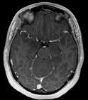
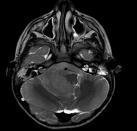
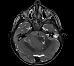
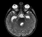
7 Steroid treatment Cerebellar anaplastic astrocytoma Follow up after treatment, leptomeningeal and local dissemination 2 nd /3 rd most frequent tumor, 20-25% First decade, peak at 7yrs 1: % in cerebellar vermis 10-15% in cerebellar hemispheres: lateral medulloblastomas (older children) IV ventricle compression with obstructive hydrocephalus Overall 5 year survival 60%, depending on histology and risk factors higher survival rates (90%) Kleihus P, Tumours of the Nervous System CT Classic MB +C Desmoplastic MB Posterior to fourth ventricle MBEN High density male 7y, headache, vomiting, ataxia Desmoplastic medulloblastoma Unsharp posterior margins T1 T1 T1 Enhancement Variable enhancement 7


")
 Focal tectal glioma (5%) Brainstem glioma Diffuse")


")
8 Brainstem glioma 2 nd /3 rd most frequent tumor, 20-25% First decade, peak at 7-9yrs 1:1 Kleihus P, Tumours of the Nervous System CSF-metastases in 30-50% of children on initial presentation Many classification systems Most frequently according to primary location & neuroimaging characteristics Diffuse intrinsic brainstem glioma (80%) Posterior exophytic glioma of cervicomedullary junction (15%) Focal tectal glioma (5%) Brainstem glioma Diffuse intrinsic brainstem glioma Depending on localization, prognosis and treatment vary significantly Tectal glioma excellent prognosis compared to diffuse intrinsic brainstem glioma Exophytic glioma may be operated, diffuse brainstem glioma cannot be operated Clinical presentation depends on primary location and involved neurofunctional structures Most frequently centered within pons Involve > 50% of cross-sectional area Triad: ataxia, long tract signs, multiple cranial nerve deficits Mood change and irritability Fibrillary astrocytoma WHO III-IV Poor prognosis, most children die < 2 yrs No effective treatment Radiotherapy may relieve symptoms temporarily Neuroimaging is specific, no biopsy necessary Diffuse intrinsic brainstem glioma Diffuse intrinsic brainstem glioma T1 Kleihus P, Tumours of the Nervous System Variable enhancement on follow up, dedifferentiation (WHO III ~> IV) Boy, 9yrs Basliary artery embracement, preserved fiber tracts, minimal enhancement 8


9 Posterior exophytic glioma Posterior exophytic glioma Located at cervicomedullary junction More favourable prognosis, median survival > 5yrs Most frequently, pilocytic astrocytoma Extend into IV ventricle Almost no infiltrative components Long history of non-specific headache and vomiting. Lower cranial nerve deficits, impaired speech and swollowing. Torticollis due to tonsillar herniation At least partial surgical resection is possible T1 Focal tectal glioma Well demarcated low grade glioma Tectal plate Good prognosis, median survival > 7 yrs Focal tectal glioma Present with increased ICP due to obstructive hydrocephalus Internuclear ophtalmoplegia, Parinaud s syndrome Hydrocephalus usually treated with 3rd ventriculostomy TAGM Focal tectal glioma Treatment: 3 rd Ventriculostomy Ependymoma 4th most common posterior fossa tumor (10-15%) Peak incidence 3-5 yrs, up to 18 yrs. 1:1.5 Arise from ependymal lining of IV ventricle (esp. velum medullare posterior) Typically respect ventricular system Tumor extension along ventricles and their outlets (Magendie/Luschka) Present with signs of increased intracranial pressure (obstructive hydrocephalus) Ataxia and cranial nerve palsy 9
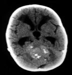
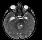
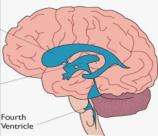
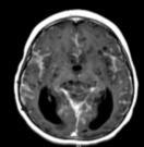
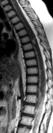
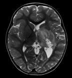
10 Ependymoma Ependymoma CSF-seeding to spinal canal may occur If CSF-seeding is seen, anaplastic ependymoma should be suspected High cellularity ~> hyperdense on CT Epicenter in fourth ventricle Perivascular pseudorosets Ependymoma Ependymoma Ependymoma frequently originates from velum medullare posterior Extension along ventricular system (even into internal auditory canal) Ependymoma Finally Metastases ~> primary brain tumors Moderate, patchy contrast enhancement M, 4Y, malignant neuroectodermal tumor 10


11 Summary Is it really a tumor? What is the most likely diagnosis? Is surgical resection an option? Use your statistics Be prepared for the unexpected Consider non-neoplastic etiologies Do not forget the spine Get the best anatomical and functional image quality Rene Margrit 11
Tumors of the Nervous System
 Tumors of the Nervous System Peter Canoll MD. PhD. What I want to cover What are the most common types of brain tumors? Who gets them? How do they present? What do they look like? How do they behave? 1
CNS TUMORS. D r. Ali Eltayb ( U. of Omdurman. I ). M. Path (U. of Alexandria)
. M. Path (U. of Alexandria)") CNS TUMORS D r. Ali Eltayb ( U. of Omdurman. I ). M. Path (U. of Alexandria) CNS TUMORS The annual incidence of intracranial tumors of the CNS ISmore than intraspinal tumors May be Primary or Secondary
CNS TUMORS D r. Ali Eltayb ( U. of Omdurman. I ). M. Path (U. of Alexandria) CNS TUMORS The annual incidence of intracranial tumors of the CNS ISmore than intraspinal tumors May be Primary or Secondary
Peter Canoll MD. PhD.
 Tumors of the Nervous System Peter Canoll MD. PhD. What I want to cover What are the most common types of brain tumors? Who gets them? How do they ypresent? What do they look like? How do they behave?
Pediatric CNS Tumors. Disclosures. Acknowledgements. Introduction. Introduction. Posterior Fossa Tumors. Whitney Finke, MD
 Pediatric CNS Tumors Disclosures Whitney Finke, MD Neuroradiology Fellow PGY-6 University of Utah Health Sciences Center Salt Lake City, Utah None Acknowledgements Introduction Nicholas A. Koontz, MD Luke
Metastasis. 57 year old with progressive Headache and Right Sided Visual Loss
 Metastasis 1% of sellar/parasellar masses Usually occurs with known primary Can involve third ventricle, hypothalamus, infundibular stalk May be both supra-, intrasellar 57 year old with progressive Headache
General: Brain tumors are lesions that have mass effect distorting the normal tissue and often result in increased intracranial pressure.
 1 Lecture Objectives Know the histologic features of the most common tumors of the CNS. Know the differences in behavior of the different tumor types. Be aware of the treatment modalities in the various
CNS pathology Third year medical students. Dr Heyam Awad 2018 Lecture 12: CNS tumours 2/3
 CNS pathology Third year medical students Dr Heyam Awad 2018 Lecture 12: CNS tumours 2/3 Pilocytic astrocytoma Relatively benign ( WHO grade 1) Occurs in children and young adults Mostly: in the cerebellum
Brain tumors: tumor types
 Brain tumors: tumor types Tumor types There are more than 120 types of brain tumors. Today, most medical institutions use the World Health Organization (WHO) classification system to identify brain tumors.
Pathologic Analysis of CNS Surgical Specimens
 2015 Kenneth M. Earle Memorial Neuropathology Review Pathologic Analysis of CNS Surgical Specimens Peter C. Burger, MD Interdisciplinary Quality Control Familiarity with entities Use of diagnostic algorithm
Site Specific Coding Rules MALIGNANT CENTRAL NERVOUS SYSTEM TUMORS
 Multiple Primary and Histology Site Specific Coding Rules MALIGNANT CENTRAL NERVOUS SYSTEM TUMORS 1 Prerequisites 2 Completion of Multiple Primary and Histology General Coding Rules 3 There are many ways
Pediatric Brain Tumors: Updates in Treatment and Care
 Pediatric Brain Tumors: Updates in Treatment and Care Writer Classroom Rishi R. Lulla, MD MS Objectives Introduce the common pediatric brain tumors Discuss current treatment strategies for pediatric brain
Pediatric Brain Tumors Pre, Intra & Post Op Evaluation and Management. Timothy M. George, MD, FACS, FAAP
 Pediatric Brain Tumors Pre, Intra & Post Op Evaluation and Management Timothy M. George, MD, FACS, FAAP PEDIATRIC BRAIN TUMORS BACKGROUND: Incidence: Third most common pediatric tumor type (leukemia, neuroblastoma,
Tumors of the Central Nervous System
 Tumors of the Central Nervous System 1 Financial Disclosures I have NO SIGNIFICANT FINANCIAL, GENERAL, OR OBLIGATION INTERESTS TO REPORT Introduction General: Brain tumors are lesions that have mass effect
CT & MRI Evaluation of Brain Tumour & Tumour like Conditions
 CT & MRI Evaluation of Brain Tumour & Tumour like Conditions Dr. Anjana Trivedi 1, Dr. Jay Thakkar 2, Dr. Maulik Jethva 3, Dr. Ishita Virda 4 1 M.D. Radiology, Professor and Head, P.D.U. Medical College
PITUITARY PARASELLAR LESIONS. Kim Learned, MD
 PITUITARY PARASELLAR LESIONS Kim Learned, MD DIFFERENTIALS Pituitary Sella Clivus, Sphenoid Sinus Suprasellar Optic chiasm, Hypothalamus, Circle of Willis Parasellar Cavernous Sinus Case 1 17 YEAR-OLD
Optic Pathway Gliomas, Germinomas, Spinal Cord Tumours. Colin Kennedy March 2015
 Optic Pathway Gliomas, Germinomas, Spinal Cord Tumours Colin Kennedy March 2015 Glioma of the optic chiasm. T1-weighted MRI with gadolinium enhancement, showing intense irregular uptake of contrast. The
Brain Tumors. Medulloblastoma. Pilocytic astrocytoma: Ahmed Koriesh, MD. Pathological finding
 NeuroPathology Page 8 Brain Tumors Pathological finding Pseudorosette Rosenthal fibers Rosettes Wet Keratin Psammoma bodies Fried egg Tumor Ependymoma, SEGA Pilocytic astrocytoma Medulloblastoma Craniopharyngioma
Update on Pediatric Brain Tumors
 Update on Pediatric Brain Tumors David I. Sandberg, M.D. Director of Pediatric Neurosurgery & Associate Professor Dr. Marnie Rose Professorship in Pediatric Neurosurgery Pre-talk Questions for Audience
Pediatric Spine Tumors (and other masses)
") Pediatric Spine Tumors (and other masses) Francisco A Perez, MD, PhD Assistant Professor Neuroradiology and Pediatric Radiology Seattle Children s Hospital University of Washington, Seattle Commercial
Pediatric Spine Tumors (and other masses) Francisco A Perez, MD, PhD Assistant Professor Neuroradiology and Pediatric Radiology Seattle Children s Hospital University of Washington, Seattle Commercial
Introduction to Neurosurgical Subspecialties:
 Introduction to Neurosurgical Subspecialties: Pediatric Neurosurgery Brian L. Hoh, MD 1 and Gregory J. Zipfel, MD 2 1 University of Florida, 2 Washington University Pediatric Neurosurgery Pediatric neurosurgeons
STUDY OFPAEDIATRIC CNS TUMORS IN TERTIARY CARE CENTER
 IJCRR Section: Healthcare Sci. Journal Impact Factor 4.016 Original Article STUDY OFPAEDIATRIC CNS TUMORS IN TERTIARY CARE CENTER Grishma P. Jobanputra Tutor, Department of Pathology, B.J. Medical College,
Neuro-oncology Update Andrew Kokkino, MD Medical Director, The Neurosciences Institute at Sacred Heart at Riverbend May 20, 2013
 Neuro-oncology Update 2013 Andrew Kokkino, MD Medical Director, The Neurosciences Institute at Sacred Heart at Riverbend May 20, 2013 Case 1 58 year old man with recent facial droop and HA s Thin, cachectic
EXPERT DIFFERENTIAL DIAGNOSIS:
 EXPERT DIFFERENTIAL DIAGNOSIS: Sellar Region Anne G. Osborn, M.D. DISCLOSURE: Published RSNA 2008 SELLA, PITUITARY: Normal Gross, 3T Anatomy SELLA, PITUITARY: Anatomically-Based Differential Diagnoses
Central Nervous System Tumors in Children. Dr.Mina Tajvidi
 Central Nervous System Tumors in Children. Dr.Mina Tajvidi CNS Central nervous system (CNS) tumors account for 20% to 25% of all malignancies that occur in childhood(united States) The incidence is highest
Posterior fossa tumors: clues to differential diagnosis with case-based review
 Posterior fossa tumors: clues to differential diagnosis with case-based review Poster No.: C-0323 Congress: ECR 2017 Type: Educational Exhibit Authors: H. A. Aboughalia, M. Abdelhady; Doha/QA Keywords:
Histopathological Study and Categorisation of Brain Tumors
 Histopathological Study and Categorisation of Brain Tumors Ruchira Wadhwa 1*, Purvi Patel 2, Hansa Goswami 3 1 Third Year Resident, 2 Assistant Professor, 3 Professor and Head, Department of Pathology,
What Are We Going to Do? Fourth Year Meds Clinical Neuroanatomy. Hydrocephalus and Effects of Interruption of CSF Flow. Tube Blockage Doctrine
 Fourth Year Meds Clinical Neuroanatomy Ventricles, CSF, Brain Swelling etc. David A. Ramsay, Neuropathologist, LHSC What Are We Going to Do? Hydrocephalus and some effects of the interruption of CSF flow
CHAPTER 11 Tumors Originating in the Brain Medulloblastomas, PNETs and Ependymomas
 Tumors Originating in the Brain Medulloblastomas, PNETs and Ependymomas Foolishly, I waited 7 months before I joined this (or any) group. By that time, my son had radiation, chemo, and a recurrence of
Adult Brain Tumours: an approach based on imaging findings
 Adult Brain Tumours: an approach based on imaging findings Robert J Sevick, MD, FRCPC, FACR Professor, Radiology and Clinical Neurosciences Cumming School of Medicine University of Calgary Learning objectives:
BRAIN TUMORS IN INFANTS
 BRAIN TUMORS IN INFANTS Dr Sergio Valenzuela M.D-( ISPN-ESPN-FLANC)&cols. Head Pediatric Neurosurgery Unit I Instituto de NeurocirugiaAsenjo Santiago CHILE RATE OF MENINGEAL,BRAIN AND OTHER CNS MALIGNANT
TUMORS of nervous system
 TUMORS of nervous system By: Shifaa Alqa qa Done By : Ola Hijjawi CNS tumors : The annual incidence of CNS tumors ranges from 10 to 17 per 100,000 persons for intracranial tumors and 1 to 2 per 100,000
Outline. Neuroradiology. Diffusion Imaging in. Clinical Applications of. Basics of Diffusion Imaging. Basics of Diffusion Imaging
 Clinical Applications of Diffusion Imaging in Neuroradiology No disclosures Stephen F. Kralik Assistant Professor of Radiology Indiana University School of Medicine Department of Radiology and Imaging
SPECIAL SLIDE SEMINAR CASE 3
 SPECIAL SLIDE SEMINAR CASE 3 Tihana Džombeta, MD Leo Pažanin, MD, PhD Department of Pathology, School of Medicine, University of Zagreb Department of Pathology, Clinical Hospital Centre Sestre milosrdnice
Joana Ramalho, MD C. Ryan Miller, MD, PhD
 Joana Ramalho, MD C. Ryan Miller, MD, PhD Case 1 3 month old baby girl Presented with new onset of seizures Newborn. Questionable blurring of the gray-white junction within the right occipital lobe. Findings
Unknown Cases. Financial Disclosures & Disclaimers. Unknown Cases Case #2. Unknown Cases Case #1. Differentials in Pediatric Brain Imaging
 Financial Disclosures & Disclaimers Differentials in Pediatric Brain Imaging William T. O Brien, Sr., D.O. Program Director, Diagnostic Radiology Residency, DGMC Associate Clinical Professor of Radiology
Patients Treated with Leksell Gamma Knife
 Patients Treated with Leksell Gamma Knife 1968-2016 TREATMENTS REPORTED 2016 BY REGION AND INDICATION INDICATION Asia excl. Europe Latin Middle East & Africa North Grand Total Benign Tumors 12283 9778
NEURORADIOLOGY DIL part 5
 NEURORADIOLOGY DIL part 5 Masses and tumors K. Agyem MD, G. Hall MD, D. Palathinkal MD, Alexandre Menard March/April 2015 OVERVIEW Introduction to Neuroimaging - DIL part 1 Basic Brain Anatomy - DIL part
Case 7391 Intraventricular Lesion
 Case 7391 Intraventricular Lesion Bastos Lima P1, Marques C1, Cabrita F2, Barbosa M2, Rebelo O3, Rio F1. 1Neuroradiology, 2Neurosurgery, 3Neuropathology, Coimbra University Hospitals, Portugal. University
Childhood Brain and Spinal Cord Tumors Treatment Overview (PDQ )
") 1 di 8 04/03/2017 07.31 NCBI Bookshelf. A service of the National Library of Medicine, National Institutes of Health. PDQ Cancer Information Summaries [Internet]. Bethesda (MD): National Cancer Institute
1 di 8 04/03/2017 07.31 NCBI Bookshelf. A service of the National Library of Medicine, National Institutes of Health. PDQ Cancer Information Summaries [Internet]. Bethesda (MD): National Cancer Institute
Financial Disclosures I have no financial interests to disclose. Templar Eye Foundation Oppenheimer Family Foundation
 Financial Disclosures I have no financial interests to disclose. Templar Eye Foundation Oppenheimer Family Foundation 2 Case 7 year old girl Initially parents noticed photophobia Then started to complain
Anaplastic Pilocytic Astrocytoma: The fusion of good and bad
 Anaplastic Pilocytic Astrocytoma: The fusion of good and bad Alexandrina Nikova 1, Charalampos-Chrysovalantis Chytoudis-Peroudis 2, Penelope Korkolopoulou 3 and Dimitrios Kanakis 4 Abstract 5 Pilocytic
H Haloes cautions, 57 neurocytomas, perinuclear, 56 Headache blue cell tumors, 147 cautions, 135, 147, 152 clinical history, 132, 144, 148
 Index A ADC. See Apparent diffusion coefficient Adult. See also Supratentorial mass, adult cerebral tumor, 1 headache and ataxia cysts, mural nodules, 118 sporadic tumors, 118 headaches and visual changes,
Case Report Atypical Presentation of Atypical Teratoid Rhabdoid Tumor in a Child
 Case Reports in Oncological Medicine Volume 2013, Article ID 815923, 4 pages http://dx.doi.org/10.1155/2013/815923 Case Report Atypical Presentation of Atypical Teratoid Rhabdoid Tumor in a Child Y. T.
Five Most Common Problems in Surgical Neuropathology
 Five Most Common Problems in Surgical Neuropathology If the brain were so simple that we could understand it, we would be so simple that we couldn t Emerson Pugh What is your greatest difficulty in neuropathology?
Astroblastoma: Radiologic-Pathologic Correlation and Distinction from Ependymoma
 AJNR Am J Neuroradiol 23:243 247, February 2002 Case Report Astroblastoma: Radiologic-Pathologic Correlation and Distinction from Ependymoma John D. Port, Daniel J. Brat, Peter C. Burger, and Martin G.
Laurie A. Loevner, MD
 Laurie A. Loevner, MD Chief, Division of Neuroradiology UPHS Professor of Radiology, Otorhinolaryngology: Head & Neck Surgery, Neurosurgery, and Ophthalmology University of Pennsylvania Health System Disclosures
Collecting Cancer Data: Central Nervous System Prizes! Question of the Month! Tip of the Month! Q&A
 Collecting Cancer Data: Central Nervous NAACCR 2008 2009 Webinar Series April 2, 2009 Prizes! Question of the Month! The participant that submits the best question of the session will receive a fbl fabulous
Brain and Spine Tumors
 Brain and Spine Tumors Andrew J. Fabiano, MD FAANS Associate Professor of Neurosurgery Roswell Park Cancer Institute SUNY at Buffalo School of Medicine Brain Tumors Brain Tumor Basics Types of Tumors Cases
Introduction. In adults, the commonest tumors are gliomas, metastases and meniongiomas; most lie in the supratentorial compartment
 Introduction Primary brain tumor 6 persons/100000/year Metastatic brain tumor 6 persons/100000/year 1 in 15 primary brain tumors occur in children under 15 years In adults, the commonest tumors are gliomas,
Management of pediatric brain tumors, strategies and long term outcome
 Management of pediatric brain tumors, strategies and long term outcome SAN The Sudanese association of neurosurgeons By Dr. Abubakr Darrag Salim Ahmed Dr. Mohammed Awad Elzain Khartoum Sudan Pediatric
INTRACRANIAL ARACHNOID CYSTS: CLASSIFICATION AND MANAGEMENT. G. Tamburrini, Rome
 INTRACRANIAL ARACHNOID CYSTS: CLASSIFICATION AND MANAGEMENT G. Tamburrini, Rome Incidence 2% of occasional neuroradiological findings From clinical studies (1960 s): 0.4-1% of intracranial space occupying
SURGICAL MANAGEMENT OF BRAIN TUMORS
 SURGICAL MANAGEMENT OF BRAIN TUMORS LIGIA TATARANU, MD, Ph D NEUROSURGICAL CLINIC, BAGDASAR ARSENI CLINICAL HOSPITAL BUCHAREST, ROMANIA SURGICAL INDICATIONS CONFIRMING HISTOLOGIC DIAGNOSIS REDUCING TUMOR
RINGS N THINGS: Imaging Patterns in Differential Diagnosis. Anne G. Osborn, M.D.
 RINGS N THINGS: Imaging Patterns in Differential Diagnosis Anne G. Osborn, M.D. ExpDDxs: Intra-axial (Parenchymal) Lesions Ring-enhancing lesions, solitary 1 Ring-enhancing lesion crossing corpus callosum
We are IntechOpen, the first native scientific publisher of Open Access books. International authors and editors. Our authors are among the TOP 1%
 We are IntechOpen, the first native scientific publisher of Open Access books 3,350 108,000 1.7 M Open access books available International authors and editors Downloads Our authors are among the 151 Countries
Brainstem diffuse gliomas: radiologic findings.
 Brainstem diffuse gliomas: radiologic findings. Poster No.: C-2220 Congress: ECR 2013 Type: Educational Exhibit Authors: E. GARCIA MARTINEZ 1, D. H. Jiménez 1, L. Navarro Vilar 2, C. P. Fernandez Ruiz
Brain Tumors. Andrew J. Fabiano, MD FAANS. Associate Professor of Neurosurgery Roswell Park Cancer Institute SUNY at Buffalo School of Medicine
 Brain Tumors Andrew J. Fabiano, MD FAANS Associate Professor of Neurosurgery Roswell Park Cancer Institute SUNY at Buffalo School of Medicine Brain Tumors Brain Tumor Basics Types of Tumors Cases Brain
Dr. Shashi Ranjani Dr Thangavelu unit Mehta children hospital
 Diagnosis after death Dr. Shashi Ranjani Dr Thangavelu unit Mehta children hospital Management team Dr.V.P.Anitha (consultant PICU) Dr.Mahesh (paediatric neurologist) Dr. Thirumaran (Neurosurgeon) 1 4
Childhood brain tumours
 Childhood brain tumours Our bodies are made up of billions of cells. Normally, these cells reproduce and repair themselves in a controlled way and do not cause us any problems. If for some reason this
CASE OF THE WEEK PROFESSOR YASSER METWALLY
 CLINICAL PICTURE CLINICAL PICTURE 15 years old male patient presented with symptoms of nausea, vomiting, and ataxia, with evidence of torticollis, papilledema, nystagmus, and palsies of the sixth and seventh
Aria Fallah MD, MSc, FRCSC
 Aria Fallah MD, MSc, FRCSC Department of Neurosurgery David Geffen School of Medicine at UCLA Pineal Region Tumors Brain Tumor Symposium August 22, 2015 Disclosures None Pineal Gland Arises from an invagination
Complex Hydrocephalus
 2012 Hydrocephalus Association Conference Washington, DC - June 27-July1, 2012 Complex Hydrocephalus Marion L. Walker, MD Professor of Neurosurgery & Pediatrics Primary Children s Medical Center University
Case Studies in Sella/Parasellar Region. Child thirsty, increased urination. Imaging. Suprasellar Germ Cell Tumor (Germinoma) No Disclosures
 No Disclosures") Case Studies in Sella/Parasellar Region No Disclosures 2018 Head and Neck Imaging Conference Child thirsty, increased urination Suprasellar Germ Cell Tumor (Germinoma) Midline Pineal >> Suprasellar > Other
Case Studies in Sella/Parasellar Region No Disclosures 2018 Head and Neck Imaging Conference Child thirsty, increased urination Suprasellar Germ Cell Tumor (Germinoma) Midline Pineal >> Suprasellar > Other
Childhood Brain and Spinal Cord Tumors Treatment Overview (PDQ )
") 1 di 14 27/11/2016 17.42 NCBI Bookshelf. A service of the National Library of Medicine, National Institutes of Health. PDQ Cancer Information Summaries [Internet]. Bethesda (MD): National Cancer Institute
1 di 14 27/11/2016 17.42 NCBI Bookshelf. A service of the National Library of Medicine, National Institutes of Health. PDQ Cancer Information Summaries [Internet]. Bethesda (MD): National Cancer Institute
Cross sectional imaging of Intracranial cystic lesions Abdel Razek A
 Cross sectional imaging of Intracranial cystic lesions Abdel Razek A Department of Radiology. Mansoura Faculty of Medicine, Mansoura. Egypt. arazek@mans.edu.eg Introduction Intracranial cystic lesions
Challenging Paediatric Brain Tumours. ASP Belfast March 2017 Dr Jane Pears Consultant Paediatric Oncologist, Dublin
 Challenging Paediatric Brain Tumours ASP Belfast March 2017 Dr Jane Pears Consultant Paediatric Oncologist, Dublin Overview (i) Paediatric malignancy (ii) Central nervous system tumours (iii) Diffuse Intrinsic
Q&A. Fabulous Prizes. Collecting Cancer Data:CNS 2/7/12. NAACCR Webinar Series Collecting Cancer Data Central Nervous System
 Collecting Cancer Data Central Nervous System NAACCR 2012 2013 Webinar Series 2/7/2013 Q&A Please submit all questions concerning webinar content through the Q&A panel. Reminder: If you have participants
Neuroimaging Core Curriculum
 Neuroimaging Core Curriculum Program Content The purpose of the training program is to prepare the physician for the independent practice of neuroimaging. Neuroimaging is the subspecialty of Neurology
Dr.Dafalla Ahmed Babiker Jazan University
 Dr.Dafalla Ahmed Babiker Jazan University Brain tumors are the second commonest malignancy in children Infratentorial tumors are more common As a general rule they do not metastasize out of the CNS, but
MRI OF THE THALAMUS. Mohammed J. Zafar, MD, FAAN Kalamazoo, MI
 1 MRI OF THE THALAMUS Mohammed J. Zafar, MD, FAAN Kalamazoo, MI Objectives: The thalamic nuclei can be involved in a wide variety of conditions. A systematic imaging approach would be useful for narrowing
THE BRAIN PATHWAYS GUIDELINE: A GUIDELINE TO ASSIST HEALTHCARE PROFESSIONALS IN THE ASSESSMENT OF CHILDREN WHO MAY HAVE A BRAIN TUMOUR
 THE BRAIN PATHWAYS GUIDELINE: A GUIDELINE TO ASSIST HEALTHCARE PROFESSIONALS IN THE ASSESSMENT OF CHILDREN WHO MAY HAVE A BRAIN TUMOUR Version 2 February 2017 (revision of version 1 Published 2008) NICE
EEG IN FOCAL ENCEPHALOPATHIES: CEREBROVASCULAR DISEASE, NEOPLASMS, AND INFECTIONS
 246 Figure 8.7: FIRDA. The patient has a history of nonspecific cognitive decline and multiple small WM changes on imaging. oligodendrocytic tumors of the cerebral hemispheres (11,12). Electroencephalogram
Chapter 1 Introduction
 Chapter 1 Introduction Men think epilepsy divine, merely because they do not understand it. But if they called everything divine which they do not understand, why, there would be no end to divine things.
Case 1. Maysa Al-Hussaini MD FRCPath
 Case 1 Maysa Al-Hussaini MD FRCPath MAYSA King AL-HUSSAINI Hussein Cancer MD Center MRCPATH KING HUSSEIN Amman CANCER Jordan CENTER Clinical history 4 year old boy History of frontal headache, sleepiness.
I have no conflicts of interest in relation to this presentation. Vogel FS & Burger PC 3/28/2016
 IF THIS IS NOT GLIOBLASTOMA, THEN WHAT IS IT? Murat Gokden, MD Department of Pathology/Neuropathology University of Arkansas for Medical Sciences Little Rock, AR mgokden@uams.edu I have no conflicts of
The Brain and Spinal Cord 33
 The Brain and Spinal Cord 33 shiao Y. Woo TOLERANCE OF THE CENTRAL NERVOUS SYSTEM TO IRRADIATION Postirradiation Cerebral Necrosis Postirradiation Myelopathy Other Late Effects CENTRAL NERVOUS SYSTEM NEOPLASMS
Neurosurgical Management of Brain Tumours. Nicholas Little Neurosurgeon RNSH
 Neurosurgical Management of Brain Tumours Nicholas Little Neurosurgeon RNSH General Most common tumours are metastatic 10x more common than primary Incidence of primary neoplasms is 20 per 100000 per year
Differential diagnosis of intracranial cystic lesions.
 Differential diagnosis of intracranial cystic lesions. Poster No.: C-0215 Congress: ECR 2015 Type: Educational Exhibit Authors: S. P. G. Alandete, M. A. Meseguer, E. De la Via, D. Uceda, C. Poyatos; Valencia/ES
Supplementary Information
 Rise in Glioblastoma Multiforme incidence in England 1995 2015 suggests an adverse environmental or lifestyle factor Alasdair Philips, Denis L Henshaw, Graham Lamburn, Michael J O Carroll Supplementary
Small and Big Operations: New Tools of the Trade for Brain Tumors. Disclosure. Incidence of Childhood Cancer
 Small and Big Operations: New Tools of the Trade for Brain Tumors Nalin Gupta MD PhD Chief, Division of Pediatric Neurosurgery Departments of Neurosurgery and Pediatrics University of California San Francisco
2. Subependymal giant cell astrocytoma:
 I. Astrocytomas: A. Diffusely infiltrating ( astrocytoma, anaplastic astrocytoma, GBM) B. Localised (pilocytic astrocytoma, pleomorphic xanthoastrocytoma, SGCA) *Grading: Diffuse: 1. Astrocytoma WHO grade
Computed Tomographic Evaluation of Posterior Fossa Lesions
 ORIGINAL ARTICLE Computed Tomographic Evaluation of Posterior Fossa Lesions Gupta MK 1, Rauniyar RK 1, Bhatta N 2, Raja S 3, Ahmad K 1 1 Department of Radiodiagnosis & Imaging, 2 Department of Internal
Pleomorphic Xanthoastrocytoma
 Pleomorphic Xanthoastrocytoma Christine E. Fuller Keywords Pleomorphic xanthoastrocytoma; Pleomorphic xanthoastrocytoma with anaplastic features 2.1 OVERVIEW Pleomorphic xanthoastrocytoma (PXA) is an uncommon
Neoplasms of the BRAIN and CNS
 Neoplasms of the BRAIN and CNS 2015-21016 FCDS Educational Webcast Series Steven Peace, BS, CTR October 15, 2015 2015 Focus Anatomy SSS 2000 MPH Rules AJCC TNM Presentation Outline Overview Reportable
intracranial anomalies
 Chapter 5: Fetal Central Nervous System 84 intracranial anomalies Hydrocephaly Dilatation of ventricular system secondary to an increase in the amount of CSF. Effects of hydrocephalus include flattening
Year 2003 Paper two: Questions supplied by Tricia
 question 43 A 42-year-old man presents with a two-year history of increasing right facial numbness. He has a history of intermittent unsteadiness, mild hearing loss and vertigo but has otherwise been well.
Clinical, radiological, and histopathological features and prognostic factors of brain tumors in children
 Journal of Physics: Conference Series PAPER OPEN ACCESS Clinical, radiological, and histopathological features and prognostic factors of brain tumors in children To cite this article: M H Siregar et al
Shared Care & Survival CTYA SSCRG (Childhood Cancer Research Group)
") Shared Care & Survival CTYA SSCRG (Childhood Cancer Research Group) January 2013 The NCIN is a UK-wide initiative, working to drive improvements in standards of cancer care and clinical outcomes by improving
Shared Care & Survival CTYA SSCRG (Childhood Cancer Research Group) January 2013 The NCIN is a UK-wide initiative, working to drive improvements in standards of cancer care and clinical outcomes by improving
Masses of the Corpus Callosum
 Masses of the Corpus Callosum Kesav Raghavan, HMS Year III Dr. Agenda Corpus Callosum Development and Anatomy Our Patient: Clinical Presentation Differential Diagnosis of Masses in the Corpus Callosum
Disclosures. Posterior Fossa Masses. I m from the Government. and I here to help! Differential Diagnosis
 Posterior Fossa Masses Differential Diagnosis James G. Smirniotopoulos, M.D. Radiology, Neurology, Biomedical Informatics Uniformed Services University Bethesda, Maryland http://rad.usuhs.edu http://medpix.usuhs.edu
Nature and Science 2017;15(7) Surgical Options for Treatment of Posterior Fossa Tumors with Hydrocephalus
 Surgical Options for Treatment of Posterior Fossa Tumors with Hydrocephalus") Surgical Options for Treatment of Posterior Fossa Tumors with Hydrocephalus Mohamed Mahmoud Abohashima; Ahmed Mohamed Hasan Salem; Magdy Asaad El-Hawary Neurosurgery department, Faculty of Medicine, Al-azhar
Surgical Options for Treatment of Posterior Fossa Tumors with Hydrocephalus Mohamed Mahmoud Abohashima; Ahmed Mohamed Hasan Salem; Magdy Asaad El-Hawary Neurosurgery department, Faculty of Medicine, Al-azhar
SCIENTIFIC PROGRAMME SNOLA THE STATE OF THE ART ON NEURO-ONCOLOGY th March
 SCIENTIFIC PROGRAMME SNOLA THE STATE OF THE ART ON NEURO-ONCOLOGY 2018 15th March 13h 13h45 ROOM 1 ROOM 2 ROOM 3 Imaging and pathology case discussion Lymphomas case discussion- Meningeomas Moderator:
Anatomy, Histology and general pathology of the Pineal gland. Uri Shiri 1 st year, Int. medicine B
 Anatomy, Histology and general pathology of the Pineal gland Uri Shiri 1 st year, Int. medicine B The Pineal gland A small, ~8mm sized Endocrine gland. The Pineal gland A small, ~8mm sized Endocrine gland.
Neuropathology Evening Session: Case 3
 Neuropathology Evening Session: Case 3 Christine E. Fuller, MD Cincinnati Children s Hospital Medical Center Disclosure of Relevant Financial Relationships USCAP requires that all faculty in a position
Intra-Fourth Ventricular Schwannoma With Obstructive Hydrocephalus A Rare Case Report
 ISPUB.COM The Internet Journal of Neurosurgery Volume 7 Number 1 Intra-Fourth Ventricular Schwannoma With Obstructive Hydrocephalus A Rare Case Report A Babbu, R Katheerayson Citation A Babbu, R Katheerayson..
Dr. Mammohan Singh. Dr. G.D. Satyarthee
 Moderator : Dr. Mammohan Singh Dr. G.D. Satyarthee Presenter : Dr. Anand Gupta Introduction Primary brain tumor 6 persons/100000/year Metastatic brain tumor 6 persons/100000/year 1 in 15 primary brain
Neuroanatomy. Assistant Professor of Anatomy Faculty of Medicine The University of Jordan Dr Maha ELBeltagy
 Neuroanatomy Dr. Maha ELBeltagy Assistant Professor of Anatomy Faculty of Medicine The University of Jordan 2018 Development of the Central Nervous System Development of the nervous system Development
Dosimetry, see MAGIC; Polymer gel dosimetry. Fiducial tracking, see CyberKnife radiosurgery
 Subject Index Acoustic neuroma, neurofibromatosis type 2 complications 103, 105 hearing outcomes 103, 105 outcome measures 101 patient selection 105 study design 101 tumor control 101 105 treatment options
Detection of Leptomeningeal CNS Metastases in Children
 Detection of Leptomeningeal CNS Metastases in Children Noah D. Sabin, M.D. Julie H. Harreld M.D. Kathleen J. Helton M.D. Zoltan Patay M.D., Ph.D. St. Jude Children s Research Hospital Memphis, TN Leptomeningeal
Protocol for management of patients with pineal region tumours v1
 Protocol for management of patients with pineal region tumours v1 West Midlands Cancer Alliance Coversheet for Cancer Alliance Expert Advisory Group Agreed Documentation This sheet is to accompany all
Classification of spontaneous brain tumors in rats
 Classification of spontaneous brain tumors in rats Central Nervous System Neoplasms in the Rat (Solleveld HA, et al., 1991) 1. Tumors of Neuroepithelial Tissue A. Astrocytic and oligodendroglial tumors