Advances In Orbital Neuropathology
|
|
|
- Daniella West
- 5 years ago
- Views:
Transcription


1 Advances In Orbital Neuropathology Charles G. Eberhart, MD PhD Associate Professor of Pathology, Ophthalmology and Oncology Johns Hopkins University School of Medicine
2 Overview Non-neoplastic lesions Microphthalmos/pseudoglioma Cephaloceles Neuroma Tumors Neurofibroma Schwannoma Meningioma Optic Nerve Glioma Genetic advances in Pilocytic Astrocytoma

3 Podos and Yanoff (1993)
4 Non-neoplastic lesions Microphthalmos/pseudoglioma Cephaloceles Neuroma Tumors Neurofibroma Schwannoma Meningioma Optic Nerve Glioma Genetic advances in Pilocytic Astrocytoma
5 Microphthalmia Caused by incomplete closure of fetal cleft Usually unilateral Often with large cyst May be associated with chromosomal deletions (13q or 18) Eye can be relatively normal or totally disorganized. Proliferating neuroectodermal tissue in the cyst can simulate neoplasm (pseudogliomatous hyperplasia)
6 Microphthalmia with cyst
7 Cephaloceles Developmental malformations with brain or meninges present in orbit. Sometimes retain communication with brain, but this often closes. Meningocele only meninges Encephalocele only brain Meningoencephalocele both present
 Amputation")
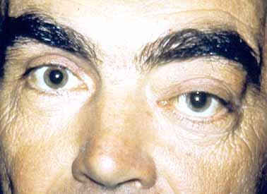
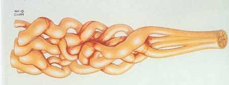
8 Rare Haphazardly entangled regenerating nerve fibers growing from end of disrupted peripheral ciliary nerve(s) Amputation Neuroma
9
10 Non-neoplastic lesions Microphthalmos/pseudoglioma Cephaloceles Neuroma Tumors Neurofibroma Schwannoma Meningioma Optic Nerve Glioma Genetic advances in Pilocytic Astrocytoma
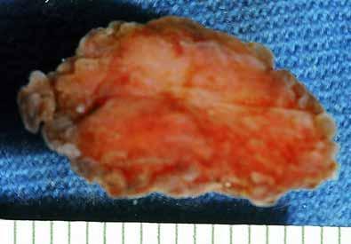
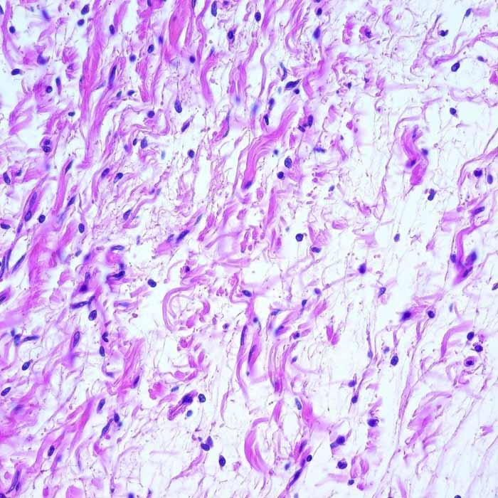
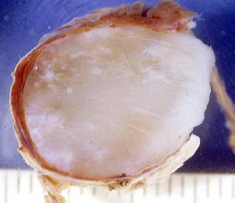
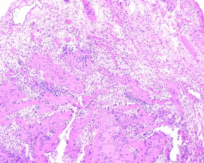
11 Neurofibroma Sporadic or NF1 associated Localized, diffuse and plexiform types Usually arise from sensory nerves 6 of 1,264 (<1%) of orbital lesions (Shields) Localized lesions can be well circumscribed, but are not encapsulated Treatment is surgical excision; recurrence is frequent with larger lesions
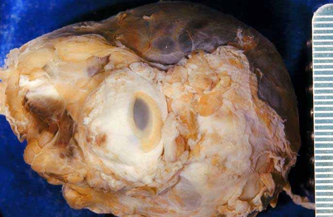
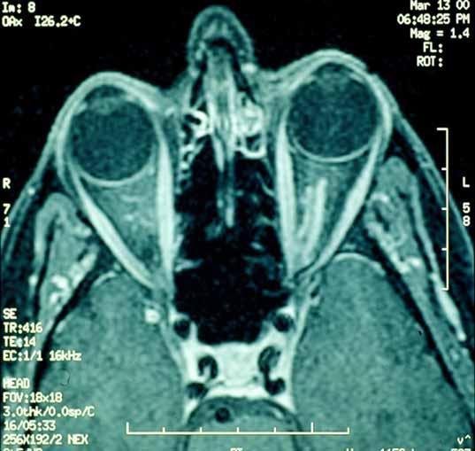
12 Neurofibroma
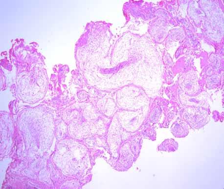
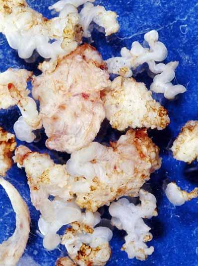
13 Plexiform Neurofibroma
14
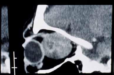
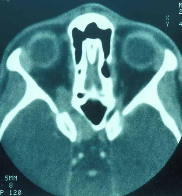
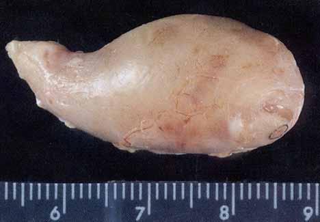
15 Schwannoma (Neurilemoma) Often arise from trigeminal nerves; less often from ocular motor nerves 14 of 1,264 (1%) of orbital lesions (Shields). Presentation at years of age (median 37) Mostly sporadic, but can be NF2-associated Encapsulated, and can sometimes be separated from the nerve
16 Schwannoma Slow progression of proptosis (several years)
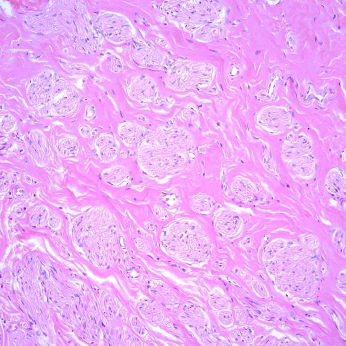
17 Schwannoma
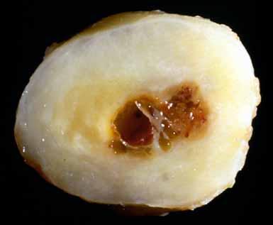
18 Cystic Schwannoma Dr. PS Rosenbaum EOPS 2003

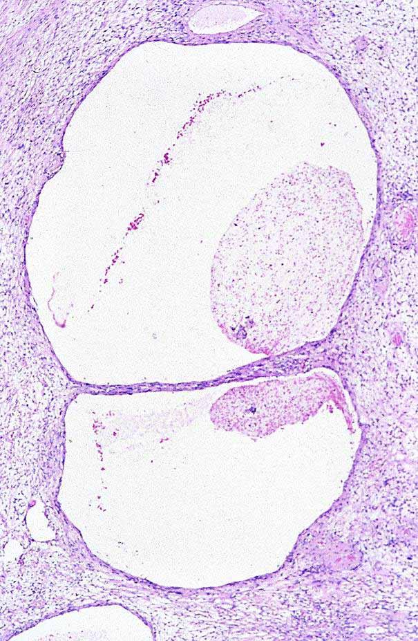
19 Schwannoma In contrast to neurofibroma and meningioma, schwannomas tend to roll along the slide and do not smear well. They also have distinctive elongated nuclei
20 Orbital Meningioma Can be derived from optic nerve sheath (primary) or extend into the orbit from the brain (secondary) 29 of 1264 (2%) of cases in one orbital tumor series were primary optic nerve menigiomas (Shields) In the same series, 24 (2%) were secondary intraorbital meningiomas Primary tumors unilateral in 95% of cases; bilateral cases usually in young adults Primary tumors may be localized or extend along much of orbital-canalicular optic nerve length

21 Dr. N. Miller

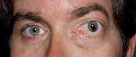
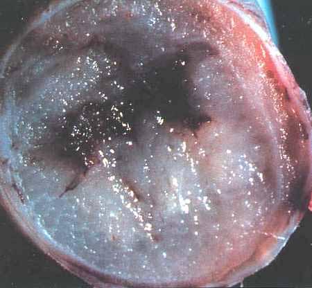
22 Diffuse Process with Tram-Tracking Dr. N Miller


23 Primary ONSMs usually not surgically curable unless accompanied by removal of optic nerve Dr. N Miller
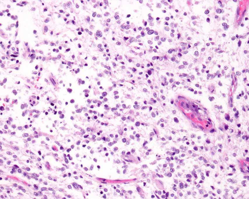
24 Review of Johns Hopkins Cases (Jain et al) 51 cases (21 Primary, 30 secondary) Mean age 45 years; 5 in children 2 patients with NF2 25 Meningothelial, 23 Transitional, 2 Angiomatous 1 Chordoid, 4 WHO grade II (elevated mitotic activity, brain invasion, Chordoid subtype)


25 Mitotic Activity In Grade II Tumor


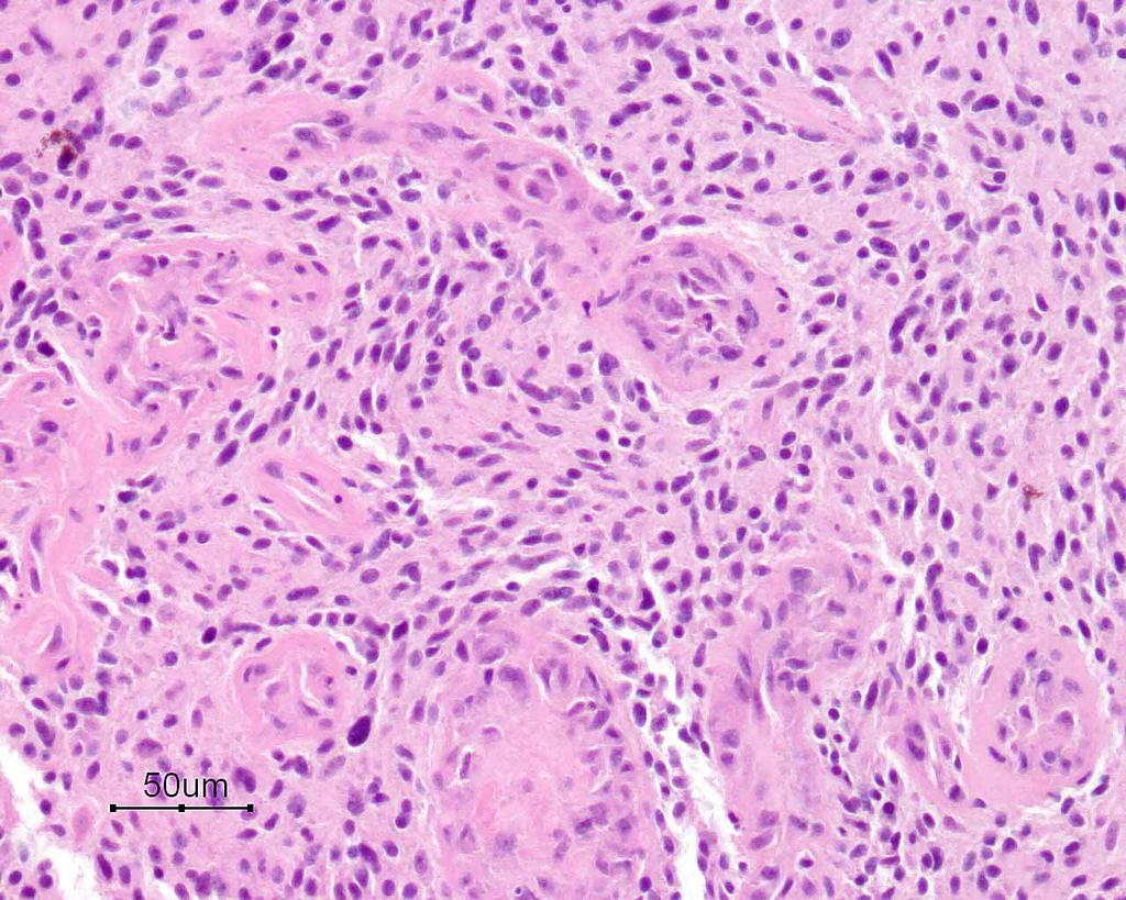
26 Invasion of Lacrimal Gland and Muscle

27 Invasion of Optic Nerve Equivalent to brain invasion? Probably not

28 Chordoid Intraorbital Mengioma (Secondary)

29 Meningioma Frozen Section Diagnosis Menigiomas smear easily and have crisp nuclear outlines with delicate chromatin Dr. P Burger
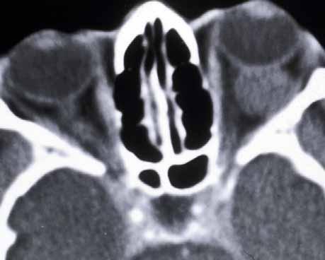
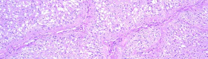
30 Optic Nerve Gliomas Almost all Pilocytic Astrocytomas 48 of 1,284 (4%) of orbital lesions (Shields) Can arise from orbital, optic canal or intracranial portions of the nerve Most in children in first decade of life 25% or more have evidence of neurofibromatosis type 1 (can be bilateral) 15% of children with NF 1 have ONGs


31 Anterior Orbital Presentation: Proptosis and optic disc swelling Neil Miller
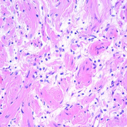
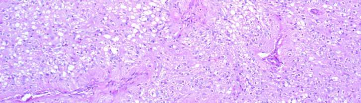
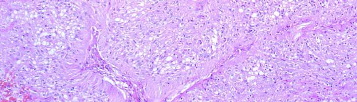
32 Posterior Orbital/Canalicular Presentation Decreased vision (variable) No (minimal) proptosis Relative afferent pupillary defect Asymptomatic Found during screening for NF1 Found during evaluation for pale disc

33 Neil Miller


34 CT scans showing diffuse enlargement of optic nerve Neil Miller

35 MRI Showing Intracranial Extension


36 Diffuse involvement of optic nerve parenchyma N. Miller

37

38 Subarachnoid Proliferation of Tumor

39 Dr. P Burger

40 Rosenthal Fibers

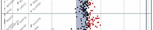
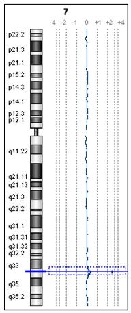
41 Eosinophilic Granular Body (EGB)
42 Natural History of PA Treatment generally not required Most remain stable throughout life and do not produce progressive visual loss Some spontaneously regress, often with improvement in visual function Rare cases increase in size associated with worsening vision Sporadic (non-nf1) tumors generally arise earlier and are more likely to grow Virtually no risk of spontaneous malignant transformation

43 Pilomyxoid Astrocytoma WHO grade II entity that is clinically more aggressive than Pilocytic Astrocytoma Frequently involves hypothalamus/chiasm Relatively monomorphous cells with mucoid background Perivascular orientation No Rosenthal fibers or EGBs

44 Malignant Optic Nerve Glioma 55 year old man Presented with visual Sx Chiasmal/optic nerve mass Died within 1 year of intracranial spread.
45
46
47 Non-neoplastic lesions Microphthalmos/pseudoglioma Cephaloceles Neuroma Tumors Neurofibroma Schwannoma Meningioma Optic Nerve Glioma Genetic advances in Pilocytic Astrocytoma
48
49 Chr
 Bar et al,")
50 17 of 25 PA had 7q34 gains 3 cases had activating BRAF mutations (V600E) Bar et al, JNEN 2008


51 Activation of BRAF/MEK/ERK Signaling in Pilocytic Astrocytoma pmek 81% Bar et al, JNEN 2008
52 The BRAF gene dupliction/amplification forms a fusion gene with unregulated kinase activity Kinase Domain RAS Binding Domain C-Term N-Term Exons Predicted telomeric end of 7q34 gain KIA
53


54 Samples 800bp- 350bp- We have identified BRAF/KIA fusion transcripts in all PA with 7q34 gains Sample 23 KIAA154 9 BRAF A Y I G C P D L I R D
55 Ras/Raf Signaling Can Be Activated At Multiple Points In PA RASSF1 RAS NF1 BRAF MEK ERK Bar et al, JNEN 2008
Meningiomas account for approximately 4% of all
 Intraorbital Meningiomas A Pathologic Review Using Current World Health Organization Criteria Deepali Jain, MD; Katayoon B. Ebrahimi, MD; Neil R. Miller, MD; Charles G. Eberhart, MD, PhD N Context. Meningiomas
General: Brain tumors are lesions that have mass effect distorting the normal tissue and often result in increased intracranial pressure.
 1 Lecture Objectives Know the histologic features of the most common tumors of the CNS. Know the differences in behavior of the different tumor types. Be aware of the treatment modalities in the various
CNS pathology Third year medical students. Dr Heyam Awad 2018 Lecture 12: CNS tumours 2/3
 CNS pathology Third year medical students Dr Heyam Awad 2018 Lecture 12: CNS tumours 2/3 Pilocytic astrocytoma Relatively benign ( WHO grade 1) Occurs in children and young adults Mostly: in the cerebellum
Year 2003 Paper two: Questions supplied by Tricia
 question 43 A 42-year-old man presents with a two-year history of increasing right facial numbness. He has a history of intermittent unsteadiness, mild hearing loss and vertigo but has otherwise been well.
Tumors of the Central Nervous System
 Tumors of the Central Nervous System 1 Financial Disclosures I have NO SIGNIFICANT FINANCIAL, GENERAL, OR OBLIGATION INTERESTS TO REPORT Introduction General: Brain tumors are lesions that have mass effect
Tumors of the Nervous System
 Tumors of the Nervous System Peter Canoll MD. PhD. What I want to cover What are the most common types of brain tumors? Who gets them? How do they present? What do they look like? How do they behave? 1
Five Most Common Problems in Surgical Neuropathology
 Five Most Common Problems in Surgical Neuropathology If the brain were so simple that we could understand it, we would be so simple that we couldn t Emerson Pugh What is your greatest difficulty in neuropathology?
Biomarkers in Neuro-Ophthalmic Tumors
 Biomarkers in Neuro-Ophthalmic Tumors Fausto J. Rodríguez MD Department of Pathology Johns Hopkins University School of Medicine Disclosure of Relevant Financial Relationships Disclosure of Relevant Financial
Plexiform Tumor of the Orbit
 Plexiform Tumor of the Orbit Anat Stemmer-Rachamimov, MD Department of Pathology Massachusetts General Hospital Harvard Medical School Disclosure of Relevant Financial Relationships USCAP requires that
Optic Pathway Gliomas, Germinomas, Spinal Cord Tumours. Colin Kennedy March 2015
 Optic Pathway Gliomas, Germinomas, Spinal Cord Tumours Colin Kennedy March 2015 Glioma of the optic chiasm. T1-weighted MRI with gadolinium enhancement, showing intense irregular uptake of contrast. The
Pathologic Analysis of CNS Surgical Specimens
 2015 Kenneth M. Earle Memorial Neuropathology Review Pathologic Analysis of CNS Surgical Specimens Peter C. Burger, MD Interdisciplinary Quality Control Familiarity with entities Use of diagnostic algorithm
Peter Canoll MD. PhD.
 Tumors of the Nervous System Peter Canoll MD. PhD. What I want to cover What are the most common types of brain tumors? Who gets them? How do they ypresent? What do they look like? How do they behave?
Anaplastic Pilocytic Astrocytoma: The fusion of good and bad
 Anaplastic Pilocytic Astrocytoma: The fusion of good and bad Alexandrina Nikova 1, Charalampos-Chrysovalantis Chytoudis-Peroudis 2, Penelope Korkolopoulou 3 and Dimitrios Kanakis 4 Abstract 5 Pilocytic
CNS TUMORS. D r. Ali Eltayb ( U. of Omdurman. I ). M. Path (U. of Alexandria)
. M. Path (U. of Alexandria)") CNS TUMORS D r. Ali Eltayb ( U. of Omdurman. I ). M. Path (U. of Alexandria) CNS TUMORS The annual incidence of intracranial tumors of the CNS ISmore than intraspinal tumors May be Primary or Secondary
CNS TUMORS D r. Ali Eltayb ( U. of Omdurman. I ). M. Path (U. of Alexandria) CNS TUMORS The annual incidence of intracranial tumors of the CNS ISmore than intraspinal tumors May be Primary or Secondary
Brain tumors: tumor types
 Brain tumors: tumor types Tumor types There are more than 120 types of brain tumors. Today, most medical institutions use the World Health Organization (WHO) classification system to identify brain tumors.
Dr. T. Venkat Kishan Asst. Prof Department of Radiodiagnosis
 Dr. T. Venkat Kishan Asst. Prof Department of Radiodiagnosis Schwannomas (also called neurinomas or neurilemmomas) constitute the most common primary cranial nerve tumors. They are benign slow-growing
NON MALIGNANT BRAIN TUMOURS Facilitator. Ros Taylor Advanced Neurosurgical Nurse Practitioner Southmead Hospital Bristol
 NON MALIGNANT BRAIN TUMOURS Facilitator Ros Taylor Advanced Neurosurgical Nurse Practitioner Southmead Hospital Bristol Neurosurgery What will be covered? Meningioma Vestibular schwannoma (acoustic neuroma)
TUMORS of nervous system
 TUMORS of nervous system By: Shifaa Alqa qa Done By : Ola Hijjawi CNS tumors : The annual incidence of CNS tumors ranges from 10 to 17 per 100,000 persons for intracranial tumors and 1 to 2 per 100,000
Pediatric Ocular Sonography
 Pediatric Ocular Sonography Cicero J Torres A Silva, MD Associate Professor of Radiology 2016 SPR Pediatric Ultrasound Course Yale University School of Medicine None Disclosures Objectives of Presentation
Malignant Peripheral Nerve Sheath Tumor
 C H A P T E R 120 Malignant Peripheral Nerve Sheath Tumor Currently, malignant peripheral nerve sheath tumor (MPNST) is the most commonly used generic name for the neoplasms known in the past as neurosarcoma,
PRINCESS MARGARET CANCER CENTRE CLINICAL PRACTICE GUIDELINES
 PRINCESS MARGARET CANCER CENTRE CLINICAL PRACTICE GUIDELINES CENTRAL NERVOUS SYSTEM MENINGIOMA CNS Site Group Meningioma Author: Dr. Norm Laperriere Date: February 20, 2018 1. INTRODUCTION 3 2. PREVENTION
CLINICAL PEARLS IN OCULAR ONCOLOGY
 CLINICAL PEARLS IN OCULAR ONCOLOGY IRIS NEVUS - Two kinds circumscribed and diffuse - Photodocumentation important to monitor growth - Risk Factors for iris nevus growth to melanoma (ABCDEF) A Age (young),
Novel BRAF alteration in a sporadic pilocytic astrocytoma
 Washington University School of Medicine Digital Commons@Becker Open Access Publications 2012 Novel BRAF alteration in a sporadic pilocytic astrocytoma Sonika Dahiya Jinsheng Yu Aparna Kaul Jeffrey R.
Early detection of Retinoblastoma in children. Max Mantik
 Early detection of Retinoblastoma in children Max Mantik Introduction The most common primary intraocular malignancy of childhood 10 to 15 % of cancers that occur within the first year of life Typical
Imaging of Hearing Loss
 Contemporary Imaging of Sensorineural Hearing Loss Imaging of Hearing Loss Discussion Outline (SNHL) Imaging Approaches Anatomic Relationships Lesions: SNHL KL Salzman, MD University of Utah School of
Imaging features of orbital neoplasm developed in pediatrics
 Imaging features of orbital neoplasm developed in pediatrics Poster No.: C-1119 Congress: ECR 2015 Type: Educational Exhibit Authors: J. H. Yoo; Seoul/KR Keywords: Eyes, Head and neck, Paediatric, CT,
Case Presentation 主治醫師 : 宋文鑫日期 :
 Case Presentation 主治醫師 : 宋文鑫日期 : 2015-2-28 General Data Name:OOO Chart Number:OOOOOOO Date of Admission:2014 年 08 月 04 日 Age: 33 y/o Sex:female Occupation : 會計 Chief Complaint Palpable soft tissue mass
Essentials of Clinical MR, 2 nd edition. 51. Primary Neoplasms
 51. Primary Neoplasms As with spinal central canal neoplasms in other regions, those of the lumbar spine may be classified as extradural, intradural extramedullary, and medullary. If an extradural lesion
N EOPLASMS of the optic nerves occur
 Tumors of the optic nerve and optic chiasm COLLINS. MAcCARTY~ M.D., ALLEN S. BOYD, JR., M.D., AND DONALD S. CHILDS, JR,, M.D. Departments of Neurologic Surgery and Therapeutic Radiology, Mayo Clinic and
Imaging the Spinal Cord & Intradural Disease
 Department of Radiology University of California San Diego Imaging the Spinal Cord & Intradural Disease John R. Hesselink, M.D. Spinal Cord Diseases Tumors Syringohydromyelia Trauma Ischemia / Infarction
JMSCR Vol 05 Issue 03 Page March 2017
 www.jmscr.igmpublication.org Impact Factor 5.84 Index Copernicus Value: 83.27 ISSN (e)-2347-176x ISSN (p) 2455-0450 DOI: https://dx.doi.org/10.18535/jmscr/v5i3.201 Etiological Profile of Proptosis A Prospective
Pleomorphic Xanthoastrocytoma
 Pleomorphic Xanthoastrocytoma Christine E. Fuller Keywords Pleomorphic xanthoastrocytoma; Pleomorphic xanthoastrocytoma with anaplastic features 2.1 OVERVIEW Pleomorphic xanthoastrocytoma (PXA) is an uncommon
Brain Tumors. Medulloblastoma. Pilocytic astrocytoma: Ahmed Koriesh, MD. Pathological finding
 NeuroPathology Page 8 Brain Tumors Pathological finding Pseudorosette Rosenthal fibers Rosettes Wet Keratin Psammoma bodies Fried egg Tumor Ependymoma, SEGA Pilocytic astrocytoma Medulloblastoma Craniopharyngioma
DISORDERS OF THE SALIVARY GLANDS Neoplasms Dr.M.Baskaran Selvapathy S IV
 DISORDERS OF THE SALIVARY GLANDS Neoplasms Dr.M.Baskaran Selvapathy S IV NEOPLASMS A) Epithelial I. Benign Pleomorphic adenoma( Mixed tumour) Adenolymphoma (Warthin s tumour) Oxyphil adenoma (Oncocytoma)
The Relevance of Cytologic Atypia in Cutaneous Neural Tumors
 The Relevance of Cytologic Atypia in Cutaneous Neural Tumors Recent Findings - New Developments New Problems Zsolt B. Argenyi, M.D. Professor of Pathology & Dermatology Director of Dermatopathology Department
Financial disclosures
 Mesenchymal Neoplasms with Melanocytic Differentiation By Konstantinos Linos MD, FCAP, FASDP Bone, Soft Tissue and Dermatopathology Assistant Professor of Pathology Dartmouth-Hitchcock Medical Center Geisel
Imaging in neurofibromatosis type 1: An original research article with focus on spinal lesions
 Original Research Article Imaging in neurofibromatosis type 1: An original research article with focus on spinal lesions Kalpesh Patel 1*, Siddharth Zala 2, C. Raychaudhuri 3 1 Assistant Professor, 2 1
BILATERAL OPTIC MALIGNANT ASTROCYTOMA IN A 3 YEAR OLD CHILD WITH NFI CASE PRESENTATION
 BILATERAL OPTIC MALIGNANT ASTROCYTOMA IN A 3 YEAR OLD CHILD WITH NFI CASE PRESENTATION BOGDAN ILIESCU 1, M. VUKIC 2, ZIYAD FAIYAD 1, RAMONA FILIPESCU*, ION POEATA 1 1 3rd Neurosurgery Department, Prof.
Management of optic nerve gliomas
 Management of optic nerve gliomas British Journal of Ophthalmology, 1980, 64, 545-552 J. E. WRIGHT, W. I. McDONALD, AND N. B. CALL* From the Orbital Clinic, Moorfields Eye Hospital, City Road, London EC]
Von Recklinghausen s Disease with a Giant Lipoma
 Von Recklinghausen s Disease with a Giant Lipoma Daiki Iwana¹( ) Kazutaka Izawa¹ Mitsuhiro Kawamura¹ Takaharu Nabeshima¹ Hideki Yoshikawa² ¹Department of Orthopaedic Surgery, Toneyama National Hospital,
A Case of Carotid-Cavernous Fistula
 A Case of Carotid-Cavernous Fistula By : Mohamed Elkhawaga 2 nd Year Resident of Ophthalmology Alexandria University A 19 year old male patient came to our outpatient clinic, complaining of : -Severe conjunctival
Case Studies in Sella/Parasellar Region. Child thirsty, increased urination. Imaging. Suprasellar Germ Cell Tumor (Germinoma) No Disclosures
 No Disclosures") Case Studies in Sella/Parasellar Region No Disclosures 2018 Head and Neck Imaging Conference Child thirsty, increased urination Suprasellar Germ Cell Tumor (Germinoma) Midline Pineal >> Suprasellar > Other
Case Studies in Sella/Parasellar Region No Disclosures 2018 Head and Neck Imaging Conference Child thirsty, increased urination Suprasellar Germ Cell Tumor (Germinoma) Midline Pineal >> Suprasellar > Other
Primary optic nerve and peripheral nerve tumors are common
 REVIEW ARTICLe Neural Tumors of the Orbit What Is New? Mark P. Ghassibi, MD, Jan P. Ulloa-Padilla, MD, and Sander R. Dubovy, MD Abstract: Primary neural tumors of the orbit account for approximately 10%
Dosimetry, see MAGIC; Polymer gel dosimetry. Fiducial tracking, see CyberKnife radiosurgery
 Subject Index Acoustic neuroma, neurofibromatosis type 2 complications 103, 105 hearing outcomes 103, 105 outcome measures 101 patient selection 105 study design 101 tumor control 101 105 treatment options
Rare case of multiple meningiomas in nonneurofibromatosis
 Romanian Neurosurgery Volume XXXI Number 2 2017 April-June Article Rare case of multiple meningiomas in nonneurofibromatosis patient at unusual locations Vikrant Setia, Deepashu Sachdeva, Shrinivas Odugoudar,
Case Report Atypical Presentation of Idiopathic Bilateral Optic Perineuritis in a Young Patient
 Case Reports in Ophthalmological Medicine Volume 2016, Article ID 6741925, 4 pages http://dx.doi.org/10.1155/2016/6741925 Case Report Atypical Presentation of Idiopathic Bilateral Optic Perineuritis in
Diplomate of the American Board of Pathology in Anatomic and Clinical Pathology
 A 33-year-old male with a left lower leg mass. Contributed by Shaoxiong Chen, MD, PhD Assistant Professor Indiana University School of Medicine/ IU Health Partners Department of Pathology and Laboratory
Orbit Deformities in Craniofacial Neurofibromatosis Type 1
 AJNR Am J Neuroradiol 24:1678 1682, September 2003 Orbit Deformities in Craniofacial Neurofibromatosis Type 1 Claude Jacquemin, Thomas M. Bosley, and Helena Svedberg BACKGROUND AND PURPOSE: The possible
Pediatric Orbital Tumors and Lacrimal Drainage System. Peter MacIntosh, MD University of Illinois
 Pediatric Orbital Tumors and Lacrimal Drainage System Peter MacIntosh, MD University of Illinois No financial disclosures Dermoid Cyst Congenital Keratinized epidermis Dermal appendage Trapped during embryogenesis
Neurocutaneous Syndromes. Phakomatoses
 Neurocutaneous Syndromes Phakomatoses Financial Disclosures I have NO SIGNIFICANT FINANCIAL, GENERAL, OR OBLIGATION INTERESTS TO REPORT Neurocutaneous Syndomes Definition Entities Diagnosis/ Presentation
Orbital facia. Periororbital facia Orbital septum Bulbar facia Muscular facia
 Anatomy Orbital facia Periororbital facia Orbital septum Bulbar facia Muscular facia Physiology of symptoms 1) Proptosis ( exophthalmos) Pseudoproptosis Axial Non axial Pulsating Positional Intermittent
Neuro-Ocular Grand Rounds
 Neuro-Ocular Grand Rounds Anthony B. Litwak,OD, FAAO VA Medical Center Baltimore, Maryland Dr. Litwak is on the speaker and advisory boards for Alcon and Zeiss Meditek COMMON OPTIC NEUROPATHIES THAT CAN
Recurrence of astrocytoma of optic nerve after 48 years
 Brit. J. Ophthal. (1976) 6o, 539 Recurrence of astrocytoma of optic nerve after 48 years JOAN MULLANEY, J. WALSH, W. R. LEE, AND J. H. ADAMS From the National Ophthalmic Pathology Laboratory of Ireland,
Supra- and infratentorial brain tumors from childhood to maternity
 Supra- and infratentorial brain tumors from childhood to maternity What to expect? I am going to show you the characteristic imaging findings of following tumors: Thierry A.G.M. Huisman, MD, FICIS, EQNR
Orbital Tumor and Tumorlike. Thiparom Sananmuang, MD. Neuroradiologist Ramathibodi hospital, Mahidol University
 Orbital Tumor and Tumorlike lesion Thiparom Sananmuang, MD. Neuroradiologist Ramathibodi hospital, Mahidol University Outline Principle of orbital imaging Case-based approach tumor & tumor-like lesion
2017 Diagnostic Slide Session Case 3
 2017 Diagnostic Slide Session Case 3 Andrew Gao, MD Lili-Naz Hazrati, MD, PhD Cynthia Hawkins, MD, PhD Hospital for Sick Children and University of Toronto, Toronto, Canada Disclosures: none Clinical History
Rare melanoma: Are the options improving? Dr Neil Steven Consultant in Medical Oncology University Hospital Birmingham University of Birmingham
 Rare melanoma: Are the options improving? Dr Neil Steven Consultant in Medical Oncology University Hospital Birmingham University of Birmingham Classifying melanoma Melanoma (site of origin, thickness,
13/02/1440 بسم ا هلل ا لرحمن ا لر حيم
 بسم ا هلل ا لرحمن ا لر حيم 1 Slowly progressive versus rapidly progressive proptosis by Ali M ISMAIL professor of ophthalmology @SOHAG U H Occuloplastic fellow @NNUH Occuloplastic fellow @Cambridge UH
Trigeminal Nerve (V)
") Trigeminal Nerve (V) Lecture Objectives Discuss briefly how the face is developed. Follow up the course of trigeminal nerve from its point of central connections, exit and down to its target areas. Describe
Trigeminal Nerve (V) Lecture Objectives Discuss briefly how the face is developed. Follow up the course of trigeminal nerve from its point of central connections, exit and down to its target areas. Describe
A Journey Down The Canal
 A Journey Down The Canal Radiological Assessment of Spinal Cord Masses John Berry-Candelario HMS III Gillian Lieberman, MD BIDMC Objectives Patient review Anatomy of the spine Imaging techniques Classification
Orbital Tumors - A Clinico Pathological Study
 Orbital Tumors - A Clinico Pathological Study Radha. J. DO, Ani Sreedhar. MS. Little Flower Hospital, Angamaly, Kerala ORIGINAL ARTICLES Abstract: Aim. To study the clinical and histopathological profiles
Neuro-Ocular Grand Rounds Anthony B. Litwak,OD, FAAO VA Medical Center Baltimore, Maryland
 Neuro-Ocular Grand Rounds Anthony B. Litwak,OD, FAAO VA Medical Center Baltimore, Maryland Dr. Litwak is on the speaker and advisory boards for Alcon and Zeiss Meditek COMMON OPTIC NEUROPATHIES THAT CAN
Corporate Medical Policy
 Corporate Medical Policy Genetic Testing for Neurofibromatosis File Name: Origination: Last CAP Review: Next CAP Review: Last Review: genetic_testing_for_neurofibromatosis 4/2016 7/2017 7/2018 1/2018 Description
Astroblastoma: Radiologic-Pathologic Correlation and Distinction from Ependymoma
 AJNR Am J Neuroradiol 23:243 247, February 2002 Case Report Astroblastoma: Radiologic-Pathologic Correlation and Distinction from Ependymoma John D. Port, Daniel J. Brat, Peter C. Burger, and Martin G.
Enterprise Interest None
 Enterprise Interest None Heterogeneous chromosomal profiles in a unique series of DIPG in children and young adults European Congress of Pathology Amsterdam, 6 th September 2017 Charlotte Dufour, Romain
Cranial Computed Tomographic Findings of Neurofibromatosis Type 2
 JOURNAL OF CASE REPORTS 2013;3(1):101-105 Cranial Computed Tomographic Findings of Neurofibromatosis Type 2 Mukesh Kumar Gupta, Kanchan Dhungel, Kaleem Ahmad, Raj Kumar Rauniyar, Sajid Ansari, Sangeeta
Kidney Case 1 SURGICAL PATHOLOGY REPORT
 Kidney Case 1 Surgical Pathology Report February 9, 2007 Clinical History: This 45 year old woman was found to have a left renal mass. CT urography with reconstruction revealed a 2 cm medial mass which
PRINCESS MARGARET CANCER CENTRE CLINICAL PRACTICE GUIDELINES
 PRINCESS MARGARET CANCER CENTRE CLINICAL PRACTICE GUIDELINES CENTRAL NERVOUS SYSTEM LOW GRADE GLIOMAS CNS Site Group Low Grade Gliomas Author: Dr. Norm Laperriere 1. INTRODUCTION 3 2. PREVENTION 3 3. SCREENING
Neurofibromatosis Research Program
 Neurofibromatosis Research Program Strategic Plan INTRODUCTION The Congressionally Directed Medical Research Programs (CDMRP) represents a unique partnership among the U.S. Congress, the military, and
Visual pathways in the chiasm
 Visual pathways in the chiasm Intracranial relationships of the optic nerve Fixation of the chiasm Chiasmatic pathologies The function of the optic chiasm may be altered by the presence of : 4) Artero
Complex Hydrocephalus
 2012 Hydrocephalus Association Conference Washington, DC - June 27-July1, 2012 Complex Hydrocephalus Marion L. Walker, MD Professor of Neurosurgery & Pediatrics Primary Children s Medical Center University
Differential Diagnosis of Radiolucent Lesions of the Jaws
 Differential Diagnosis of Radiolucent Lesions of the Jaws Multilocular Multilocular Radiolucencies Odontogenic Keratocyst Botryoid Odontogenic Cyst Glandular odontogenic Cyst Invasive Ameloblastoma Central
Outline. Brief history and principles of ophthalmic ultrasound. Types of ocular ultrasound. Examination techniques. Types of Ultrasound
 Ultrasound and Intraocular Tumors 2015 Ophthalmic Photographers' Society Mid-Year Program Cagri G. Besirli MD, PhD Kellogg Eye Center University of Michigan Outline Brief history and principles of ophthalmic
CT & MRI Evaluation of Brain Tumour & Tumour like Conditions
 CT & MRI Evaluation of Brain Tumour & Tumour like Conditions Dr. Anjana Trivedi 1, Dr. Jay Thakkar 2, Dr. Maulik Jethva 3, Dr. Ishita Virda 4 1 M.D. Radiology, Professor and Head, P.D.U. Medical College
Optic Nerve Disorders: Structure and Function and Causes
 Optic Nerve Disorders: Structure and Function and Causes Using Visual Fields, OCT and B-scan Ultrasound to Diagnose and Follow Optic Nerve Visual Losses Ohio Ophthalmological Society and Ophthalmic Tech
Decreased Vision and Junctional Scotoma from Pituicytoma
 Decreased Vision and Junctional Scotoma from Pituicytoma The Harvard community has made this article openly available. Please share how this access benefits you. Your story matters. Citation Published
Case Report Multiple Intracranial Meningiomas: A Review of the Literature and a Case Report
 Case Reports in Surgery Volume 2013, Article ID 131962, 4 pages http://dx.doi.org/10.1155/2013/131962 Case Report Multiple Intracranial Meningiomas: A Review of the Literature and a Case Report F. Koech,
JOURNAL OF CASE REPORTS 2014;4(2): Giant Upper Eyelid Schwannoma with Total Upper Lid Reconstruction
: Giant Upper Eyelid Schwannoma with Total Upper Lid Reconstruction") JOURNAL OF CASE REPORTS 2014;4(2):278-282 Giant Upper Eyelid Schwannoma with Total Upper Lid Reconstruction Salil Kumar Mandal 1, Aparna Mandal 2, Biplab Kumar Biswas 3 From the Department of Ophthalmology
JOURNAL OF CASE REPORTS 2014;4(2):278-282 Giant Upper Eyelid Schwannoma with Total Upper Lid Reconstruction Salil Kumar Mandal 1, Aparna Mandal 2, Biplab Kumar Biswas 3 From the Department of Ophthalmology
Selumetinib (AZD6244; ARRY ) Pediatric MATCH. George Kirk June 2016
 Pediatric MATCH. George Kirk June 2016") Selumetinib (AZD6244; ARRY-142886) Pediatric MATCH George Kirk June 2016 MEK activation and selumetinib MOA MEK is a fundamental component of the MAPK pathway a central oncogenic signalling pathway frequently
Selumetinib (AZD6244; ARRY-142886) Pediatric MATCH George Kirk June 2016 MEK activation and selumetinib MOA MEK is a fundamental component of the MAPK pathway a central oncogenic signalling pathway frequently
IMAGE OF THE MOMENT PRACTICAL NEUROLOGY
 178 PRACTICAL NEUROLOGY IMAGE OF THE MOMENT Gawn G. McIlwaine*, James H. Vallance* and Christian J. Lueck *Princess Alexandra Eye Pavilion, Chalmers Street, Edinburgh UK; The Canberra Hospital, P.O. Box
USCAP Neuropathology. Case No. 3 Elisabeth J. Rushing, MD Armed Forces Institute of Pathology Washington, DC
 USCAP Neuropathology Case No. 3 Elisabeth J. Rushing, MD Armed Forces Institute of Pathology Washington, DC Clinical history The patient is a 9 year-old boy who has had seizures since age 2, at which time
CNS third year med students Summary of midterm material H Awad
 CNS third year med students 2018 Summary of midterm material H Awad Dear All This presentation summaries the main important topics covered in the midterm material ( lectures 1-6) There will be two questions
section 2 What Is Neurofibromatosis Type 1?
 section 2 What Is Neurofibromatosis Type 1? What Is Neurofibromatosis Type 1? The term neurofibromatosis covers three different genetic disorders that cause tumors to form around the nerves: neurofibromatosis
Anna Maria Buccoliero Department of Biomedicine, Careggi Hospital Florence
 PEDIATRIC RHABDOID MENINGIOMA Anna Maria Buccoliero Department of Biomedicine, Careggi Hospital Florence CLINICAL HISTORY A 3-year-old boy, with a recent history of seizures, was admitted to the Neurosurgery
CASE REPORT What Is It? A Rare Presentation of a Meningioma
 CASE REPORT What Is It? A Rare Presentation of a Meningioma Matthew A. Applebaum, MS, a Connor Barnes, MD, b and Michael Harrington, MD MPH b a University of South Florida Morsani College of Medicine,
Ocular Manifestations of Intracranial Space Occupying Lesions A Clinical Study
 248 Kerala Journal of Ophthalmology Vol. XXI, No. 3 ORIGINAL ARTICLE Ocular Manifestations of Intracranial Space Occupying Lesions A Clinical Study Dr.Sandhya somasundaran.ms, Dr. K.V.Raju.MS Abstract
ADRENAL MEDULLARY DISORDERS: PHAEOCHROMOCYTOMAS AND MORE
 ADRENAL MEDULLARY DISORDERS: PHAEOCHROMOCYTOMAS AND MORE DR ANJU SAHDEV READER AND CONSULTANT RADIOLOGIST QUEEN MARY UNIVERSITY AND ST BARTHOLOMEW S HOSPITAL BARTS HEALTH, LONDON, UK DISCLOSURE OF CONFLICT
Spinal Neoplasms. First Things First!! Localize the Lesion!! Ependymomas. Common Intramedullary Lesions
 Acta Radiológica Portuguesa, Vol.XXIII, nº 90, pág. 101-114, Abr.-Jun., 2011 Spinal Neoplasms Bruno A Policeni University of Iowa Hospitals and Clinics Assistant Professor of Radiology Disclosure of Commercial
Metastasis. 57 year old with progressive Headache and Right Sided Visual Loss
 Metastasis 1% of sellar/parasellar masses Usually occurs with known primary Can involve third ventricle, hypothalamus, infundibular stalk May be both supra-, intrasellar 57 year old with progressive Headache
A Rare Case of Optic Nerve Schwannoma in Neurofibromatosis
 www.jmscr.igmpublication.org Impact Factor 3.79 ISSN (e)-2347-176x A Rare Case of Optic Nerve Schwannoma in Neurofibromatosis ABSTRACT Authors Dr. P. Rawat 1, Dr. U.Srivastava 2, Dr. Sachin Tammannavar
Case Report Atypical Presentation of Atypical Teratoid Rhabdoid Tumor in a Child
 Case Reports in Oncological Medicine Volume 2013, Article ID 815923, 4 pages http://dx.doi.org/10.1155/2013/815923 Case Report Atypical Presentation of Atypical Teratoid Rhabdoid Tumor in a Child Y. T.
CASE OF THE WEEK PROFESSOR YASSER METWALLY
 CLINICAL PICTURE CLINICAL PICTURE: A 22 years old male patient presented clinically with bilateral tinnitus, headache, bilateral diminution of hearing, bilateral papilledema, bilateral facial nerve palsy,
USCAP Pediatrics Evening Subspecialty Conference 2015
 USCAP Pediatrics Evening Subspecialty Conference 2015 Sunday 22 March 2015 Alexander Lazar MD/PhD Department of Pathology S Section of Bone Soft TIssue Pathology Sarcoma Research Center The Case Patient
Pediatric CNS Tumors. Disclosures. Acknowledgements. Introduction. Introduction. Posterior Fossa Tumors. Whitney Finke, MD
 Pediatric CNS Tumors Disclosures Whitney Finke, MD Neuroradiology Fellow PGY-6 University of Utah Health Sciences Center Salt Lake City, Utah None Acknowledgements Introduction Nicholas A. Koontz, MD Luke
Site Specific Coding Rules Benign and Borderline Intracranial and CNS Tumors
 Multiple Primary and Histology Site Specific Coding Rules Benign and Borderline Intracranial and CNS Tumors 1 Prerequisites 2 Completion of Multiple Primary and Histology General Coding Rules 3 There are
MALIGNANT GLIOMAS: TREATMENT AND CHALLENGES
 MALIGNANT GLIOMAS: TREATMENT AND CHALLENGES DISCLOSURE No conflicts of interest to disclose Patricia Bruns APRN, CNS Givens Brain Tumor Center Abbott Northwestern Hospital October 12, 2018 OBJECTIVES THEN
Capt. Nazim ATA Aerospace Medicine Specialist Turkish Air Force AAMIMO 2013
 F-15 Pilot with ACOUSTIC NEUROMA Capt. Nazim ATA Aerospace Medicine Specialist Turkish Air Force AAMIMO 2013 Disclosure Information 84 th Annual AsMA Scientific Meeting Nazim ATA I have no financial relationships
Meningioma tumor. Meningiomas are named according to their location (Fig. 1) and cause various symptoms: > 1
 and cause various symptoms: > 1") Meningioma tumor Overview A meningioma is a type of tumor that grows from the protective membranes, called meninges, which surround the brain and spinal cord. Most meningiomas are benign (not cancer) and
Meningioma tumor Overview A meningioma is a type of tumor that grows from the protective membranes, called meninges, which surround the brain and spinal cord. Most meningiomas are benign (not cancer) and
Otolaryngologist s Perspective of Stereotactic Radiosurgery
 Otolaryngologist s Perspective of Stereotactic Radiosurgery Douglas E. Mattox, M.D. 25 th Alexandria International Combined ORL Conference April 18-20, 2007 Acoustic Neuroma Benign tumor of the schwann
Chapter 2 Nonmalignant Tumors of the Orbit
 Chapter 2 Nonmalignant Tumors of the Orbit Eric M. Hink and Vikram Durairaj Abstract Most orbital tumors are nonmalignant. Nonmalignant orbital tumors can arise from any of the structures within the orbit,