CUTANEOUS PSEUDONEOPLASTIC MESENCHYMAL LESIONS ( PSEUDOSARCOMAS )
|
|
|
- Elijah Conley
- 5 years ago
- Views:
Transcription

1 CUTANEOUS PSEUDONEOPLASTIC MESENCHYMAL LESIONS ( PSEUDOSARCOMAS ) Mark R. Wick, M.D. University of Virginia Health System Charlottesville, VA mrw9c@virginia.edu
2 PSEUDONEOPLASMS & PSEUDOTUMORS : Philosophical & Practical Issues The images of disease that are seen by clinicians, radiologists, & pathologists are often quite different, and this fact pertains especially to pseudotumors. A palpable lump in the breast felt by a surgeon, but not corresponding to any mammographic abnormality or any proliferative lesion on fine needle aspiration [Probable anatomical variation] Observation of histologically atypical squamoid cell nests in a biopsy of the oral mucosa done for evaluation of a non-neoplastic process without radiographic abnormalities [Organ of Chievitz]


3 Organ of Chievitz Squamous CA
4 PSEUDONEOPLASTIC PROLIFERATIONS: Anatomic Distribution & Clinical Frequency Virtually all body sites are potential hosts to at least some pseudoneoplastic proliferations Those locations where pseudotumors are seen with greatest frequency include the lung, urinary tract, skin, & gastrointestinal tract The latter point undoubtedly reflects the relative impact of common etiologic causes of pseudoneoplastic proliferations on the organ sites in question
5 LESIONS THAT ARE RELATED ETIOLOGICALLY TO TRAUMA & REPAIR
6 Pseudosarcomatous Polyp Acroangiodermatitis Nodular Fasciitis Proliferating Scar Reaction to Monsel Solution Posttraumatic Spindle-Cell Proliferation Variations on a Theme: Cutaneous Pseudosarcomatous Over-reactions to Injury
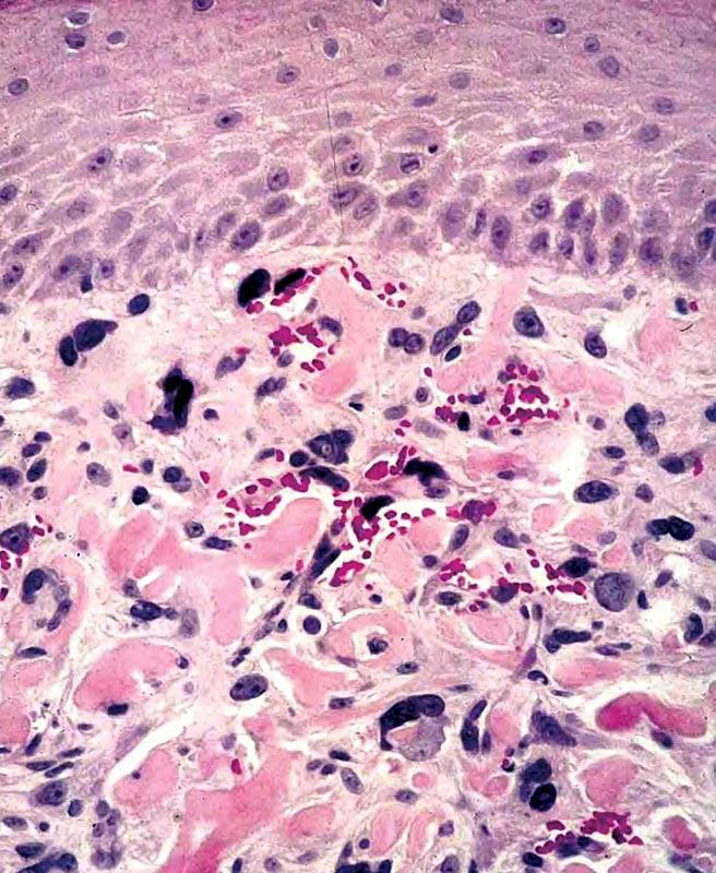
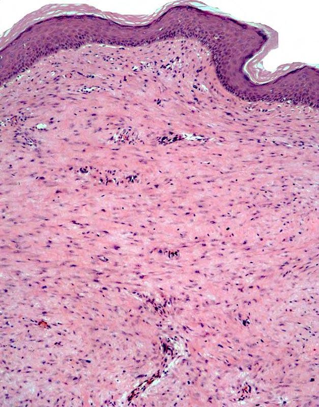
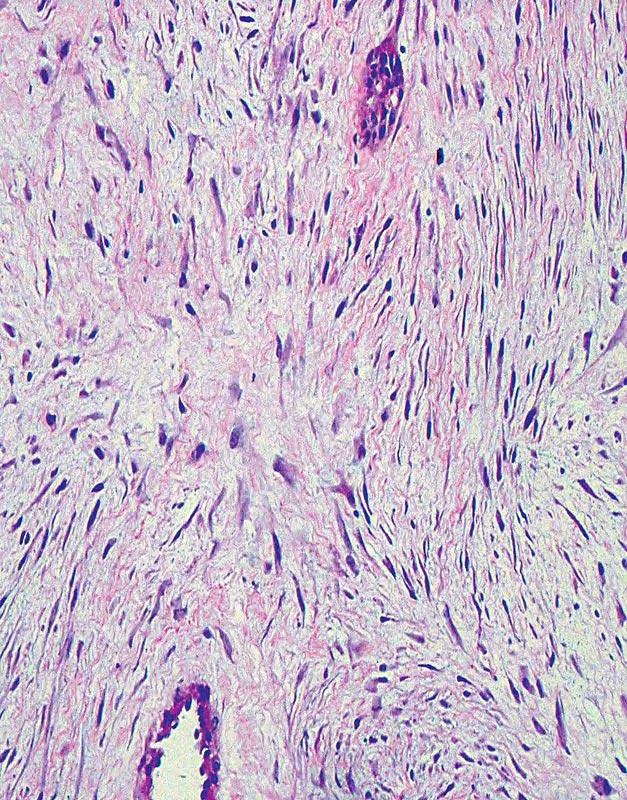
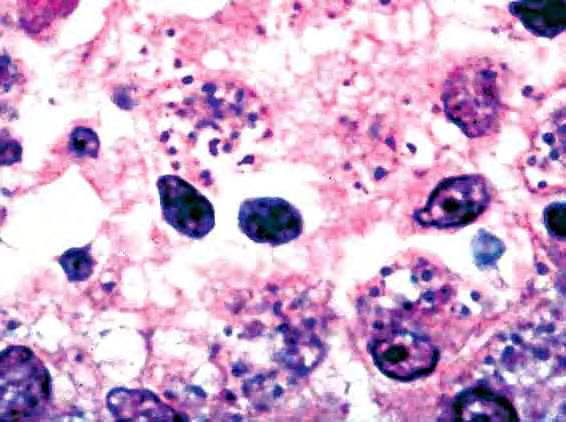
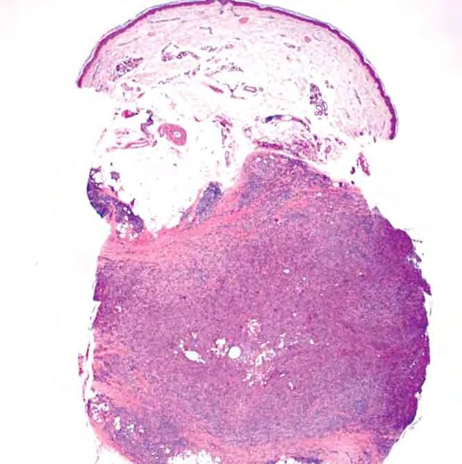
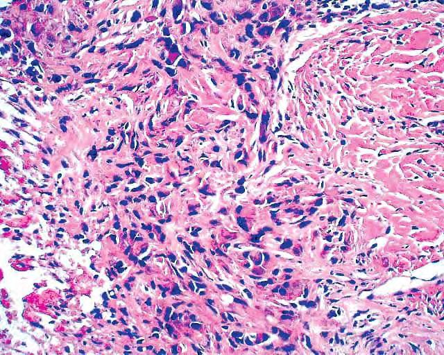
7 Williams BT, Barr RJ, Barrett TL, Everett MA, Lin F: Cutaneous pseudosarcomatous polyp: a histological & immunohistochemical study. J Cutan Pathol 1996; 23: Two unusual acquired polypoid skin lesions exhibited prominent histological atypia, but were biologically benign. Both patients were elderly females. The lesions clinically mimicked fibroepithelial polyp or nevus lipomatosus. Both had been present for about 20 years. One lesion was located on the back, the other on the posterior thigh. Each lesion exhibited dilated, hyalinized vessels in the dermis with focal fibrin deposits, myxoid stroma, and a population of bizarre, pleomorphic spindle to stellate cells, some of which were multinucleated. Occasional atypical mitoses were present. One lesion had abundant admixed fat. Immunohistochemical staining was strongly positive only for vimentin. The lesions share features with degenerating angiofibroma and vaginal pseudosarcomatous polyp. As in those lesions, the atypia is most probably reactive and degenerative.
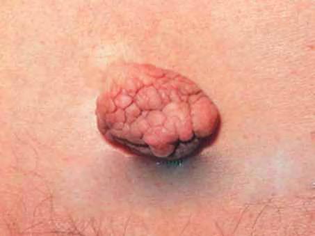
8 Pseudosarcomatous polyp
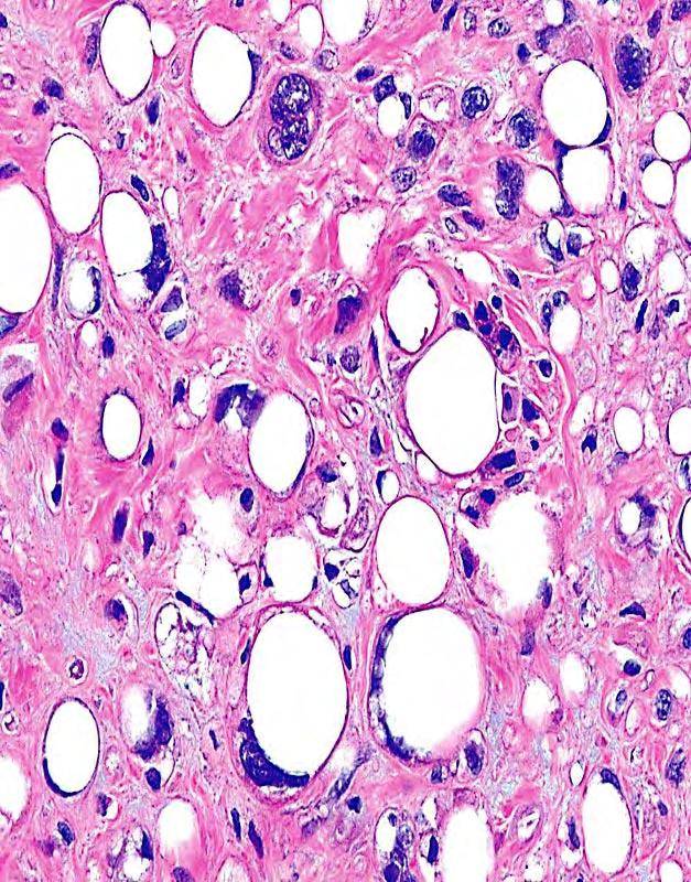
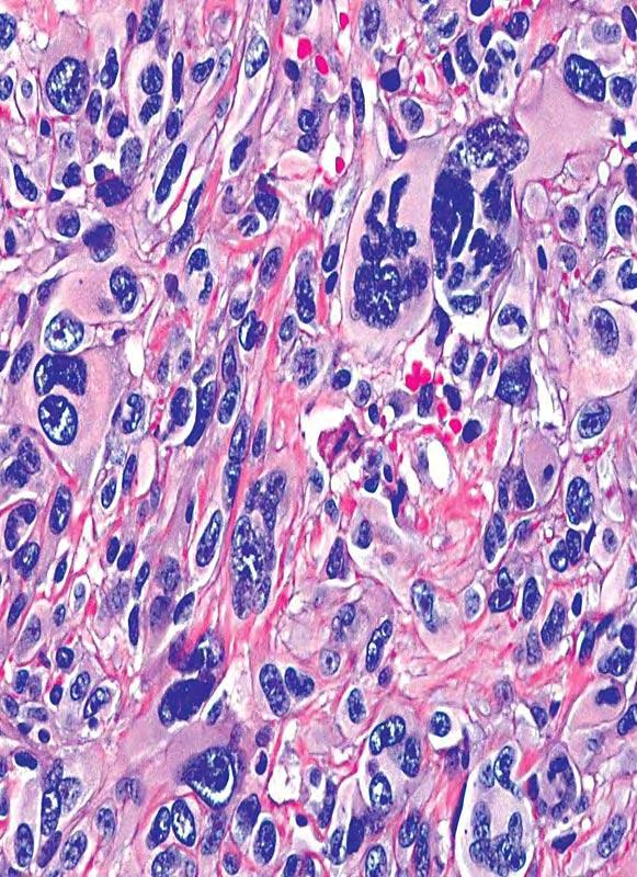
9 Pseudosarcomatous polyp


10 REPARATIVE PSEUDONEOPLASTIC PROLIFERATIONS OF THE SKIN THAT SIMULATE FIBROBLASTIC, SMOOTH-MUSCLE, OR VASCULAR TUMORS Nodular & proliferative fasciitis Proliferating vascular scars & acroangiodermatitis Postoperative-posttraumatic spindle cell nodules Reactions to Monsel s solution
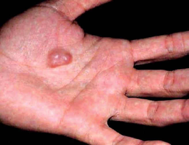
 are preferentially affected by nodular fasciitis RAPID growth of lesion over days to weeks.")
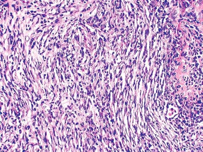
11 NODULAR FASCIITIS (NF): Clinical Features Most often observed in areas of soft tissue that are prone to injury, in line with the premise that NF is an idiosyncratic proliferative reaction to trauma As such, it is understandable that young individuals (< 25 yrs of age; male dominance) are preferentially affected by nodular fasciitis RAPID growth of lesion over days to weeks... too rapid to be malignant! Also reported in salivary glands, mouth, nasal cavity, and scalp

12 Nodular fasciitis

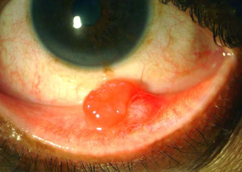
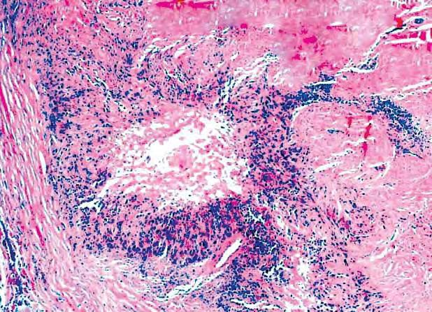
13 Postoperative/Posttraumatic Spindle Cell Nodule of the Skin Analogous to similar lesions seen in the genitourinary tract after recent surgery Clinically has the appearance of polypoid granulation tissue or pyogenic granuloma Microscopically, PSCN shows random proliferation of spindle cells with variable nuclear atypia and brisk mitotic activity Red cell extravasation & stromal edema analogous to nodular fasciitis in deeper tissue May demonstrate aberrant immunoreactivity for keratin, leading to potential misdiagnosis as sarcomatoid carcinoma; also DES+/MSA+


14 Excision of junctional nevus 1 mo. prior Postop spindle cell nodule


15 Postop spindle cell nodule Aberrant keratin immunostaining

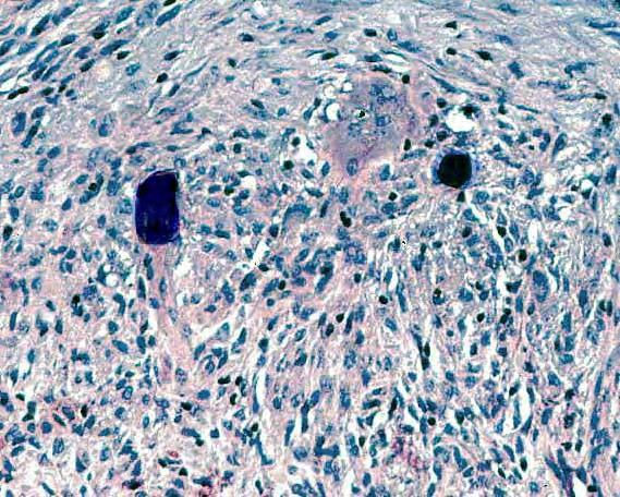
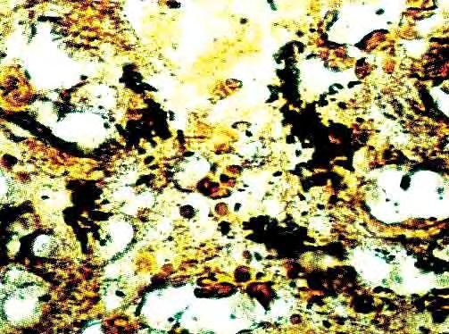
16 Reactions to Monsel Solution Monsel solution is ferric subsulphate solution, used as a styptic for minor surgical procedures In selected patients, it idiosyncratically incites a proliferative fibroblastic response that may simulate a neoplasm


17 Monsel reaction Perl Stain
18 Reaction to Monsel Solution (RMS) Clues to the diagnosis of RMS: Knowledge of recent surgical procedure (biopsy, excision) at the site of the current lesion Presence of foreign-body-type giant cells Large deposits of iron in the lesion, stainable with Perl s method
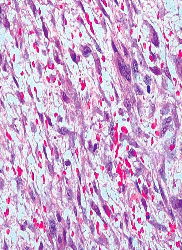
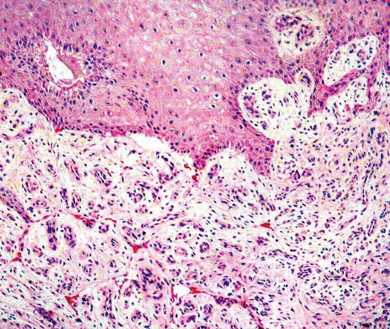
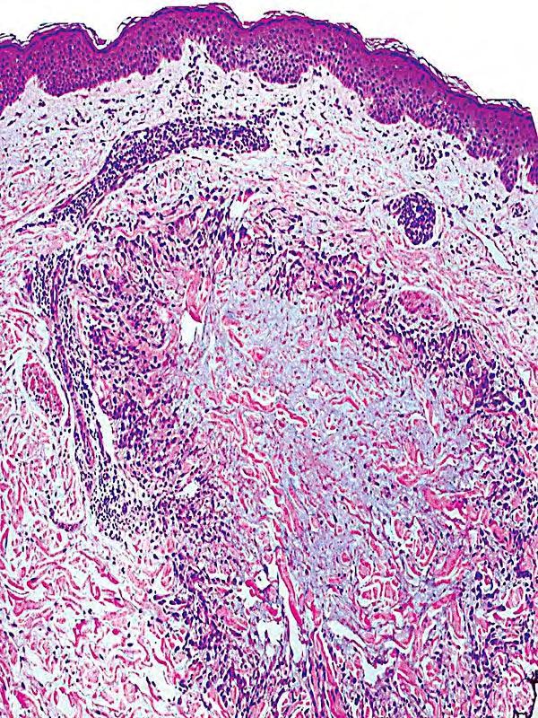
19 Acroangiodermatitis: Clinical Also known as dermatitis hemostatica, & pseudo-kaposi s sarcoma Proliferative response to stasis dermatitis, seen in older individuals Clinically and sometimes histologically confused with KS
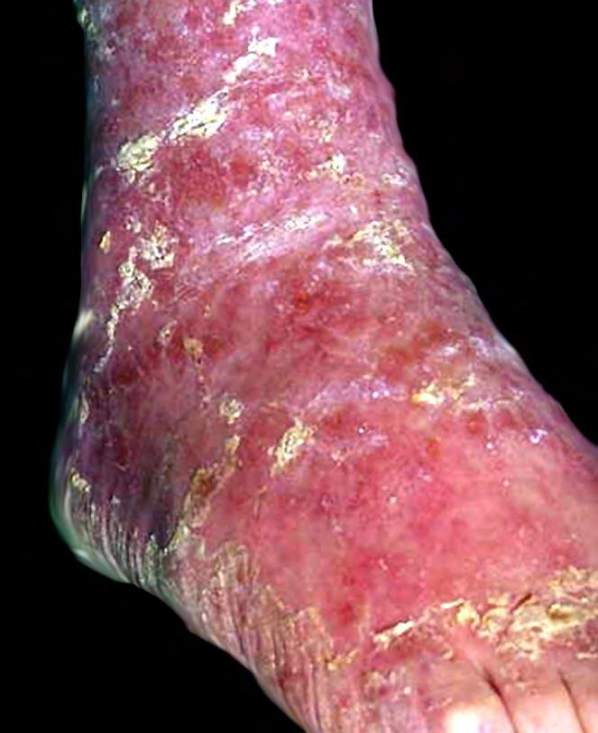
20 Acroangiodermatitis
21 Acroangiodermatitis: Histologic Proliferating venule-sized vessels, admixed with extravasated erythrocytes, hemosiderin, and stromal spindle cells No promontory sign, dilated lymphatics, plasma cell infiltrates, or eosinophilic globules, as seen in KS
22 Acroangiodermatitis
23 HISTOLOGICAL FEATURES OF MESENCHYMAL PROLIFERATIONS THAT TEND TO EXCLUDE A DIAGNOSIS OF A REPARATIVE LESION Marked nuclear hyperchromasia or pleomorphism in proliferating cells Spontaneous ulceration of overlying skin or mucosa by the lesion Pathologically-shaped mitoses Prominent spontaneous intralesional geographic necrosis
 Desmin (+/-) S100 protein (-) CD57 (-) Collagen type IV (+/-) CD68/163 (+/-) Nuclear")
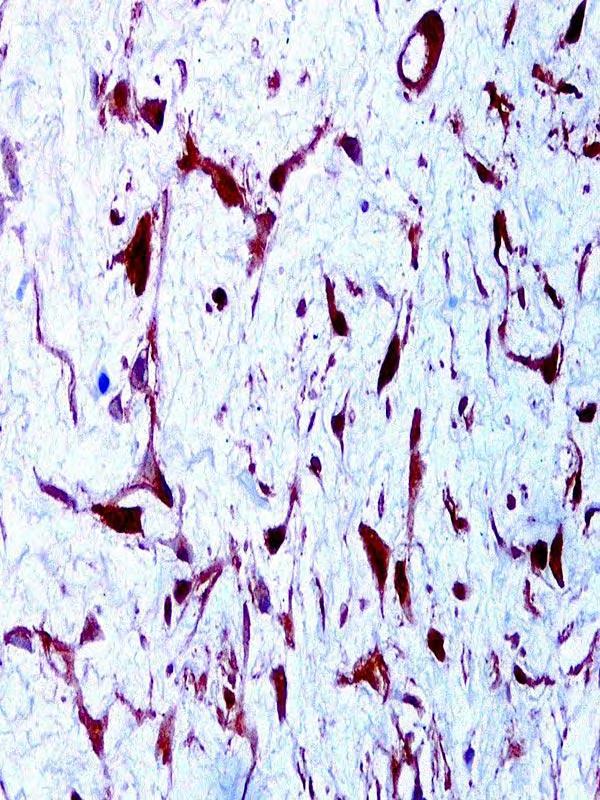
24 Reparative Spindle-Cell Proliferations in the Skin-- Immunophenotype Vimentin (+) Muscle-specific actin (+) Alpha-isoform actin (+) Desmin (+/-) S100 protein (-) CD57 (-) Collagen type IV (+/-) CD68/163 (+/-) Nuclear beta-catenin (-)
25 MESENCHYMAL PROLIFERATIONS WITH IDENTICAL IMMUNOPHENOTYPES Nodular fasciitis & related lesions Myofibroblastoma Leiomyoma & Leiomyosarcoma Fibromatoses (except for desmoid nuclear beta-catenin)
26 Nuclear Beta-catenin in Desmoid Fibromatosis
27 LESIONS THAT ARE RELATED ETIOLOGICALLY TO INFECTION
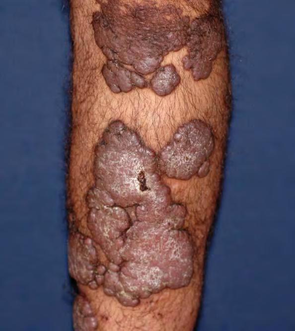
28 BACILLARY ANGIOMATOSIS: Clinical Features Recognized in HIV+ patients in 1983 Cutaneous red-violet nodules and papules Mistaken clinically for Kaposi s sarcoma, but pathologically for epithelioid hemangioma Eminently treatable with antibiotics; causative organism is currently classified as a Bartonella sp. (formerly Rochilamaea)
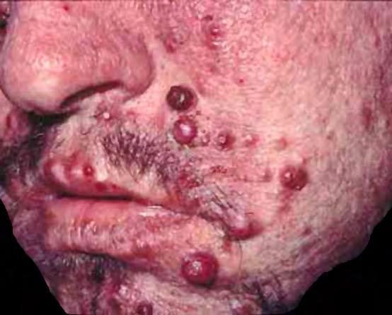
29 Bacillary angiomatosis
 Simulates epithelioid (a.k.a. histiocytoid) hemangioma or low-grade angiosarcoma Lobules of epithelioid")
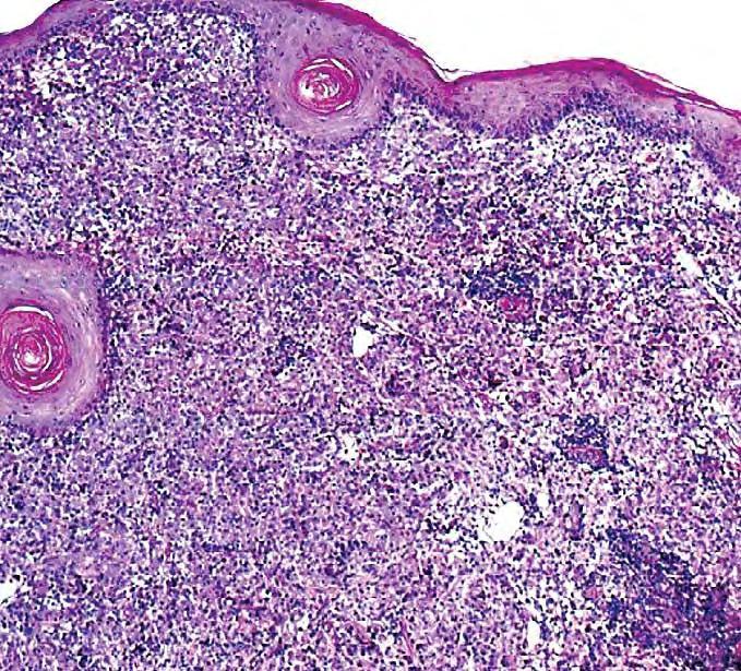
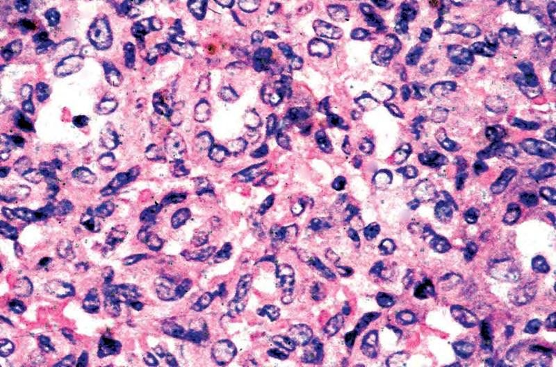
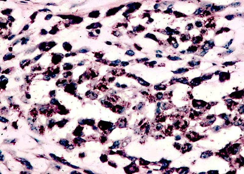
30 BACILLARY ANGIOMATOSIS: Histological Features Histologically-identical to verruga peruana of Bartonellosis (Carrion s disease) Simulates epithelioid (a.k.a. histiocytoid) hemangioma or low-grade angiosarcoma Lobules of epithelioid endothelial cells forming variably-distinct intercellular lumina Interspersed neutrophils common Proliferating epithelioid endothelial cells often contain numerous Warthin-Starry+ bacilliform bacteria
31 Bacillary angiomatosis
32 Bacillary angiomatosis Steiner Stain
33 Histoid Leprosy & Other Mycobacterial Pseudotumors: Mimics of Fibrous Histiocytomas Fibroblastic & histiocytic proliferations are seen in the skin and viscera in association with infections by M. leprae, M. avium-intracellulare, and, rarely, other mycobacteria Cytoplasm of proliferating cells is frequently foamy or granular Lesions may be well-demarcated or pseudoinfiltrative Histochemical stains for AFB (e.g., Ziehl- Neelsen; Fite) are floridly positive


34 Histoid lepromatous leprosy Mycobacterial pseudotumor
35 Mycobacterial pseudotumor Fite Stain
36 IDIOPATHIC & IMMUNOLOGICAL PSEUDOSARCOMATOUS SKIN LESIONS
37 Isolated Necrobiotic Granulomas of the Skin Some examples of deep granuloma annulare & rheumatoid nodule demonstrate nuclear atypia & mitotic activity in constituent histiocytic cells, simulating the appearance of epithelioid sarcoma Central areas of necrosis/necrobiosis also possible in GA and RN Immunohistologic differences: Epithelioid sarcoma: CK+/EMA+/CD45-/CD34 +/CD163-/- Granulomas: CK-/EMA-/CD45+/CD34-/CD163+

38 Granuloma annulare
39 Epithelioid sarcoma


40 Granuloma annulare: Colloidal iron stain CD163 immunostain


41 Epithelioid sarcoma: Keratin immunostain EMA immunostain
42 PSEUDONEOPLASTIC DISORDERS: Methods for Avoiding Misdiagnosis 1.Close collegial interaction, with detailed sharing of all available clinicopathologic information-- physical & historical findings, radiographic data, and macroscopic & histopathologic observations 2. Special focus on diagnostic disagreements between specialists involved in the care of a given patient

43
Spindle Cell Lesions Of The Breast. Emad Rakha Professor of Breast Pathology and Consultant Pathologist
 Spindle Cell Lesions Of The Breast Emad Rakha Professor of Breast Pathology and Consultant Pathologist * SCLs comprise a wide spectrum of diseases, ranging from reactive processes to aggressive malignant
Case year female. Routine Pap smear
 Case 1 57 year female Routine Pap smear Diagnosis? 1. Atypical glandular cells of unknown significance (AGUS) 2. Endocervical AIS 3. Endocervical adenocarcinoma 4. Endometrial adenocarcinoma 5. Adenocarcinoma
أملس عضلي غرن = Leiomyosarcoma. Leiomyosarcoma 1 / 5
 Leiomyosarcoma 1 / 5 EPIDEMIOLOGY Exact incidence is unknown, but older studies suggest that leiomyosarcomas comprise approximately 3 percent of soft-tissue sarcomas. Superficial leiomyosarcoma occurs
Classification (1) Classification (3) Classification (2) Spindle cell lesions. Spindle cell lesions of bladder (Mills et al.
 Classification (3) Classification (2) Spindle cell lesions. Spindle cell lesions of bladder (Mills et al.") Non-epithelial tumours and nonepithelial tumour-like lesions of the bladder Dr Jonathan H Shanks The Christie NHS Foundation Trust, Manchester, UK Classification (1) Myofibroblastic proliferations and
Non-epithelial tumours and nonepithelial tumour-like lesions of the bladder Dr Jonathan H Shanks The Christie NHS Foundation Trust, Manchester, UK Classification (1) Myofibroblastic proliferations and
SESSION 1: GENERAL (BASIC) PATHOLOGY CONCEPTS Thursday, October 16, :30am - 11:30am FACULTY COPY
 PATHOLOGY CONCEPTS Thursday, October 16, :30am - 11:30am FACULTY COPY") SESSION 1: GENERAL (BASIC) PATHOLOGY CONCEPTS Thursday, October 16, 2008 9:30am - 11:30am FACULTY COPY GOAL: Describe the basic morphologic (structural) changes which occur in various pathologic conditions.
SESSION 1: GENERAL (BASIC) PATHOLOGY CONCEPTS Thursday, October 16, 2008 9:30am - 11:30am FACULTY COPY GOAL: Describe the basic morphologic (structural) changes which occur in various pathologic conditions.
Newer soft tissue entities
 Newer soft tissue entities Examples among fibroblastic tumors Turku, May 6, 2010 Markku Miettinen, M.D. AFIP, Washington, DC Fibroblastic neoplasms Solitary fibrous tumor /Hemangiopericytoma Low-grade
Selected Pseudomalignant Soft Tissue Tumors of the Skin and Subcutis
 Selected Pseudomalignant Soft Tissue Tumors of the Skin and Subcutis Andrew L. Folpe, M.D. Professor of Laboratory Medicine and Pathology Mayo Clinic, Rochester, MN folpe.andrew@mayo.edu 2016 MFMER slide-1
Desmoplastic Melanoma R/O BCC. Clinical Information. 74 y.o. man with lesion on left side of neck r/o BCC
 R/O BCC Sabine Kohler, M.D. Professor of Pathology and Dermatology Dermatopathology Service Stanford University School of Medicine Clinical Information 74 y.o. man with lesion on left side of neck r/o
Normal endometrium: A, proliferative. B, secretory.
 Normal endometrium: A, proliferative. B, secretory. Nội mạc tử cung Nội mạc tử cung Cyclic changes in endometrium.. Approximate relationship of useful microscopic changes. Arias-Stella reaction in endometrial
العصوي الوعاي ي الورام = angiomatosis Bacillary
 1 / 7 BACILLARY ANGIOMATOSIS Epidemiology BA is most commonly seen in patients with acquired immunodeficiency syndrome (AIDS) and a CD4 count less than 50 cells/mm 3, with an incidence of 1.2 cases per
IN THE NAME OF GOD Dr. Kheirandish Oral and maxillofacial pathology
 IN THE NAME OF GOD Dr. Kheirandish Oral and maxillofacial pathology ORAL FOCAL MUCINOSIS Uncommon Tumorlike Cutaneous myxoid cyst Overproduction of hyaluronic acid by firoblasts Young adults Female Gingiva
Inflammatory pseudotumor
 Inflammatory pseudotumor Inflammatory pseudotumor (IPT) Heterogeneous group of lesions of obscure etiology On physical and radiographic examination often confused with malignancy Synonyms Plasma cell granuloma
Gross appearance of nodular hyperplasia in material obtained from suprapubic prostatectomy. Note the multinodular appearance and the admixture of
 Tiền liệt tuyến Tiền liệt tuyến Gross appearance of nodular hyperplasia in material obtained from suprapubic prostatectomy. Note the multinodular appearance and the admixture of solid and microcystic areas.
Division of Pathology
 Case 38 Adult woman with a 35mm right breast lump at the 10 o clock position. Excision performed. (Case contributed by Dr Mihir Gudi, KKH) Division of Pathology Merlion, One Fullerton Singapore Diagnosis
Enterprise Interest Nothing to declare
 Enterprise Interest Nothing to declare Diagnoses one would not like to miss in soft tissue pathology early in your career Marta Sbaraglia, MD Department of Pathology Hospital of Treviso University of Padua
Malignant Peripheral Nerve Sheath Tumor
 C H A P T E R 120 Malignant Peripheral Nerve Sheath Tumor Currently, malignant peripheral nerve sheath tumor (MPNST) is the most commonly used generic name for the neoplasms known in the past as neurosarcoma,
21/07/2017. Hobnail endothelial cells are not the same as epithelioid endothelial cells
 UPDATE IN CUTANEOUS VASCULAR S DERMATOPATHOLOGY SESSION BELFAST PATHOLOGY JUNE 21/2017 Dr E Calonje St John s Institute of Dermatology, London, United Kingdom THE FAMILY OF VASCULAR S WITH EPITHELIOID
A 25 year old female with a palpable mass in the right lower quadrant of her abdomen
 May 2016 A 25 year old female with a palpable mass in the right lower quadrant of her abdomen Contributed by: Paul Ndekwe, MD, Resident Physician, Indiana University School of Department of Pathology and
An Overview of Genital Stromal Tumors
 An Overview of Genital Stromal Tumors By Konstantinos Linos MD, FCAP, FASDP Bone, Soft Tissue and Dermatopathology Assistant Professor of Pathology Dartmouth-Hitchcock Medical Center Geisel School of Medicine
Diplomate of the American Board of Pathology in Anatomic and Clinical Pathology
 A 33-year-old male with a left lower leg mass. Contributed by Shaoxiong Chen, MD, PhD Assistant Professor Indiana University School of Medicine/ IU Health Partners Department of Pathology and Laboratory
Atypical Palisaded Myofibroblastoma of Lymph Node: Report of a rare case.
 ISPUB.COM The Internet Journal of Pathology Volume 10 Number 1 Atypical Palisaded Myofibroblastoma of Lymph Node: Report of a rare case. V Kinnera, R Nandyala, M Yootla, K Mandyam Citation V Kinnera, R
Prostatic stromal hyperplasia with atypia (PSHA) is a
 is a") Prostatic Stromal Hyperplasia With Atypia Follow-up Study of 18 Cases Deloar Hossain, MD, FRCPC; Isabelle Meiers, MD; Junqi Qian, MD; Gregory T. MacLennan, MD; David G. Bostwick, MD, MBA Context. Prostatic
Prostatic Stromal Hyperplasia With Atypia Follow-up Study of 18 Cases Deloar Hossain, MD, FRCPC; Isabelle Meiers, MD; Junqi Qian, MD; Gregory T. MacLennan, MD; David G. Bostwick, MD, MBA Context. Prostatic
The Relevance of Cytologic Atypia in Cutaneous Neural Tumors
 The Relevance of Cytologic Atypia in Cutaneous Neural Tumors Recent Findings - New Developments New Problems Zsolt B. Argenyi, M.D. Professor of Pathology & Dermatology Director of Dermatopathology Department
Clinicopathologic Self- Assessment S003 AAD 2017
 Clinicopathologic Self- Assessment S003 AAD 2017 Clay J. Cockerell, M.D. Director, Cockerell Dermatopathology Director, Division of Dermatopathology UT Southwestern Medical Center July 2017 No relevant
5/10. Pathology Soft tissue tumors. Farah Bhani. Mohammed Alorjani
 5/10 Pathology Soft tissue tumors Mohammed Alorjani Farah Bhani Slides are included in this sheet. Objectives: Soft tissue tumors 1. Describe soft tissue tumors. 2. Understand the classification of soft
5/21/2018. Prostate Adenocarcinoma vs. Urothelial Carcinoma. Common Differential Diagnoses in Urological Pathology. Jonathan I.
 Common Differential Diagnoses in Urological Pathology Jonathan I. Epstein Prostate Adenocarcinoma vs. Urothelial Carcinoma 1 2 NKX3.1 NKX3.1 3 4 5 6 Proposed ISUP Recommendations Option to use PSA as a
Fine-needle aspiration (FNA) has been used increasingly
 has been used increasingly") Worrisome Histologic Alterations Following Fine-Needle Aspiration of Benign Parotid Lesions Shiyong Li, MD, PhD; Zubair W. Baloch, MD, PhD; John E. Tomaszewski, MD; Virginia A. LiVolsi, MD Objective. To
Worrisome Histologic Alterations Following Fine-Needle Aspiration of Benign Parotid Lesions Shiyong Li, MD, PhD; Zubair W. Baloch, MD, PhD; John E. Tomaszewski, MD; Virginia A. LiVolsi, MD Objective. To
USCAP 2011: ASDP companion meeting. Steven D. Billings 1
 USCAP 2011: ASDP companion meeting. Steven D. Billings (billins@ccf.org) 1 Spindle cell tumors that make you say, Oh $*&%! This lecture will focus on examples of cutaneous tumors that present particular
An Overview of Cutaneous Vascular Neoplasms
 An Overview of Cutaneous Vascular Neoplasms By Konstantinos Linos MD, FCAP, FASDP Bone, Soft Tissue and Dermatopathology Assistant Professor of Pathology Dartmouth-Hitchcock Medical Center Geisel School
SOFT TISSUE TUMOR PATHOLOGY: AN UPDATE
 SOFT TISSUE TUMOR PATHOLOGY: AN UPDATE Jason L. Hornick, MD, PhD July 18, 2013 Department of Pathology Brigham and Women s Hospital Harvard Medical School Boston, MA, USA I have no disclosures. New Soft
TitleSarcomatoid carcinoma of the bladde.
 TitleSarcomatoid carcinoma of the bladde Author(s) Takashi, Munehisa; Sakata, Takao; N Tatsuya; Miyake, Koji Citation 泌尿器科紀要 (1992), 38(1): 67-70 Issue Date 1992-01 URL http://hdl.handle.net/2433/117446
Mody. AIS vs. Invasive Adenocarcinoma of the Cervix
 Common Problems in Gynecologic Pathology Michael T. Deavers, M.D. Houston Methodist Hospital, Houston, Texas Common Problems in Gynecologic Pathology Adenocarcinoma in-situ (AIS) of the Cervix vs. Invasive
All authors abide by the Association for Medical Ethics (AME) ethical rules of disclosure.
 ethical rules of disclosure.") Longo F, Musumeci G, Parenti R, Vecchio G, Magro G. Atypical cell leiomyoma of the uterus with amianthoid-like fibers: A case report. OA Case Reports 2013 Nov 15;2(14):137. Licensee OA Publishing London
Longo F, Musumeci G, Parenti R, Vecchio G, Magro G. Atypical cell leiomyoma of the uterus with amianthoid-like fibers: A case report. OA Case Reports 2013 Nov 15;2(14):137. Licensee OA Publishing London
History A 89 year old gentleman presenting with a scalp/forehead nodule. Patient had squamous cell carcinoma 18 m at same site, excised. Outside diagn
 Case III History A 89 year old gentleman presenting with a scalp/forehead nodule. Patient had squamous cell carcinoma 18 m at same site, excised. Outside diagnoses: Squamous cell carcinoma. R/O: SCC, Melanoma,
Gross appearance of peritoneal cysts. They have a thin, translucent wall and contain a clear fluid.
 Gross appearance of peritoneal cysts. They have a thin, translucent wall and contain a clear fluid. So-called multicystic benign mesothelioma. A, Gross appearance. So-called multicystic benign mesothelioma.
Salivary Glands 3/7/2017
 Salivary Glands 3/7/2017 Goals and objectives Focus on the entities unique to H&N Common board type facts Information for your future practice Salivary Glands Salivary Glands Major gland. Paratid. Submandibular.
Hemangioendothelioma with a Prominent Lymphoid Infiltrate Mimicking Follicular Dendritic Cell Tumor: Report of a Case
 Journal of Cancer Research Updates, 2013, 2, 135-139 135 Hemangioendothelioma with a Prominent Lymphoid Infiltrate Mimicking Follicular Dendritic Cell Tumor: Report of a Case Justin Kerstetter 1, Mia Perez
3/27/2017. Disclosure of Relevant Financial Relationships
 Ophthalmic Pathology Evening Specialty Conference USCAP 2017 5 th March, 2017 Mukul K. Divatia, MD Assistant Professor Department of Pathology & Genomic Medicine Weill Cornell Medical College Houston Methodist
Cellular Neurothekeoma
 Cellular Neurothekeoma Scott W Binder, MD Pritzker Professor of Pathology & Dermatology Sr. Vice Chair Director, Pathology Clinical Services Chief, Dermatopathology Geffen/UCLA School of Medicine Clinical
Case Report A Rare Cutaneous Adnexal Tumor: Malignant Proliferating Trichilemmal Tumor
 Case Reports in Medicine Volume 2015, Article ID 742920, 4 pages http://dx.doi.org/10.1155/2015/742920 Case Report A Rare Cutaneous Adnexal Tumor: Malignant Proliferating Trichilemmal Tumor Omer Alici,
Case Report Clear Cell Basal Cell Carcinoma
 SAGE-Hindawi Access to Research Volume 2011, Article ID 386921, 4 pages doi:10.4061/2011/386921 Case Report Clear Cell Basal Cell Carcinoma Deba P. Sarma, 1 Daniel Olson, 1 Jennifer Olivella, 1 Tracey
Case Presentation. Maha Akkawi, MD, Fatima Obeidat, MD, Tariq Aladily, MD. Department of Pathology Jordan University Hospital Amman, Jordan
 Case Presentation Maha Akkawi, MD, Fatima Obeidat, MD, Tariq Aladily, MD Department of Pathology Jordan University Hospital Amman, Jordan The 25th Annual Congress of the ADIAP The 8/11/2013 1 5th International
Benign Mimics of Malignancy in Breast Pathology
 Arthur Purdy Stout Society of Surgical Pathologists Companion Meeting Benign Mimics of Malignancy in Breast Pathology Stuart J. Schnitt, M.D. Beth Israel Deaconess Medical Center and Harvard Medical School,
An unusual case of a large fibroepithelial stromal polyp presenting as a nipple mass
 Shaaban et al. BMC Research tes 2013, 6:345 CASE REPORT Open Access An unusual case of a large fibroepithelial stromal polyp presenting as a nipple mass Abeer M Shaaban 1*, EPL Turton 2 and William Merchant
Fun with Fat. General Rules. Case
 Fun with Fat General Rules Imaging: location (deep vs. superficial) Superficial lesions are seldom liposarcomas Deep lesions may be benign or malignant Myxoid stroma is common in benign and malignant lesions
2015 Descriptive Vet Path Course. Histo Exam #3 KEY
 2015 Descriptive Vet Path Course Histo Exam #3 KEY Test 3, Slide 1 Tissue from a guinea pig. MORPHOLOGIC DIAGNOSIS: Heart: Multifocally and randomly (1 pt), within the left and right ventricular myocardium
Abid Irshad, MD Director Breast Imaging. Medical University of South Carolina Charleston
 Abid Irshad, MD Director Breast Imaging Medical University of South Carolina Charleston Cases Financial disclosure: I or my family have no financial interest related to the material discussed in this presentation
Post-test Self-assessment Cases
 Post-test Self-assessment Cases Ibrahim Khalifeh, M.D. Associate Professor Department of Pathology American University of Beirut Medical Center Beirut, Lebanon Case I History A 69 year old gentleman presenting
59 yo male with past medical history of prostate carcinoma, presented with upper abdominal pain
 December 2016 59 yo male with past medical history of prostate carcinoma, presented with upper abdominal pain Contributed by: Divya Sharma, MD. Fellow, Gastrointestinal Pathology, Department of Pathology
Inflammatory and Reactive Lesions of the Breast
 Inflammatory and Reactive Lesions of the Breast Laura C. Collins, M.D. Vice Chair of Anatomic Pathology Professor of Pathology Beth Israel Deaconess Medical Center and Harvard Medical School Boston, MA
Pleomorphic adenoma of breast - a case report and distinction with metaplastic carcinoma D Gupta, S Agrawal, N Trivedi, A Tewari
 of breast - a case report and distinction with metaplastic carcinoma D Gupta, S Agrawal, N Trivedi, A Tewari Introduction, also known as mixed tumour, is a benign tumour which typically presents as a painless,
Mousa. Israa Ayed. Abdullah AlZibdeh. 0 P a g e
 1 Mousa Israa Ayed Abdullah AlZibdeh 0 P a g e Breast pathology The basic histological units of the breast are called lobules, which are composed of glandular epithelial cells (luminal cells) resting on
6/5/2010. Outline of Talk. Endometrial Alterations That Mimic Cancer & Vice Versa: Metaplastic / reactive changes. Problems in Biopsies/Curettages
 Outline of Talk Endometrial Alterations That Mimic Cancer & Vice Versa: Problems in Biopsies/Curettages Metaplastic / reactive changes Mucinous change Microglandular hyperplasia-like change Squamous metaplasia
Basal cell carcinoma 5/28/2011
 Goal of this Presentation A practical approach to the diagnosis of cutaneous carcinomas and their mimics Thaddeus Mully, MD University of California San Francisco To review common non-melanoma skin cancers
Diseases of the breast (1 of 2)
") Diseases of the breast (1 of 2) Introduction A histology introduction Normal ducts and lobules of the breast are lined by two layers of cells a layer of luminal cells overlying a second layer of myoepithelial
Diseases of the breast (1 of 2) Introduction A histology introduction Normal ducts and lobules of the breast are lined by two layers of cells a layer of luminal cells overlying a second layer of myoepithelial
Financial disclosures
 Mesenchymal Neoplasms with Melanocytic Differentiation By Konstantinos Linos MD, FCAP, FASDP Bone, Soft Tissue and Dermatopathology Assistant Professor of Pathology Dartmouth-Hitchcock Medical Center Geisel
Update on Cutaneous Mesenchymal Tumors. Thomas Brenn
 Update on Cutaneous Mesenchymal Tumors Thomas Brenn Cutaneous Mesenchymal Tumours Wide morphological and biological spectrum Myofibroblastic, smooth muscle, neural, vascular, apidocytic, undifferentiated;
JOURNAL OF CLINICAL AND DIAGNOSTIC RESEARCH
 JOURNAL OF CLINICAL AND DIAGNOSTIC RESEARCH How to cite this article : PAI K,GUPTA A.INTRAVASCULAR EPITHELIOID HAEMANGIOMA OF TEMPORAL ARTERY: A DIAGNOSTIC DIFFICULTY Journal of Clinical and Diagnostic
Self assessment case. Dr Saleem Taibjee Dorset County Hospital, Dorchester
 Self assessment case Dr Saleem Taibjee saleemtaibjee@gmail.com Dorset County Hospital, Dorchester Clinical details 34-year-old man: Shave excision Skin tag / papilloma left thigh The best diagnosis is:
Special slide seminar
 Special slide seminar Tomáš Rozkoš The Fingerland Department of Pathology Charles University Medical Faculty and Faculty Hospital in Hradec Králové Czech Republic Case history, 33 years old resistance
Case Report Inflammatory Myofibroblastic Tumor of the Nasal Septum
 Case Reports in Otolaryngology Volume 2013, Article ID 670105, 4 pages http://dx.doi.org/10.1155/2013/670105 Case Report Inflammatory Myofibroblastic Tumor of the Nasal Septum Yuri Okumura, 1 Kazuhiro
57th Annual HSCP Spring Symposium 4/16/2016
 An Unusual Malignant Spindle Cell Lesion to Involve the Breast Erinn Downs-Kelly, D.O. Associate Professor of Pathology University of Utah & ARUP Laboratories No disclosures Case 39 y/o female with no
BREAST PATHOLOGY. Fibrocystic Changes
 BREAST PATHOLOGY Lesions of the breast are very common, and they present as palpable, sometimes painful, nodules or masses. Most of these lesions are benign. Breast cancer is the 2 nd most common cause
Sarcomatoid (spindle cell) carcinoma of the cricopharynx presenting as dysphagia
 carcinoma of the cricopharynx presenting as dysphagia") Case Report Sarcomatoid (spindle cell) carcinoma of the cricopharynx presenting as dysphagia Jagtap Sunil V. 1, Shukla Dhirajkumar B. 2, Jagtap Swati S. 3, Havle Abhay D. 4 1 Associate Professor, Department
Case Report Sarcomatoid (spindle cell) carcinoma of the cricopharynx presenting as dysphagia Jagtap Sunil V. 1, Shukla Dhirajkumar B. 2, Jagtap Swati S. 3, Havle Abhay D. 4 1 Associate Professor, Department
Case: The patient is a 62 year old woman with a history of renal cell carcinoma that was removed years ago. A 2.4 cm liver mass was found on CT
 Case: The patient is a 62 year old woman with a history of renal cell carcinoma that was removed years ago. A 2.4 cm liver mass was found on CT during follow- up. ALT, AST, Alk Phos and bilirubin were
ACCME/Disclosures ALK FUSION-POSITIVE MESENCHYMAL TUMORS. Tumor types with ALK rearrangements. Anaplastic Lymphoma Kinase. Jason L.
 Companion Meeting of the International Society of Bone and Soft Tissue Pathology The Evolving Concept of Mesenchymal Tumors ALK FUSION-POSITIVE MESENCHYMAL TUMORS Jason L. Hornick, MD, PhD March 13, 2016
المركب النموذج--- سبيتز وحمة = Type Spitz's Nevus, Compound SPITZ NEVUS 1 / 7
 SPITZ NEVUS 1 / 7 Epidemiology An annual incidence rate of 1.4 cases of Spitz nevus per 100,000 individuals has been estimated in Australia, compared with 25.4 per 100,000 individuals for cutaneous melanoma
Solitary Fibrous Tumor of the Kidney with Massive Retroperitoneal Recurrence. A Case Presentation
 246) Prague Medical Report / Vol. 113 (2012) No. 3, p. 246 250 Solitary Fibrous Tumor of the Kidney with Massive Retroperitoneal Recurrence. A Case Presentation Sfoungaristos S., Papatheodorou M., Kavouras
Salivary gland tumor cytologic and histologic correlation: Algorithmic and risk stratification based approaches
 Salivary gland tumor cytologic and histologic correlation: Algorithmic and risk stratification based approaches Christopher C. Griffith, MD, PhD Raja R. Seethala, MD 1. Salivary gland tumor cytology: A
Postoperative spindle cell nodule after thyroidectomy: A case mimicking recurrence with anaplastic transformation of thyroid cancer
 CASE REPORT Postoperative spindle cell nodule after thyroidectomy: A case mimicking recurrence with anaplastic transformation of thyroid cancer Sun Wook Kim, MD, PhD, 1 Young Lyun Oh, MD, PhD, 2 Joon Young
Diagnosis of a granular cell tumour at the abdominal wall using fine needle aspiration cytology and histology: Case report
 Case Report Diagnosis of a granular cell tumour at the abdominal wall using fine needle aspiration cytology and histology: Case report Journal of International Medical Research 2015, Vol. 43(4) 592 596!
CHRONIC INFLAMMATION
 CHRONIC INFLAMMATION Chronic inflammation is an inflammatory response of prolonged duration often for months, years or even indefinitely. Its prolonged course is proved by persistence of the causative
This case presentation reviews a challenging case of. Metaplastic Carcinomas of the Breast: Diagnostic Challenges and New Translational Insights
 Metaplastic Carcinomas of the Breast: Diagnostic Challenges and New Translational Insights Comprising less than 1% of invasive carcinomas of the breast, metaplastic carcinomas are a heterogeneous group
Myxo-inflammatory Fibroblastic sarcoma
 AKA Myxo-inflammatory Fibroblastic sarcoma Acral Myxoinflammatory fibroblastic sarcomaam.j.surg.path1998; 22; 911-924 Inflammatory myxoid tumour of soft parts with bizarre giant cells [Pathol.Res.Pract.
Contents. vii. Preface... Acknowledgments... v xiii
 Contents Preface... Acknowledgments... v xiii SECTION I 1. Introduction... 3 Knowledge-Based Diagnosis... 4 Systematic Examination of the Lymph Node... 7 Cell Type Identification... 9 Cell Size and Cellularity...
Diagnostic problems in uterine smooth muscle tumors
 Diagnostic problems in uterine smooth muscle tumors Marina Kos Ljudevit Jurak Clinical Department of Pathology, Clinical Hospital Center Sestre milosrdnice, Zagreb Institute of Pathology, University of
Differential Diagnosis of Oral Masses. Palatal Lesions
 Differential Diagnosis of Oral Masses Palatal Lesions Palatal Masses Periapical Abscess Torus Palatinus Mucocele Lymphoid Hyperplasia Adenomatous Hyperplasia Benign Salivary Neoplasms Malignant Salivary
Evaluating and Reporting Gastrointestinal Stromal Tumors after Imatinib Mesylate Treatment
 The Open Pathology Journal, 2009, 3, 53-57 53 Open Access Evaluating and Reporting Gastrointestinal Stromal Tumors after Imatinib Mesylate Treatment Katie L. Dennis * and Ivan Damjanov Department of Pathology
Slide seminar. Asist. Prof. Jože Pižem, MD, PhD Institute of Pathology Medical Faculty, University of Ljubljana
 Slide seminar Asist. Prof. Jože Pižem, MD, PhD Institute of Pathology Medical Faculty, University of Ljubljana Case 5 A 57-year-old man with a dermal/subcutaneous lesion on the scalp, which was interpreted
Inflammatory Myofibroblastic Tumor of the Bladder
 Inflammatory Myofibroblastic Tumor of the Bladder We illustrate a case of an inflammatory myofibroblastic tumor (IMT) involving the bladder in a woman with dysuria and review the literature and differential
Diagnostically Challenging Cases in Gynecologic Pathology
 Diagnostically Challenging Cases in Gynecologic Pathology Eric C. Huang, M.D., Ph.D. Department of Pathology and Laboratory Medicine University of California, Davis Medical Center Case 1 Presentation 38
Self assesment Case 21
 17-18 MAY 2018 London Dermatopathology Symposium 2018 Self assesment Case 21 MARC HASPESLAGH CASE 21 1802-50585 48 year old lady with eczematous lesions at ear helix and red patch on nose bridge since
A neoplasm is defined as "an abnormal tissue proliferation, which exceeds that of adjacent normal tissue. This proliferation continues even after
 NEOPLASIA Neoplasia is a very important topic in pathology because neoplasms are both common and serious diseases. A neoplasm literally means a new growth, and this term is used interchangeably with a
Gastrointestinal stromal tumor
 Gastrointestinal stromal tumor 영남의대병리학교실 최준혁 Classification of gastrointestinal mesenchymal tumor Gastrointestinal stromal tumor(gist) Smooth muscle tumors : leiomyoma, leiomyosarcoma Neurogenic tumors
Histopathology: skin pathology
 Histopathology: skin pathology These presentations are to help you identify, and to test yourself on identifying, basic histopathological features. They do not contain the additional factual information
A case of giant cell tumour of soft parts in a horse Francesco Cian 1, Sarah Whiteoak 2, Jennifer Stewart 1
 A case of giant cell tumour of soft parts in a horse Francesco Cian 1, Sarah Whiteoak 2, Jennifer Stewart 1 1 Animal Health Trust, Newmarket, UK 2 608 Equine and Farm Vets, Rowington, UK Signalment: Horse,
Cellular Pseudosarcomatous Fibroepithelial Stromal Polyp of the Vagina during Pregnancy: A Lesion That Is Overdiagnosed as a Malignant Tumor
 The Korean Journal of Pathology 2012; 46: 494-498 CASE REPORT Cellular Pseudosarcomatous Fibroepithelial Stromal Polyp of the Vagina during Pregnancy: A Lesion That Is Overdiagnosed as a Malignant Tumor
A case of fat-free pleomorphic lipoma occurring in the upper back and axilla simultaneously
 Wang et al. World Journal of Surgical Oncology 2013, 11:145 WORLD JOURNAL OF SURGICAL ONCOLOGY CASE REPORT Open Access A case of fat-free pleomorphic lipoma occurring in the upper back and axilla simultaneously
number Done by Corrected by Doctor Maha Shomaf
 number 16 Done by Waseem Abo-Obeida Corrected by Zeina Assaf Doctor Maha Shomaf MALIGNANT NEOPLASMS The four fundamental features by which benign and malignant tumors can be distinguished are: 1- differentiation
Benign, Reactive and Inflammatory Lesions of the Breast
 Benign, Reactive and Inflammatory Lesions of the Breast Marilin Rosa, MD Associate Member Section Head of Breast Pathology Department of Anatomic Pathology Program Director, Breast Pathology Fellowship
Dermatopathology. Dr. Rafael Botella Estrada. Hospital La Fe de Valencia
 Dermatopathology Dr. Rafael Botella Estrada. Hospital La Fe de Valencia Melanoma and mimics Dr. Martin Mihm Malignant lesions result from the accumulation of mutations Class I lesions (benign) Class II
Challenges to diagnose metaplastic carcinoma of the breast through cytologic methods: an eight-case series
 CASE REPORT Open Access Challenges to diagnose metaplastic carcinoma of the breast through cytologic methods: an eight-case series Seema Lale 1, Kiyoe Kure 2, Daniel Lingamfelter 3* Abstract Because metaplastic
Vascular Tumors. Case 1: Papillary endothelial hyperplasia
 Vascular Tumors Case 1: Papillary endothelial hyperplasia Papillary endothelial hyperplasia (PEH) essentially represents a stage in the recanalization of a thrombosed blood vessel. It may occur in any
Brief History. Identification : Past History : HTN without regular treatment.
 Brief History Identification : Name : 陳 x - Admission : 94/10/06 Gender : male Age : 75 y/o Chief Complaint : Urinary difficulty for months. Past History : HTN without regular treatment. Brief History
Article begins on next page
 Bizarre stromal cells in an endometrial polyp Rutgers University has made this article freely available. Please share how this access benefits you. Your story matters. [https://rucore.libraries.rutgers.edu/rutgers-lib/48967/story/]
MALIGNANT PHYLLODES TUMOUR OF BREAST WITH LIPOSARCOMATOUS DIFFERENTIATION PRESENTING CLINICALLY AS FIBROADENOMA REPORT OF A RARE CASE
 IJCRR Vol 06 issue 10 Section: Healthcare Category: Case Report Received on: 13/04/14 Revised on: 28/04/14 Accepted on: 13/05/14 MALIGNANT PHYLLODES TUMOUR OF BREAST WITH LIPOSARCOMATOUS DIFFERENTIATION
HEAD AND NECK PATHOLOGY
 Bosnian-British School of Pathology November 2012 HEAD AND NECK PATHOLOGY Slide seminar: Oral Pathology Preferred Diagnoses Dr A Sandison, Slide seminar: Pathology of the Oral Cavity Page 1 of 5 1. Female
Inflammatory Myofibroblastic Tumor of the Bladder:
 Inflammatory Myofibroblastic Tumor of the Bladder: Report of Two Cases 1 Hanna Kim, M.D., Soon Nam Oh, M.D., Sung Eun Rha, M.D., Seung Eun Jung, M.D., Young Joon Lee, M.D., Jae Young Byun, M.D., Chan-Kwon
No financial or other disclosures
 Case 2014-5 Esther N. Bit-Ivan, DO Northwestern University Jason Wang, MD Jason Park, MD Korgun Koral, MD Children s Medical Center Charles Timmons, MD Veena Rajaram, MD No financial or other disclosures
Cerebral Parenchymal Lesions: I. Metastatic Neoplasms
 Chapter 4 Cerebral Parenchymal Lesions: I. Metastatic Neoplasms After one has reasonably ruled out the possibility of a nonneoplastic diagnosis (see Chap. 3), one is left with considering a diagnosis of