Brachytherapy The use of radioactive sources in close proximity to the target area for radiotherapy
|
|
|
- May Baldwin
- 5 years ago
- Views:
Transcription


1 Brachytherapy The use of radioactive sources in close proximity to the target area for radiotherapy Interstitial Seven 192-Ir wires Interstitial implant for breast radiotherapy


2 Intracavitary Three 137-Cs sources Intracavitary gynecological implant
3 Brachytherapy overview Brachytherapy uses encapsulated radioactive sources to deliver a high dose to tissues near the source brachys (Greek) = short (distance) Inverse square law determines most of the dose distribution
4 Brachytherapy Characterized by strong dose gradients Many different techniques and sources available Implants are highly customized for individual patients
5 Brachytherapy Use of radioactive materials in direct contact with patients - more radiation safety issues than in external beam radiotherapy Less than 10% of radiotherapy patients are treated with brachytherpay Per patient treated the number of accidents in brachytherapy is considerably higher than in EBT
6 Contents Brachytherapy Sources and equipment Brachytherapy techniques
7 Brachytherapy Sources and Equipment Objectives To understand the concept of sealed source To know the most common isotopes used for brachytherapy To be familiar with general rules for source handling and testing To be aware of differences between permanent implants, low (LDR) and high dose rate (HDR) applications To understand the basic fundamentals of brachytherapy equipment design.

8 1. Sealed sources IAEA BSS glossary: Radioactive material that is a) permanently sealed in a capsule or b) closely bound and in a solid form. In other words: the activity is fixed to its carrier and contamination of the environment is not possible as long as the source is intact Have an activity which can be derived from a calibration certificate and the half life of the isotope (nothing is lost) MUST be checked for integrity regularly - a good means of doing this is by wipe tests
9 Sealed and unsealed sources in radiotherapy Both are used to treat cancer Sealed sources are used for EBT and Brachytherapy - the Brachytherapy sources are discussed here Unsealed sources may be used for systemic treatments (Nuclear Medicine) as: 131-I for thyroid treatment 89-Sr and 153-Sm for treatment of bone metastasis.
10 2. The ideal source in Brachytherapy What do you think one would expect from and ideal Brachytherapy source? Clinical usefulness determined by Half life = the time after which half of the original activity is still present in the source Specific activity = activity per gram of material. The higher the smaller a source of a particular activity can be made Radiation energy determines the range of radiation in tissue (AND the requirements for shielding)
11 The Ideal Brachytherapy source Pure gamma emitter - betas or alphas are too short in range and result in very high doses to small volumes around the source Medium gamma energy high enough to treat the target with homogenous dose low enough to avoid normal tissues and reduce shielding requirements High specific activity suitable also for high dose rate applications small
12 The Ideal Brachytherapy source Stable daughter product For مؤقتtemporary implants: long half life allows economical re-use of sources For permanent implants: medium half life
13 3. Real brachytherapy Sources A variety of source types and isotopes are currently in use They differ for different applications because of half life, size (specific activity) and radiation energy When deciding on a source one must also keep the shielding requirements in mind.
 2.5 198 Au 2.7 days 0.412 2.5 125 I 60.2 days 0.028 ave 0.025 103 Pd 17.0 days 0.021 ave 0.008")
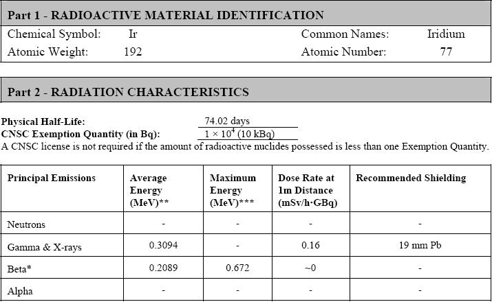
14 Brachytherapy Sources Radionuclide Half-life Photon Energy (MeV) Half-value Layer (mm lead) 226 Ra 1600 years (0.83 ave) Rn 3.83 days (0.83 ave) Co 5.26 years 1.17, Cs 30.0 years Ir 74.2 days (0.38 ave) Au 2.7 days I 60.2 days ave Pd 17.0 days ave 0.008

15 Brachytherapy sources The first isotope used clinically was radium around 1903 However, radium and radon have only historical importance - they should not be used in a modern radiotherapy department
16 Brachytherapy sources Because: wide energy spectrum leading to high dose close to the source and still high dose around the patient - shielding difficult Radon, the daughter product of radium, is a noble gas which is very difficult to contain - contamination risk The long half life means disposal is very difficult
17 Popular sources: 137-Cs Cesium 137 Main substitute for radium Mostly used in gynecological applications Long half life of 30 years ---> decay correction necessary every 6 months Sources are expensive and must be replaced every 10 to 15 years
18 Popular sources: 192-Ir Iridium 192 Many different forms available Most important source for HDR applications Medium half life (75 days) - decay correction necessary for each treatment Needs to be replaced every 3 to 4 months to maintain effective activity and therefore an acceptable treatment time
19 Popular sources: 192-Ir Iridium 192 High specific activity - therefore even high activity sources can be miniaturized essential for HDR applications A bit easier to shield than 137-Cs - because the gamma energies of 192-Ir range from 136 to 1062keV (effective energy around 350keV)
20

21 Popular sources: 125-I Very low energy - therefore shielding is easy and radiation from an implant is easily absorbed in the patient: permanent implants are possible Mostly used in the form of seeds

22 125-I seeds Many different designs

23 125-I seeds Design aims and features: sealed source non-toxic tissue compatible encapsulation isotropic dose distribution radio-opaque for localization Mentor

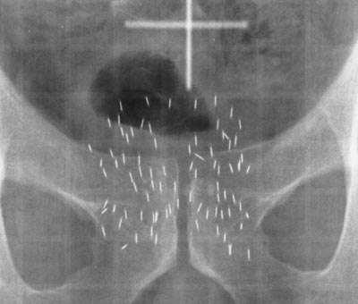
24 X-ray visibility of 125-I seeds
25 Other isotopes used for seeds Palladium 103 Half Life = 17 days - dose rate about 2.5 times larger than for 125-I Energy = 22 kev TVL lead = 0.05mm Gold 198 Half Life = 2.7 days - short enough to let activity decay in the patient Energy = 412 kev TVL lead = around 8mm
26 Brachytherapy Sources A variety of source shapes and forms: pellets = balls of approximately 3 mm diameter seeds = small cylinders about 1 mm diameter and 4 mm length needles = between 15 and 45 mm active length tubes = about 14 mm length, used for gynaecological implants hairpins = shaped as hairpins, approximately 60 mm active length wire = any length, usually customised in the hospital - inactive ends may be added HDR sources = high activity miniature cylinder sources approximately 1mm diameter, 10mm length

27 Source form examples Seeds (discussed before): small containers for activity usually 125-I, 103-Pd or 198-Au for permanent implant such as prostate cancer Needles and hairpins: Scale in mm for life implants in the operating theatre - activity is directly introduced in the target region of the patient usually 192-Ir for temporary implants eg. of the tongue

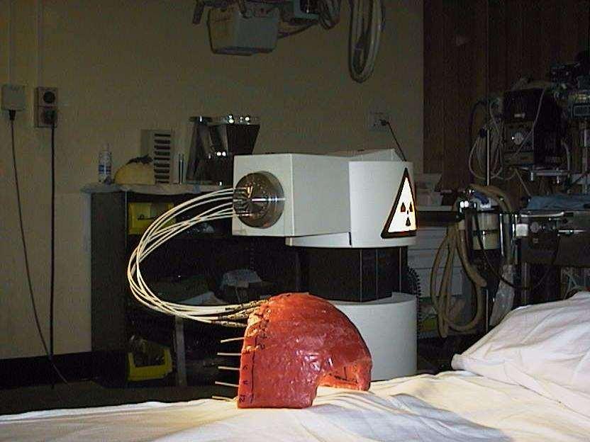
28 Source form: 192-Ir wire Used for LDR interstitial implants Cut to appropriate length prior to implant to suit individual patient Cutting using manual technique or cutter...
29 Source form 192-Ir wires 192-Ir wire: activity between 0.5 and 10mCi per cm used for interstitial implants low to medium dose rate can be cut from 50 cm long coils to the desired length for a particular patient Length measurement Movement controls Shielding Wire cutter
30 Source form example 192-Ir wire: activity between 0.5 and 10mCi per cm used for interstitial implants low to medium dose rate can be cut from 50 cm long coils to the desired length for a particular patient Length measurement Movement controls Shielding Wire cutter
31 Question: Why would people use 198-Au for brachytherapy?
32 Some clues for an answer Key features of 198-Au are: small sources (seed) short half life (2.7 days) inert material photon energy 412keV Therefore, ideal for permanent implant
33 Brachytherapy Brachytherapy installations cover direct source loading 137-Cs sources for gynaecological applications (radium should not be used) permanent seed implants (gold or 125-I) surface applicators (moulds, 125-I, strontium and ruthenium plaques manual afterloading (137-Cs, 192-Ir) automatic afterloading (LDR, PDR and HDR)
34 Brachytherapy Highly customized treatment techniques - each patient is treated differently Techniques depend on Disease site and stage Operator/clinician Technology/equipment available Many of the points covered for External Beam installations also apply to Brachytherapy installations, particularly for automatic afterloading systems
35 Preparation of sources for brachytherapy Choosing the correct sources is an important part of the implant optimization This is applicable for situations when: there are several different sources available (eg 137-Cs source with slightly different length and activity for gynecological implants) sources are ordered and customized for an individual patient (eg. 192-Ir wire)

36 Require a pre-implant plan...

37 Choosing the correct sources Prepare a plan for a particular implant following the prescription Select appropriate sources If existing sources are to be used select sources from the safe and place in transport container Document what is done source safe shielding
38 Interstitial implants For LDR usually use 192-Ir wire (compare part VI) Optimization is possible as the length of the wire can be adjusted for a particular implant
39 HDR sources No preparation necessary Ensure source calibration optimized plan
40 Implant techniques Permanent implants patient discharged with implant in place Temporary implants implant removed before patient is discharged Here particular emphasis on radiation protection issues in medical exposures
41 Permanent Implants: Radiation protection issues Implant of activity in theatre: Radiation protection of staff from a variety of professional backgrounds - radiation safety training is essential RSO or physicist should be present Source transport always necessary Potential of lost sources

42 Problems with handling activity in the operating theatre The time to place the sources in the best possible locations is typically limited Work behind shields or with other protective equipment may prolong procedure and result in sub-optimal access to the patient

43 Working behind shields
44 Permanent Implants: Radiation protection issues Patients are discharged with radioactive sources in place: lost sources exposure of others issues with accidents to the patient, other medical procedures, death, autopsies and cremation - compare part XV of the course
45 Temporary implants Mostly done in afterloading technique Radiation safety issues for staff: Source handling and preparation Exposure of nursing staff in manual afterloading Radiation safety issues for patients: Source placement and removal
46 Afterloading Manual The sources are placed manually usually by a physicist The sources are removed only at the end of treatment Remote The sources are driven from an intermediate safe into the implant using a machine ( afterloader ) The sources are withdrawn every time someone enters the room
47 Afterloading advantages No rush to place the sources in theatre - more time to optimize the implant Treatment is verified and planned prior to delivery Significant advantage in terms of radiation safety (in particular if a remote afterloader is used)

48 Use of lead shield reduces scatter to the patient
49 High Dose Rate Brachytherapy Most modern brachytherapy is delivered using HDR Reasons? Outpatient procedure Optimization possible
50 HDR brachytherapy In the past possible using 60-Co pellets Today, virtually all HDR brachytherapy is delivered using a 192-Ir stepping source Source moves step by step through the applicator - the dwell times in different locations determine the dose distribution
51 HDR unit interface
52 4. Brachytherapy equipment Design considerations often similar to external beam therapy Nucletron

53 Remote Afterloading Equipment The most complex pieces of equipment in brachyhterapy Low dose rate units High dose rate units Many important design consideration in IEC standard
54 Low dose rate brachytherapy Selectron for gynecological brachytherapy 137-Cs pellets pushed into the applicators using compressed air Location of active and inactive pellets can be chosen by the operator to optimize the source loading for an individual patient Shown are 6 channels - the red lights indicate the location of an active source Nucletron
55 Other features No computer required Two independent timers Optical indication of source locations Permanent record through printout Key to avoid unauthorized use
56 HDR brachytherapy units Must be located in a bunker Have multiple channels to allow the same source to drive into many catheters/needles MDS Nordion

57 Nucletron HDR unit control Emergency off button Keypad Printout = permanent record Display Key Key for source out Memory card for transfer of the dwell positions for the treatment of a particular patient - labeled
58 Catheters are indexed to avoid mixing them up Transfer catheters are locked into place during treatment - green light indicates the catheters in use
59 Regular maintenance is required Source drive must be working within specified accuracy (typically 1-2mm) Emergency buttons must work Manual retraction of the source in case of power failure must work

60 Regular maintenance is required Maintenance work should follow manufacturers recommendations All modifications MUST be documented A physicists should be notified to perform appropriate tests
61 LDR and HDR units are not all... Other brachytherapy equipment: PDR (pulsed dose rate) units Seed implant equipment Endovascular brachytherapy
62 LDR and HDR units are not all... Other brachytherapy equipment: PDR units - similar to HDR Seed implant equipment - discussed in more detail in the second lecture of part VI Endovascular brachytherapy
63 Typical Radiation Levels Selectron LDR (Cs-137) Cervix insertion 10 pellets of 15 mci/seed = 150 mci 20 mr/h at 1m 0.2 msv/h 5 days for 1 msv (Background) this is inside the room! microselectron HDR (Ir-192) turned ON! 10 Ci source = mci 4700 mr/h at 1m 47 msv/h 1.3 minutes for 1 msv (Background) door interlock ensures that no-one is in room
64 Brachytherapy Techniques 1. Clinical brachytherapy applications 2. Implant techniques and applicators 3. Delivery modes and equipment
65 Brachytherapy Very flexible radiotherapy delivery Source position determines treatment success Depends on operator skill and experience In principle the ultimate conformal radiotherapy Highly individualized for each patient Typically an inpatient procedure as opposed to external beam radiotherapy which is usually administered in an outpatient setting
66 Clinical brachytherapy
67 History Brachytherapy has been one of the earliest forms of radiotherapy After discovery of radium by M Curie, radium was used for brachytherapy already late 19th century There is a wide range of applications - this versatility has been one of the most important features of brachytherapy

68 Today Many different techniques and a large variety of equipment Less than 10% of radiotherapy patients receive brachytherapy Use depends very much on training and skill of clinicians and access to operating theatre
69 A brachytherapy patient Typically localized cancer Often relatively small tumor Often good performance status (must tolerate the operation) Sometimes pre-irradiated with external beam radiotherapy (EBT) Often treated with combination brachytherapy and EBT
70 1. Clinical brachytherapy applications A. Surface moulds B. Intracavitary (gynaecological, bronchus,..) C. Interstitial (Breast, Tongue, Sarcomas, )

71 A. Surface moulds Treatment of superficial lesions with radioactive sources in close contact with the skin Hand A mould for the back of a hand including shielding designed to protect the patient during treatment Catheters for source transfer
72

73 Surface mould advantages Fast dose fall off in tissues Can conform the activity to any surface Flaps available

74 B. Intracavitary implants Introduction of radioactivity using an applicator placed in a body cavity Gynaecological implants Bronchus Oesophagus Rectum

75 Gynaecological implants Most common brachytherapy application - cervix cancer Many different applicators Either as monotherapy or in addition to external beam brachytherapy as a boost

76 Gynecological applicators Different design - all Nucletron


77 Vaginal applicators Single source line Different diameters and length Gammamed - on the right with shielding Nucletron

78 Bronchus implants Often palliative to open air ways Usually HDR brachytherapy Most often single catheter, however also dual catheter possible


79 Dual catheter bronchus implant Catheter placement via bronchoscope Bifurcation may create complex dosimetry
80 C. Interstitial implants Implant of needles or flexible catheters directly in the target area Breast Head and Neck Sarcomas Requires surgery - often major
81 Interstitial implants - tongue implant Catheter loop tongue tongue Button

82 Breast implants Typically a boost Often utilizes templates to improve source positioning Catheters or needles
83 2. Implant techniques and applicators Permanent implants patient discharged with implant in place Temporary implants implant removed before patient is discharged from hospital
84 Source requirement for permanent implants Low energy gammas or betas to minimize radiation levels outside of the patient (125-I is a good isotope) May be short-lived to reduce dose with time (198-Au is a good isotope) More details on most common 125-I prostate implants in section 4A of the lecture
85 Temporary implants Implant of activity in theatre Manual afterloading Remote afterloading
 Pulsed Dose Rate")
86 3. Delivery modes and equipment Low Dose Rate (LDR) Medium Dose Rate High Dose Rate (HDR) Pulsed Dose Rate (PDR)
87 Delivery modes - different classifications are in use Low Dose Rate Medium Dose Rate High Dose Rate Pulsed Dose Rate < 1Gy/hour around 0.5Gy/hour > 1Gy/hour not often used >10Gy/hour pulses of around 1Gy/hour
88 Low dose rate brachytherapy The only type of brachytherapy possible with manual afterloading Most clinical experience available for LDR brachytherapy Performed with remote afterloaders using 137-Cs or 192-Ir
89 Low dose rate brachytherapy Selectron for gynecological brachytherapy 137-Cs pellets pushed into the applicators using compressed air 6 channels for up to two parallel treatments Nucletron
90 Simple design No computer required Two independent timers Optical indication of source locations Permanent record through printout Key to avoid unauthorized use
")

91 Treatment process Implant of applicator (typically in the operating theatre) Verification of applicator positioning using diagnostic X-rays (eg radiotherapy simulator)
92 Treatment planning Most commercial treatment planning systems have a module suitable for brachytherapy planning: Choosing best source configuration Calculate dose distribution Determine time required to give desired dose at prescription points Record dose to critical structures

93 Treatment planning of different brachytherapy implants

94 High Dose Rate Brachytherapy Most modern brachytherapy is delivered using HDR Reasons? Outpatient procedure Optimization possible
95 HDR brachytherapy In the past possible using 60-Co pellets Today, virtually all HDR brachytherapy is delivered using a 192-Ir stepping source Source moves step by step through the applicator - the dwell times in different locations determine the dose distribution

96 HDR 192-Ir source Source length 5mm, diameter 0.6mm Activity: around 10Ci From presentation by Pia et al

97 Optimization of dose distribution adjusting the dwell times of the source in an applicator Nucletron

98 HDR brachytherapy procedure Implant of applicators, catheters or needles in theatre For prostate implants as shown here use transrectal ultrasound guidance


99 HDR brachytherapy procedure Localization using diagnostic X-rays HDR prostate implant: Simulator image Scout image for CT scan


100 Treatment planning Definition of the desired dose distribution (usually using many points) Computer optimization of the dwell positions and times for the treatment

101 Treatment Transfer of date to treatment unit Connecting patient Treat... Gammamed Nucletron

102 HDR unit interface
103 HDR brachytherapy Usually fractionated (eg. 6 fractions of 6Gy) Either patient has new implant each time or stays in hospital for bi-daily treatments Time between treatments should be >6hours to allow normal tissue to repair all damage

104 HDR units: different designs available


105 Catheters are indexed to avoid mixing them up Transfer catheters are locked into place during treatment - green light indicates the catheters in use
106 HDR systems Can be moved eg between different facilities or into theatre for intraoperative work
107 Pulsed dose rate Unit has a similar design as HDR, however the activity is smaller (around 1Ci instead of 10Ci) Stepping source operation - same optimization possible as in HDR Treatment over same time as LDR treatment to mimic favorable radiobiology In-patient treatment: hospitalization required Source steps out for about 10 minutes per hour and then retracts. Repeats this every hour to deliver minifractions ( pulses ) of about 1Gy
108 Feras Mansour Jargon فراس منصور جرغون IAEA Training Course: Radiation Protection in Radiotherapy slide 109
Brachytherapy an Overview
 Brachytherapy an Overview Yakov Pipman, D Sc North Shore LIJ Health System Monterrey, Nov30-Dec1, 2007 Brachytherapy A procedure in therapeutic radiology that involves the irradiation of a target with
Brachytherapy. What is brachytherapy and how is it used?
 Scan for mobile link. Brachytherapy Brachytherapy places radioactive sources inside the patient on a temporary or permanent basis to damage cancer cells DNA and destroy their ability to divide and grow.
Brachytherapy Planning and Quality Assurance w Classical implant systems and modern computerized dosimetry w Most common clinical applications w
 Brachytherapy Planning and Quality Assurance w Classical implant systems and modern computerized dosimetry w Most common clinical applications w Quality assurance Classical implant systems w Manchester
Brachytherapy Planning and Quality Assurance
 Brachytherapy Planning and Quality Assurance Classical implant systems Most common clinical applications and modern dosimetry methods Quality assurance Classical implant systems Manchester (Paterson-Parker)
Regulatory Guidelines and Computational Methods for Safe Release of Radioactive Patients II. Brachytherapy
 Regulatory Guidelines and Computational Methods for Safe Release of Radioactive Patients II. Brachytherapy Firas Mourtada, Ph.D., DABR Chief of Clinical Physics Helen F. Graham Cancer Center Christiana
How ICD-10 Affects Radiation Oncology. Presented by, Lashelle Bolton CPC, COC, CPC-I, CPMA
 How ICD-10 Affects Radiation Oncology Presented by, Lashelle Bolton CPC, COC, CPC-I, CPMA ICD-10 ICD-10-CM has added new challenges to the radiation oncology specialty. Approximately 220 ICD-9-CM codes
Radioactive sources in brachytherapy
 Radioactive sources in brachytherapy Janez Burger Institute of Oncology, Department of Radiophysics, Brachytherapy Unit, Ljubljana, Slovenia Background. In modern brachytherapy, a great step forward was
Definitions. Brachytherapy in treatment of cancer. Implantation Techniques and Methods of Dose Specifications. Importance of Brachytherapy in GYN
 Implantation Techniques and Methods of Dose Specifications Brachytherapy Course Lecture V Krishna Reddy, MD, PhD Assistant Professor, Radiation Oncology Brachytherapy in treatment of cancer GYN Cervical
FEE RULES RADIATION ONCOLOGY FEE SCHEDULE CONTENTS
 Tel: +27-21-9494060 Fax: +27-21-9494112 E-mail: leon.gouws@cancercare.co.za FEE RULES RADIATION ONCOLOGY FEE SCHEDULE CONTENTS 1. EXTERNAL BEAM RADIATION... 2 2. PLANNING OF TREATMENT... 2 3. DELIVERY
INTRODUCTION TO BRACHYTHERAPY, HISTORY AND INDICATIONS. Christine Haie-Meder Brachytherapy Unit
 INTRODUCTION TO BRACHYTHERAPY, HISTORY AND INDICATIONS Christine Haie-Meder Brachytherapy Unit HISTORY OF BRACHYTHERAPY 8/11/1895 Röentgen : X-ray discovery 22/11/1895 Mrs Röentgen s hand X-ray 1896 Becquerel
2015 Radiology Coding Survival Guide
 2015 Radiology Coding Survival Guide Chapter 31: Clinical Brachytherapy (77750-77799) Clinical brachytherapy involves applying radioelements into or around a treatment field. CPT guidelines clarify that
BRACHYTHERAPY IN HORSES
 Vet Times The website for the veterinary profession https://www.vettimes.co.uk BRACHYTHERAPY IN HORSES Author : DAVID DONALDSON Categories : Vets Date : June 16, 2014 DAVID DONALDSON BVSc(Hons), DipECVO,
Radiotherapy. Marta Anguiano Millán. Departamento de Física Atómica, Molecular y Nuclear Facultad de Ciencias. Universidad de Granada
 Departamento de Física Atómica, Molecular y Nuclear Facultad de Ciencias. Universidad de Granada Overview Introduction Overview Introduction Brachytherapy Radioisotopes in contact with the tumor Overview
Chapter 13: Brachytherapy: Physical and Clinical Aspects
 Chapter 13: Brachytherapy: Physical and Clinical Aspects Set of 163 slides based on the chapter authored by N. Suntharalingam, E.B. Podgorsak, H. Tolli of the IAEA publication (ISBN 92-0-107304-6): Radiation
A Study on Dosimetry of Gynaecological Cancer and Quality Assurance of HDR Brachytherapy in BPKMCH, Nepal
 A Study on Dosimetry of Gynaecological Cancer and Quality Assurance of HDR Brachytherapy in BPKMCH, Nepal SB Chand, PP Chaursia, MP Adhikary, AK Jha, and S Shrestha Dept. of Radiation Oncology, BPKM Cancer
Varian Acuity BrachyTherapy Suite One Room Integrated Image-Guided Brachytherapy
 Varian Acuity BrachyTherapy Suite One Room Integrated Image-Guided Brachytherapy The Acuity BrachyTherapy Suite Integrating Imaging, Planning, and Treatment in a Single Room Each component draws on the
BRACHYTHERAPY IN HEAD & NECK CANCERS DR. GIRI G.V.
 BRACHYTHERAPY IN HEAD & NECK CANCERS DR. GIRI G.V. BASICS High dose to tumor = local control Spare normal structures i.e. salivary gland, mandible and muscles of mastication. Seed 100 Relative dose 10
Radiotherapy physics & Equipments
 Radiotherapy physics & Equipments RAD 481 Lecture s Title: An Overview of Radiation Therapy for Health Care Professionals Dr. Mohammed Emam Vision :IMC aspires to be a leader in applied medical sciences,
MultiSource. for a higher standard in HDR brachytherapy.
 MultiSource for a higher standard in HDR brachytherapy www.ibt-bebig.eu MultiSource HDR afterloader Multiple use: The MultiSource radiation sources have two things in common a small size and a favourable
Review of brachytherapy in
 Medical Physics in the Baltic States 2017 Review of brachytherapy in Klaipėda University Hospital Romas Vilkas Rasa Dagienė Klaipėda University Hospital 1 Aims of presentation: To show progress of brachytherapy
Comprehensive and Practical Brachytherapy March 04-8 March 2018, Ljubljana, Slovenia Day 1 Sunday 4 March 2018
 Day 1 Sunday 4 March 2018 Welcome and Summary of the course 13:00-13:20 15 introduction 13:20-13:50 30 Radioactivity Radioactivity: What we need to know Characteristics of LDR-PDR-HDR, radiobiological
OPPORTUNITIES IN BRACHYTHERAPY
 OPPORTUNITIES IN BRACHYTHERAPY EDITION 2014 A report written by Paul-Emmanuel Goethals & Richard Zimmermann www.medraysintell.com Brachytherapy THE FIRST COMPREHENSIVE EVALUATION OF THE BRACHYTHERAPY INDUSTRY
I. Equipments for external beam radiotherapy
 I. Equipments for external beam radiotherapy 5 linear accelerators (LINACs): Varian TrueBeam 6, 10 & 18 MV photons, 6-18 MeV electrons, image-guided (IGRT) and intensity modulated radiotherapy (IMRT),
Inter-society standards for the performance of brachytherapy: a joint report from ABS, ACMP and ACRO
 Critical Reviews in Oncology/Hematology 48 (2003) 1 17 Inter-society standards for the performance of brachytherapy: a joint report from ABS, ACMP and ACRO Subir Nag a,, Ralph Dobelbower b, Glenn Glasgow
The Future of Brachytherapy. Alex Rijnders Europe Hospitals Brussels, Belgium
 The Future of Brachytherapy Alex Rijnders Europe Hospitals Brussels, Belgium a.rijnders@europehospitals.be Sarajevo, May 21, 2014 Brachytherapy = application of sealed sources inside or in close proximity
Therapeutic Medical Physics. Stephen J. Amadon Jr., Ph.D., DABR
 Therapeutic Medical Physics Stephen J. Amadon Jr., Ph.D., DABR Outline 1. Why physicists are needed in medicine 2. Branches of medical physics 3. Physics in Radiation Oncology 4. Treatment types and Treatment
ICRT รศ.พญ.เยาวล กษณ ชาญศ ลป
 ICRT รศ.พญ.เยาวล กษณ ชาญศ ลป Brachytherapy การร กษาด วยร งส ระยะใกล Insertion การสอดใส แร Implantation การฝ งแร Surface application การวางแร physical benefit of brachytherapy - very high dose of radiation
 https://patient.varian.com/sit es/default/files/videos/origin al/imrt.mp4 brachy- from Greek brakhys "short" Historically LDR has been used. Cs-137 at 0.4-0.8 Gy/h With optimally placed device, dose
Radiation Biology in Brachytherapy
 Journal of Surgical Oncology 1997;65:66 70 REVIEW ARTICLE Radiation Biology in Brachytherapy DAVID J. BRENNER, DSc Center for Radiological Research, College of Physicians and Surgeons, Columbia University,
RADIOTHERAPY: TECHNOLOGIES AND GLOBAL MARKETS
 RADIOTHERAPY: TECHNOLOGIES AND GLOBAL MARKETS HLC176A February 2015 Neha Maliwal Project Analyst ISBN: 1-62296-043-2 BCC Research 49 Walnut Park, Building 2 Wellesley, MA 02481 USA 866-285-7215 (toll-free
GYNECOLOGIC CANCER and RADIATION THERAPY. Jon Anders M.D. Radiation Oncology
 GYNECOLOGIC CANCER and RADIATION THERAPY Jon Anders M.D. Radiation Oncology Brachytherapy Comes from the Greek brakhus meaning short Brachytherapy is treatment at short distance Intracavitary vs interstitial
Basics of Cervix Brachytherapy. William Small, Jr., MD Professor and Chairman Loyola University Chicago
 Gynecologic Cancer InterGroup Cervix Cancer Research Network Basics of Cervix Brachytherapy William Small, Jr., MD Professor and Chairman Loyola University Chicago Cervix Cancer Education Symposium, January
Can we deliver the dose distribution we plan in HDR-Brachytherapy of Prostate Cancer?
 Can we deliver the dose distribution we plan in HDR-Brachytherapy of Prostate Cancer? Dimos Baltas Dept. of Medical Physics & Engineering, Strahlenklinik, Klinikum Offenbach GmbH 63069 Offenbach, Germany
Intravascular Brachytherapy Dosimetry Techniques for Gamma systems
 Intravascular Brachytherapy Dosimetry Techniques for Gamma systems Shirish K. Jani, Ph.D., FACR Scripps Clinic La Jolla, California Physics Parameters-1: IVBT Gamma or X ray Isotopes Energy [ kev ] Half
MEDICAL MANAGEMENT POLICY
 PAGE: 1 of 8 This medical policy is not a guarantee of benefits or coverage, nor should it be deemed as medical advice. In the event of any conflict concerning benefit coverage, the employer/member summary
3D CONFORMATIONAL INTERSTITIAL BRACHYTHERAPY PLANNING FOR SOFT TISSUE SARCOMA
 3D CONFORMATIONAL INTERSTITIAL BRACHYTHERAPY PLANNING FOR SOFT TISSUE SARCOMA Alina TĂNASE 1,3, M. DUMITRACHE 2,3, O. FLOREA 1 1 Emergency Central Military Hospital Dr. Carol Davila Bucharest, Romania,
12 Physics and Clinical Aspects of Brachytherapy
 Physics and Clinical Aspects of Brachytherapy 255 12 Physics and Clinical Aspects of Brachytherapy Zuofeng Li CONTENTS 12.1 Introduction 255 12.2 Classifications of Brachytherapy 256 12.2.1 Permanent Versus
Pulsed Dose Rate for GYN Brachytherapy
 Pulsed Dose Rate for GYN Brachytherapy Firas Mourtada,, Ph.D. Department of Radiation Physics Dose equivalency to LDR Brief Introduction Radiobiology Dose Distribution: Radial dose function Source anisotropy
The Canadian National System for Incident Reporting in Radiation Treatment (NSIR-RT) Taxonomy March 11, 2015
 Taxonomy March 11, 2015") The Canadian National System for Incident Reporting in Radiation Treatment (NSIR-RT) Taxonomy March 11, 2015 Taxonomy Data Category Number Description Data Fields and Menu Choices 1. Impact 1.1 Incident
The Canadian National System for Incident Reporting in Radiation Treatment (NSIR-RT) Taxonomy March 11, 2015 Taxonomy Data Category Number Description Data Fields and Menu Choices 1. Impact 1.1 Incident
Clinical Applications of Brachytherapy Radiobiology. Radiobiology is Essential
 Clinical Applications of Brachytherapy Radiobiology Dr Alexandra Stewart University of Surrey St Luke s Cancer Centre Guildford, England Radiobiology is Essential Knowledge of radiobiological principles
HDR vs. LDR Is One Better Than The Other?
 HDR vs. LDR Is One Better Than The Other? Daniel Fernandez, MD, PhD 11/3/2017 New Frontiers in Urologic Oncology Learning Objectives Indications for prostate brachytherapy Identify pros/cons of HDR vs
Radio-isotopes in Clinical Medicine
 Radio-isotopes in Clinical Medicine Radiactive isotopes, whether naturally occurring or artificially produced, have a number of different uses in clinical medicine. These include: (1) Diagnostic and research
Who Should Know Radiation Oncology Coding?
 Why Should We Learn Radiation Oncology Coding? Terry Wu, Ph.D. Chief Physicist Radiation Oncology Department Willis-Knighton Cancer Center Who Should Know Radiation Oncology Coding? Radiation Oncologist
Brachytherapy, Radionuclide Therapy Medical Physics in the Clinic. Raymond K. Wu, PhD Chairman AAPM Exchange Scientist Program
 Brachytherapy, Radionuclide Therapy Medical Physics in the Clinic Raymond K. Wu, PhD Chairman AAPM Exchange Scientist Program TG 43 of 1995 New dose calculation formalism for brachytherapy Consensus data
Can we deliver the dose distribution we plan in HDR-Brachytherapy of Prostate Cancer?
 Can we deliver the dose distribution we plan in HDR-Brachytherapy of Prostate Cancer? Dimos Baltas 1,3, Natasa Milickovic 1, Nikolaos Zamboglou 2 1 Dept. of Medical Physics & Engineering, 2 Strahlenklinik,
DOSIMETRIC OPTIONS AND POSSIBILITIES OF PROSTATE LDR BRACHYTHERAPY WITH PERMANENT I-125 IMPLANTS
 DOSIMETRIC OPTIONS AND POSSIBILITIES OF PROSTATE LDR BRACHYTHERAPY WITH PERMANENT I-125 IMPLANTS Andrius IVANAUSKAS*, Eduardas ALEKNAVIČIUS*, Arvydas BURNECKIS*, Albert MILLER *Institute of Oncology Vilnius
Prostate Cancer. What is prostate cancer?
 Scan for mobile link. Prostate Cancer Prostate cancer is a tumor of the prostate gland, which is located in front of the rectum and below the bladder. Your doctor may perform a physical exam, prostate-specific
Prostate Cancer Treatment
 Scan for mobile link. Prostate Cancer Treatment Prostate cancer overview Prostate cancer is the most common form of cancer in American men, most prevalent in men over age 65 and fairly common in men 50-64
Brachytherapy:General Principles
 Brachytherapy:General Principles Dr. S.C.Sharma Professor & Head Department foradiotherapy and Oncology, Regional Cancer Centre, Postgraduate Institute of Medical Education and Research Chandigarh Brachytherapy:
PRINCIPLES and PRACTICE of RADIATION ONCOLOGY. Matthew B. Podgorsak, PhD, FAAPM Department of Radiation Oncology
 PRINCIPLES and PRACTICE of RADIATION ONCOLOGY Matthew B. Podgorsak, PhD, FAAPM Department of Radiation Oncology OUTLINE Physical basis Biological basis History of radiation therapy Treatment planning Technology
Medical Use of Radioisotopes
 Medical Use of Radioisotopes Therapy Radioisotopes prove to be useful in the application of brachytherapy, the procedure for using temporary irradiation close to the area of disease (i.e. cancer) 10% Medical
X-ray Safety Discussion and Tour for Health Physicists
 X-ray Safety Discussion and Tour for Health Physicists April 1, 2016 Health Physics Society Spring Meeting Jerry Bai Environmental Administrator, Field Operations Bureau of Radiation Control Florida Department
EORTC Member Facility Questionnaire
 Page 1 of 9 EORTC Member Facility Questionnaire I. Administrative Data Name of person submitting this questionnaire Email address Function Phone Institution Address City Post code Country EORTC No Enter
National System for Incident Reporting in Radiation Therapy (NSIR-RT) Taxonomy
 Taxonomy") Canadian Partnership for Quality Radiotherapy (CPQR) National System for Incident Reporting in Radiation Therapy (NSIR-RT) National System for Incident Reporting in Radiation Therapy (NSIR-RT) Taxonomy
Canadian Partnership for Quality Radiotherapy (CPQR) National System for Incident Reporting in Radiation Therapy (NSIR-RT) National System for Incident Reporting in Radiation Therapy (NSIR-RT) Taxonomy
Cervical Cancer Treatment
 Scan for mobile link. Cervical Cancer Treatment Cervical cancer overview Cervical cancer occurs in the cervix, the part of the female reproductive system that connects the vagina and uterus. Almost all
Practice and Risk at Medical Facilities in Agency Operations
 Practice and Risk at Medical Facilities in Agency Operations Igor Gusev Radiation Protection Unit IAEA International Atomic Energy Agency Outline What is medical radiation exposure? Radiation sources and
MEDICAL POLICY SUBJECT: BRACHYTHERAPY OR RADIOACTIVE SEED IMPLANTATION FOR PROSTATE CANCER
 MEDICAL POLICY SUBJECT: BRACHYTHERAPY OR PAGE: 1 OF: 6 If the member's subscriber contract excludes coverage for a specific service it is not covered under that contract. In such cases, medical policy
ADVANCES IN RADIATION TECHNOLOGIES IN THE TREATMENT OF CANCER
 ADVANCES IN RADIATION TECHNOLOGIES IN THE TREATMENT OF CANCER Bro. Dr. Collie Miller IARC/WHO Based on trends in the incidence of cancer, the International Agency for Research on Cancer (IARC) and WHO
Overview of Clinical and Research Activities at Georgetown University Hospital
 Overview of Clinical and Research Activities at Georgetown University Hospital Dalong Pang, Ph.D. Department of Radiation Medicine Georgetown University Hospital Clinical Operation Two Varian linear accelerators
University Medical Center Utrecht The Netherlands
 University Medical Center Utrecht The Netherlands Metha Maenhout, MD: PHD student Maaike Koelink: Specialist RTT Wilfred de Vries: Physicist Assistant MED-00005 [00] UMC Utrecht Radiotherapy Department
Advances in Treatment of Cancer by Brachytherapy in Kenya, in Particular, Prostate Cancer
 Research Article imedpub Journals www.imedpub.com Journal Of Medical Physics And Applied Sciences ISSN 2574-285X DOI: 1.21767/2574-285X.12 Advances in Treatment of Cancer by Brachytherapy in Kenya, in
Trina Lynd, M.S. Medical Physicist Lifefirst Imaging & Oncology Cullman, AL Tri-State Alabama, Louisiana and Mississippi Spring 2016 Meeting April
 Trina Lynd, M.S. Medical Physicist Lifefirst Imaging & Oncology Cullman, AL Tri-State Alabama, Louisiana and Mississippi Spring 2016 Meeting April 17, 2016 Discuss permanent prostate brachytherapy and
Interstitial Breast Brachytherapy
 Patient Education Questions? Your questions are important. Call your doctor or health care provider if you have questions or concerns. UWMC clinic staff are also available to help at any time. During normal
Inpatient Admission for Radiation Therapy
 Medical Coverage Policy Effective Date... 8/15/2017 Next Review Date... 8/15/2018 Coverage Policy Number... 0408 Inpatient Admission for Radiation Therapy Table of Contents Coverage Policy... 1 Overview...
MEDICAL POLICY. SUBJECT: BRACHYTHERAPY OR RADIOACTIVE SEED IMPLANTATION FOR PROSTATE CANCER POLICY NUMBER: CATEGORY: Technology Assessment
 MEDICAL POLICY SUBJECT: BRACHYTHERAPY OR PAGE: 1 OF: 5 If the member's subscriber contract excludes coverage for a specific service it is not covered under that contract. In such cases, medical policy
JOURNAL OF APPLIED CLINICAL MEDICAL PHYSICS, VOLUME 6, NUMBER 2, SPRING 2005
 JOURNAL OF APPLIED CLINICAL MEDICAL PHYSICS, VOLUME 6, NUMBER 2, SPRING 2005 Advantages of inflatable multichannel endorectal applicator in the neo-adjuvant treatment of patients with locally advanced
Cigna Medical Coverage Policy
 Cigna Medical Coverage Policy Subject Inpatient Admission for Radiation Therapy Table of Contents Coverage Policy... 1 General Background... 1 Coding/Billing Information... 3 References... 3 Effective
20 Prostate Cancer Dan Ash
 20 Prostate Cancer Dan Ash 1 Introduction Prostate cancer is a disease of ageing men for which the aetiology remains unknown. The incidence rises up to 30 to 40% in men over 80. The symptoms of localised
PROGRESS IN RADIATION ONCOLOGY. Fox Chase Cancer Center. Jean Holland RN MSN AOCN Department of Radiation Oncology April 2017
 PROGRESS IN RADIATION ONCOLOGY Fox Chase Cancer Center Jean Holland RN MSN AOCN Department of Radiation Oncology April 2017 Radiation Therapy Therapeutic use of electromagnetic energy from a machine or
Prostate Cancer. What is prostate cancer?
 Scan for mobile link. Prostate Cancer Prostate cancer is a tumor of the prostate gland, which is located in front of the rectum, below the bladder and above the base of the penis. Your doctor may perform
APPLICATIONS OF RADIATION IN MEDICINE. Kirstin Murray BSc (Hons) Radiotherapy & Oncology
 Radiotherapy & Oncology") APPLICATIONS OF RADIATION IN MEDICINE Kirstin Murray BSc (Hons) Radiotherapy & Oncology CONTENTS Principles of Radiation Therapy Radiobiology Radiotherapy Treatment Modalities Special Techniques Nuclear
APPLICATIONS OF RADIATION IN MEDICINE Kirstin Murray BSc (Hons) Radiotherapy & Oncology CONTENTS Principles of Radiation Therapy Radiobiology Radiotherapy Treatment Modalities Special Techniques Nuclear
TRANSRECTAL ULTRASOUND-GUIDED PROSTATE BRACHYTHERAPY
 TRANSRECTAL ULTRASOUND-GUIDED PROSTATE BRACHYTHERAPY 1 TRANSRECTAL ULTRASOUND-GUIDED PROSTATE BRACHYTHERAPY BRENDAN CAREY, MD TRANSRECTAL ULTRASOUND-GUIDED PROSTATE BRACHYTHERAPY 2 TRANSRECTAL ULTRASOUND-GUIDED
Online in vivo dosimetry in conformal radiotherapies with MOSkin detectors
 Online in vivo dosimetry in conformal radiotherapies with MOSkin detectors G.Gambarini 1, C.Tenconi 1, N.Mantaut 1 M.Carrara 2, M.Borroni 2, E.Pignoli 2 D.Cutajar 3, M.Petasecca 3, I.Fuduli 3, M.Lerch
IAEA-TECDOC Implementation of microsource high dose rate (mhdr) brachytherapy in developing countries
 brachytherapy in developing countries") IAEA-TECDOC-1257 Implementation of microsource high dose rate (mhdr) brachytherapy in developing countries November 2001 The originating Section of this publication in the IAEA was: Applied Radiation Biology
IAEA-TECDOC-1257 Implementation of microsource high dose rate (mhdr) brachytherapy in developing countries November 2001 The originating Section of this publication in the IAEA was: Applied Radiation Biology
Understanding Radiation Therapy. For Patients and the Public
 Understanding Radiation Therapy For Patients and the Public Introduction to Radiation Oncology Radiation has been an effective tool for treating cancer for more than 100 years. Radiation oncologists are
Dosimetric Characteristics of the Brachytherapy Sources Based on Monte Carlo Method
 10 Dosimetric Characteristics of the Brachytherapy Sources Based on Monte Carlo Method Mahdi Sadeghi 1, Pooneh Saidi 2 and Claudio Tenreiro 3 1 Agricultural, Medical and Industrial School, P.O. Box 31485-498,
3D ANATOMY-BASED PLANNING OPTIMIZATION FOR HDR BRACHYTHERAPY OF CERVIX CANCER
 SAUDI JOURNAL OF OBSTETRICS AND GYNECOLOGY VOLUME 11 NO. 2 1430 H - 2009 G 3D ANATOMY-BASED PLANNING OPTIMIZATION FOR HDR BRACHYTHERAPY OF CERVIX CANCER DR YASIR BAHADUR 1, DR CAMELIA CONSTANTINESCU 2,
Patient Safety Focused QA. LDR Brachytherapy Vrinda Narayana
 Patient Safety Focused QA LDR Brachytherapy Vrinda Narayana D < 2 Gy/h Old LDR Brachytherapy? Ra-226; Cs-137; Ir-192 New Gynecological; interstitial Pd-103; I-125; Cs-131 Prostate implants Eye plaques
Regions Hospital Delineation of Privileges Radiation Oncology
 Regions Hospital Delineation of Privileges Radiation Oncology Applicant s Last First M. Instructions: Place a check-mark where indicated for each core group you are requesting. Review education and basic
NIA MAGELLAN HEALTH RADIATION ONCOLOGY CODING STANDARD. Dosimetry Planning
 NIA MAGELLAN HEALTH RADIATION ONCOLOGY CODING STANDARD Dosimetry Planning CPT Codes: 77295, 77300, 77301, 77306, 77307, 77321, 77316, 77317, 77318, 77331, 77399 Original Date: April, 2011 Last Reviewed
RADIATION ONCOLOGY AT UNIVERSITY OF COLORADO HOSPITAL
 RADIATION ONCOLOGY AT UNIVERSITY OF COLORADO HOSPITAL Cancer treatment requires a team effort. The Radiation Oncology team may include radiation oncology doctors, registered nurses, radiation therapists,
Radiation Safety Information for Students in Courses given by the Nuclear Physics Group at KTH, Stockholm, Sweden
 Radiation Safety Information for Students in Courses given by the Nuclear Physics Group at KTH, Stockholm, Sweden September 2006 The aim of this text is to explain some of the basic quantities and units
Wireless In-Vivo Dosimetry of High Dose Rate Brachytherapy for Prostate Cancer using MOSkin Detectors
 University of Wollongong Research Online University of Wollongong Thesis Collection 2017+ University of Wollongong Thesis Collections 2018 Wireless In-Vivo Dosimetry of High Dose Rate Brachytherapy for
Dosimetric characteristics of 137 Cs sources used in after loading Selectron system by Monte Carlo method
 Iran. J. Radiat. Res., 2007; 5 (3): 147-152 Dosimetric characteristics of Cs sources used in after loading Selectron system by Monte Carlo method M.B.Tavakoli, D. Shahbazi-Gahrouei *, M. Hosseinpour Department
Bell in 1903 was first suggested implanting. First Report on Study of Quality Control Program of High Dose Rate (Hdr) Brachytherapy.
 Brachytherapy.") Original Article First Report on Study of Quality Control Program of High Dose Rate (Hdr) Brachytherapy. Adhikari KP*, Dhakal BP**, Prasiko G*** * Sr. Medical Physicist & Radiation Safety Officer, NAMS,
Original Article First Report on Study of Quality Control Program of High Dose Rate (Hdr) Brachytherapy. Adhikari KP*, Dhakal BP**, Prasiko G*** * Sr. Medical Physicist & Radiation Safety Officer, NAMS,
Option D: Medicinal Chemistry
 Option D: Medicinal Chemistry Basics - unstable radioactive nuclei emit radiation in the form of smaller particles alpha, beta, positron, proton, neutron, & gamma are all used in nuclear medicine unstable
Breast Cancer. What is breast cancer?
 Scan for mobile link. Breast Cancer Breast cancer is a malignant tumor in or around breast tissue. It usually begins as a lump or calcium deposit that develops from abnormal cell growth. Most breast lumps
Quality Assurance of Ultrasound Imaging in Radiation Therapy. Zuofeng Li, D.Sc. Murty S. Goddu, Ph.D. Washington University St.
 Quality Assurance of Ultrasound Imaging in Radiation Therapy Zuofeng Li, D.Sc. Murty S. Goddu, Ph.D. Washington University St. Louis, Missouri Typical Applications of Ultrasound Imaging in Radiation Therapy
Ritu Raj Upreti, S. Dayananda, R. L. Bhalawat*, Girish N. Bedre*, D. D. Deshpande
 60 Original Article Evaluation of radiograph-based interstitial implant dosimetry on computed tomography images using dose volume indices for head and neck cancer Ritu Raj Upreti, S. Dayananda, R. L. Bhalawat*,
Radiation Therapy for Cancer: Questions and Answers
 Radiation Therapy for Cancer: Questions and Answers Key Points Radiation therapy uses ionizing radiation to kill cancer cells and shrink tumors (see Question 1). About half of all people with cancer are
Radiation Protection in Laboratory work. Mats Isaksson, prof. Department of radiation physics, GU
 Radiation Protection in Laboratory work Mats Isaksson, prof. Department of radiation physics, GU mats.isaksson@radfys.gu.se Fundamental principles (ICRP) Justification Optimisation Application of dose
Atoms for Health. Atoms for Health The. Atoms for Health - Division of Nuclear Health - Dept of Nuclear Aplications P Andreo DIR-NAHU 1
 International Atomic Energy Agency Atoms for Health The human side of nuclear applications NUCLEAR APPLICATIONS IN HEALTH A UNIQUE MANDATE OF THE UN SYSTEM The Agency shall seek to accelerate and enlarge
PHYS 383: Applications of physics in medicine (offered at the University of Waterloo from Jan 2015)
") PHYS 383: Applications of physics in medicine (offered at the University of Waterloo from Jan 2015) Course Description: This course is an introduction to physics in medicine and is intended to introduce
PHYS 383: Applications of physics in medicine (offered at the University of Waterloo from Jan 2015) Course Description: This course is an introduction to physics in medicine and is intended to introduce
Image based Brachytherapy- HDR applications in Gynecological Tumors
 Image based Brachytherapy- HDR applications in Gynecological Tumors Yakov Pipman, D. Sc. North Shore LIJ Health System Sites amenable to treatment with HDR Brachytherapy GYN Breast Prostate Head and Neck
Evaluation of Normal Tissue Complication Probability and Risk of Second Primary Cancer in Prostate Radiotherapy
 Evaluation of Normal Tissue Complication Probability and Risk of Second Primary Cancer in Prostate Radiotherapy Rungdham Takam Thesis submitted for the degree of Doctor of Philosophy in The School of Chemistry
WN MEDICAL IMAGING. RADIOTHERAPY. MEDICAL PHYSICS. NUCLEAR MEDICINE. RADIOACTIVITY
 WN MEDICAL IMAGING. RADIOTHERAPY. MEDICAL PHYSICS. NUCLEAR MEDICINE. RADIOACTIVITY Definitions : (from various online encyclopaedias/dictionaries) Medical Imaging/Radiology/ Diagnostic imaging: the use
PROSTATE CANCER BRACHYTHERAPY. Kazi S. Manir MD,DNB,PDCR RMO cum Clinical Tutor Department of Radiotherapy R. G. Kar Medical College
 PROSTATE CANCER BRACHYTHERAPY Kazi S. Manir MD,DNB,PDCR RMO cum Clinical Tutor Department of Radiotherapy R. G. Kar Medical College Risk categorization Very Low Risk Low Risk Intermediate Risk High Risk
Application of the Commission's Recommendations for the Protection of People in
 ICRP Publication 127 ICRP Publication 126 ICRP Publication 125 ICRP Publication 124 ICRP Publication 123 ICRP Publication 122 ICRP Publication 121 ICRP Publication 120 ICRP 2011 Proceedings Radiological
Accelerated Partial Breast Irradiation. Dr Patricia Lillis MD, MHA,MSS Marshfield Clinic Radiation Oncology
 Accelerated Partial Breast Irradiation Dr Patricia Lillis MD, MHA,MSS Marshfield Clinic Radiation Oncology Outline 1. Rationale 2. Review of selected literature 3. Technical aspects 4. Selection criteria