for the TCGA Breast Phenotype Research Group
|
|
|
- Bethanie Bailey
- 5 years ago
- Views:
Transcription

1 Decoding Breast Cancer with Quantitative Radiomics & Radiogenomics: Imaging Phenotypes in Breast Cancer Risk Assessment, Diagnosis, Prognosis, and Response to Therapy Maryellen Giger & Yuan Ji The University of Chicago, NorthShore University for the TCGA Breast Phenotype Research Group Analysis funded by The University of Chicago Dean Bridge Fund Images hosted by NCI TCIA COI: M L Giger is a stockholder in R2/Hologic, a co-founder and equity holder in Quantitative Insights, and receives royalties from Hologic, GE Medical Systems, MEDIAN Technologies, Riverain Medical, Mitsubishi, and Toshiba
2 NCI TCGA/TCIA Breast Phenotype Research Group Mapping of Breast MRI Phenotypes to Histopathology and Genomics Computer-Extracted Phenotypes & Data analysis/associations University of Chicago Maryellen Giger Hui Li Karen Drukker Li Lan NorthShore University Yuan Ji Yitan Zhu Wentian Guo NCI: Carl Jaffe John Freymann Erich Huang Justin Kirby Brenda Fevrier-Sullivan Radiologists: Elizabeth Morris MSKCC Ermelinda Bonaccio Roswell Kathleen Brandt Mayo Elizabeth Burnside U Wisconsin Madison Basak Dogan MD Anderson Marie Ganott Magee Jose Net U Miami Elizabeth Sutton MSKCC Gary Whitman MD Anderson Margarita Zuley U Pittsburgh H. Carisa Le-Petross MD Anderson Human-Extracted Phenotypes Analysis -- MD Anderson Arvind Rao
3 Decoding Breast Cancer with Quantitative Radiomics & Radiogenomics: Imaging Phenotypes in Breast Cancer Risk Assessment, Diagnosis, Prognosis, and Response to Therapy Purpose: To demonstrate, using the TCGA TCIA breast cancer dataset of MRI images, the role of quantitative radiomics in characterizing the molecular subtypes of breast cancer and associating the magenetic resonance imaging (MRI) computer-extracted image phenotypes with genomic data.
 - developed from CAD research Investigations in the applications of these techniques to gain knowledge in (a) the management of the cancer patient and in (b) the understanding of")
4 Decoding Breast Cancer with Imaging Involves interdisciplinary research: Development and/or customization of mathematical image analysis methods for extracting information from biomedical image data (computer vision) - developed from CAD research Investigations in the applications of these techniques to gain knowledge in (a) the management of the cancer patient and in (b) the understanding of cancer Quantitatively Extract Lesion Characteristics (Computer Vision) Patient-Specific Image-based Tumor Signature for Precision Medicine Data-mining of Computer- Extracted Features on Large Datasets for Population-based Cancer Discovery
5 Definitions Radiomics: High throughput conversion of images to mineable data Radiogenomics (imaging genomics): association of radiomic features with genomics and other -omics data
6 Imaging Genomics Asks questions about the relationships between features seen in medical images and the biology of cancer Data Sources Medical Images Computer Segmentation of Lesions Computer-extracted Lesion Features (size, morphology, texture, kinetics) Radiologist Descriptors Histopathology, Molecular Classification Genomics Associations and/or Classification Relevant to Clinical or Biological Questions Develop Predictive Models
 Which correlate and which are complementary?")
7 Imaging Genomics Asks questions about the relationships between features seen in medical images and the biology of cancer Data Sources Medical Images Computer Segmentation of Lesions Radiologist Descriptors Histopathology, Molecular Classification Genomics Computer-extracted Lesion Features (size, morphology, texture, kinetics) Which correlate and which are complementary??? Associations and/or Classification Relevant to Clinical or Biological Questions Develop Predictive Models
8 Imaging Genomics Asks questions about the relationships between features seen in medical images and the biology of cancer Data Sources Medical Images Computer Segmentation of Lesions Computer-extracted Lesion Features (size, morphology, texture, kinetics) Radiologist Descriptors Histopathology, Molecular Classification Genomics Lead to Personalized Screening and Personalized Treatment Which correlate and which are complementary??? Associations and/or Classification Relevant to Clinical or Biological Questions Develop Predictive Models
9 Imaging Genomics Asks questions about the relationships between features seen in medical images and the biology of cancer Radiomics Data Sources Medical Images Computer Segmentation of Lesions Computer-extracted Lesion Features (size, morphology, texture, kinetics) Radiologist Descriptors Histopathology, Molecular Classification Genomics Lead to Personalized Screening and Personalized Treatment Which correlate and which are complementary??? Associations and/or Classification Relevant to Clinical or Biological Questions Develop Predictive Models
10 University of Chicago Analysis High-Throughput & Output of MRI Tumor Phenotyping Signature System (Quantitative Image Analysis Workstation) Automated Lesion Segmentation, Feature Extraction [volumetrics, morphological, texture, kinetics] and Estimation of the Probability of Malignancy Giger et al., RSNA 2010


11 Dataset cancergenome.nih.gov Breast Cancer cases cancerimagingarchive.net Clinical /Histopathology /Genomic data downloaded by TCGA Assembler & Molecular subtyping / risk of recurrence values by Perou Lab MRIs of 91 cases (GE 1.5T) collected by TCIA MRIs of 91 cases downloaded to UChicago for computational MRI tumor phenotyping (radiomics) Tumor location on MRI determined by consensus of three of the TCIA radiologists
12 Dataset cancergenome.nih.gov Breast Cancer cases cancerimagingarchive.net Clinical /Histopathology /Genomic data downloaded by TCGA Assembler & Molecular subtyping / risk of recurrence values by Perou Lab MRIs of 91 cases (GE 1.5T) collected by TCIA MRIs of 91 cases downloaded to UChicago for computational MRI tumor phenotyping (radiomics) Tumor location on MRI determined by consensus of three of the TCIA radiologists


13 Distribution of the 91 MRI cases 77 Negative Positive ER PR HER2 TN


14 Distribution of the 91 MRI cases
15 Dataset cancergenome.nih.gov Breast Cancer cases cancerimagingarchive.net Clinical /Histopathology /Genomic data downloaded by TCGA Assembler & Molecular subtyping / risk of recurrence values by Perou Lab MRIs of 91 cases (GE 1.5T) collected by TCIA MRIs of 91 cases downloaded to UChicago for computational MRI tumor phenotyping (radiomics) Tumor location on MRI determined by consensus of three of the TCIA radiologists


16 Contrast-enhanced MR images of breast Tumors have increased blood vessels and differ in microvascular density and vessel permeability Gd-DTPA shortens T1 relaxation time which leads to increase of signal in T1-weighted images Precontrast Postcontrast Subtraction


17 Dynamic Contrast-Enhanced MRI & Tumor Segmentation 4D image analysis Increasing time Across slices


18 University of Chicago High-Throughput MRI Phenotyping System (Segmentation of the Tumor within the Breast MR image) 4D DCE MRI images Radiologist-indicated Tumor Center Computerized Tumor Segmentation ER-negative ER-positive

19 3D Breast MRI image

20 Computer-extracted Breast Cancer on MRI (can analyze as a virtual biopsy of the tumor)
21 University of Chicago High-Throughput MRI Phenotyping System 4D DCE MRI images Radiologist-indicated Tumor Center Computerized Tumor Segmentation Computer-Extracted Image Phenotypes (CEIP) Size Shape Morphology Contrast Enhancement CAD pipeline = radiomics pipeline Texture Curve Variance
 Size Shape Morphology Contrast Enhancement CAD pipeline = radiomics pipeline Texture Curve")
22 University of Chicago High-Throughput MRI Phenotyping System 4D DCE MRI images Volume Effective diameter Maximum linear size Surface Area Radiologist-indicated Tumor Center Computerized Tumor Segmentation Computer-Extracted Image Phenotypes (CEIP) Size Shape Morphology Contrast Enhancement CAD pipeline = radiomics pipeline Texture Curve Variance
 Size Shape Morphology Contrast Enhancement CAD pipeline = radiomics pipeline Texture Curve")
23 University of Chicago High-Throughput MRI Phenotyping System 4D DCE MRI images Radiologist-indicated Tumor Center Sphericity Irregularity Surface area/volume Computerized Tumor Segmentation Computer-Extracted Image Phenotypes (CEIP) Size Shape Morphology Contrast Enhancement CAD pipeline = radiomics pipeline Texture Curve Variance
 Size Shape Morphology Contrast Enhancement CAD pipeline = radiomics pipeline Texture Curve")
24 University of Chicago High-Throughput MRI Phenotyping System 4D DCE MRI images Margin sharpness Variance of margin sharpness Variance of radial gradient histogram Radiologist-indicated Tumor Center Computerized Tumor Segmentation Computer-Extracted Image Phenotypes (CEIP) Size Shape Morphology Contrast Enhancement CAD pipeline = radiomics pipeline Texture Curve Variance
 Size Shape Morphology Contrast Enhancement CAD pipeline = radiomics pipeline Texture")
25 University of Chicago High-Throughput MRI Phenotyping System 4D DCE MRI images Enhancement heterogeneity & kinetics of the uptake and washout of the contrast agent during the imaging time Radiologist-indicated Tumor Center Computerized Tumor Segmentation Computer-Extracted Image Phenotypes (CEIP) Size Shape Morphology Contrast Enhancement CAD pipeline = radiomics pipeline Texture Curve Variance


26 Tumors are Heterogeneous: Contrast Enhancement Heterogeneity & Kinetics Heterogeneity of Tumors:


27 University of Chicago High-Throughput MRI Phenotyping System 4D DCE MRI images Radiologist-indicated Tumor Center Contrast enhancement texture characterizing heterogeneity Computerized Tumor Segmentation Computer-Extracted Image Phenotypes (CEIP) Size Shape Morphology Contrast Enhancement Chen W, Giger ML, et al. Volumetric Texture Analysis of Breast Lesions on Contrast-Enhanced Magnetic Resonance Images Magn. Reson. Med. 58: , 2007 Texture Curve Variance
 Size Shape Morphology Contrast Enhancement Chen W, Giger ML, et al.")
28 University of Chicago High-Throughput MRI Phenotyping System 4D DCE MRI images Radiologist-indicated Tumor Center Kinetic curve assessment based on mostenhancing voxels within tumor: Uptake, washout, curve shape Computerized Tumor Segmentation Computer-Extracted Image Phenotypes (CEIP) Size Shape Morphology Contrast Enhancement Chen W, Giger ML, et al.: Automatic identification and classification of characteristic kinetic curves of breast lesions on DCE-MRI. Medical Physics, 33: ,2006 Texture Curve Variance
29 University of Chicago High-Throughput MRI Phenotyping System 4D DCE MRI images Radiologist-indicated Tumor Center Computerized Tumor Segmentation Enhancement variance kinetics Computer-Extracted Image Phenotypes (CEIP) Size Shape Morphology Contrast Enhancement Chen W, Giger ML, et al.: Computerized interpretation of breast MRI: Investigation of enhancement-variance dynamics, Medical Physics 31: , 2004 Texture Curve Variance
 Size Shape Morphology Contrast Enhancement Can be thought of as a non-invasive virtual biopsy Texture")
30 University of Chicago High-Throughput MRI Phenotyping System For Breast Tumors 4D DCE MRI images Radiologist-indicated Tumor Center Computerized Tumor Segmentation Computer-Extracted Image Phenotypes (CEIP) Size Shape Morphology Contrast Enhancement Can be thought of as a non-invasive virtual biopsy Texture Curve Variance
31 Virtual biopsy yielding tumor phenotypes & signatures Relating Computer-extracted MRI Phenotypes to: Classification & Association Tasks: 1. Clinical Tumor Status 1. Tumor Stage 2. Presence or Absence of Positive Lymph Nodes 2. Molecular Classification & Cancer Subtype 1. ER- vs. ER+ 2. PR- vs. PR+ 3. Her2- vs. Her2+ 4. Triple Negative vs. Others 3. Risk of Recurrence 1. OncotypeDX 2. PAM50 3. MammaPrint 4. Genomic Pathways
32 Virtual biopsy yielding tumor phenotypes & signatures Relating Computer-extracted MRI Phenotypes to: Classification & Association Tasks: 1. Clinical Tumor Status 1. Tumor Stage 2. Presence or Absence of Positive Lymph Nodes 2. Molecular Classification & Cancer Subtype 1. ER- vs. ER+ 2. PR- vs. PR+ 3. Her2- vs. Her2+ 4. Triple Negative vs. Others 3. Risk of Recurrence & Cancer Subtype (Normal, Luminal A..) 1. OncotypeDX 2. PAM50 3. MammaPrint 4. Genomic Pathways
33 MRI-based Phenotypes of Size predictive of breast cancer tumor stage TCGA/TCIA Breast Cancer Group cases; University of Chicago Giger Lab Giger computer-extracted TCGA 2015 image phenotypes
34 Virtual biopsy yielding tumor phenotypes & signatures Relating Computer-extracted MRI Phenotypes to: Classification & Association Tasks: 1. Clinical Tumor Status 1. Tumor Stage 2. Presence or Absence of Positive Lymph Nodes 2. Molecular Classification & Cancer Subtype 1. ER- vs. ER+ 2. PR- vs. PR+ 3. HER2- vs. HER2+ 4. Triple Negative vs. Others 3. Risk of Recurrence 1. OncotypeDX 2. PAM50 3. MammaPrint 4. Genomic Pathways
35 From TCIA MRI Radiomics -- ER Negative Breast Cancers tended to have larger size, a more irregular shape, and more heterogeneous in terms of contrast enhancement
36 From TCIA Radiomics- Triple Negative Breast Cancers tended to have a more irregular shape, and more heterogeneous in terms of contrast enhancement
37 From the TCIA Radiomics -- Enhancement Texture of Tumor Heterogeneity appears Predictive of Molecular Subtype Kendall test results for trends; p-value= Molecular Subtyping from C. Perou
38 From the TCIA Radiomics -- Enhancement Texture of Tumor Heterogeneity appears Predictive of Molecular Subtype size < 2 cm tumors Kendall test for trends; p-value= cm < size < 5 cm Kendall test for trends; p-value=0.016 Molecular Subtyping from C. Perou
39 Virtual biopsy yielding tumor phenotypes & signatures Relating Computer-extracted MRI Phenotypes to: Classification & Association Tasks: 1. Clinical Tumor Status 1. Tumor Stage 2. Presence or Absence of Positive Lymph Nodes 2. Molecular Classification & Cancer Subtype 1. ER- vs. ER+ 2. PR- vs. PR+ 3. Her2- vs. Her2+ 4. Triple Negative vs. Others 3. Risk of Recurrence from multi-gene assays 1. OncotypeDX 2. PAM50 3. MammaPrint 4. Genomic Pathways


40 Computer analysis of Breast MRIs of tumors Multi-gene assays of risk of recurrence Radiomics for virtual biopsy

41 Radiomics Virtual Biopsy & Risk of Recurrence
 ROC curves for leave-one-out LDA classifier using computer-extracted MRI phenotypes")
![as decision variable in the tasks of distinguishing between [low+medium] and high risk levels](/docs-images/86/93499254/images/42-3.jpg "of recurrence for MammaPrint, PAM50 ROR-S (Subtype), and PAM50 ROR-P (Subtype+Proliferation)")
42 Performance of the MRI Tumor Signatures in the task of predicting Risk of Recurrence (ROC analysis ) ROC curves for leave-one-out LDA classifier using computer-extracted MRI phenotypes as decision variable in the tasks of distinguishing between [low+medium] and high risk levels of recurrence for MammaPrint, PAM50 ROR-S (Subtype), and PAM50 ROR-P (Subtype+Proliferation) from Perou
43 Virtual biopsy yielding tumor phenotypes & signatures Relating Computer-extracted MRI Phenotypes to: Classification & Association Tasks: 1. Clinical Tumor Status 1. Tumor Stage 2. Presence or Absence of Positive Lymph Nodes 2. Molecular Classification & Cancer Subtype 1. ER- vs. ER+ 2. PR- vs. PR+ 3. Her2- vs. Her2+ 4. Triple Negative vs. Others 3. Risk of Recurrence 1. OncotypeDX 2. PAM50 3. MammaPrint 4. Genomic Pathways
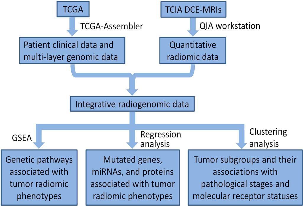
44 Radiogenomics Flowchart

45 Exploratory Cluster Analysis of the MRI Tumor Phenotypes Significant associations between radiomic features and clinical outcomes evaluated by t-tests.

46 Radiomics from the MRI tumor Virtual Biopsy shows association with Pathway Transcriptional Activities Giger lab Ji lab Zhu et al. submitted
47 Identified significant associations


48 Identified significant associations Size Phenotypes Gene expressions of pathways


49 Identified significant associations Enhancement Texture Heterogeneity Phenotypes mirna expressions
50 Summary & Conclusion Computational quantitative MRI analysis shows promise as a means for high-throughput image-based phenotyping and appears to predict breast cancer molecular subtypes Radiomics of tumor size and enhancement heterogeneity appear as dominant MRI phenotypes in classifiying tumor subtypes and risk of recurrence. Significant associations were identified between the MRI phenotypes (such as tumor size, shape, margin, enhancement texture, blood flow kinetics) and molecular features involved in multiple regulation layers (including DNA mutation, mirna expression, protein expression, pathway gene expression and copy number variation).
51 Summary & Conclusion Limitations included a small dataset of only 91 cancers TCIA is collecting additional images Investigators are organizing a multi-institutional radiomics network to collect beyond the TCGA/TCIA Identification of radiomics of molecular subtypes of breast tumors is expected to allow for virtual biopsies Ongoing research involves relating and merging MRI phenotypes with genomic data to develop improved predictive models
52 Questions Is it possible to decide targeted therapy based on imaging-genomics association results? Can imaging features inform important genomics features? Can integration of imaging and genomics features lead to higher power in prediction? Can imaging serve as a virtual biopsy? non-invasive, covers complete tumor, & repeatable
53 Thank you & please attend our related Workshop & Posters Workshop: Imaging Resources for the TCGA: Radiology and Pathology Tools for Enabling Science; May 11; 4-5pm and repeated 5-6pm Poster 91 Poster 79 Poster 105
7/21/2014. Image-based Phenotyping and Genomics
 AAPM Annual Meeting - 2014 Joint Imaging Education Quantitative Imaging Symposium: Genomics and Image-Omics for Medical Physicists Image-based Phenotyping and Genomics Maryellen L. Giger, The University
Quantitative MRI radiomics in the prediction of molecular classifications of breast cancer subtypes in the TCGA/TCIA data set
 www.nature.com/npjbcancer All rights reserved 2374-4677/16 ARTICLE OPEN in the prediction of molecular classifications of breast cancer subtypes in the TCGA/TCIA data set Hui Li 1,12, Yitan Zhu 2,12, Elizabeth
Breast MRI radiomics: comparison of computer- and human-extracted imaging phenotypes
 Sutton et al. European Radiology Experimental (2017) 1:22 DOI 10.1186/s41747-017-0025-2 European Radiology Experimental ORIGINAL ARTICLE Open Access Breast MRI radiomics: comparison of computer- and human-extracted
MRI-Based Biomarkers of Therapeutic Response in Triple-Negative Breast Cancer
 MRI-Based Biomarkers of Therapeutic Response in Triple-Negative Breast Cancer Daniel Golden Postdoctoral Scholar (Radiology) Stanford University Daniel Rubin Laboratory NCI Cancer Imaging Fellowship Seminar
CaPTk: Cancer Imaging Phenomics Toolkit
 CaPTk: Cancer Imaging Phenomics Toolkit Interaction Personalized Diagnostics CaPTk Quantitative Imaging Phenomic Features Radiogenomics Image Analysis Machine Learning What is CaPTk? A dynamically growing
Using lesion washout volume fraction as a biomarker to improve suspicious breast lesion characterization
 JOURNAL OF APPLIED CLINICAL MEDICAL PHYSICS, VOLUME 16, NUMBER 5, 2015 Using lesion washout volume fraction as a biomarker to improve suspicious breast lesion characterization Jie Huang, a Sarah M. Schafer,
Computerized Segmentation and Classification of Breast Lesions Using Perfusion Volume Fractions in Dynamic Contrast-enhanced MRI
 28 International Conference on BioMedical Engineering and Informatics Computerized Segmentation and Classification of Breast Lesions Using Perfusion Volume Fractions in Dynamic Contrast-enhanced MRI Sang
8/1/2017. Imaging and Molecular Biomarkers of Lung Cancer Prognosis. Disclosures. The Era of Precision Oncology
 Imaging and Molecular Biomarkers of Lung Cancer Prognosis Ruijiang Li, PhD Assistant Professor of Radiation Oncology 08/01/2017 Stanford University Department of Radiation Oncology School of Medicine Disclosures
Contemporary Classification of Breast Cancer
 Contemporary Classification of Breast Cancer Laura C. Collins, M.D. Vice Chair of Anatomic Pathology Professor of Pathology Beth Israel Deaconess Medical Center and Harvard Medical School Boston, MA Outline
Triple-negative breast cancer: which typical features can we identify on conventional and MRI imaging?
 Triple-negative breast cancer: which typical features can we identify on conventional and MRI imaging? Poster No.: C-1862 Congress: ECR 2013 Type: Educational Exhibit Authors: V. Bertani 1, A. Gualano
Automated Analysis of Breast Tumour in the Breast DCE-MR Images Using Level Set Method and Selective Enhancement of Invasive Regions
 Automated Analysis of Breast Tumour in the Breast DCE-MR Images Using Level Set Method and Selective Enhancement of Invasive Regions Atsushi Teramoto 1(&), Satomi Miyajo 2, Hiroshi Fujita 3, Osamu Yamamuro
Breast cancer classification: beyond the intrinsic molecular subtypes
 Breast cancer classification: beyond the intrinsic molecular subtypes Britta Weigelt, PhD Signal Transduction Laboratory CRUK London Research Institute Summary Breast cancer heterogeneity Molecular classification
Mike Becich, MD PhD Distinguished University Professor and Chair, Department of Biomedical Informatics University of Pittsburgh School of Medicine
 Affiliated with the University of Pittsburgh School of Medicine Mike Becich, MD PhD Distinguished University Professor and Chair, Department of Biomedical Informatics University of Pittsburgh School of
Case Studies on High Throughput Gene Expression Data Kun Huang, PhD Raghu Machiraju, PhD
 Case Studies on High Throughput Gene Expression Data Kun Huang, PhD Raghu Machiraju, PhD Department of Biomedical Informatics Department of Computer Science and Engineering The Ohio State University Review
Oncology - Evolution of imaging From helpful to essential
 Oncology - Evolution of imaging From helpful to essential 1990s 2000 1980s 18 FDG PET/CT MRI/MRSI 2010 1970s MRI 1960s CT 2015 HP13C-MRSI Ultrasound Nuc Med X-Ray - IVU MRI/PET Imaging 2016: Essential
AAPM Medical Physics Tutorial Session 1. Digital Breast Tomosynthesis. CT Breast Imaging. TMIST Update. Stereotactic Breast Biopsy
 Saturday SPPH01 AAPM Medical Physics Tutorial Session 1 Saturday, Nov. 25 12:00PM - 2:00PM Room: E351 BR PH AMA PRA Category 1 Credits : 2.00 ARRT Category A+ Credits: 2.00 FDA Discussions may include
Computer Science, Biology, and Biomedical Informatics (CoSBBI) Outline. Molecular Biology of Cancer AND. Goals/Expectations. David Boone 7/1/2015
 Outline. Molecular Biology of Cancer AND. Goals/Expectations. David Boone 7/1/2015") Goals/Expectations Computer Science, Biology, and Biomedical (CoSBBI) We want to excite you about the world of computer science, biology, and biomedical informatics. Experience what it is like to be a
Goals/Expectations Computer Science, Biology, and Biomedical (CoSBBI) We want to excite you about the world of computer science, biology, and biomedical informatics. Experience what it is like to be a
QUANTITATIVE IMAGING ANALYTICS
 QUANTITATIVE IMAGING ANALYTICS the future of radiology enabling evidence based care for oncology September 2015 Madison, Wisconsin HealthMyne.com Page 1 quantitative imaging analytics the future of radiology
Participants John M. Boone, PhD, Sacramento, CA (Presenter) Patent agreement, Isotropic Imaging Corporation; Consultant, RadSite;
 Patent agreement, Isotropic Imaging Corporation; Consultant, RadSite;") Saturday SPPH01 AAPM Medical Physics Tutorial Session 1 Saturday, Nov. 25 12:00PM - 2:00PM Room: E351 BR PH AMA PRA Category 1 Credits : 2.00 ARRT Category A+ Credits: 2.25 FDA Discussions may include
Saturday SPPH01 AAPM Medical Physics Tutorial Session 1 Saturday, Nov. 25 12:00PM - 2:00PM Room: E351 BR PH AMA PRA Category 1 Credits : 2.00 ARRT Category A+ Credits: 2.25 FDA Discussions may include
Educator Navigation Guide
 Decoding Breast Cancer Virtual Lab Educator Navigation Guide Decoding Cancer Nav Guide 2 Introduction In this virtual lab, students test tissue samples from different patients with breast cancer in order
Differentiating Tumor and Edema in Brain Magnetic Resonance Images Using a Convolutional Neural Network
 Original Article Differentiating Tumor and Edema in Brain Magnetic Resonance Images Using a Convolutional Neural Network Aida Allahverdi 1, Siavash Akbarzadeh 1, Alireza Khorrami Moghaddam 2, Armin Allahverdy
Imaging Decisions Start Here SM
 Owing to its high resolution and wide anatomic coverage, dynamic first-pass perfusion 320-detector-row CT outperforms PET/CT for distinguishing benign from malignant lung nodules, researchers from Japan
POC Brain Tumor Segmentation. vlife Use Case
 Brain Tumor Segmentation vlife Use Case 1 Automatic Brain Tumor Segmentation using CNN Background Brain tumor segmentation seeks to separate healthy tissue from tumorous regions such as the advancing tumor,
ncounter Assay Automated Process Immobilize and align reporter for image collecting and barcode counting ncounter Prep Station
 ncounter Assay ncounter Prep Station Automated Process Hybridize Reporter to RNA Remove excess reporters Bind reporter to surface Immobilize and align reporter Image surface Count codes Immobilize and
OPTO-ACOUSTIC BREAST IMAGING
 OPTO-ACOUSTIC BREAST IMAGING A Novel Fusion of Functional and Morphologic Imaging Reni S. Butler, MD A. Thomas Stavros, MD F. Lee Tucker, MD Michael J. Ulissey, MD PURPOSE 1. Explain opto-acoustic (OA)
Breast cancer: Molecular STAGING classification and testing. Korourian A : AP,CP ; MD,PHD(Molecular medicine)
") Breast cancer: Molecular STAGING classification and testing Korourian A : AP,CP ; MD,PHD(Molecular medicine) Breast Cancer Theory: Halsted Operative breast cancer is a local-regional disease The positive
Breast cancer: Molecular STAGING classification and testing Korourian A : AP,CP ; MD,PHD(Molecular medicine) Breast Cancer Theory: Halsted Operative breast cancer is a local-regional disease The positive
Radiological assessment of neoadjuvent chemotherapy for breast cancer
 XV th Balkan Congress of Radiology Budapest, Hungary, October 12 15, 2017 Radiological assessment of neoadjuvent chemotherapy for breast cancer V. Bešlagić C l i n i c o f R a d i o l o g y, U n i v e
Early Detection of Lung Cancer
 Early Detection of Lung Cancer Aswathy N Iyer Dept Of Electronics And Communication Engineering Lymie Jose Dept Of Electronics And Communication Engineering Anumol Thomas Dept Of Electronics And Communication
Scientific Exhibit Authors: A. Shimauchi, H. Abe, N. Mori, H. Ota, K. Takase, S.
 MRI- Detected Breast Masses: Are CAD-Measured Kinetic Diversity Analyses Useful for Differential Diagnosis? A New Approach with Whole Lesion Curve Distribution Analysis. Poster No.: C-1901 Congress: ECR
I SPY 2 TRIAL. How it Works April 9, 2013
 I SPY 2 TRIAL How it Works April 9, 2013 1 The Challenge in Breast Cancer Breast Cancer is a common and serious disease Over 200,000 new cases of invasive breast cancer each year Over 40,000 women will
8/1/2017. PROSTATEx-2 Overview. Disclosures. Joint Imaging-Therapy Scientific Symposium: PROSTATEx-2 Challenge
 Joint Imaging-Therapy Scientific Symposium: -2 Challenge -2 Overview S.G. Armato III Disclosures Royalties and licensing fees for CAD technology from the University of Chicago Consultant, Aduro Biotech,
8/10/2016. PET/CT Radiomics for Tumor. Anatomic Tumor Response Assessment in CT or MRI. Metabolic Tumor Response Assessment in FDG-PET
 PET/CT Radiomics for Tumor Response Evaluation August 1, 2016 Wei Lu, PhD Department of Medical Physics www.mskcc.org Department of Radiation Oncology www.umaryland.edu Anatomic Tumor Response Assessment
Triple Negative Breast Cancer: Clinical Presentation and Multimodality Imaging Characteristics
 Triple Negative Breast Cancer: Clinical Presentation and Multimodality Imaging Characteristics Poster No.: R-0141 Congress: RANZCR-AOCR 2012 Type: Scientific Exhibit Authors: O. H. Woo, S. Jang, K. R.
Only Estrogen receptor positive is not enough to predict the prognosis of breast cancer
 Young Investigator Award, Global Breast Cancer Conference 2018 Only Estrogen receptor positive is not enough to predict the prognosis of breast cancer ㅑ Running head: Revisiting estrogen positive tumors
Effect of intravenous contrast medium administration on prostate diffusion-weighted imaging
 Effect of intravenous contrast medium administration on prostate diffusion-weighted imaging Poster No.: C-1766 Congress: ECR 2015 Type: Authors: Keywords: DOI: Scientific Exhibit J. Bae, C. K. Kim, S.
Radiologic and pathologic correlation of non-mass like breast lesions on US and MRI: Benign, high risk, versus malignant
 Radiologic and pathologic correlation of non-mass like breast lesions on US and MRI: Benign, high risk, versus malignant Poster No.: C-1161 Congress: ECR 2013 Type: Educational Exhibit Authors: J. Kwak,
Since its introduction in 2000, digital mammography has become
 Review Article Smith A, PhD email : Andrew.smith@hologic.com Since its introduction in 2000, digital mammography has become an accepted standard of care in breast cancer screening and has paved the way
AB MR Interpretation Overview
 AB MR Interpretation Overview Goal of AB MR interpretation is to maintain high sensitivity and specificity In order to minimize false positives and short term follow ups, it is fundamental to focus only
Visual interpretation in pathology
 13 Visual interpretation in pathology Tissue architecture (alteration) evaluation e.g., for grading prostate cancer Immunohistochemistry (IHC) staining scoring e.g., HER2 in breast cancer (companion diagnostic
What Radiologists do?
 Multimodality Imaging in Oncology 2018 March 5 th 9th Diagnostic Imaging in Oncology What Radiologists do? Chikako Suzuki, MD, PhD Department of Diagnostic Radiology, KS Solna Department of Molecular Medicine
Role of Genomic Profiling in (Minimally) Node Positive Breast Cancer
 Node Positive Breast Cancer") Role of Genomic Profiling in (Minimally) Node Positive Breast Cancer Kathy S. Albain, MD, FACP Professor of Medicine Dean s Scholar Loyola University Chicago Stritch School of Medicine Cardinal Bernardin
Role of Genomic Profiling in (Minimally) Node Positive Breast Cancer Kathy S. Albain, MD, FACP Professor of Medicine Dean s Scholar Loyola University Chicago Stritch School of Medicine Cardinal Bernardin
MRI BI-RADS: How to make it out?
 MRI BI-RADS: How to make it out? Poster No.: C-1850 Congress: ECR 2016 Type: Educational Exhibit Authors: M. Ben Ammar, A. Ben Miled, O. Ghdes, S. Harguem, A. Gaja, N. Mnif; Tunis/TN Keywords: Breast,
Quantitative Radiomics System: Decoding the Tumor Phenotype
 HARVARD MEDICAL SCHOOL Quantitative Radiomics System: Decoding the Tumor Phenotype Hugo Aerts Director, Computational Imaging and Bioinformatics Lab (CIBL) Dana-Farber Cancer Institute, Brigham and Women
MRI/MRS Biomarkers. Robert E. Lenkinski, Ph.D.
 MRI/MRS Biomarkers Robert E. Lenkinski, Ph.D. Disclosure GE Healthcare-Research Grant Aspect MR-Scientific Advisor Aposense-Scientific Advisor Brainwatch-Scientific Advisor I will be discussing off-label
Triple Negative Breast Cancer
 Triple Negative Breast Cancer Prof. Dr. Pornchai O-charoenrat Division of Head-Neck & Breast Surgery Department of Surgery Faculty of Medicine Siriraj Hospital Breast Cancer Classification Traditional
MR Functional Imaging to Guide Radiotherapy: Challenges and Opportunities
 Abstract No. 1234 MR Functional Imaging to Guide Radiotherapy: Challenges and Opportunities Michael Milosevic, MD Department of Radiation Oncology, University of Toronto Radiation Medicine Program, Princess
Computer Aided Detection (CAD) for Breast MRI
 for Breast MRI") Technology in Cancer Research & Treatment ISSN 1533-0346 Volume 4, Number 1, February (2005) Adenine Press (2005) Computer Aided Detection (CAD) for Breast MRI www.tcrt.org Since 1999, there has been a
Technology in Cancer Research & Treatment ISSN 1533-0346 Volume 4, Number 1, February (2005) Adenine Press (2005) Computer Aided Detection (CAD) for Breast MRI www.tcrt.org Since 1999, there has been a
Leveraging Interaction between Genetic Variants and Mammographic Findings for Personalized Breast Cancer Diagnosis
 Leveraging Interaction between Genetic Variants and Mammographic Findings for Personalized Breast Cancer Diagnosis Jie Liu, PhD 1, Yirong Wu, PhD 1, Irene Ong, PhD 1, David Page, PhD 1, Peggy Peissig,
University of Pittsburgh Cancer Institute UPMC CancerCenter. Uma Chandran, MSIS, PhD /21/13
 University of Pittsburgh Cancer Institute UPMC CancerCenter Uma Chandran, MSIS, PhD chandran@pitt.edu 412-648-9326 2/21/13 University of Pittsburgh Cancer Institute Founded in 1985 Director Nancy Davidson,
Breast Cancer. What is breast cancer?
 Scan for mobile link. Breast Cancer Breast cancer is a malignant tumor in or around breast tissue. It usually begins as a lump or calcium deposit that develops from abnormal cell growth. Most breast lumps
CDIS: what's beyond microcalcifications? - Pictorial essay
 CDIS: what's beyond microcalcifications? - Pictorial essay Poster No.: C-1096 Congress: ECR 2014 Type: Educational Exhibit Authors: R. N. Lucas, C. A. S. Ruano, I. Oliveira, J. M. G. Lourenco, Z. 1 1 1
Radiomics for outcome modeling: state-of-the-art and challenges
 Workshop Avancées récentes en analyse d images médicales multi-modales Radiomics for outcome modeling: state-of-the-art and challenges Mathieu Hatt, PhD, HDR CR INSERM mathieu.hatt@inserm.fr Laboratoire
A Novel Method For Automatic Screening Of Nonmass Lesions In Breast DCE-MRI
 Volume 3 Issue 2 October 2015 ISSN: 2347-1697 International Journal of Informative & Futuristic Research A Novel Method For Automatic Screening Of Paper ID IJIFR/ V3/ E2/ 046 Page No. 565-572 Subject Area
MRI features of Triple-negative breast cancer: our experience.
 MRI features of Triple-negative breast cancer: our experience. Poster No.: C-1852 Congress: ECR 2013 Type: Scientific Exhibit Authors: V. Bertani, A. Gualano, V. Londero, A. Dal Col, M. Marcon, P. 1 2
Armed Forces Institute of Pathology.
 Armed Forces Institute of Pathology www.radpath.com Armed Forces Institute of Pathology Breast Disease www.radpath.org Armed Forces Institute of Pathology Interpretation of Breast MRI Leonard M. Glassman
Objectives. Image analysis and Informatics. Cancer is not being cured. The bad news. One of the problems
 Image analysis and Informatics Robert J. Gillies, Dept. Radiology H. Lee Moffitt Cancer Center Tampa, FLORIDA Objectives Describe the motivation underlying analyses of tumor heterogeneity Describe the
TECHNICAL, SCIENTIFIC
 SPECIAL ISSUE TECHNICAL, SCIENTIFIC AND RESEARCH REPORTS VOL.9 (2017) Prostate cancer Radiomics using multiparametric MR imaging TSRR IFAC-TSRR vol. 9 (2017) 1-51 Prostate cancer Radiomics using multiparametric
Quantitative Radiomics System Decoding the Tumor Phenotype. John Quackenbush and Hugo Aerts
 Quantitative Radiomics System Decoding the Tumor Phenotype John Quackenbush and Hugo Aerts The Radiomics Hypothesis The tumor s structural phenotype reflects its molecular and clinical properties. This
How to Use MRI Following Neoadjuvant Chemotherapy (NAC) in Locally Advanced Breast Cancer
 in Locally Advanced Breast Cancer") Global Breast Cancer Conference 2016 & 5 th International Breast Cancer Symposium April 29 th 2016, 09:40-10:50 How to Use MRI Following Neoadjuvant Chemotherapy (NAC) in Locally Advanced Breast Cancer
Global Breast Cancer Conference 2016 & 5 th International Breast Cancer Symposium April 29 th 2016, 09:40-10:50 How to Use MRI Following Neoadjuvant Chemotherapy (NAC) in Locally Advanced Breast Cancer
Breast Cancer. What is breast cancer?
 Scan for mobile link. Breast Cancer Breast cancer is a malignant tumor in or around breast tissue. It usually begins as a lump or calcium deposit that develops from abnormal cell growth. Most breast lumps
Here are examples of bilateral analog mammograms from the same patient including CC and MLO projections.
 Good afternoon. It s my pleasure to be discussing Diagnostic Breast Imaging over the next half hour. I m Wei Yang, Professor of Diagnostic Radiology and Chief, the Section of Breast Imaging as well as
LUNG CANCER continues to rank as the leading cause
 1138 IEEE TRANSACTIONS ON MEDICAL IMAGING, VOL. 24, NO. 9, SEPTEMBER 2005 Computer-Aided Diagnostic Scheme for Distinction Between Benign and Malignant Nodules in Thoracic Low-Dose CT by Use of Massive
COMPARATIVE STUDY ON FEATURE EXTRACTION METHOD FOR BREAST CANCER CLASSIFICATION
 COMPARATIVE STUDY ON FEATURE EXTRACTION METHOD FOR BREAST CANCER CLASSIFICATION 1 R.NITHYA, 2 B.SANTHI 1 Asstt Prof., School of Computing, SASTRA University, Thanjavur, Tamilnadu, India-613402 2 Prof.,
Contrast-enhanced Breast MRI RSSA 2013
 Contrast-enhanced Breast MRI RSSA 2013 Prof. dr. Maurice van den Bosch University Medical Center Utrecht, the Netherlands Index 1) Breast cancer 2) Why MRI of the breast 3) Technique 4) Interpretation
Cancer Informatics Lecture
 Cancer Informatics Lecture Mayo-UIUC Computational Genomics Course June 22, 2018 Krishna Rani Kalari Ph.D. Associate Professor 2017 MFMER 3702274-1 Outline The Cancer Genome Atlas (TCGA) Genomic Data Commons
Computer-Aided Evaluation of Malignancy with Magnetic Resonance Imaging of the Breast. Original Policy Date
 MP 6.01.36 Computer-Aided Evaluation of Malignancy with Magnetic Resonance Imaging of the Breast Medical Policy Section Radiology Issue 12:2013 Original Policy Date 12:2013 Last Review Status/Date Reviewed
Leveraging Expert Knowledge to Improve Machine-Learned Decision Support Systems
 Leveraging Expert Knowledge to Improve Machine-Learned Decision Support Systems Finn Kuusisto, MS 1 ; Inês Dutra, PhD 2 ; Mai Elezaby, MD 1 ; Eneida Mendonça, MD, PhD 1 ; Jude Shavlik, PhD 1 ; Elizabeth
Pitfalls and Limitations of Breast MRI. Susan Orel Roth, MD Professor of Radiology University of Pennsylvania
 Pitfalls and Limitations of Breast MRI Susan Orel Roth, MD Professor of Radiology University of Pennsylvania Objectives Review the etiologies of false negative breast MRI examinations Discuss the limitations
Imaging and radiotherapy physics topics for project and master thesis
 Imaging and radiotherapy physics topics for project and master thesis Supervisors: Assoc. Professor Kathrine Røe Redalen and PhD candidates Franziska Knuth and Kajsa Fridström. Contact: kathrine.redalen@ntnu.no,
RECENT ADVANCES IN CLINICAL MR OF ARTICULAR CARTILAGE
 In Practice RECENT ADVANCES IN CLINICAL MR OF ARTICULAR CARTILAGE By Atsuya Watanabe, MD, PhD, Director, Advanced Diagnostic Imaging Center and Associate Professor, Department of Orthopedic Surgery, Teikyo
MolEcular Taxonomy of BReast cancer International Consortium (METABRIC)
") PERSPECTIVE 1 LARGE SCALE DATASET EXAMPLES MolEcular Taxonomy of BReast cancer International Consortium (METABRIC) BC Cancer Agency, Vancouver Samuel Aparicio, PhD FRCPath Nan and Lorraine Robertson Chair
PERSPECTIVE 1 LARGE SCALE DATASET EXAMPLES MolEcular Taxonomy of BReast cancer International Consortium (METABRIC) BC Cancer Agency, Vancouver Samuel Aparicio, PhD FRCPath Nan and Lorraine Robertson Chair
FISH mcgh Karyotyping ISH RT-PCR. Expression arrays RNA. Tissue microarrays Protein arrays MS. Protein IHC
 Classification of Breast Cancer in the Molecular Era Susan J. Done University Health Network, Toronto Why classify? Prognosis Prediction of response to therapy Pathogenesis Invasive breast cancer can have
Leonard M. Glassman MD
 BI-RADS The New BI-RADS Leonard M. Glassman MD FACR Former Chief of Breast Imaging American Institute for Radiologic Pathology Washington Radiology Associates, PC Breast Imaging Reporting and Data System
National Surgical Adjuvant Breast and Bowel Project (NSABP) Foundation Annual Progress Report: 2009 Formula Grant
 Foundation Annual Progress Report: 2009 Formula Grant") National Surgical Adjuvant Breast and Bowel Project (NSABP) Foundation Annual Progress Report: 2009 Formula Grant Reporting Period July 1, 2011 June 30, 2012 Formula Grant Overview The National Surgical
National Surgical Adjuvant Breast and Bowel Project (NSABP) Foundation Annual Progress Report: 2009 Formula Grant Reporting Period July 1, 2011 June 30, 2012 Formula Grant Overview The National Surgical
MRI in breast cancer: diagnosis and intervention. Dr Sue Barter Addenbrookes Hospital, Cambridge UK
 MRI in breast cancer: diagnosis and intervention Dr Sue Barter Addenbrookes Hospital, Cambridge UK Intervention will be discussed in High Risk Screening! Indications UK and Europe: Breast MRI is well established
Anatomic Imaging of Prostate Cancer
 Masoom Haider, MD, FRCP(C) Professor of Radiology, University of Toronto Clinician Scientist, Ontario Institute of Cancer Research Senior Scientist, Sunnybrook Research Institute Chief, Dept of Medical
BI-RADS and Breast MRI. Kathy Borovicka, M.D. Thursday February 15, 2018
 BI-RADS and Breast MRI Kathy Borovicka, M.D. Thursday February 15, 2018 Learning Objectives Be familiar with the Breast Imaging Reporting and Data System (BI-RADS) Understand the components of a breast
Perfusion Physics. ICMRI2018 March 29-31, 2018 Grand Hilton Hotel, Seoul, Korea. Asian Forum Ⅱ: Perfusion MRI SY24-1.
 SY24-1 Perfusion Physics Hiroyuki Kabasawa MR Collaborations and Development, GE Healthcare, Tokyo, Japan Perfusion is referred as the blood supply to micro capillary in tissue. Perfusion parameter such
PLACE LABEL HERE. ACRIN 6657 MRI Form: Pre-Treatment (MRI-1)
") M3 ACRIN 6657 MRI Form: Pre-Treatment (MRI-1) If this is a revised or corrected form,indicate by checking box. ACRIN Study 6657 Case # Instructions: In accordance with the protocol, four MRI exams are
M3 ACRIN 6657 MRI Form: Pre-Treatment (MRI-1) If this is a revised or corrected form,indicate by checking box. ACRIN Study 6657 Case # Instructions: In accordance with the protocol, four MRI exams are
Detailed Program of the second BREAST IMAGING AND INTERVENTIONS PROGRAM am am : Clinician s requirements from breast imaging
 Detailed Program of the second BREAST IMAGING AND INTERVENTIONS PROGRAM 2012 Day one, 2 nd November BREAST IMAGING AND INTERVENTIONS PROGRAM 2012 9.00 AM 9.10 am Introduction 9.10 am - 9.30 am : Clinician
An Accurate Segmentation Method for Volumetry of Brain Tumor in 3D MRI
 An Accurate Segmentation Method for Volumetry of Brain Tumor in 3D MRI Jiahui Wang* a, Qiang Li a, Toshinori Hirai b, Shigehiko Katsuragawa c, Feng Li d, and Kunio Doi d a Department of Radiology, Duke
Non-Discrimination Statement and Multi-Language Interpreter Services information are located at the end of this document.
 MRI OF THE BREAST Non-Discrimination Statement and Multi-Language Interpreter Services information are located at the end of this document. Coverage for services, procedures, medical devices and drugs
SSQ01-01 SSQ SSQ01 Breast Imaging (Breast Density and Risk Assessment) Participants. Sub-Events
 Participants. Sub-Events") SSQ01 Breast Imaging (Breast Density and Risk Assessment) Scientific Papers BQ BR AMA PRA Category 1 Credits : 1.50 ARRT Category A+ Credits: 1.50 Thu, Dec 4 10:30 AM - 12:00 PM Location: E450A Participants
SSQ01 Breast Imaging (Breast Density and Risk Assessment) Scientific Papers BQ BR AMA PRA Category 1 Credits : 1.50 ARRT Category A+ Credits: 1.50 Thu, Dec 4 10:30 AM - 12:00 PM Location: E450A Participants
Automatic Computer Aided Diagnosis of Breast Cancer in Dynamic Contrast Enhanced Magnetic Resonance Images. Hongbo Wu
 Automatic Computer Aided Diagnosis of Breast Cancer in Dynamic Contrast Enhanced Magnetic Resonance Images by Hongbo Wu A thesis submitted in conformity with the requirements for the degree of Master of
The role of apparent diffusion coefficient (ADC) and relative ADC in the evaluation of breast masses
 and relative ADC in the evaluation of breast masses") The role of apparent diffusion coefficient (ADC) and relative ADC in the evaluation of breast masses Poster No.: C-1749 Congress: ECR 2014 Type: Scientific Exhibit Authors: U. Aksoy Ozcan 1, A. Öz 2, S.
The role of apparent diffusion coefficient (ADC) and relative ADC in the evaluation of breast masses Poster No.: C-1749 Congress: ECR 2014 Type: Scientific Exhibit Authors: U. Aksoy Ozcan 1, A. Öz 2, S.
Breast Imaging: Multidisciplinary Approach. Madelene Lewis, MD Assistant Professor Associate Program Director Medical University of South Carolina
 Breast Imaging: Multidisciplinary Approach Madelene Lewis, MD Assistant Professor Associate Program Director Medical University of South Carolina No Disclosures Objectives Discuss a multidisciplinary breast
The latest developments - Automated Breast Volume Scanning. Dr. med. M. Golatta
 The latest developments - Automated Breast Volume Scanning Dr. med. M. Golatta Automated Breast Volume US: Why? o Mammography is limited in dense breasts: high false negative rate o Many of these tumors
Automatic Lung Cancer Detection Using Volumetric CT Imaging Features
 Automatic Lung Cancer Detection Using Volumetric CT Imaging Features A Research Project Report Submitted To Computer Science Department Brown University By Dronika Solanki (B01159827) Abstract Lung cancer
Bayesian Prediction Tree Models
 Bayesian Prediction Tree Models Statistical Prediction Tree Modelling for Clinico-Genomics Clinical gene expression data - expression signatures, profiling Tree models for predictive sub-typing Combining
Clasificación Molecular del Cáncer de Próstata. JM Piulats
 Clasificación Molecular del Cáncer de Próstata JM Piulats Introduction The Gleason score is the major method for prostate cancer tissue grading and the most important prognostic factor in this disease.
Multigene Testing in NCCN Breast Cancer Treatment Guidelines, v1.2011
 Multigene Testing in NCCN Breast Cancer Treatment Guidelines, v1.2011 Robert W. Carlson, M.D. Professor of Medicine Stanford University Chair, NCCN Breast Cancer Treatment Guidelines Panel Selection of
Type: Evidence Based Evidence Quality: High Strength of Recommendation: Strong
 Clinical Question 1: For women with early-stage invasive breast cancer and with known estrogen and progesterone receptor (ER/PgR) and human epidermal growth factor receptor 2 (HER2 status), which other
Breast Imaging: From Predictive to Participatory
 Breast Imaging: From Predictive to Participatory T. Helbich Department of Radiology Division of Molecular and Gender Imaging Medical University of Vienna P4 Medicine in Breast Imaging: What can we offer
Background Information
 Background Information Erlangen, November 26, 2017 RSNA 2017 in Chicago: South Building, Hall A, Booth 1937 Artificial intelligence: Transforming data into knowledge for better care Inspired by neural
Spectrum of findings of sclerosing adenosis at breast MRI.
 Spectrum of findings of sclerosing adenosis at breast MRI. Poster No.: C-0738 Congress: ECR 2012 Type: Scientific Exhibit Authors: F. Vasselli 1, F. Pediconi 2, M. Telesca 2, M. Luciani 2, V. Casali 2,
FrP1B4.6 I. INTRODUCTION
 Proceedings of the 9th Annual International Conference of the IEEE EMBS Cité Internationale, Lyon, France August 3-6, 007. Optimal Clustering of Kinetic Patterns on Malignant Breast Lesions: Comparison
Classifying Breast Masses in Volumetric Whole Breast Ultrasound Data: A 2.5-Dimensional Approach
 Classifying Breast Masses in Volumetric Whole Breast Ultrasound Data: A 2.5-Dimensional Approach Gobert N. Lee 1,*, Toshiaki Okada 2, Daisuke Fukuoka 3, Chisako Muramatsu 2, Takeshi Hara 2, Takako Morita
Molecular Methods in the Diagnosis and Prognostication of Melanoma: Pros & Cons
 Molecular Methods in the Diagnosis and Prognostication of Melanoma: Pros & Cons Ben J. Friedman, MD Senior Staff Physician Department of Dermatology Department of Pathology and Laboratory Medicine Henry
Balancing Evidence and Clinical Practice in the Treatment of Localized Breast Cancer May 5, 2006
 Balancing Evidence and Clinical Practice in the Treatment of Localized Breast Cancer May 5, 2006 Deborah Hamolsky MS, RN : DCIS Carol Franc Buck Breast Care Center UCSF Comprehensive Cancer Center Jane