A 24 year old male patient presented with a swelling on the dorsal aspect of left foot since 3 years. He was operated thrice before, outside, for
|
|
|
- Nathan Daniel
- 5 years ago
- Views:
Transcription

1
2 A 24 year old male patient presented with a swelling on the dorsal aspect of left foot since 3 years. He was operated thrice before, outside, for same. Came to us with recurrence since last one year with gradually increasing swelling with dull aching pain.
3 Dimension of 10x10 cm swelling on the dorsum of the left foot freely movable, non tender, lobulated, not attached to underlying bone, firm to hard in consistency. No local rise of temperature No signs of any infection Skin over the swelling was freely movable.


4 Large soft tissue swelling with no bone involvement Large soft tissue swelling

5 USG findings shows solid, homogenous, hypoechoic masses with detectable internal vascularity that are associated with the extensor tendon.

6 Transverse Colour Power Doppler shows that the dorsalis pedis vessels are intermingled in mass.

7 NO BONY INVOLVEMENT T1 WI sag Appearance of the focal form is generally decreased signal intensity on both T1-and T2-weighted MR imaging.
8 Excisional biopsy was performed. The surgery was performed using a tourniquet and under spinal anaesthesia.
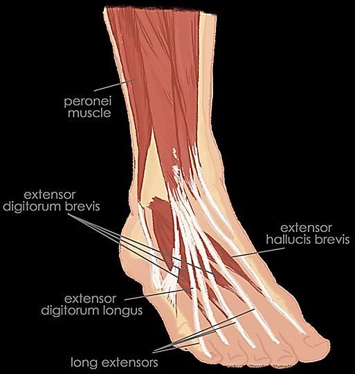
9 Tumor was found to be arising from Extensor Digitorum Brevis tendon sheath Dorsalis pedis vessels and anterior tibial nerve was adherent to the tumor Tendons were intact. We managed to remove it without removing the tendon.
10
11

12 EXTENSOR TENDONS OF FOOT DORSALIS PEDIS VESSELS
13 Giant cell tumors of the tendon sheath have a well-circumscribed multilobular appearance and often possess shallow grooves along their deep surfaces and are usually with a diameter of cms. Compared with other lesions, giant cell tumors in the hand digits are usually smaller and have a more regular appearance while those of the feet and elsewhere are often larger and more irregular in appearance.
14 On cut sections, these tumors have a mottled appearance, varying in color from grayish-brown to yellow-orange. The coloration depends on the amount of hemosiderin, collagen, and histiocytes in the sample. Tumors with more hemosiderin deposition due to bleeding have more of the yellow-orange or even reddish-brown color
15
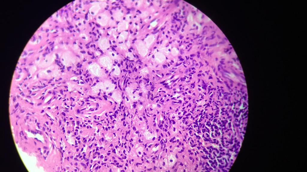

16 MULTINUCLEATED GIANT CELLS
17 Characterized by Proliferating histiocytes, moderately cellular (sheets of rounded or polygonal cells) Hemosiderin (brown color) may be present, but typically less than seen with PVNS (pigmented villonodular tumor of the tendon sheath) PVNS- a condition that causes the synovium to thicken and overgrow. The mass is not cancerous and does not spread. It is a progressive disease that can lead to bone damage and arthritis later on. Multinucleated giant cells are common

18
19 FINAL DIAGNOSIS : GIANT CELL TUMOUR of the tendon sheath
20 Giant cell tumors of the tendon sheath are the second most common tumors of the hand, with simple ganglion cysts being the most common. Giant cell tumors of the soft tissue are classified into the following two types: Localized (common) Diffuse (rare)
21 The rare diffuse form (often locally aggressive) is considered the soft-tissue counterpart of diffuse pigmented villonodular synovitis (PVNS) and affects the lower extremities, most commonly found around the knee, followed by the ankle and foot. Typically, these lesions, like those of PVNS, occur in young patients; 50% of cases are diagnosed in patients younger than 40 years. The diffuse form probably represents an extraarticular extension of a primary intra-articular PVNS process.
22 As is true for most soft-tissue tumors, the etiology of giant cell tumors of the tendon sheath is unknown. Pathogenetic theories have included Trauma, Disturbed lipid metabolism, Osteoclastic proliferation, Infection, Vascular disturbances, Immune mechanisms, Inflammation, Neoplasia, and metabolic disturbances. Probably the most widely accepted theory, as Jaffe et al proposed, is that of a reactive or regenerative hyperplasia associated with an inflammatory process.
23 Age distribution: years (peak incidence in those aged years) Rarely are these tumors found in patients younger than 10 years or older than 60 years. M:F ratio- 3:2 Giant cell tumors of the tendon sheath are associated with degenerative joint disease, especially in the distal interphalangeal (DIP) joint. An occasional association with rheumatoid arthritis has also been reported.
24 The incidence of local recurrence is high, ranging from 9% to 44%. Researchers have reported the following rates: Phalen et al, 9% recurrence rate in 56 cases Moore et al, 9% recurrence rate in 115 cases Jones et al, 17% recurrence rate in 95 cases Reilly et al, 27% recurrence rate in 70 cases Wright, 44% recurrence rate in 69 cases
25 Tumor recurrence Deep infection
26 Giant cell tumor of tendon sheath Malignant fibrous histiocytoma Plexiform fibrohistiocytic tumor:
27 As of now the patient is absolutely fit and fine. It has not recurred in the last 5 months. A meticulously planned surgery gives complete relief and no recurrence with full function and no morbidity.
28 THANK YOU.
Ultrasound Evaluation of Masses
 Ultrasound Evaluation of Masses Jon A. Jacobson, M.D. Professor of Radiology Director, Division of Musculoskeletal Radiology University of Michigan Disclosures: Consultant: Bioclinica Advisory Panel: GE,
GIANT CELL TUMOR OF TENDON SHEATH A CYTO HISTO CORRELATION
 GIANT CELL TUMOR OF TENDON SHEATH A CYTO HISTO CORRELATION Dr.S.SRIKANTH, Assistant Professor.Dept of Patholgy. Dr.SMITHA VADANA, Resident.Dept of pathology. Dr.R.SUHELA. Resident.Dept Of Pathology. Prathima
Multicentric localized giant cell tumor of the tendon. sheath
 Multicentric localized giant cell tumor of the tendon sheath Toshihiro Akisue, Tetsuji Yamamoto ( ), Teruya Kawamoto, Toshiaki Hitora, Takashi Marui, Tetsuya Nakatani, Takafumi Onga, and Masahiro Kurosaka.
Pigmented Villonodular Synovitis PVNS
 February 2002 Pigmented Villonodular Synovitis PVNS Amy Gillis, Harvard Medical School Year III 47 year old female Our Patient Right hip pain since age 20 No history of trauma Diagnosed with DJD of R hip
Giant Cell Tumor of the Tendon Sheath: Experience With 65 Cases
 Giant Cell Tumor of the Tendon Sheath: Experience With 65 Cases Erin L. Adams, BS, a Eric M. Yoder, MD, MBA, a and Morton L. Kasdan, MD, FACS b a University of Louisville School of Medicine, Louisville,
Giant-cell tumor of the tendon sheath: when must we suspect it?
 Giant-cell tumor of the tendon sheath: when must we suspect it? Poster No.: C-0538 Congress: ECR 2014 Type: Educational Exhibit Authors: C. Santos Montón, J. M. Alonso Sánchez, D. C. Cuellar, P. A. Chaparro
Dupuytren's Contracture Assessment
 Dupuytren's Contracture Assessment Link to guidance: http://www.enhertsccg.nhs.uk/ bedfordshire-and-hertfordshire-priorities-forum Dupuytren's contracture - clinical presentation for patients History Examination
Case Report Multiple Giant Cell Tumors of Tendon Sheath Found within a Single Digit of a 9-Year-Old
 Case Reports in Orthopedics Volume 2016, Article ID 1834740, 4 pages http://dx.doi.org/10.1155/2016/1834740 Case Report Multiple Giant Cell Tumors of Tendon Sheath Found within a Single Digit of a 9-Year-Old
Ultrasound in Rheumatology
 Arthritis Research UK Primary Care Centre Winner of a Queen s Anniversary Prize For Higher and Further Education 2009 Ultrasound in Rheumatology Alison Hall Consultant MSK Sonographer/Research Fellow Primary
Why? Ultrasound of the Foot. Ultrasound of the Foot. General Rules. Plantar Fascia. Plantar Fasciitis 18/09/2018
 Ultrasound of the Foot Why? Ultrasound of the Foot Plantar fasciitis Plantar fascia fibromatosis Morton s neuroma Intermetatarsal bursitis Adventitial bursitis Plantar plate tears MTP joint synovitis Ganglia
ELENI ANDIPA General Hospital of Athens G. Gennimatas
 ELENI ANDIPA General Hospital of Athens G. Gennimatas Technological advances over the last years have caused a dramatic improvement in ultrasound quality and resolution An established imaging modality
Urgent Cases and Foreign Bodies
 Urgent Cases and Foreign Bodies Catherine J. Brandon, MD, MS University of Michigan Ann Arbor, MI, USA Introduction: Patients added on to the schedule from the emergency department or as urgent add-on
The plantar aponeurosis
 Anatomy of the foot The plantar aponeurosis Is a triangular thickening of the deep fascia Its apex is attached to the medial and lateral tubercles of the calcaneum. The base of the aponeurosis divides
Ankle Arthroscopy.
 Ankle Arthroscopy Key words: Ankle pain, ankle arthroscopy, ankle sprain, ankle stiffness, day case surgery, articular cartilage, chondral injury, chondral defect, anti-inflammatory medication Our understanding
Anatomy MCQs Week 13
 Anatomy MCQs Week 13 1. Posterior to the medial malleolus of the ankle: The neurovascular bundle lies between Tibialis Posterior and Flexor Digitorum Longus The tendon of Tibialis Posterior inserts into
PIGMENTED VILLONODULAR SYNOVITIS OF THE WRIST
 PIGMENTED VILLONODULAR SYNOVITIS OF THE WRIST WITH PENETRATION INTO BONE FRITZ SCHAJOWICZ and ISIDORO BLUMENFELD, BUENOS AIRES, ARGENTINA From the Latin-American Register of Bone Pathology, Hospital Italiano,
Interesting Case Series. Ganglion Cyst of the Peroneus Longus
 Interesting Case Series Ganglion Cyst of the Peroneus Longus Andrew A. Marano, BA, Paul J. Therattil, MD, Dare V. Ajibade, MD, PhD, MPH, and Ramazi O. Datiashvili, MD, PhD Division of Plastic and Reconstructive
Femoral Artery. Its entrance to the thigh Position Midway between ASIS and pubic symphysis
 Lower Limb Vessels Lecture Objectives Describe the major arteries of the lower limb. Describe the deep and superficial veins of the lower limb. Describe the topographical relationships of the arteries
musculoskeletal system anatomy muscles of foot sheet done by: dina sawadha & mohammad abukabeer
 musculoskeletal system anatomy muscles of foot sheet done by: dina sawadha & mohammad abukabeer Extensor retinaculum : A- superior extensor retinaculum (SER) : originates from the distal ends of the tibia
Gross Anatomy Coloring Book Series. Lower Extremity Arteries
 Gross Anatomy Coloring Book Series Lower Extremity Arteries 1 Femoral Artery and Associated Branches For the life of the flesh is in the blood. Leviticus 17:11 Femoral Artery and Associated Branches After
2013/10/18 GANGLION CYSTS ENDOSCOPIC GANGLIONECTOMY ARTHROSCOPIC GANGLIONECTOMY OPEN GANGLIONECTOMY COMPARED TO FOOT AND ANKLE
 GANGLION CYSTS gelatinous fluid filled, encapsulated soft tissue masses adjacent to a joint or tendon ENDOSCOPIC GANGLIONECTOMY OF THE FOOT AND ANKLE Dr TH Lui North District Hospital HKSAR Pain, mass
Types of osteoarthritis
 ARTHRITIS Osteoarthritis is a degenerative joint disease is the most common joint disorder. It is a frequent part of aging and is an important cause of physical disability in persons older than 65 years
Case 8 Soft tissue swelling
 Case 8 Soft tissue swelling 26-year-old female presented with a swelling on the back of the left knee joint since the last 6 months and chronic pain in the calf and foot since the last 2 months. Pain in
Welcome to the: Orthopaedic Opinion Online Website The website for the answer to all your Orthopaedic Questions
 Welcome to the: Orthopaedic Opinion Online Website The website for the answer to all your Orthopaedic Questions Orthopaedic Opinion Online is a website designed to provide information to patients who have
The Lower Limb VI: The Leg. Anatomy RHS 241 Lecture 6 Dr. Einas Al-Eisa
 The Lower Limb VI: The Leg Anatomy RHS 241 Lecture 6 Dr. Einas Al-Eisa Muscles of the leg Posterior compartment (superficial & deep): primary plantar flexors of the foot flexors of the toes Anterior compartment:
A Case Of Primary Intra-Articular And Extra Articular Synovial Chondromatosis Of Ankle And Foot
 ISPUB.COM The Internet Journal of Orthopedic Surgery Volume 4 Number 1 A Case Of Primary Intra-Articular And Extra Articular Synovial Chondromatosis Of Ankle And Foot S Pathak, C Joseph, M Aravinda, S
CASE STUDY: PRO-DENSE Injectable Regenerative Graft Used to Backfill a Bone Cavity Following Resection of a Giant Cell Tumor
 : PRO-DENSE Used to Backfill a Bone Cavity Following Resection of a Giant Cell Tumor Contributed by: Matthew J. Seidel, MD* Lauren A. Schwartz, NP Scottsdale, AZ *Dr. Seidel is a paid consultant for Wright
Case Study. Your Diagnosis?
 Case Study 35 year old man twisted his ankle while playing and was surgically for treated for ankle sprain for instability symptoms 2 years. Increasing pain around the ankle and pain present all the time
Extra articular pigmented villonodular synovitis of knee
 A Case Report Extra articular pigmented villonodular synovitis of knee Sandeep tripathi 1, Brajesh nandan 2, Manish dhawan 3, VK nijhawan 4 ABSTRACT This study is a case report of a 30 year old male with
Keywords Giant Cell Tumors; Bone and Bones; Radiography; Magnetic Resonance. Marcelo de Pinho Teixeira Alves
 CASE REPORT Excision of giant cell tumor of tendon sheath with bone involvement by means of double access approach: case report Marcelo de Pinho Teixeira Alves Abstract Giant cell tumors of the tendon
The University Of Jordan Faculty Of Medicine FOOT. Dr.Ahmed Salman Assistant Prof. of Anatomy. The University Of Jordan
 The University Of Jordan Faculty Of Medicine FOOT Dr.Ahmed Salman Assistant Prof. of Anatomy. The University Of Jordan Tarsal Tunnel Syndrome Due to compression of Tibial nerve as it travels through the
Lecture 09. Popliteal Fossa. BY Dr Farooq Khan Aurakzai
 Lecture 09 Popliteal Fossa BY Dr Farooq Khan Aurakzai Dated: 14.02.2018 What is popliteus? Introduction Anything relating to, or near the part of the leg behind the knee. From New Latin popliteus the muscle
The Painful Elbow, Wrist, and Hand. Jennifer R Marks, MD
 The Painful Elbow, Wrist, and Hand Jennifer R Marks, MD The Painful Elbow A 44 yo M presents to clinic complaining of a sore elbow What further questions do you have for this patient? What is on your differential
Unicompartmental Knee Resurfacing
 Disclaimer This movie is an educational resource only and should not be used to manage knee pain. All decisions about the management of knee pain must be made in conjunction with your Physician or a licensed
Describing and interpreting gross lesions. Prepared for VPM 4600, May 2018; Shannon Martinson
 Describing and interpreting gross lesions Prepared for VPM 4600, May 2018; Shannon Martinson How to Describe (and Interpret) Lesions Step 1 Step 2 Step 3 Step 4 Look at the specimen: Is it normal or abnormal
First & second layers of muscles of the sole
 The FOOT First & second layers of muscles of the sole introduction The muscles acting on the foot can be divided into two distinct groups; extrinsic and intrinsic muscles. The extrinsic muscles arise from
Foot. Dr. Heba Kalbouneh Associate Professor of Anatomy and Histology
 Foot Dr. Heba Kalbouneh Associate Professor of Anatomy and Histology Dorsum of the Foot Sole of the Foot Plantar aponeurosis It is a triangular thickening of deep fascia in the sole of the foot Attachments:
Myxo-inflammatory Fibroblastic sarcoma
 AKA Myxo-inflammatory Fibroblastic sarcoma Acral Myxoinflammatory fibroblastic sarcomaam.j.surg.path1998; 22; 911-924 Inflammatory myxoid tumour of soft parts with bizarre giant cells [Pathol.Res.Pract.
Psoriatic arthritis: early ultrasound findings
 Psoriatic arthritis: early ultrasound findings Poster No.: C-0399 Congress: ECR 2014 Type: Educational Exhibit Authors: R. Persechino 1, L. Cristiano 1, A. Bartoloni 1, C. Cantone 2, A. Keywords: DOI:
Swelling. Size: measure exact size in cm using a tape measure (measure longitudinal and transverse axis and if possible the depth)
") Swelling Inspection Site: exact anatomic position Number: single or multiple Shape: spherical, oval, kidney-shaped or irregular Size: measure exact size in cm using a tape measure (measure longitudinal
Swelling Inspection Site: exact anatomic position Number: single or multiple Shape: spherical, oval, kidney-shaped or irregular Size: measure exact size in cm using a tape measure (measure longitudinal
Plantar fasciopathy (PFs)
") Plantar fasciopathy (PFs) 2016. 04. 30. Jung-Soo Lee, M.D., Ph.D. Department of Rehabilitation Medicine, Uijeongbu St. Mary's Hospital, College of Medicine, The Catholic University of Korea Anatomy of
Plantar fasciopathy (PFs) 2016. 04. 30. Jung-Soo Lee, M.D., Ph.D. Department of Rehabilitation Medicine, Uijeongbu St. Mary's Hospital, College of Medicine, The Catholic University of Korea Anatomy of
Patient #1. Rheumatoid Arthritis. Rheumatoid Arthritis. 45 y/o female Morning stiffness in her joints >1 hour
 Patient #1 Rheumatoid Arthritis Essentials For The Family Medicine Physician 45 y/o female Morning stiffness in her joints >1 hour Hands, Wrists, Knees, Ankles, Feet Polyarticular, symmetrical swelling
Dr Nabil khouri MD. MSc. Ph.D
 Dr Nabil khouri MD. MSc. Ph.D Foot Anatomy The foot consists of 26 bones: 14 phalangeal, 5 metatarsal, and 7 tarsal. Toes are used to balance the body. Metatarsal Bones gives elasticity to the foot in
This presentation is the intellectual property of the author. Contact them for permission to reprint and/or distribute.
 Introduction Compartment Syndromes of the Leg Related to Athletic Activity Mark M. Casillas, M.D. Consequences of a misdiagnosis persistence of a performance limitation loss of function/compartment loss
FOOT AND ANKLE ARTHROSCOPY
 FOOT AND ANKLE ARTHROSCOPY Information for Patients WHAT IS FOOT AND ANKLE ARTHROSCOPY? The foot and the ankle are crucial for human movement. The balanced action of many bones, joints, muscles and tendons
fig fig For the following diagrams
 fig. 1271 For the following diagrams Please draw small circles at the following points (pts in bold are main syllabus pts): Liver-1 Liver-2 Liver-3 Liver-4 Spleen-4 Spleen-5 Stomach-41 Stomach-42 Stomach-43
Ultrasound in Rheumatology
 Ultrasound in Rheumatology Alison Hall Consultant MSK Sonographer Research Institute for Primary Care & Health Sciences, Keele University Department of Rheumatology, Cannock Hospital, Royal Wolverhampton
Physical therapy of the wrist and hand
 Physical therapy of the wrist and hand Functional anatomy wrist and hand The wrist includes distal radius, scaphoid, lunate, triquetrum, pisiform, trapezium, trapezoid, capitate, and hamate. The hand includes
Copyright 2004 Lippincott Williams & Wilkins. 2. Bone Structure. Copyright 2004 Lippincott Williams & Wilkins
 Chapter 7 The Skeleton: Bones and Joints The Skeleton Skeletal system is made up of bones and joints and supporting connective tissue. 1. Bone Functions 1. To store calcium salts 2. To protect delicate
A case of giant cell tumour of soft parts in a horse Francesco Cian 1, Sarah Whiteoak 2, Jennifer Stewart 1
 A case of giant cell tumour of soft parts in a horse Francesco Cian 1, Sarah Whiteoak 2, Jennifer Stewart 1 1 Animal Health Trust, Newmarket, UK 2 608 Equine and Farm Vets, Rowington, UK Signalment: Horse,
Located Deep to Flexor Retinaculum on medial aspect of ankle. Posterior to Posterior Tibial Artery. Tom, Dick, and Very Nervous Harry
 ANKLE BLOCK ANESTHESIA GREGORY CLARK D.P.M. HEAD, SECTION OF PODIATRY SCRIPPS CLINIC LA JOLLA, CALIFORNIA A METHOD BY WHICH ONE MAY PROVIDE AN ANESTHETIC BLOCK TO THE FOOT OR ANKLE WITH A MINIMUM OF PATIENT
ORTHOPAEDIC INJECTION AND ASPIRATION TECHNIQUES
 ORTHOPAEDIC INJECTION AND ASPIRATION TECHNIQUES OAAPN October 20, 2016 David H. Sohn, JD MD Chief, Shoulder and Sports Medicine University of Toledo Medical Center When to aspirate? To rule out infection
4 2 Osteoarthritis 1
 Osteoarthritis 1 Osteoarthritis ( OA) Osteoarthritis is a chronic disease and the most common of all rheumatological disorders. It particularly affects individuals over the age of 65 years. The prevalence
Selected Pseudomalignant Soft Tissue Tumors of the Skin and Subcutis
 Selected Pseudomalignant Soft Tissue Tumors of the Skin and Subcutis Andrew L. Folpe, M.D. Professor of Laboratory Medicine and Pathology Mayo Clinic, Rochester, MN folpe.andrew@mayo.edu 2016 MFMER slide-1
Monophasic Synovial Carcinoma of knee joint- A Case Report and Review of Literature
 IOSR Journal of Dental and Medical Sciences (IOSR-JDMS) e-issn: 2279-0853, p-issn: 2279-0861.Volume 17, Issue 3 Ver.5 March. (2018), PP 13-17 www.iosrjournals.org Monophasic Synovial Carcinoma of knee
Clinical examination of the wrist, thumb and hand
 Clinical examination of the wrist, thumb and hand 20 CHAPTER CONTENTS Referred pain 319 History 319 Inspection 320 Functional examination 320 The distal radioulnar joint.............. 320 The wrist.......................
Pigmented Villonodular Synovitis (PVNS) A Case Report
 A Case Report") Case Report: Pigmented Villonodular Synovitis (PVNS) A Case Report Abhishek Patil 1, Sanjay Mulay 2, Chandrashekar Jaiswal 1, Uday Mahajan 1, Shriniwas Yadkikar 3, Vishnu Yadkikar 4 1Junior Resident, 2
Case Report: Pigmented Villonodular Synovitis (PVNS) A Case Report Abhishek Patil 1, Sanjay Mulay 2, Chandrashekar Jaiswal 1, Uday Mahajan 1, Shriniwas Yadkikar 3, Vishnu Yadkikar 4 1Junior Resident, 2
Figuring out the "fronds"-synovial proliferative disorders of the knee.
 Figuring out the "fronds"-synovial proliferative disorders of the knee. Poster No.: C-1209 Congress: ECR 2014 Type: Educational Exhibit Authors: S. Sivasubramanian; Tamil Nadu/IN Keywords: Imaging sequences,
Anatomy of the lower limb
 Anatomy of the lower limb Arches & sole of the foot Dr. Hayder ARCHES OF THE FOOT The foot as a mechanical unit performs two major functions: - It acts as a pliable platform to support the body weigh during
Systemic forms of stiffness
 Systemic forms of stiffness ANNA LITWIC CONSULTANT RHEUMATOLOGIST SALISBURY DISTRICT HOSPITAL CLINICAL RESEARCH FELLOW MRC LIFECOURSE EPIDEMIOLOGY UNIT Overview Rheumatoid arthritis Know it when you see
Unusual Lateral Presentation of Popliteal Cyst
 Unusual Lateral Presentation of Popliteal Cyst Tarek Hemmali,* Abstract: The most common cyst occurs in the popliteal region is the popliteal cyst and over the past years it has been received much clinical
Message of the Month for GPs June 2013
 Message of the Month for GPs June 2013 Dr Winn : Consultant Musculoskeletal Radiologist, Manchester Royal Infirmary Imaging of the musculoskeletal system Musculoskeletal pain is a common problem in the
Arthroscopy Of the Ankle.
 Arthroscopy Of the Ankle www.fisiokinesiterapia.biz Ankle Arthroscopy Anatomy Patient setup Portal placement Procedures Complications Anatomy Portals Anterior Anteromedial Anterolateral Anterocentral Posterior
The Foot. Dr. Wegdan Moh.Mustafa Medicine Faculty Assistant Professor Mob:
 The Foot Dr. Wegdan Moh.Mustafa Medicine Faculty Assistant Professor Mob: 0127155717 The skeleton of the foot Cutaneous innervations Sole of foot layers of muscles First layer -Abductor hallucis -Flexor
Intra-articular soft tissue masses of the knee: An imaging review of biopsy proven diagnoses
 Intra-articular soft tissue masses of the knee: An imaging review of biopsy proven diagnoses Poster No.: P-0114 Congress: ESSR 2014 Type: Scientific Poster Authors: A. Kirwadi 1, S. Raniga 2, R. Hargunani
Case Studies. A. Kent Allen, DVM LAMENESS AND IMAGING IN THE SPORT HORSE
 Case Studies A. Kent Allen, DVM Author s address: Virginia Equine Imaging, 2716 Landmark School Road, The Plains, VA 20198; e-mail: vaequine@aol.com. 2007 AAEP. 1. Case Study #1: Medial Collateral Desmitis
fitting shoes, or repetitive stress. It also frequently arises from unknown causes.
 43 Thames Street, St Albans, Christchurch 8013 Phone: (03) 356 1353. Website: philip-bayliss.com Morton's Neuroma Morton's Neuroma, also sometimes referred to as plantar Neuroma or intermetatarsal Neuroma,
CLINICAL PRESENTATION AND RADIOLOGY QUIZ QUESTION
 Donald L. Renfrew, MD Radiology Associates of the Fox Valley, 333 N. Commercial Street, Suite 100, Neenah, WI 54956 11/24/2012 Radiology Quiz of the Week # 100 Page 1 CLINICAL PRESENTATION AND RADIOLOGY
Ganglion of the peroneal nerve. CULLY A. COBB, III, M.D., AND RICHARD H. MoIr4 M.D.
 Ganglion of the peroneal nerve Report of two cases CULLY A. COBB, III, M.D., AND RICHARD H. MoIr4 M.D. Division of Neurological Surgery, Baylor College of Medicine, Houston, Texas ~" Peroneal nerve ganglion
Wrist Ganglion. Did not notice Suddenly. Over several hours Over several days Over several weeks Over several months
 Onset, Duration and Frequency Are you left handed or right handed? Left Right Both Which side of your body is affected? Left Right Both If both, which side is more severe? Left Right Varies About the same
Joint Disorders. Musculoskeletal Disorders (Part B-2) Module 7 -Chapter 10. Overview Disorders of the Muscular System Disorders of the Skeletal System
 Module 7 -Chapter 10. Overview Disorders of the Muscular System Disorders of the Skeletal System") Musculoskeletal Disorders (Part B-2) Module 7 -Chapter 10 Overview Disorders of the Muscular System Disorders of the Skeletal System Susie Turner, MD 1/9/13 Joint Disorders Arthritis Inflammation of Joint
Musculoskeletal Disorders (Part B-2) Module 7 -Chapter 10 Overview Disorders of the Muscular System Disorders of the Skeletal System Susie Turner, MD 1/9/13 Joint Disorders Arthritis Inflammation of Joint
A Patient s Guide to Ankle Anatomy
 A Patient s Guide to Ankle Anatomy 1436 Exchange Street Middlebury, VT 05753 Phone: 802-388-3194 Fax: 802-388-4881 cvo@champlainvalleyortho.com DISCLAIMER: The information in this booklet is compiled from
I-A-1) Non-specific thickening of synovial membrane
 Non-specific thickening of synovial membrane") I-A-1) Non-specific thickening of synovial membrane Grayscale Metatarsal Power Doppler Dorsal aspect of metatarsophalangeal joint in right 1 st toe, longitudinal view Asterisks indicate non-specific thickening
I-A-1) Non-specific thickening of synovial membrane Grayscale Metatarsal Power Doppler Dorsal aspect of metatarsophalangeal joint in right 1 st toe, longitudinal view Asterisks indicate non-specific thickening
Localized Nodular Tenosynovitis in the Horse
 Path. vet. 5: 436-441 (1968) From the Department of Veterinary Pathology, School of Veterinary Medicine, Washington State University, Pullman Localized Nodular Tenosynovitis in the Horse W.L. RAGLAND 1x1
1. A worker falls from a height and lands on his feet. Radiographs reveal a fracture of the sustentaculum tali. The muscle passing immediately
 1. A worker falls from a height and lands on his feet. Radiographs reveal a fracture of the sustentaculum tali. The muscle passing immediately beneath it that would be adversely affected is the: fibularis
Answers to Pre-Lab Quiz (p. 171) Answers to Activity Questions
 Answers to Activity Questions") Answers to Pre-Lab Quiz (p. 171) 1. Holds bones together; allows the rigid skeleton some flexibility so that gross body movements can occur 2. c, amount of movement allowed by the joint 3. synovial 4.
Answers to Pre-Lab Quiz (p. 171) 1. Holds bones together; allows the rigid skeleton some flexibility so that gross body movements can occur 2. c, amount of movement allowed by the joint 3. synovial 4.
LOCALIZED NODULAR SYNOVITIS OF THE KNEE: A REPORT OF TWO CASES WITH ABNORMAL ARTHROGRAMS*
 MARcH, i6 LOCALIZED NODULAR SYNOVITIS OF THE KNEE: A REPORT OF TWO CASES WITH ABNORMAL ARTHROGRAMS* ABSTRACT: By THOMAS G. GOERGEN, M.D.,t DONALD RESNICK, M.D.,t and GEM NIWAYAMA, M.D4 SAN DiEGO, CALIFORNIA
Therapeutic Foot Care Certificate Program Part I: Online Home Study Program
 Therapeutic Foot Care Certificate Program Part I: Online Home Study Program 1 Anatomy And Terminology Of The Lower Extremity Joan E. Edelstein, MA, PT, FISPO Associate Professor of Clinical Physical Therapy
A Patient s Guide to Ankle Anatomy
 A Patient s Guide to Ankle Anatomy 245 North College Lafayette, LA 70506 Phone: 337.232.5301 Fax: 337.237.6504 DISCLAIMER: The information in this booklet is compiled from a variety of sources. It may
Radiological Reasoning: Acutely Painful Swollen Finger. Musculoskeletal Imaging Chew and Richardson Benign-Appearing Bone Mass.
 Musculoskeletal Imaging Chew and Richardson Benign-Appearing Bone Mass AJR Integrative Imaging LIFELONG LEARNING FOR RADIOLOGY This Radiological Reasoning article is available for SAM credit and CME credits
Musculoskeletal Imaging of the Digits. Arash David Tehranzadeh, MD UCSD MSK Radiology May 11 th, 2006
 Musculoskeletal Imaging of the Digits Arash David Tehranzadeh, MD UCSD MSK Radiology May 11 th, 2006 Musculoskeletal Imaging of the Digit Anatomy & Internal Derangement The Extensor System The Flexor System
TENDINOSIS: TRIGGER FINGER DE QUERVAIN S TENOSYNOVITIS. Renita Sirisena Mark Puhaindran
 TENDINOSIS: TRIGGER FINGER DE QUERVAIN S TENOSYNOVITIS Renita Sirisena Mark Puhaindran Tendinosis vs Tendinitis Tendinosis: Degeneration of the tendon s collagen Related to chronic use Tendinitis Tendon
Ultrasound of the Knee
 Ultrasound of the Knee Jon A. Jacobson, M.D. Professor of Radiology Director, Division of Musculoskeletal Radiology University of Michigan Disclosures: Consultant: Bioclinica Book Royalties: Elsevier Advisory
MUSCULOSKELETAL LOWER LIMB
 MUSCULOSKELETAL LOWER LIMB Spinal Cord Lumbar and Sacral Regions Spinal cord Dorsal root ganglion Conus medullaris Cauda equina Dorsal root ganglion of the fifth lumbar nerve End of subarachnoid space
Organization of the Lower Limb
 Organization of the Lower Limb Limb Development Lower limb develops in an aterolateral position at the level of the L2 to S3 trunk segments Great toe positioned cephalic direction with the soles of the
GIANT CELL TUMOUR OF PATELLA
 GIANT CELL TUMOUR OF PATELLA Pages with reference to book, From 279 To 281 Younus Soomro, Asim Hussain ( Department of Orthopaedics, Civil Hospital and Dow Medical College, Karachi. ) The giant cell tumour
Peripheral Vascular Examination. Dr. Gary Mumaugh Western Physical Assessment
 Peripheral Vascular Examination Dr. Gary Mumaugh Western Physical Assessment Competencies 1. Inspection of upper extremity for: size symmetry swelling venous pattern color Texture nail beds Competencies
inerve Guide to Nerves 2009
 inerve Guide to Nerves 2009 A guide to self learning and self assessment Context: The following guide is intended to help interpret the sono-anatomy and follow a systematic stepwise approach to the practice
Department of Plastic Surgery, Royal Melbourne Hospital, Australia
 ARTICULAR CARTILAGE LOSS IN LONG-STANDING IMMOBILISATION OF INTERPHALANGEAL JOINTS By P. L. FIELD, F.R.C.S., and J. T. HUESTON,/Vi.S., F.R.C.S., F.R.A.C.S. Department of Plastic Surgery, Royal Melbourne
MRI KNEE WHAT TO SEE. Dr. SHEKHAR SRIVASTAV. Sr.Consultant KNEE & SHOULDER ARTHROSCOPY
 MRI KNEE WHAT TO SEE Dr. SHEKHAR SRIVASTAV Sr.Consultant KNEE & SHOULDER ARTHROSCOPY MRI KNEE - WHAT TO SEE MRI is the most accurate and frequently used diagnostic tool for evaluation of internal derangement
Chapter 5 The Skeletal System
 Chapter 5 The Skeletal System The Skeletal System Parts of the skeletal system Bones (skeleton) Joints Cartilages Ligaments (bone to bone)(tendon=bone to muscle) Divided into two divisions Axial skeleton:
Injuries to the Hands and Feet
 Injuries to the Hands and Feet Chapter 26 Injuries to the Hands and Feet Introduction Combat injuries to the hands and feet differ from those of the arms and legs in terms of mortality and morbidity. Death
Venous Insufficiency Ulcers. Patient Assessment: Superficial varicosities. Evidence of healed ulcers. Dermatitis. Normal ABI.
 Venous Insufficiency Ulcers Patient Assessment: Superficial varicosities Evidence of healed ulcers Dermatitis Normal ABI Edema Eczematous skin changes 1. Scaling 2. Pruritus 3. Erythema 4. Vesicles Lipodermatosclerosis
Pigmented villonodular synovitis of the knee joint in a 5-year-old girl treated with combined open and arthroscopic surgery: a case report
 APP Template V1.03 Article id: JPOB_07_1853 This final peer-reviewed manuscript version is made available under the CC-BY-NC 4.0 license http://creativecommons.org/licenses/by-nc/4.0/ Case report 1 Pigmented
SIGNIFICANT OTHERS. Miscellaneous Benign Breast Conditions
 SIGNIFICANT OTHERS Miscellaneous Benign Breast Conditions Epworth HealthCare 1 FAT NECROSIS TRAUMATIC Cell rupture Seat-Belt injury Blunt trauma Iatrogenic injury Surgery, Flaps, Radiotherapy Pathology
A Patient s Guide to Ankle Anatomy
 A Patient s Guide to Ankle Anatomy Pond View Professional Park 301 Professional View Drive Freehold, NJ 07728 Phone: 732-720-2555 DISCLAIMER: The information in this booklet is compiled from a variety
Examination of the Hand
 1 Examination of the Hand R E Page History 3 Examination 4 Movement 8 Features of Specific Conditions Affecting the Hand and Special Tests 8 Examination of the Hand 3 History General Establish important
Where should you palpate the pulse of different arteries in the lower limb?
 Where should you palpate the pulse of different arteries in the lower limb? The femoral artery In the femoral triangle, its pulse is easily felt just inferior to the inguinal ligament midway between the
Trigger Finger Release
 Trigger Finger Release Trigger finger, also known as stenosing tenosynovitis, occurs when one of the tendons responsible for bending a finger or the thumb develops a thickening, known as a nodule, and
Index. Note: Page numbers of article titles are in boldface type.
 Magn Reson Imaging Clin N Am 12 (2004) 185 189 Index Note: Page numbers of article titles are in boldface type. A Acromioclavicular joint, MR imaging findings concerning, 161 Acromion, types of, 77 79