Vascular Tumors in Children and Adults. Thuy Phung, MD, PhD Houston Methodist Hospital Texas Children s Hospital Baylor College of Medicine
|
|
|
- Julie Carter
- 5 years ago
- Views:
Transcription

1 Vascular Tumors in Children and Adults Thuy Phung, MD, PhD Houston Methodist Hospital Texas Children s Hospital Baylor College of Medicine


2 What are these lesions? (Marcelo Hochman, MD)

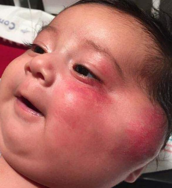
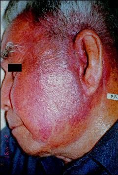
3 What are these lesions?
4 Outline Clinical presentation, histopathology and molecular genetics Congenital hemangioma (NICH/RICH) Kaposiform hemangioendothelioma Epithelioid angiosarcoma
5 ISSVA classification for vascular anomalies (Approved at the 20th ISSVA Workshop, Melbourne, April 2014) Vascular anomalies Vascular tumors Vascular malformations Benign Locally aggressive or borderline Simple Combined of major named vessels associated with other anomalies Malignant Capillary malformations CVM, CLM LVM, See details See list Lymphatic malformations CLVM CAVM* Venous malformations CLAVM* Arteriovenous malformations* Arteriovenous fistula* others
6 ISSVA Classification of Vascular Tumors Benign vascular tumors Infantile hemangioma / Hemangioma of infancy see details Congenital hemangioma Rapidly involuting (RICH) * Non-involuting (NICH) Partially involuting (PICH) Tufted angioma * Spindle-cell hemangioma Epithelioid hemangioma Pyogenic granuloma (aka lobular capillary hemangioma) Others Locally aggressive or borderline vascular tumors Kaposiform hemangioendothelioma * Retiform hemangioendothelioma Papillary intralymphatic angioendothelioma (PILA), Dabska tumor Composite hemangioendothelioma Kaposi sarcoma Others Malignant vascular tumors Angiosarcoma Epithelioid hemangioendothelioma Others
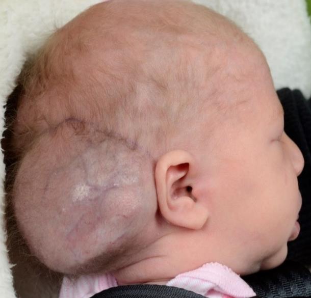
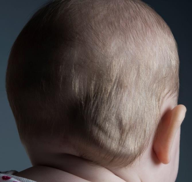
7 Congenital Hemangiomas
 -can be gone by 12-18 months of age Non-involuting congenital hemangiomas (NICH) GLUT 1")
8 Congenital Hemangiomas (distinct from infantile hemangiomas) Fully formed at birth More common in males Can occur at any body site, commonly on head and neck Rapidly involuting congenital hemangiomas (RICH) -can be gone by months of age Non-involuting congenital hemangiomas (NICH) GLUT 1 negative

9 Rapidly-involuting congenital hemangioma (RICH) 23 weeks in utero Neck mass Soon after birth 8 months old 17 months old (Michele Ramien, MD, University of Ottawa)

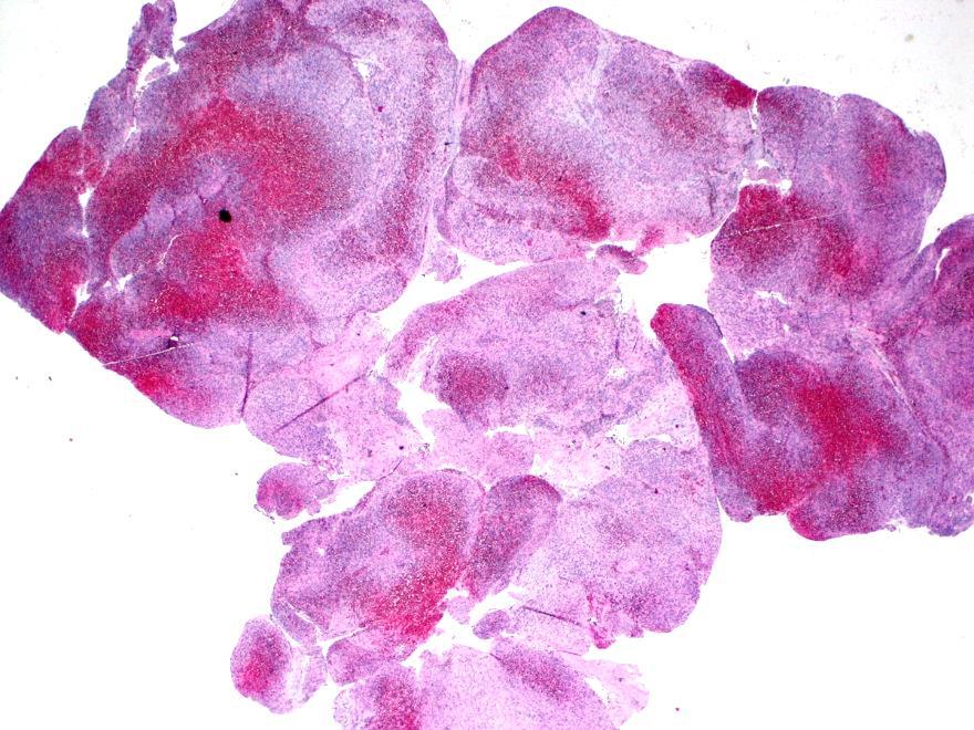
10 Rapidly involuting congenital hemangioma (RICH) Discreet hypercellular lobules with slit-like channels Fibrous tissue separating lobules Lobules with enlarged central draining vessels

11 Rapidly involuting congenital hemangioma (RICH) Slit-like channels Fibrous tissue separating lobules Lobules with enlarged central draining vessels

12 RICH Hobnail nuclei Small vessels surrounding enlarged central draining vessels

13 RICH Small vessels surrounding enlarged central draining vessels
 Lesion is fully")

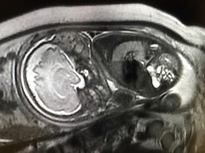
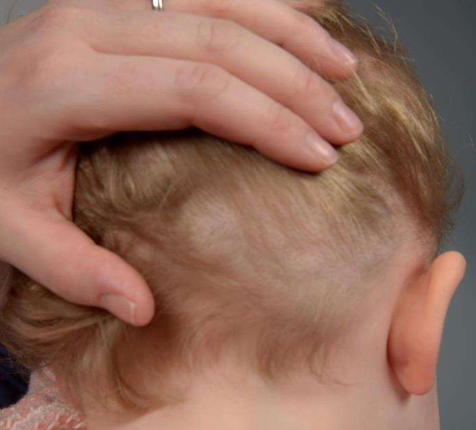
14 Non-involuting congenital hemangioma (NICH) Lesion is fully formed at birth Does NOT involute as the child grows In Utero ultrasound Newborn

15 Non-involuting congenital hemangioma (NICH) Blue halo Firm rubbery swelling with telangiectasia and a blue halo, warm to touch, non-compressible, non-tender mass


16 NICH: Ultrasound Imaging Color Doppler shows pulsatile arteries and veins within a well-circumscribed, non-compressible soft tissue mass

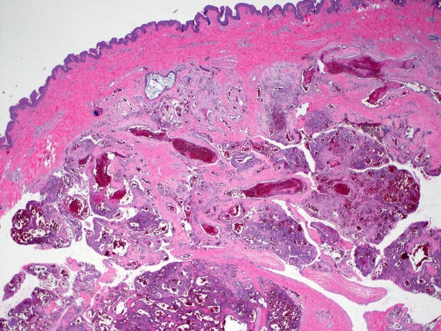
17 Fibrous tissue separating large lobules NICH Large lobules with enlarged stellate central draining vessels and curvelinear channels

18 Non-involuting congenital hemangioma (NICH) Lobules of cells with dilated central vessels

19 NICH Lobules of cells with dilated central vessels

20 NICH Hobnail nuclei Lobules of small vessels and large central draining vessels
21 NICH

22 Thrombi NICH


23 Congenital hemangioma GLUT-1 negative Infantile hemangioma GLUT-1 positive
24 Lesion size Congenital hemangioma vs. Infantile hemangioma Growth curves NICH RICH Infantile hemangioma Prenatal Birth Post-natal (Wassef et al., Pediatrics Jul;136(1):e203-14)
25 Kaposiform hemangioendothelioma (KHE)
26 Kaposiform hemangioendothelioma Solitary firm tumor in the skin or soft tissue with red-purple ecchymosis Presents during infancy (58%) or early childhood (32%); rare in adults Present in head and neck, extremities and trunk. Extracutaneous sites are bone, mediastinum and retroperitoneum A locally aggressive, borderlinemalignant tumor; metastasis is very rare Major clinical complication is Kasabach-Merritt phenomenon a severe consumptive coagulopathy
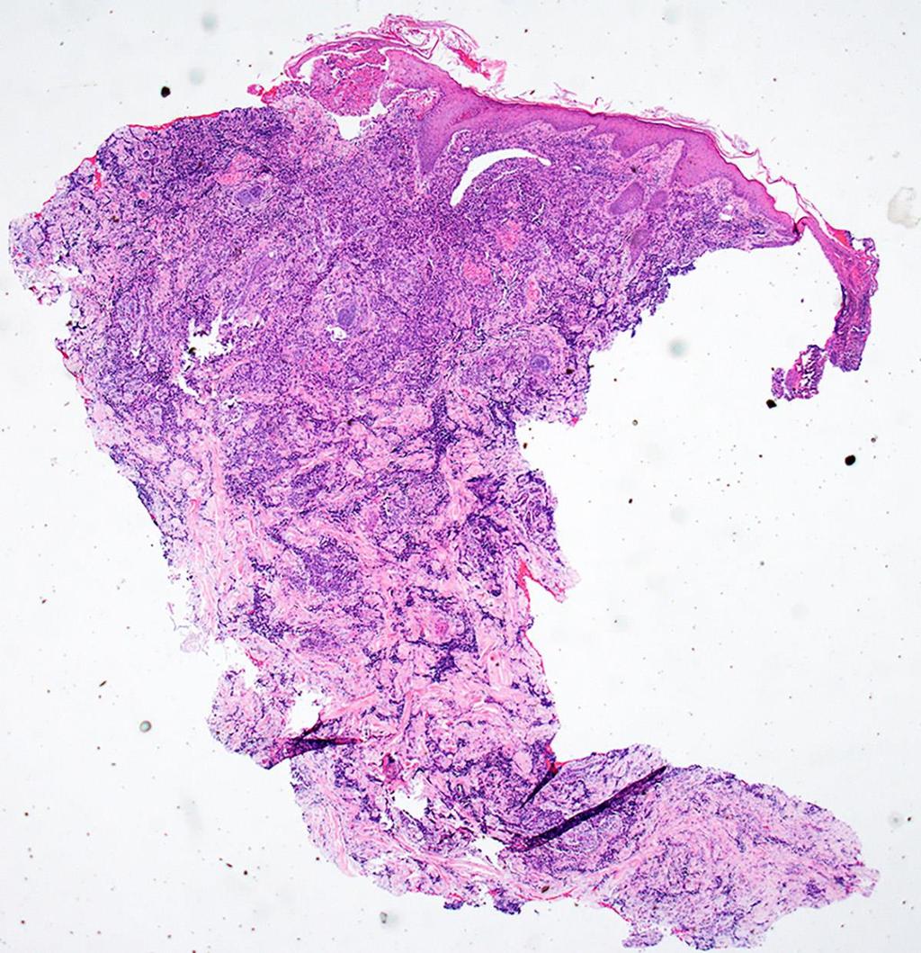
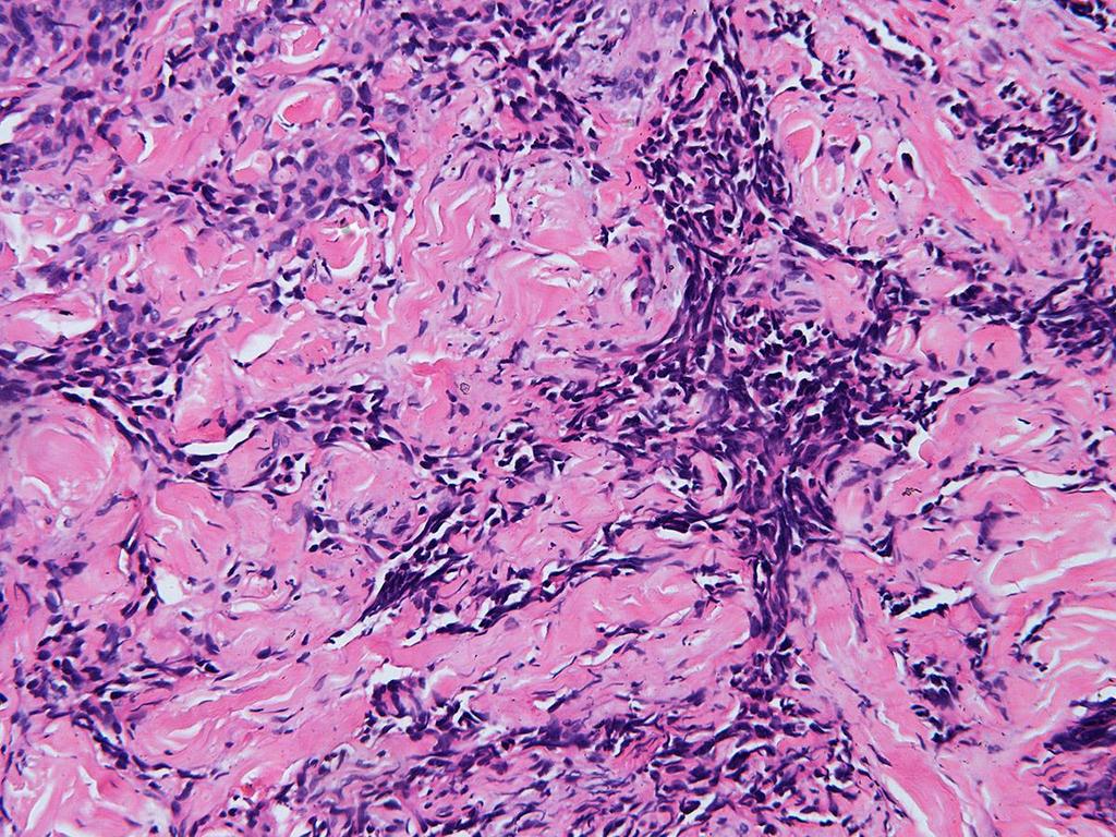
27 Kaposiform hemangioendothelioma
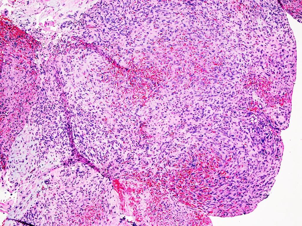
28 Kaposiform hemangioendothelioma
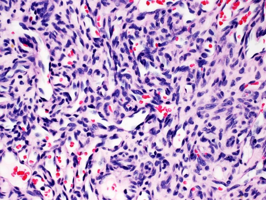
29 Kaposiform hemangioendothelioma
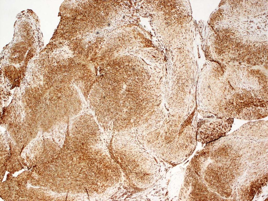
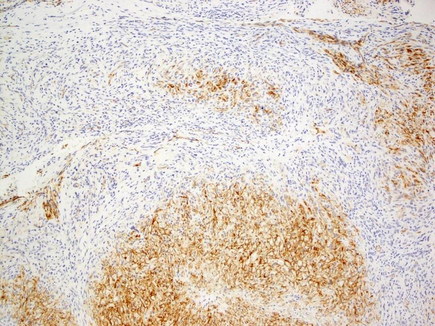
30 Kaposiform hemangioendothelioma CD31
31 Kaposiform hemangioendothelioma CD31
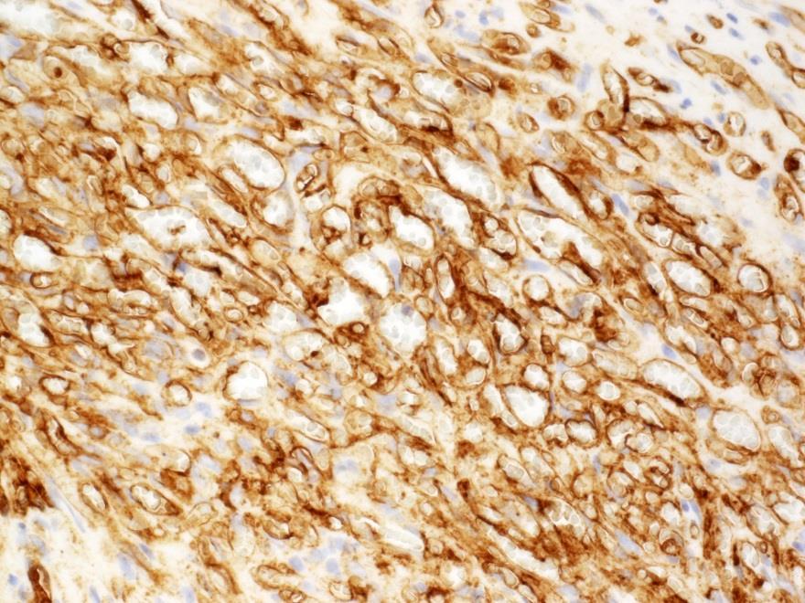
32 Kaposiform hemangioendothelioma D2-40
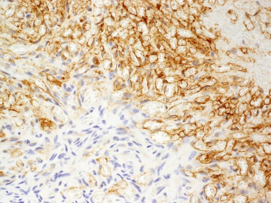
33 Kaposiform hemangioendothelioma D2-40
34 Angiosarcoma
35 Often occurs in the head and neck in elderly men Angiosarcoma Lymphedema-associated angiosarcoma (Stewart-Treves syndrome) Post-radiation angiosarcoma Exposure to vinyl chloride Poor prognosis, median survival is 2-3 years; 5-year survival rate is 30% Epithelioid angiosarcoma has worse prognosis
36 Angiosarcoma
37 Angiosarcoma
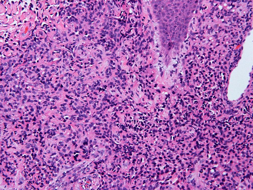
38 Angiosarcoma

39 Angiosarcoma

40 CD31+, CD45-, CK- Angiosarcoma
 Occurs in the spleen and liver Presents as sudden splenic rupture (Stuart C.")
41 Hemangiosarcoma in Dogs Dogs are a unique species in which spontaneous hemangiosarcoma commonly occurs. Accounts for ~7% of malignant tumors in dogs Common in certain dog strains (e.g., German shepherds) Occurs in the spleen and liver Presents as sudden splenic rupture (Stuart C. Helfand et al, Neoplasia, 2004)


42 Genetic Mutations in Vascular Tumors Infantile hemangioma VEGFR-2/TEM8 Angiosarcoma VEGFR-2 G-protein Coupled Receptors PTEN β γ α Congenital hemangioma GNAQ, GNA11 KHE and tufted angioma GNA14 PI3K Ras Raf Angiosarcoma PTEN, K-Ras, PTPRB, PLCG1, p53 NUP160-SLC43A3 gene fusion
43 Some Key Take Home Points Vascular anomalies (i.e., vascular malformations and tumors) in children and adults represent a number of unique entities with diverse etiologies, many with distinctive clinical, imaging and histologic features. Some lesions continue to defy classification and are best viewed as complex, dynamic biological processes yet to be defined. For these, it is best not to pigeon hole the lesion as a hemangioma but to describe it as accurately as possible and correlate with clinical, imaging and histologic features of the lesion.
44 Thank You!
Update on Pediatric Vascular Tumors
 Update on Pediatric Vascular Tumors Jim Treat, MD Associate Professor of Clinical Pediatrics and Dermatology No Relevant Conflicts of Interest PHOTOGRAPHY & VIDEOTAPING ARE STRICTLY PROHIBITED IN ALL EDUCATIONAL
Hemangiomas and Other Vascular Tumors
 facebook.com/cincykidsrad Hemangiomas and Other Vascular Tumors Disclosures No relevant financial disclosures Bernadette L. Koch, M.D. Departments of Radiology and Pediatrics Cincinnati Children s Hospital
Ultrasound imaging of vascular anomalies: pearls and pitfalls
 Ultrasound imaging of vascular anomalies: pearls and pitfalls Oscar Navarro, MD Dept. of Medical Imaging, University of Toronto Dept. of Diagnostic Imaging, The Hospital for Sick Children Declaration of
Sonography of soft-tissue vascular lesions
 Sonography of soft-tissue vascular lesions Oscar M. Navarro Associate Professor, University of Toronto Dept. of Diagnostic Imaging, The Hospital for Sick Children Toronto, Canada Declaration of Disclosure
ISSVA classification for vascular anomalies (Approved at the 20th ISSVA Workshop, Melbourne, April 2014, last revision May 2018)
") ISSVA classification for vascular anomalies (Approved at the 20th ISSVA Workshop, Melbourne, April 2014, last revision May 2018) This classification is intended to evolve as our understanding of the biology
ISSVA classification for vascular anomalies (Approved at the 20th ISSVA Workshop, Melbourne, April 2014, last revision May 2018) This classification is intended to evolve as our understanding of the biology
An Overview of Cutaneous Vascular Neoplasms
 An Overview of Cutaneous Vascular Neoplasms By Konstantinos Linos MD, FCAP, FASDP Bone, Soft Tissue and Dermatopathology Assistant Professor of Pathology Dartmouth-Hitchcock Medical Center Geisel School
Clinical Perspective: Vascular Anomalies with revised 2014 ISSVA Classification
 Clinical Perspective: Vascular Anomalies with revised 2014 ISSVA Classification Joao uilherme Amaral Pediatric Interventional Radiologist Division Chief Image uided Therapy Centre - The Hospital for Sick
Update on Vascular Tumors
 Update on Vascular Tumors Deepti Gupta, MD Assistant Professor Department of Pediatrics and Division of Dermatology Seattle Children s Hospital University of Washington School of Medicine PHOTOGRAPHY &
Ultrasound of soft-tissue vascular anomalies
 Ultrasound of soft-tissue vascular anomalies Oscar M. Navarro Associate Professor, University of Toronto Dept. of Diagnostic Imaging, The Hospital for Sick Children Toronto, Canada Declaration of Disclosure
Every Vascular Tumor is NOT a Hemangioma What the Hematologist/Oncologist needs to know about Rare Vascular Tumors
 Welcome! To join the call dial (866) 740-1260, passcode 3754894#. All participants are placed on mute for the duration of the webinar. If you have questions, type them in the chat box at the bottom left
Classification des Malformations vasculaires
 Classification des Malformations vasculaires Gilles Soulez, MD, MSc, FSIR Professeur Titulaire et Chairman Dpt Radiologie, Radio-Oncologie et Medecine Nucléaire Université de Montréal Basic principles
Comment je réponds au clinicien qui me demande de classer (selon ISSVA) une tache de naissance?
 une tache de naissance?") Comment je réponds au clinicien qui me demande de classer (selon ISSVA) une tache de naissance? Ph. Clapuyt, L. Boon Cliniques universitaires saint Luc Université catholique de Louvain 1200 Bruxelles Les
Comment je réponds au clinicien qui me demande de classer (selon ISSVA) une tache de naissance? Ph. Clapuyt, L. Boon Cliniques universitaires saint Luc Université catholique de Louvain 1200 Bruxelles Les
Angiographic features of rapidly involuting congenital hemangioma (RICH)
") Pediatr Radiol (2003) 33: 15 19 DOI 10.1007/s00247-002-0726-3 CASE REPORT Orhan Konez Patricia E. Burrows John B. Mulliken Steven J. Fishman Harry P.W. Kozakewich Angiographic features of rapidly involuting
Pediatr Radiol (2003) 33: 15 19 DOI 10.1007/s00247-002-0726-3 CASE REPORT Orhan Konez Patricia E. Burrows John B. Mulliken Steven J. Fishman Harry P.W. Kozakewich Angiographic features of rapidly involuting
Epithelioid Hemangioendothelioma. Ibtehal Al-Harbi Medical Intern
 Epithelioid Hemangioendothelioma Ibtehal Al-Harbi Medical Intern Hemangioendothelioma Hemangioendothelioma (HE) is the term used to describe a diverse group of vascular cancers. Very rare, accounting for
Index. oralmaxsurgery.theclinics.com. Note: Page numbers of article titles are in boldface type.
 Index Note: Page numbers of article titles are in boldface type. A Adenomatoid odontogenic tumor, pediatric, 50 51 Ameloblastic carcinoma, pediatric, 17, 49 Ameloblastic fibro-odontoma, pediatric, 54 Ameloblastic
Quick Reference Guide SUPPORTED BY:
 Quick Reference Guide SUPPORTED BY: 1 The recommendations in this publication do not indicate an exclusive course of treatment or serve as a standard of care. Variations, taking into account individual
HIGH-FLOW ARTERIOVENOUS MALFORMATION WİTHİN ENLARGED FETAL LEG (Congenital Hemangioma vs Parkes Weber Syndrome)
") HIGH-FLOW ARTERIOVENOUS MALFORMATION WİTHİN ENLARGED FETAL LEG (Congenital Hemangioma vs Parkes Weber Syndrome) DORUK CEVDI KATLAN, MD Department of Obstetrics and Gynecology / Perinatology Suleymaniye
HIGH-FLOW ARTERIOVENOUS MALFORMATION WİTHİN ENLARGED FETAL LEG (Congenital Hemangioma vs Parkes Weber Syndrome) DORUK CEVDI KATLAN, MD Department of Obstetrics and Gynecology / Perinatology Suleymaniye
Malignant Focal Liver Lesions
 Malignant Focal Liver Lesions Other Than HCC Pablo R. Ros, MD, MPH, PhD Departments of Radiology and Pathology University Hospitals Cleveland Medical Center Case Western Reserve University Pablo.Ros@UHhospitals.org
2/2/14 INTRODUCTION. Vascular Anomalies: An Introduction to Classification and Coordination of Care. VASCULAR ANOMALIES: History and Classification
 INTRODUCTION Vascular Anomalies: An Introduction to Classification and Coordination of Care Cindy Kerr,RN,MSN,CPNP Erin Spera,RN,MSN,CPNP Mary Beth Sylvia,RN,MS,FNP-BC 1in 3 newborns has a vascular birthmark
Vascular tumors & a brief discussion on varicose veins
 Vascular tumors & a brief discussion on varicose veins Classification Locally aggressive but metastasize infrequently Robbins basic pathology 9 th edition Overview Can arise from: endothelium (e.g., hemangioma,
MI Acute occlusion of the proximal left anterior descending (LAD) artery is the cause of 40% to 50% of all MIs. *
 artery is the cause of 40% to 50% of all MIs. *") MI *33% -50% die before hospital lethal arrhythmia Sudden Cardiac Death. * Arrhythmias are caused by electrical abnormalities of the ischemic myocardium and conduction system. *Acute occlusion of the proximal
MI *33% -50% die before hospital lethal arrhythmia Sudden Cardiac Death. * Arrhythmias are caused by electrical abnormalities of the ischemic myocardium and conduction system. *Acute occlusion of the proximal
Vascular Tumors. Case 1: Papillary endothelial hyperplasia
 Vascular Tumors Case 1: Papillary endothelial hyperplasia Papillary endothelial hyperplasia (PEH) essentially represents a stage in the recanalization of a thrombosed blood vessel. It may occur in any
GLUT-1: an extra diagnostic tool to differentiate between haemangiomas and vascular malformations q
 The British Association of Plastic Surgeons (2005) 58, 348 352 GLUT-1: an extra diagnostic tool to differentiate between haemangiomas and vascular malformations q J. Leon-Villapalos a, K. Wolfe b, L. Kangesu
Beta-blockers in the Treatment of Infantile Hemangiomas 5yrs experience
 Beta-blockers in the Treatment of Infantile Hemangiomas 5yrs experience Josef Mališ V.Stará, S.Klovrzová, L.Nováková, M.Kynčl, A.Sukop, B.Kocmichová, Š.Čapková, K.Bláhová Dept. of Pediatric Hematology/Oncology
QUICK REFERENCE GUIDE
 QUICK REFERENCE GUIDE The recommendations in this publication do not indicate an exclusive course of treatment or serve as a standard of care. Variations, taking into account individual circumstances,
Evaluation of Liver Mass Lesions. American College of Gastroenterology 2013 Regional Postgraduate Course
 Evaluation of Liver Mass Lesions American College of Gastroenterology 2013 Regional Postgraduate Course Lewis R. Roberts, MB ChB, PhD Division of Gastroenterology and Hepatology Mayo Clinic College of
16 Nuchal Fibroma and Gardner
 C O N T E N T S Dedication v Preface vii Acknowledgments ix SECTION 1 Fibrohistiocytic Tumors 1 1 Classification of Fibrohistiocytic Tumors 3 2 Dermatofibroma 5 3 Juvenile Xanthogranuloma 33 4 5 6 Solitary
Treatment of tongue cavernous haemangioma with direct puncture and sclerotization with ethanol
 case report 75 Treatment of tongue cavernous haemangioma with direct puncture and sclerotization with ethanol Tomaz Seruga, Jernej Lucev, Marko Jevsek Radiology Department, University Medical Centre Maribor,
Musculoskeletal Sarcomas
 Musculoskeletal Sarcomas Robert C. Orth, M.D., Ph.D. Edward B. Singleton Department of Pediatric Radiology Texas Children s Hospital Page 0 xxx00.#####.ppt 9/23/2012 9:01:18 AM No disclosures Page 1 xxx00.#####.ppt
EXTRA-ORAL HEMANGIOMA IN A 10 YEAR OLD GIRL A CASE REPORT WITH LITERATURE REVIEW
 EXTRA-ORAL HEMANGIOMA IN A 10 YEAR OLD GIRL A CASE REPORT WITH LITERATURE REVIEW 1 2 2 2 R.Kannan B. Sekar S.Murali Dominic Augustine 1 2 Department of Oral and Maxillofacial Surgery, Department of Oral
Vascular Tumors. Abstract. History and Classification. Abel Sepulveda, MD 1 Edward P. Buchanan, MD 1
 49 Abel Sepulveda, MD 1 Edward P. Buchanan, MD 1 1 Division of Plastic Surgery, Baylor College of Medicine, Houston, Texas Semin Plast Surg 2014;28:49 57. Address for correspondence Edward P. Buchanan,
Vascular Anomalies Classification: Recommendations From the International Society for the Study of Vascular Anomalies
 Vascular Anomalies Classification: Recommendations From the International Society for the Study of Vascular Anomalies Michel Wassef, MD a, Francine Blei, MD b, Denise Adams, MD c, Ahmad Alomari, MD d,
Vascular Anomalies. Dr Kurosh Parsi MBBS, Msc (MED), FACD, FACP Vascular Birthmark Clinic Sydney Children s Hospital
, FACD, FACP Vascular Birthmark Clinic Sydney Children s Hospital") Vascular Anomalies Dr Kurosh Parsi MBBS, Msc (MED), FACD, FACP Vascular Birthmark Clinic Sydney Children s Hospital Vascular Anomalies Can be divided into two types: Tumours Malformations Vascular Tumours
Vascular Anomalies Dr Kurosh Parsi MBBS, Msc (MED), FACD, FACP Vascular Birthmark Clinic Sydney Children s Hospital Vascular Anomalies Can be divided into two types: Tumours Malformations Vascular Tumours
Vascular Imaging in the Pediatric Abdomen. Jonathan Swanson, MD
 Vascular Imaging in the Pediatric Abdomen Jonathan Swanson, MD Goals and Objectives To understand the imaging approach, appearance, and clinical manifestations of the common pediatric abdominal vascular
Vascular Anomalies: From the Dermatologist s Perspective
 Vascular Anomalies: From the Dermatologist s Perspective Aimee Smidt, MD, FAAD, FAAP University of New Mexico School of Medicine, Depts. of Dermatology & Pediatrics April 2018 No conflict of interest to
Vascular anomalies in children (Part 1): Vascular malformations
: Vascular malformations") Vascular anomalies in children (Part 1): Vascular malformations Vascular anomalies are common problems in infancy and childhood. GPs need to have a good understanding about these conditions in order to
Vascular anomalies in children (Part 1): Vascular malformations Vascular anomalies are common problems in infancy and childhood. GPs need to have a good understanding about these conditions in order to
Index. Cystic lymphatic malformations, 54, 67 clinical features, 67
 INDEX Acquired acral fibrokeratoma, 279 Acquired elastotic hemangioma, 174 clinical features, 174 histopathologic features, 174-175 immunohistochemistry, 174 treatment, 175 Acral pseudolymphomatous angiokeratoma
Varicose Veins Vascular Tumors
 Varicose Veins Vascular Tumors Varicose veins: Dilated tortuous increased intraluminal pressure incompetence of the venous valves. superficial veins of the upper and lower leg (m.c, high venous pressures,
Multifocal vascular anomalies: Common, less common, and rare
 Multifocal vascular anomalies: Common, less common, and rare Ilona J. Frieden Professor of Dermatology and Pediatrics University of California, San Francisco Conflict of interests: Pierre Fabre Dermo-Cosmétique
Histopathology of Vascular Anomalies
 Histopathology of Vascular Anomalies Anita Gupta, MD a, Harry Kozakewich, MD b, * KEYWORDS Hemangioma Vascular malformation Vascular tumor Pyogenic granuloma Immunohistochemical stain Over the past decade,
IN THE NAME OF GOD Dr. Kheirandish Oral and maxillofacial pathology
 IN THE NAME OF GOD Dr. Kheirandish Oral and maxillofacial pathology ORAL FOCAL MUCINOSIS Uncommon Tumorlike Cutaneous myxoid cyst Overproduction of hyaluronic acid by firoblasts Young adults Female Gingiva
Case: The patient is a 62 year old woman with a history of renal cell carcinoma that was removed years ago. A 2.4 cm liver mass was found on CT
 Case: The patient is a 62 year old woman with a history of renal cell carcinoma that was removed years ago. A 2.4 cm liver mass was found on CT during follow- up. ALT, AST, Alk Phos and bilirubin were
Vascular Tumors of Intermediate Malignancy: A Review and Update
 Review Vascular Tumors of Intermediate Malignancy: A Review and Update Jason S. Stratton 1 Steven D. Billings 1, 2 Vascular tumors of intermediate malignancy encompass a broad range of histologic entities.
Swelling. Size: measure exact size in cm using a tape measure (measure longitudinal and transverse axis and if possible the depth)
") Swelling Inspection Site: exact anatomic position Number: single or multiple Shape: spherical, oval, kidney-shaped or irregular Size: measure exact size in cm using a tape measure (measure longitudinal
Swelling Inspection Site: exact anatomic position Number: single or multiple Shape: spherical, oval, kidney-shaped or irregular Size: measure exact size in cm using a tape measure (measure longitudinal
Histopathology of Pediatric Vascular Anomalies using the International Society of Vascular Anomalies Classification - a must know for all
 Histopathology of Pediatric Vascular Anomalies using the International Society of Vascular Anomalies Classification - a must know for all Anita Gupta, MD Assistant Professor in the Department of Pathology,
WK KWOK, NSY CHAO, MWY LEUNG, PMY TANG, BPY WONG, LC HO, KKW LIU. Abstract. Key words. Introduction. Case Report
 HK J Paediatr (new series) 2009;14:209-213 Neonatal Intestinal Obstruction and Thrombocytopenia: Sepsis or Otherwise? Neonatal Intestinal Kaposiform Haemangioendothelioma: A Case Report and Literature
Contents. 1 Embryological and Anatomical Introduction... 1
 1 Embryological and Anatomical Introduction.... 1 1.1 Preliminary Remarks.................... 1 1.2 Leptomeninges....................... 21 1.3 Subpial Space........................ 22 1.3.1 Anatomy...........................
Hamburg Classification: Vascular Malformation
 1 2 Hamburg Classification: Vascular Malformation 9 3 Dirk A. Loose and Raul E. Mattassi 4 5 6 7 8 9 10 11 12 13 14 15 16 17 18 19 20 21 22 23 24 25 26 27 28 29 30 31 32 AU1 Vascular malformations occur
PEDIATRICS WK 3 HEAD AND NECK ALISON WALLACE MD, PHD
 PEDIATRICS WK 3 HEAD AND NECK ALISON WALLACE MD, PHD Topics 1. Cervical lymphadenopathy 2. Lymphatic malformation 3. Thyroglossal duct cysts 4. Branchial cleft cysts 5. Thyroid masses CASE 1 Case 1 A 2
Kaposiform Hemangioendothelioma: Case Report and Literature Review
 288 Chinese Journal of Clinical Oncology Aug. 2007, Vol. 4, No. 4 P 288~292 Gang Zhou et al. [SpringerLink] DOI 10.1007/s11805-007-0289-z Kaposiform Hemangioendothelioma: Case Report and Literature Review
Chief complaint. A mass at right chest
 Chief complaint A mass at right chest Present illness This 1-year-5-month-old girl had a mass at right side chest since one month ago. flat and not tender at first In the recent 2 days, the mass enlarged
In the 1940s, Kasabach and Merritt
 Kaposiform hemangioendothelioma without Kasabach-Merritt phenomenon Alla Gruman, MD, a MarilynG.Liang,MD, b,g John B. Mulliken, MD, c,g Steven J. Fishman, MD, d,g Patricia E. Burrows, MD, e,g HarryP.W.Kozakewich,MD,
External Jugular Vein Vascular Malformation: Sonographic and MR Imaging Appearances
 AJNR Am J Neuroradiol 25:338 342, February 2004 Case Report External Jugular Vein Vascular Malformation: Sonographic and MR Imaging Appearances Anil T. Ahuja, Hok-Yuen Yuen, Ka-Tak Wong, Ann D. King, Victor
Hemangioendothelioma with a Prominent Lymphoid Infiltrate Mimicking Follicular Dendritic Cell Tumor: Report of a Case
 Journal of Cancer Research Updates, 2013, 2, 135-139 135 Hemangioendothelioma with a Prominent Lymphoid Infiltrate Mimicking Follicular Dendritic Cell Tumor: Report of a Case Justin Kerstetter 1, Mia Perez
Tumors of the Spleen
 Tumors of the Spleen 803-808-7387 www.gracepets.com These notes are provided to help you understand the diagnosis or possible diagnosis of cancer in your pet. For general information on cancer in pets
Contents Part I Introduction 1 General Description 2 Natural History: Importance of Size, Site, Histopathology
 Contents Part I Introduction 1 General Description... 3 1.1 Introduction... 3 1.2 Incidence and Prevalence... 5 1.3 Predisposing and Genetic Factors... 8 References... 16 2 Natural History: Importance
Vascular Anomalies: Hemangiomas and more
 Workshop Pädiatrische Dermatologie Thalwil 17. und 18. März 2016 Vascular Anomalies: Hemangiomas and more Lisa Weibel Head of Pediatric Dermatology University Children s Hospital, Zurich CONTENT 1. Classification
Mesenchymal Tumors. Cavernous Hemangioma (CH) VASCULAR TUMORS MESENCHYMAL TUMORS OF THE LIVER: WHAT S NEW AND UNUSUAL (MY PERSPECTIVE)
 VASCULAR TUMORS MESENCHYMAL TUMORS OF THE LIVER: WHAT S NEW AND UNUSUAL (MY PERSPECTIVE)") Mesenchymal Tumors MESENCHYMAL TUMORS OF THE LIVER: WHAT S NEW AND UNUSUAL (MY PERSPECTIVE) CURRENT ISSUES IN ANATOMIC PATHOLOGY MAY 23, 2014 Linda Ferrell, MD, UCSF Focus on Vascular Tumors Benign and
Mesenchymal Tumors MESENCHYMAL TUMORS OF THE LIVER: WHAT S NEW AND UNUSUAL (MY PERSPECTIVE) CURRENT ISSUES IN ANATOMIC PATHOLOGY MAY 23, 2014 Linda Ferrell, MD, UCSF Focus on Vascular Tumors Benign and
Intraosseous hemangioma is an uncommon benign
 Case Report 71 An Intraosseous Capillary Hemagioma Of The Foot In A Child Kah-Wai Ngan, MD; Hui-Ling Hsu 1, MD; Shir-Hwa Ueng, MD An 8-year-old boy presented with an osteolytic lesion at the fourth metatarsal
3/27/2017. Disclosure of Relevant Financial Relationships
 Ophthalmic Pathology Evening Specialty Conference USCAP 2017 5 th March, 2017 Mukul K. Divatia, MD Assistant Professor Department of Pathology & Genomic Medicine Weill Cornell Medical College Houston Methodist
Mesenchymal Tumors MESENCHYMAL TUMORS OF THE LIVER: WHAT S NEW AND UNUSUAL (MY PERSPECTIVE)
") MESENCHYMAL TUMORS OF THE LIVER: WHAT S NEW AND UNUSUAL (MY PERSPECTIVE) CURRENT ISSUES IN ANATOMIC PATHOLOGY MAY 23, 2014 Linda Ferrell, MD, UCSF Mesenchymal Tumors Focus on Vascular Tumors Benign and
MESENCHYMAL TUMORS OF THE LIVER: WHAT S NEW AND UNUSUAL (MY PERSPECTIVE) CURRENT ISSUES IN ANATOMIC PATHOLOGY MAY 23, 2014 Linda Ferrell, MD, UCSF Mesenchymal Tumors Focus on Vascular Tumors Benign and
3. Discussion. 2 Case Reports in Pediatrics
 Case Reports in Pediatrics Volume 2015, Article ID 537530, 4 pages http://dx.doi.org/10.1155/2015/537530 Case Report Paravertebral and Retroperitoneal Vascular Tumour Presenting with Kasabach-Merritt Phenomenon
Case # nd Annual SEVPAC May 17, Kathy-Anne Clarke
 Case # 10 42 nd Annual SEVPAC May 17, 2014 Kathy-Anne Clarke Google images Babu Babu is 10 year old spayed female French Bulldog Chronic weight loss over 4 months Febrile and lethargic at the referring
Intramuscular hemangioma of the forearm; Report of a case
 Iran. J. Radiat. Res., 2003; 1(3): 175-179 Intramuscular hemangioma of the forearm; Report of a case M. Vakilha 1, F. Farhan 1, F. Samiei 1, S. Shariat 2 H 1 Radiotherapy-Oncology Dept., Imam Hospital,
THE EVOLVING CONCEPT OF HEMANGIOENDOTHELIOMA
 THE EVOLVING CONCEPT OF HEMANGIOENDOTHELIOMA CHRISTOPHER D.M. FLETCHER, M.D., FRCPath Department of Pathology Harvard Medical School & Brigham and Women s Hospital 75 Francis Street Boston, MA 02115 Introduction
Diagnostic imaging of cervical vascular malformations
 Diagnostic imaging of cervical vascular malformations Poster No.: C-0781 Congress: ECR 2013 Type: Educational Exhibit Authors: A. Llanes Rivada, D. Dualde-Beltrán, L. Ariño Montaner, J. 1 2 1 1 1 1 1 Joudanin
Disorders of Cell Growth & Neoplasia. Histopathology Lab
 Disorders of Cell Growth & Neoplasia Histopathology Lab Paul Hanna April 2010 Case #84 Clinical History: 5 yr-old, West Highland White terrier. skin mass from axillary region. has been present for the
العصوي الوعاي ي الورام = angiomatosis Bacillary
 1 / 7 BACILLARY ANGIOMATOSIS Epidemiology BA is most commonly seen in patients with acquired immunodeficiency syndrome (AIDS) and a CD4 count less than 50 cells/mm 3, with an incidence of 1.2 cases per
Imaging features of pediatric vascular malformations of the skin and subcutaneous tissue.
 Imaging features of pediatric vascular malformations of the skin and subcutaneous tissue. Poster No.: C-1962 Congress: ECR 2012 Type: Educational Exhibit Authors: R. F. Ocete Pérez, M. Fajardo Cascos,
High-Flow Vascular Malformation of Ear: A Case Report
 256 Ear high-flow vascular malformation Case Report High-Flow Vascular Malformation of Ear: A Case Report Ankit Gupta 1*, Shyam Gupta 1, Akhil Kumar 2, Sameek Bhattacharaya 1, Manoj Jha 1, Vinay Tiwari
Disclosure of Relevant Financial Relationships
 Surgical Pathology Companion Meeting Case 5: Locally Recurrent Chest wall Mass Cristina Antonescu, MD Department of Pathology, Memorial Sloan Kettering Cancer Center Disclosure of Relevant Financial Relationships
المركب النموذج--- سبيتز وحمة = Type Spitz's Nevus, Compound SPITZ NEVUS 1 / 7
 SPITZ NEVUS 1 / 7 Epidemiology An annual incidence rate of 1.4 cases of Spitz nevus per 100,000 individuals has been estimated in Australia, compared with 25.4 per 100,000 individuals for cutaneous melanoma
Soft Tissue Sarcoma. Presley Regional Trauma Center Department of Surgery University of Tennessee Health Science Center Memphis, Tennessee
 Soft Tissue Sarcoma Presley Regional Trauma Center Department of Surgery University of Tennessee Health Science Center Memphis, Tennessee Soft Tissue Sarcoma Collective term for an unusual and diverse
Canine Histiocytic Disorders DR. MEREDITH GAUTHIER, DVM DACVIM (ONCOLOGY) OCTOBER 29, 2015
 OCTOBER 29, 2015") Canine Histiocytic Disorders DR. MEREDITH GAUTHIER, DVM DACVIM (ONCOLOGY) OCTOBER 29, 2015 Canine Histiocytes! Cells derived from CD34+ stem cells and blood monocytes! Macrophages! Dendritic cells (DC)!
Canine Histiocytic Disorders DR. MEREDITH GAUTHIER, DVM DACVIM (ONCOLOGY) OCTOBER 29, 2015 Canine Histiocytes! Cells derived from CD34+ stem cells and blood monocytes! Macrophages! Dendritic cells (DC)!
DERMATOLOGY ROTATION: COMPETENCY-BASED GOALS AND OBJECTIVES
 UNC DIVISION OF PLASTIC AND RECONSTRUCTIVE SURGERY DERMATOLOGY ROTATION: COMPETENCY-BASED GOALS AND OBJECTIVES MEDICAL KNOWLEDGE A. Anatomy/Physiology/Embryology Goal: The resident will have knowledge
Immunohistochemical Study of Some Rare Vascular Tumors
 Journal of the Egyptian Nat. Cancer Inst., Vol. 16, No. 2, June: 123-129, 2004 MONA EL-SAYED, M.D. and MOHAMMAD RAMADAN, PH.D. The Department of Pathology, Faculty of Medicine, Zagazig University. ABSTRACT
Vessel malformation vascular malformation vascular malformation vascular malformation Vascular malformation vascular malformations
 A vascular malformation is another type of birthmark, or congenital (present at birth) growth, made up of arteries, veins, capillaries, or lymphatic vessels. There are several different types of malformations
Introduction to Musculoskeletal Tumors. James C. Wittig, MD Orthopedic Oncologist Sarcoma Surgeon
 Introduction to Musculoskeletal Tumors James C. Wittig, MD Orthopedic Oncologist Sarcoma Surgeon www.tumorsurgery.org Definitions Primary Bone / Soft tissue tumors Mesenchymally derived tumors (Mesodermal)
Objectives. 1. Recognizing benign skin lesions. 2.Know which patients will likely need surgical intervention.
 The Joy of Pediatric Skin Dr. Claire Sanger University of Kentucky Plastic & Reconstructive Surgery Objectives 1. Recognizing benign skin lesions 2.Know which patients will likely need surgical intervention.
Controversies & updates in Vascular Surgery
 Controversies & updates in Vascular Surgery Paris - january 24 2018 Venous session VENOUS ODDITIES DUPLEX IMAGE Philippe LEMASLE Le Chesnay - France I have no financial relationship to disclose Case n
Nursing Implications for the Management of Lymphatic Malformation in Children
 646541JPOXXX10.1177/1043454216646541Journal of Pediatric Oncology NursingBerger research-article2016 Article Nursing Implications for the Management of Lymphatic Malformation in Children Journal of Pediatric
Can lymphangiosarcoma be resurrected? A clinicopathological and immunohistochemical study of lymphatic differentiation in 49 angiosarcomas
 Histopathology 2010, 56, 364 371. DOI: 10.1111/j.1365-2559.2010.03484.x Can lymphangiosarcoma be resurrected? A clinicopathological and immunohistochemical study of lymphatic differentiation in 49 angiosarcomas
JOURNAL OF CLINICAL AND DIAGNOSTIC RESEARCH
 JOURNAL OF CLINICAL AND DIAGNOSTIC RESEARCH How to cite this article : PAI K,GUPTA A.INTRAVASCULAR EPITHELIOID HAEMANGIOMA OF TEMPORAL ARTERY: A DIAGNOSTIC DIFFICULTY Journal of Clinical and Diagnostic
INFECTION. HIV Infection DWI
 HIV Infection INFECTION DWI Fig Axial CT and MRI images show multiple enlarged lymph nodes in the neck as well as in the parotid gland bilaterally. These nodes were suppurative with positive diffusion.
Vision Health: Conditions, Disorders & Treatments EYELID DISORDERS
 Vision Health: Conditions, Disorders & Treatments EYELID DISORDERS There are a number of disorders that can affect the eyelid. Entropion Entropion is an inward turning of the eyelid and lashes toward the
Soft Tissue Sarcomas: Questions and Answers
 Soft Tissue Sarcomas: Questions and Answers 1. What is soft tissue? The term soft tissue refers to tissues that connect, support, or surround other structures and organs of the body. Soft tissue includes
Case : A Newborn Girl with a Large Cutaneous Lesion, Thrombocytopenia, and Anemia
 The new england journal of medicine case records of the massachusetts general hospital Founded by Richard C. Cabot Nancy Lee Harris, m.d., Editor Jo-Anne O. Shepard, m.d., Associate Editor Sally H. Ebeling,
SCORCH 2018 Bone, Brain & Vessels
 To complete the SAM assessment, please use one of the methods below: Complete a paper version of the exam Go to: https://www.surveymonkey.com/r/thurssam Open your smartphone camera and scan this QR Code
KASABACH MERRITT SYNDROME
 KASABACH MERRITT SYNDROME Pages with reference to book, From 159 To 161 Shahina Qureshi, Zaheer Abbasi, Naheed Qadeer, Naila Yaqoob ( Children's Hospital, Pakistan Institute of Medical Sciences, Islamabad.
Coagulation abnormalities associated with extensive venous malformations of the limbs: differentiation from Kasabach Merritt syndrome
 Clin. Lab. Haem. 2002, 24, 243 251 E. MAZOYER*, O. ENJOLRAS, C. LAURIANà, E. HOUDART, L. DROUET* Coagulation abnormalities associated with extensive venous malformations of the limbs: differentiation from
Enlarging Congenital Soft-Tissue Mass: Venous Malformation Mimicking Hemangioma Vs Sarcoma.
 ISPUB.COM The Internet Journal of Plastic Surgery Volume 7 Number 1 Enlarging Congenital Soft-Tissue Mass: Venous Malformation Mimicking Hemangioma Vs Sarcoma. D Dockray, A Smidt, D Meredith, A Shetty
When is Limb Edema Not Heart Failure
 When is Limb Edema Not Heart Failure An Approach to the Swollen Leg Greg Harding M.D. Vascular Surgeon Faculty/Presenter Disclosure Faculty: Greg Harding M.D. Relationships with commercial interests: None
Pediatric Soft-Tissue Sarcomas. Beth McCarville, MD St. Jude Children s Research Hospital Memphis, Tn
 Pediatric Soft-Tissue Sarcomas Beth McCarville, MD St. Jude Children s Research Hospital Memphis, Tn Overview Histologic classifications Characteristic imaging features Helpful clinical characteristics
Chief Complain. Liver lesion found in routine health check 41 days ago
 Chief Complain Liver lesion found in routine health check 41 days ago Present Illness On 2005-7-26 at 台北署立醫院 he underwent a health check for the first time. Abdominal US showed suspicious of a 6*5 cm hepatoma,
Vascular Malformations
 Vascular Malformations LTC Robert Shih Chief of Neuroradiology Walter Reed Medical Center Special thanks to LTC Alice Smith (retired) Disclosures: None. This presentation reflects the personal views of
MINERVA MEDICA COPYRIGHT
 ISVI-IUA Consensus Document Diagnostic Guidelines of Vascular Anomalies: Vascular Malformations and Hemangiomas B. B. LEE, P. L. ANTIGNANI, V. BARALDINI, I. BAUMGARTNER, P. BERLIEN, F. BLEI, G. P. CARRAFIELLO,
Lab 2 CVS pathology بالتأكيد
 Lab 2 CVS pathology أعزائي الطلبة هذه الورقة تحتوي فقط عىل ما قيل عن هذه الصور زيادة عن النظري وأي معلومة ذكرت في النظري وتكررت في العمىلي لن تجدوها مذكورة هنا ولكنكم مطالبون بمعرفتها بالتأكيد The aortic
Mammary analogue secretory carcinoma of salivary gland A case report of new entity
 Case Report Mammary analogue secretory carcinoma of salivary gland A case report of new entity Vaibhav Bhika Bari 1*, Sandhya Unmesh Bholay 2 1 Assistant Professor, 2 Associate Professor Rajiv Gandhi Medical