New Immunohistochemical Markers in the Evaluation of Primary Non-Glial Central Nervous System Tumors
|
|
|
- Alban Logan Ball
- 5 years ago
- Views:
Transcription
1 New Immunohistochemical Markers in the Evaluation of Primary Non-Glial Central Nervous System Tumors Suzanne Z. Powell, M.D. The Methodist Hospital Houston, TX

2 The Methodist Hospital Research Institute
3 Recent Advances Germinoma Hemangioblastoma Meningioma Solitary Fibrous Tumor/Hemangiopericytoma Hereditary Schwannoma Craniopharyngioma vs. Rathke s cleft cyst
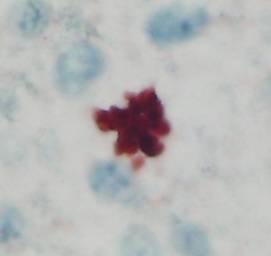
4 Germinoma Occurs predominantly in the midline Suprasellar and pineal regions Biphasic pattern large cells with prominent nucleoli admixed with lymphocytes When lymphocytes predominate, immunohistochemical analysis may be critical for diagnosis
5 Germinoma Placental alkaline phosphatase, PLAP well established marker; membranous pattern of staining C-kit proto-oncogene (CD117) Present on cell surface in almost all seminomas and dysgerminomas Rarely expressed in other germ cell tumors CD30 and C-kit in combination have been useful in separating embryonal components in gonadal tumors
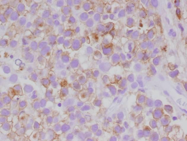
6 PLAP
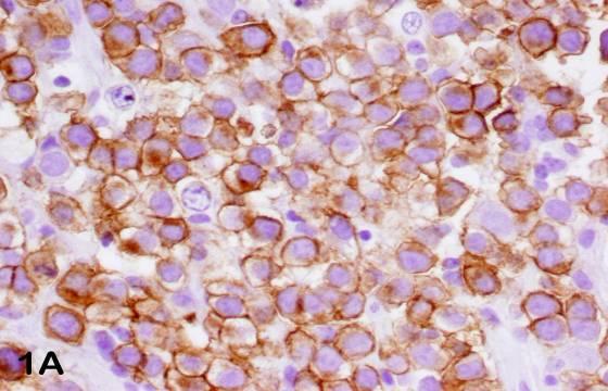
7 C-KIT
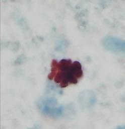
8 Germinoma OCT4 (POU5F1, OCT3 or OTF3) Nuclear transcription factor expressed in germ cells Regulation of pluripotent potential in germ cells Necessary for stem cell formation Immunoreactive in all 33 ovarian dysgerminomas and their metastases (Cheng et al, Am J Surg Path 2004) Almost 100% immunoreactivity in seminoma and embryonal carcinomas of the testes (Jones et al, Am J Surg Path 2004) negative in other germ cell components Nuclear staining pattern
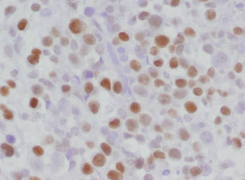
9 OCT 3/4
10 Germinoma Hattab et al (Am J Surg Path, 2005) examined intracranial germinomas and confirmed the findings of OCT4 and PLAP immunoreactivity patterns described in testicular seminomas and ovarian dysgerminomas Significantly stronger staining for OCT4 than PLAP overall

11
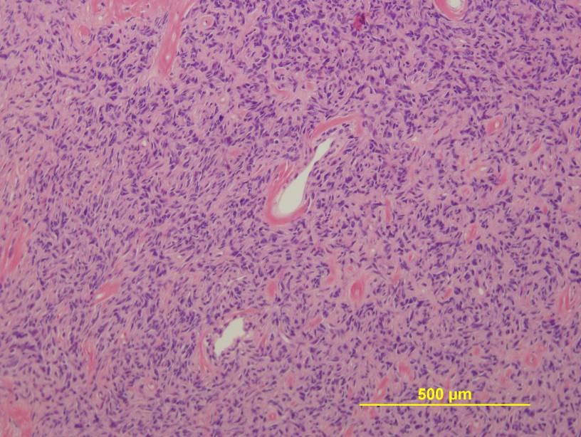
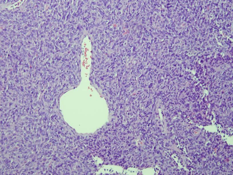
12 Hemangioblastoma WHO 2007 grade I neoplasm of uncertain histogenesis Composed of capillaries and stromal cells cerebellum and spinal cord Sporadic or associated with Von Hippel Lindau Necessity to differentiate from metastatic renal cell carcinoma in those patients with VHL Jarrel et al (J Neurosurg 2006) reported 8% of patients with VHL had metastases (most commonly RCC) within a hemangioblastoma
13 Hemangioblastoma Alpha Inhibin Produced in Sertoli cells of the testes and granulosa cells of the ovary normally Expressed in sex cord stromal tumors and adrenal cortical tumors Hoang (Am J Surg Path 2003) demonstrated cytoplasmic immunoreactivity in stromal cells in hemangioblastoma and negative staining for renal cell carcinoma
14 Hemangioblastoma Alpha-Inhibin
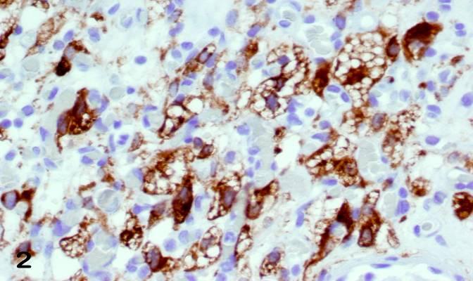
15 Expression of aquaporin 1 associated with cyst formation in hemangioblastoma (Chen et al, J Neuroonc 2006)
16 Hemangioblastoma Aquaporin 1 Weinbreck et al (Am J Surg Path, 2008) found that a panel including alpha-inhibin and aquaporin 1 were reliable positive markers for hemangioblastoma Membranous pattern aquaporin 1 AE1/AE3 and CD10 were reliable markers of clear cell renal cell carcinoma (CCRCC) Combined use of AE1/AE3 and aquaporin 1 were reliable for the differentiation of hemangioblastoma and metastatic CCRCC
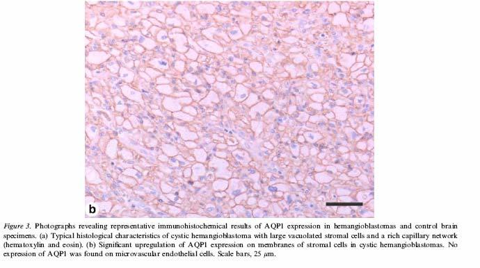
17 Aquaporin 1 in hemangioblastoma
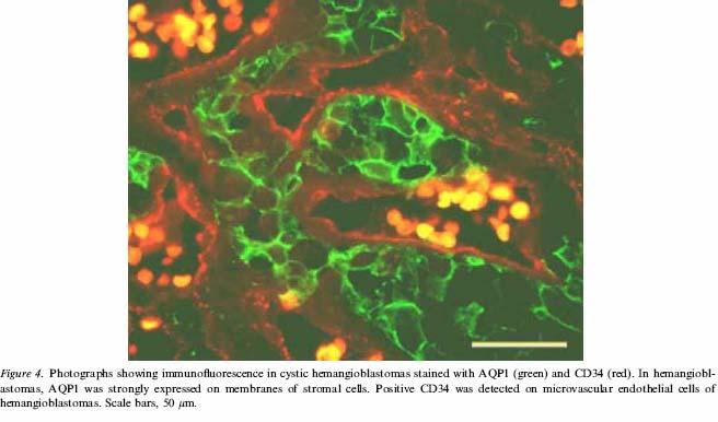
18 Immunofluorescence

19 The Texas Medical Center Facts & Figures at TMC One of the world s largest medical center complexes 46 Institutions 73,600 employees 6,500 beds 5.5M patient annually 10,000+ Intl. patients 13 Academic & RI s 10 Hospitals 26,000,000 gross sf TMC Corporation Owns land Grants restricted deeds to institutions Provides roads, utilities, parking garages
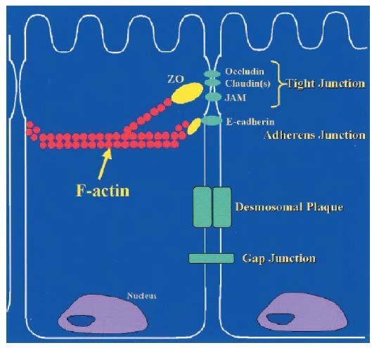
20 Meningioma Claudin-1 PHH-3 Secretory meningioma variant study
21 Meningioma Claudin-1 Tight junction-associated protein Identified in perineurial cells and soft tissue perineuriomas (Folpe et al Am J Surg Path, 2002) Bhattacharjee et al (AANP 2003) reported claudin-1 immunoreactivity in 85% of meningiomas with a punctate membranous pattern Hahn et al (AJCP 2006) found a similar pattern, but in a small percentage of cases, 53% studied
22
23
24
25 Claudin-1
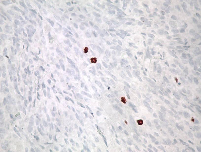
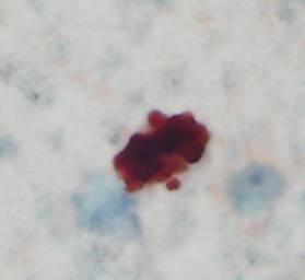
26 Phospho-Histone H3 (phh3) A Mitotic Figure Immunostain
27 Phospho-Histone H3 From: Prigent & Dimitrov. J Cell Sci 116: , 2003

28 Phospho-Histone H3 (phh3) Mitotic Figure Immunostain
29 PHH-3

30 Phospho-Histone H3 (phh3) Mitotic Figure Immunostain
31 Phospho-Histone H3 (phh3) Mitotic Figure Immunostain
32 Phosphohistone H3 (phh3)
33 Am J Clin Pathol, (1):
34
35
36

37
38 Meningioma, General Cytokeratin expression depends on meningioma subtype Focal positivity except in microcystic and anaplastic tumors CK18 positivity commonly present in all types Negativity overall for CK20 (Mieettinen and Paetau, 2002)
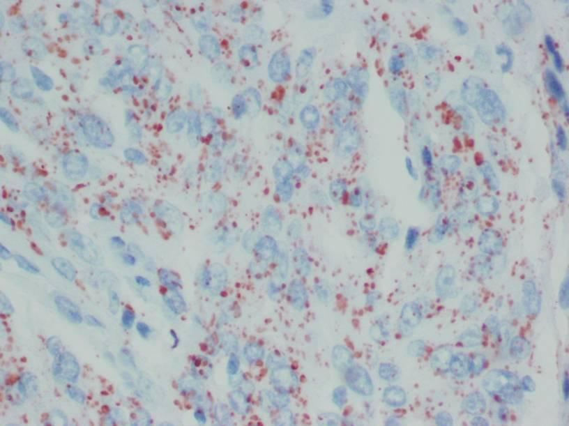
39 Secretory Meningioma CK immunoreactivity is unique, confined to cells adjacent to secretory material ( pseudopsammoma bodies ) CK18, also CK7, CK8 and CK19 CEA immunoreactivity well described in the secretory droplets Recent review of 6 cases (Caffo et al, J of Clin Neuroscience, May 2008) examining extracellular matrix proteins laminin, fibronectin and type IV collagen found staining for all three proteins in 2 of the 6 cases, and mild to absent immunoreactivity in the remaining 4 cases

40 The Methodist Hospital The Methodist Hospital is recognized as one of the leading hospitals in the United States A legacy of medical milestones and proven quality has attracted patients, healthcare professionals and institutional collaborations from around the world for more than 80 years
41 Solitary Fibrous Tumor to Hemangiopericytoma A Continuum? Panel of immunostains including EMA, CD99, BCL-2 and CD34 for distinguishing meningioma from SFT and HPC SFT and HPC having overlapping histologic features SFT and HPC remain separate entities in CNS as opposed to soft tissues in which this diagnosis has merged and HPC is a pattern
42 SFT/HPC Immunohistochemical staining patterns BCL-2 and CD34 are diffuse and strongly positive in SFT; weak and focal in HPC CD34 is strongly positive not only in vessels, but in the cytoplasm in SFT Usually diffuse, with less staining in HPC CD99 often shows a similar pattern to CD34, but may be negative EMA is often negative, distinguishing these tumors from meningiomas, but may have focal positive staining areas
43
44
45

46 Cellular SFT/HPC Reticulin Stain

47 Cellular SFT/HPC BCL-2

48 Cellular SFT/HPC EMA

49 Cellular SFT/HPC- CD34

50 Cellular SFT/HPC CD99

51 Cellular SFT/HPC MIB-1
52 The Methodist Hospital System San Jacinto Methodist Hospital 268 Licensed Beds 14,241 Inpatients 56,903 ER Visits Methodist Sugar Land Hospital 54 Licensed Beds (expanding to 300 in 2008) 5,204 Inpatients 33,495 ER Visits Methodist Willowbrook Hospital 119 Licensed Beds (expanding to 300 in 2009) 8,285 Inpatients 44,623 ER Visits Community Hospitals West Houston Methodist Hospital West Houston, co-located with new TCH 200 beds Spring 2010
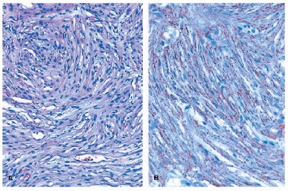
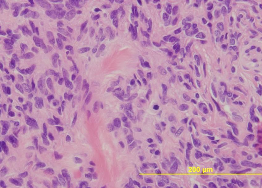
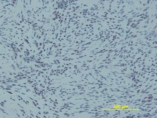
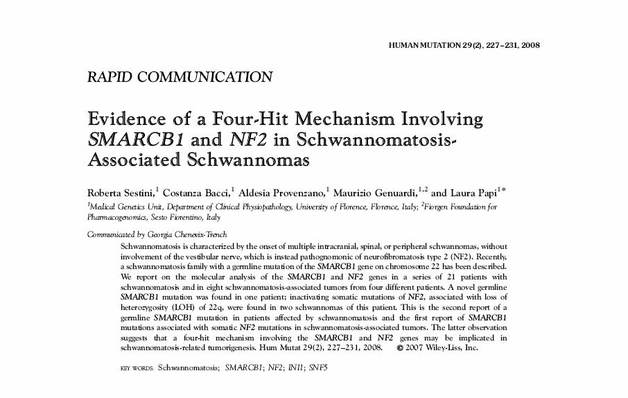
53 Hereditary Schwannoma INI-1/SMARCB1 protein recently implicated in pathogenesis of schwannoma in a family with familial schwannomatosis Mutations of SMARCB1 gene identified Mosaic loss of expression of INI1
54

55 INI-1 staining (Panel B) in hereditary schwannoma versus the diffuse positivity in a solitary, sporadic tumor (Panel A)

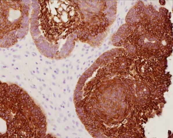
56 Sporadic Schwannoma INI 1 Sporadic Schwannoma
57

58 TMH Outpatient Facility Project Scope 24 Floor tower TPC $ 331 M 760,000 GSF occupied space 846,000 GSF parking 1370 spaces Outpatient Clinic Building
59 Craniopharyngioma vs. Rathke Cleft Cyst Sellar/suprasellar masses with differing clinical outcomes Purely cystic craniopharyngiomas vs. Rathke cleft cysts with squamous metaplasia and without significant ciliated epithelium may be extremely challenging Craniopharyngioma may also rarely demonstrate ciliated epithelium, compounding an already difficult problem
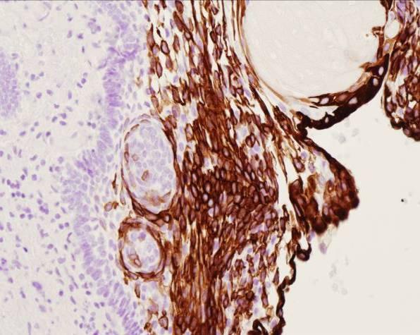
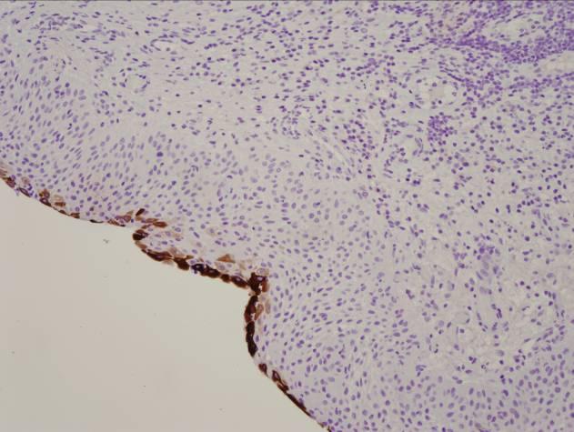
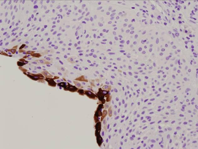
60 Beta-catenin Adamantinomatous craniopharyngiomas have been found to have an activating mutation in exon 3 of the beta-catenin gene in approximately 90% of cases examined (Buslei et al, Acta Neuropathologica, 2005) Pattern of staining is inhomogeneous and may be clustered in whorls Whorl-like immunoreactivity is associated with positive staining for CK8 (CAM5.2) and CK18
61 Beta-catenin Rathke cleft cyst (RCC) and papillary craniopharyngioma immunoreactivity is exclusively membranous Distinctive staining patterns may be useful in separating these entities, especially in small surgical specimens NOT useful for distinguishing papillary craniopharyngioma from RCC with squamous metaplasia

62 Adamantinomatous Craniopharyngioma Strong immunoreactivity for beta catenin in a whorl

63 Beta catenin negative staining of wet keratin
")
64 CAM 5.2 (CK8)
65 CK5-6
66 CK7

67 Beta-Catenin membranous staining pattern in epithelium of papillary craniopharyngioma
68 CK7 Papillary Craniopharyngioma
69 CK7 Papillary Craniopharyngioma

70 Rathke s cleft cyst

71 Rathke s Cyst Beta-catenin Membranous pattern as in papillary CP

72 CK7 Rathke s Cyst

73 Rathke s Cyst Pan Keratin
74 Utilizing the Differences Craniopharyngioma, Adamantinomatous Type Whorl-like positivity for beta-catenin Superficial staining for CK7, diffuse staining for CK5/6 Rathke s Cyst Membranous pattern of staining for beta-catenin as in Papillary craniopharyngioma Similar patterns of staining as for adamantinomatous craniopharyngioma for CK7 and CK5/6 (CAM 5.2)
 8,285 Inpatients 44,623 ER Visits Community Hospitals West Houston Methodist Hospital West")
75 The Methodist Hospital System San Jacinto Methodist Hospital 268 Licensed Beds 14,241 Inpatients 56,903 ER Visits Methodist Sugar Land Hospital 54 Licensed Beds (expanding to 300 in 2008) 5,204 Inpatients 33,495 ER Visits Methodist Willowbrook Hospital 119 Licensed Beds (expanding to 300 in 2009) 8,285 Inpatients 44,623 ER Visits Community Hospitals West Houston Methodist Hospital West Houston, co-located with new TCH 200 beds Spring 2010
Genetic Studies of Dysgerminoma
 Genetic Studies of Chromosome 12p abnormalities are characteristic of germ cell tumors isochromosome 12p and 12p overrepresentation Some can be detected by karyotyping FISH study of 21 dysgerminomas showed
"CRITICAL" IMMUNOHISTOCHEMICAL AND GENETIC MARKERS IN GLIAL NEOPLASMS
 AMERCAN ASSOCIATION OF NEUROPATHOLOGISTS USCAP COMPANION SOCIETY - 2009 "CRITICAL" IMMUNOHISTOCHEMICAL AND GENETIC MARKERS IN GLIAL NEOPLASMS Anthony T. Yachnis, M.D. Department of Pathology, Immunology,
Immunohistochemical Staining for Claudin-1 Can Help Distinguish Meningiomas From Histologic Mimics
 Anatomic Pathology / CLAUDIN-1 IN MENINGIOMAS Immunohistochemical Staining for Claudin-1 Can Help Distinguish Meningiomas From Histologic Mimics Hejin P. Hahn, MD, PhD, Elizabeth A. Bundock, MD, PhD, and
Peter Canoll MD. PhD.
 Tumors of the Nervous System Peter Canoll MD. PhD. What I want to cover What are the most common types of brain tumors? Who gets them? How do they ypresent? What do they look like? How do they behave?
Brain Tumors. Medulloblastoma. Pilocytic astrocytoma: Ahmed Koriesh, MD. Pathological finding
 NeuroPathology Page 8 Brain Tumors Pathological finding Pseudorosette Rosenthal fibers Rosettes Wet Keratin Psammoma bodies Fried egg Tumor Ependymoma, SEGA Pilocytic astrocytoma Medulloblastoma Craniopharyngioma
CNS TUMORS. D r. Ali Eltayb ( U. of Omdurman. I ). M. Path (U. of Alexandria)
. M. Path (U. of Alexandria)") CNS TUMORS D r. Ali Eltayb ( U. of Omdurman. I ). M. Path (U. of Alexandria) CNS TUMORS The annual incidence of intracranial tumors of the CNS ISmore than intraspinal tumors May be Primary or Secondary
CNS TUMORS D r. Ali Eltayb ( U. of Omdurman. I ). M. Path (U. of Alexandria) CNS TUMORS The annual incidence of intracranial tumors of the CNS ISmore than intraspinal tumors May be Primary or Secondary
Ovarian Clear Cell Carcinoma
 Ovarian Clear Cell Carcinoma Rouba Ali-Fehmi, MD Professor of Pathology The Karmanos Cancer Institute, Wayne State University School of Medicine 50 year old woman with chief complaint of shortness of breath
Note: The cause of testicular neoplasms remains unknown
 - In the 15- to 34-year-old age group, they are the most common tumors of men. - Tumors of the testis are a heterogeneous group of neoplasms that include: I. Germ cell tumors : 95%; all are malignant.
Disclosure. Relevant Financial Relationship(s) None. Off Label Usage None MFMER slide-1
 None. Off Label Usage None MFMER slide-1") Disclosure Relevant Financial Relationship(s) None Off Label Usage None 2013 MFMER slide-1 Case Presentation A 43 year old male, with partial nephrectomy for a right kidney mass 2013 MFMER slide-2 2013
Disclosure Relevant Financial Relationship(s) None Off Label Usage None 2013 MFMER slide-1 Case Presentation A 43 year old male, with partial nephrectomy for a right kidney mass 2013 MFMER slide-2 2013
Hemangioblastoma is a benign central nervous system
 ORIGINAL ARTICLE PAX2( )/PAX8( )/Inhibin A(+) Immunoprofile in Hemangioblastoma: A Helpful Combination in the Differential Diagnosis With Metastatic Clear Cell Renal Cell Carcinoma to the Central Nervous
hemangioblastoma of the retroperitoneum
 Int J Clin Exp Pathol 2014;7(4):1777-1781 www.ijcep.com /ISSN:1936-2625/IJCEP1401042 Case Report Yong Huang 1, Xiang-Chun Han 2, Guo-Shi Lv 3 1 Department of Pathology, 251 Hospital of PLA, Zhangjiakou,
Synonyms. Nephrogenic metaplasia Mesonephric adenoma
 Nephrogenic Adenoma Synonyms Nephrogenic metaplasia Mesonephric adenoma Definition Benign epithelial lesion of urinary tract with tubular, glandular, papillary growth pattern Most frequently in the urinary
Tumors of the Nervous System
 Tumors of the Nervous System Peter Canoll MD. PhD. What I want to cover What are the most common types of brain tumors? Who gets them? How do they present? What do they look like? How do they behave? 1
Mesothelioma: diagnostic challenges from a pathological perspective. Naseema Vorajee August 2016
 Mesothelioma: diagnostic challenges from a pathological perspective Naseema Vorajee August 2016 Naseema.vorajee@nhls.ac.za Pleural diseases (whether neoplastic, reactive or infective) may have similar
57th Annual HSCP Spring Symposium 4/16/2016
 An Unusual Malignant Spindle Cell Lesion to Involve the Breast Erinn Downs-Kelly, D.O. Associate Professor of Pathology University of Utah & ARUP Laboratories No disclosures Case 39 y/o female with no
Male Genital Cancers in the US in Frequency of Types
 Germ Cell Tumors of the Testis Pathology, Immunohistochemistry, and the Often Confusing Appearance of Their Metastases Charles Zaloudek, MD Department of Pathology UCSF Male Genital Cancers in the US in
Case Report Hemangioblastoma in the Lung: Metastatic or Primary Lesions?
 Hindawi Publishing Corporation Volume 2014, Article ID 468671, 5 pages http://dx.doi.org/10.1155/2014/468671 Case Report Hemangioblastoma in the Lung: Metastatic or Primary Lesions? Li Lu, Peter A. Drew,
Various hereditary, acquired and neoplastic conditions can lead to cyst formation in the kidney.
 Dr. Fatima AlAl-Hashimi Hashimi,, MD, FRCPath Salmaniya Medical Complex, Bahrain Various hereditary, acquired and neoplastic conditions can lead to cyst formation in the kidney. The most frequently encountered
Case E1. Female aged 63 years Right Nephrectomy Two separate tumours Section of each tumour
 Case E1 Female aged 63 years Right Nephrectomy Two separate tumours Section of each tumour Tumour 1 Upper pole tumour 28mm macro diameter Circumscribed Friable cut surface Tumour 2 Middle pole Part solid
Tinh hoàn
 Tinh hoàn Tinh hoàn Tinh hoàn Tiền liệt tuyến Tiền liệt tuyến Mào tinh hoàn Mào tinh hoàn Túi tinh Túi tinh Túi tinh Túi tinh So-called cystadenoma of seminal vesicle. Gross appearance of granulomatous
3/27/2017. Disclosure of Relevant Financial Relationships
 Ophthalmic Pathology Evening Specialty Conference USCAP 2017 5 th March, 2017 Mukul K. Divatia, MD Assistant Professor Department of Pathology & Genomic Medicine Weill Cornell Medical College Houston Methodist
My personal experience at University of Toronto and recent updates of
 My personal experience at University of Toronto and recent updates of Endocrine Pathology Toshitetsu Hayashi M.D. Ph.D. ¹Department of Diagnostic Pathology, Takamatsu Red Cross Hospital, Japan ²Laboratory
-The cause of testicular neoplasms remains unknown
 - In the 15- to 34-year-old age group, they are the most common tumors of men. - include: I. Germ cell tumors : (95%); all are malignant. II. Sex cord-stromal tumors: from Sertoli or Leydig cells; usually
Pathology Mystery and Surprise
 Pathology Mystery and Surprise Tim Smith, MD Director Anatomic Pathology Medical University of South Carolina Disclosures No conflicts to declare Some problem cases Kidney tumor Scalp tumor Bladder tumor
Salivary Gland FNA ATYPICAL : Criteria and Controversies
 Salivary Gland FNA ATYPICAL : Criteria and Controversies W.C. Faquin, M.D., Ph.D. Director, Head and Neck Pathology Massachusetts General Hospital Massachusetts Eye and Ear Infirmary Harvard Medical School
A 25 year old female with a palpable mass in the right lower quadrant of her abdomen
 May 2016 A 25 year old female with a palpable mass in the right lower quadrant of her abdomen Contributed by: Paul Ndekwe, MD, Resident Physician, Indiana University School of Department of Pathology and
Histopathological Study and Categorisation of Brain Tumors
 Histopathological Study and Categorisation of Brain Tumors Ruchira Wadhwa 1*, Purvi Patel 2, Hansa Goswami 3 1 Third Year Resident, 2 Assistant Professor, 3 Professor and Head, Department of Pathology,
Select problems in cystic pancreatic lesions
 Disclosure Select problems in cystic pancreatic lesions Five Prime Therapeutics shareholder Adicet Bio shareholder Bristol-Meyer Squibb advisory board grace.kim@ucsf.edu Pancreatic cystic lesions Intraductal
Craniopharyngiomas are epithelial neoplasms thought
 Differential Expression of Cytokeratins 8 and 20 Distinguishes Craniopharyngioma From Rathke Cleft Cyst Wei Xin, MD, PhD; Mark A. Rubin, MD; Paul E. McKeever, MD, PhD Background. Craniopharyngiomas are
Sporadic Hemangioblastoma of the Kidney: a rare renal tumor
 Liu et al. Diagnostic Pathology 2012, 7:49 CASE REPORT Open Access Sporadic Hemangioblastoma of the Kidney: a rare renal tumor Yang Liu 1,2, Xue-shan Qiu 1,2* and En-Hua Wang 1,2 Abstract: Hemangioblastoma
Int J Clin Exp Pathol 2016;9(8): /ISSN: /IJCEP Guobin Zhang, Yingzhi Hou, Wenqing Jia, Jun Yang, Yulun Xu
: /ISSN: /IJCEP Guobin Zhang, Yingzhi Hou, Wenqing Jia, Jun Yang, Yulun Xu") Int J Clin Exp Pathol 2016;9(8):8730-8735 www.ijcep.com /ISSN:1936-2625/IJCEP0026741 Case Report Metastasis of renal cell carcinoma to hemangioblastoma of the spinal cord in von Hippel-Lindau disease:
Int J Clin Exp Pathol 2016;9(8):8730-8735 www.ijcep.com /ISSN:1936-2625/IJCEP0026741 Case Report Metastasis of renal cell carcinoma to hemangioblastoma of the spinal cord in von Hippel-Lindau disease:
Neuroendocrine Carcinoma. Lebanon Neuroendocrine Neoplasms of H&N Nov /7/2011. Broad Classification:
 H&N Neuroendocrine Neoplasms: Classification and Diagnostic Considerations Adel K. El-Naggar, M.D., Ph.D. The University of Texas MD Anderson Cancer Center, Houston, Texas Broad Classification: A. Epithelial:
Adenocarcinoma of the Cervix
 Question 1. Each of the following statements about cervical adenocarcinoma is true except: Adenocarcinoma of the Cervix SAMS a) A majority of women with cervical adenocarcinoma have stage I tumors at diagnosis.
Disclosure of Relevant Financial Relationships
 Squamous entities of the thyroid: Reactive to Neoplastic Michelle D. Williams Associate Professor Dept of Pathology, Head & Neck Section University of Texas MD Anderson Cancer Center Disclosure of Relevant
Self assessment case. Dr Saleem Taibjee Dorset County Hospital, Dorchester
 Self assessment case Dr Saleem Taibjee saleemtaibjee@gmail.com Dorset County Hospital, Dorchester Clinical details 34-year-old man: Shave excision Skin tag / papilloma left thigh The best diagnosis is:
Updates in Urologic Pathology WHO Made Those Changes?! Peyman Tavassoli Pathology Department BC Cancer Agency
 Updates in Urologic Pathology WHO Made Those Changes?! Peyman Tavassoli Pathology Department BC Cancer Agency World Health Organization Available in Feb 2016 Frame work for reporting Major contributing
Anna Maria Buccoliero Department of Biomedicine, Careggi Hospital Florence
 PEDIATRIC RHABDOID MENINGIOMA Anna Maria Buccoliero Department of Biomedicine, Careggi Hospital Florence CLINICAL HISTORY A 3-year-old boy, with a recent history of seizures, was admitted to the Neurosurgery
The Pathology of Germ Cell Tumours of the Ovary
 The Pathology of Germ Cell Tumours of the Ovary Professor Mike Wells University of Sheffield Amman, Jordan November 2013 Professor Francisco Paco Nogales I. Primitive germ cell tumors A. Dysgerminoma
SPECIAL SLIDE SEMINAR CASE 3
 SPECIAL SLIDE SEMINAR CASE 3 Tihana Džombeta, MD Leo Pažanin, MD, PhD Department of Pathology, School of Medicine, University of Zagreb Department of Pathology, Clinical Hospital Centre Sestre milosrdnice
Kidney Case 1 SURGICAL PATHOLOGY REPORT
 Kidney Case 1 Surgical Pathology Report February 9, 2007 Clinical History: This 45 year old woman was found to have a left renal mass. CT urography with reconstruction revealed a 2 cm medial mass which
RECURRENT ADRENAL DISEASE. Megan Applewhite Endorama 2/19/2015 SR , SC
 RECURRENT ADRENAL DISEASE Megan Applewhite Endorama 2/19/2015 SR 2412318, SC 3421561 Category: Adrenal Attendings: Angelos & Grogan PATIENT #1 36yo woman with a hx of Cushing s Syndrome and right adrenalectomy
Unknown Slides Conference
 Unknown Slides Conference Jae Y. Ro, MD, PhD Weill Medical College of Cornell Univ. The Methodist Hospital, and UT MD Anderson Cancer Center Houston, TX November 9, 2013 Amman, Jordan 25 th Congress of
Renal tumours: use of immunohistochemistry & molecular pathology. Dr Lisa Browning John Radcliffe Hospital Oxford
 Renal tumours: use of immunohistochemistry & molecular pathology Dr Lisa Browning John Radcliffe Hospital Oxford Renal tumours: the use of immunohistochemistry & molecular pathology Classification of RCC
Tumors of kidney and urinary bladder
 Tumors of kidney and urinary bladder Overview of kidney tumors Benign and malignant Of the benign: papillary adenoma -cortical -small (0.5cm) -in 40% of population -clinically insignificant The most common
04/09/2018. Salivary Gland Pathology in the Molecular Era Old Friends, Old Foes, & New Acquaintances
 Salivary Gland Pathology in the Molecular Era Old Friends, Old Foes, & New Acquaintances Jennifer L. Hunt, MD, MEd Aubrey J. Hough Jr, MD, Endowed Professor of Pathology Chair of Pathology and Laboratory
Testicular Tumors: What s New, True, Important Cristina Magi-Galluzzi, MD, PhD
 Testicular Tumors: What s New, True, Important Cristina Magi-Galluzzi, MD, PhD Director, Genitourinary Pathology R.J. Tomsich Pathology & Laboratory Medicine Institute Professor of Pathology, Lerner College
Spectrum of Preneoplastic and Neoplastic Cystic Lesions of the Kidney in Adult. by dr. Banan Burhan Mohammed Lecturer in Pathology Department
 Spectrum of Preneoplastic and Neoplastic Cystic Lesions of the Kidney in Adult by dr. Banan Burhan Mohammed Lecturer in Pathology Department Various hereditary, acquired, and neoplastic conditions can
Disclosures. The Thin Red Line Between Neuropathology and Head & Neck Pathology. Introduction CASE 1. Current Issues Tihan
 Disclosures I have nothing to disclose The Thin Red Line Between Neuropathology and Head & Neck Pathology Tarik Tihan, MD, PhD UCSF, Department of Pathology Neuropathology Division Introduction Three cases
Leukaemia 35% Lymphoma 14%
 Distribution ib ti of Cancers in Children under 15 years Leukaemia 35% Lymphoma 14% Neuroblastoma 9% Other 5% Liver 1% Retinoblastoma 3% Bone and STS 15% CNS 20% Wilms' 8% 30-40% Mortality Germ Cell Tumours
American Journal of. Medical Case Reports. CAM5.2 Expression in Metastatic Tumours of CNS: A Diagnostic Tool
 American Journal of American Journals of Medical Case Reports http://ivyunion.org/index.php/ajmcr/index Medical Case Reports Mathur SK et al. American Journal of Medical Case Reports 2014, 2:1-8 Vol 2,
AMERICAN ASSOCIATION OF NEUROPATHOLOGISTS COMPANION SOCIETY MEETING at the 106 th ANNUAL MEETING OF THE USCAP San Antonio, March 4, 2017
 AMERICAN ASSOCIATION OF NEUROPATHOLOGISTS COMPANION SOCIETY MEETING at the 106 th ANNUAL MEETING OF THE USCAP San Antonio, March 4, 2017 SYLLABUS Papillary Tumor of the Pineal Region and the Differential
Papillary adenocarcinoma of the rete testis with adjacent hyperplasia: a case report
 CASE REPORT Papillary adenocarcinoma of the rete testis with adjacent hyperplasia: a case report Carolina Polanco 1, Cooley G. Pantazis 2, Rolando Prieto 2, Kenneth A. Iczkowski 1 1. Medical College of
3/27/2017. Pulmonary Pathology Specialty Conference. Disclosure of Relevant Financial Relationships. Clinical History:
 Pulmonary Pathology Specialty Conference Saul Suster, M.D. Medical College of Wisconsin Disclosure of Relevant Financial Relationships USCAP requires that all planners (Education Committee) in a position
Financial disclosures
 An update on immunohistochemical markers in mesenchymal neoplasms By Konstantinos Linos MD, FCAP, FASDP Assistant Professor of Pathology Geisel School of Medicine at Dartmouth Dartmouth-Hitchcock Medical
Testicular Germ Cell Tumors; A Simplistic Approach
 Testicular Germ Cell Tumors; A Simplistic Approach Merce Jorda, MD, PhD, MBA Professor and Vice Chair, Director of Anatomic Pathology Director of Genitourinary Pathology Service Interim Director of Cytopathology
Review and Updates of Immunohistochemistry in Selected Salivary Gland and Head and Neck Tumors
 Review and Updates of Immunohistochemistry in Selected Salivary Gland and Head and Neck Tumors. Monophasic tumors : myoepithelioma, acinic cell carcinoma, and salivary duct carcinoma. Biphasic tumors includes
Gross appearance of nodular hyperplasia in material obtained from suprapubic prostatectomy. Note the multinodular appearance and the admixture of
 Tiền liệt tuyến Tiền liệt tuyến Gross appearance of nodular hyperplasia in material obtained from suprapubic prostatectomy. Note the multinodular appearance and the admixture of solid and microcystic areas.
Dr Sanjiv Manek Oxford. Oxford Pathology Course 2010 for FRCPath Illustration-Cellular Pathology. Oxford Radcliffe NHS Trust
 Dr Sanjiv Manek Oxford Oxford Pathology Course 2010 for FRCPath Illustration-Cellular Pathology. Oxford Radcliffe NHS Trust Ovarian Endometrial Vulvo-vaginal Cervical Illustration-Cellular Pathology. Oxford
RENAL EPITHELIAL NEOPLASMS: IS THERE A ROLE OF IMMUNOSTAINS IN DIAGNOSIS?
 RENAL EPITHELIAL NEOPLASMS: IS THERE A ROLE OF IMMUNOSTAINS IN DIAGNOSIS? John C. Cheville, M.D. Mayo Clinic and Mayo Foundation Rochester, MN The majority of renal epithelial neoplasms are diagnosed on
3 cell types in the normal ovary
 Ovarian tumors 3 cell types in the normal ovary Surface (coelomic epithelium) the origin of the great majority of ovarian tumors 90% of malignant ovarian tumors Totipotent germ cells Sex cord-stromal cells
Pitfalls in the diagnosis of well-differentiated hepatocellular lesions
 2013 Colorado Society of Pathology Pitfalls in the diagnosis of well-differentiated hepatocellular lesions Sanjay Kakar, MD University of California, San Francisco Outline Hepatocellular adenoma: new WHO
BSD 2015 Case 19. Female 21. Nodule on forehead. The best diagnosis is:
 BSD 2015 Case 19 Female 21. Nodule on forehead. The best diagnosis is: A. mixed tumour of skin B. porocarcinoma C. nodular hidradenoma D. metastatic adenocarcinoma BSD 2015 Case 19 Female 21 Nodule on
Diplomate of the American Board of Pathology in Anatomic and Clinical Pathology
 A 33-year-old male with a left lower leg mass. Contributed by Shaoxiong Chen, MD, PhD Assistant Professor Indiana University School of Medicine/ IU Health Partners Department of Pathology and Laboratory
Test Bank for Robbins and Cotran Pathologic Basis of Disease 9th Edition by Kumar
 Link full download:https://getbooksolutions.com/download/test-bank-for-robbinsand-cotran-pathologic-basis-of-disease-9th-edition-by-kumar Test Bank for Robbins and Cotran Pathologic Basis of Disease 9th
Institute of Pathology First Faculty of Medicine Charles University. Ovary
 Ovary Barrett esophagus ph in vagina between 3.8 and 4.5 ph of stomach varies from 1-2 (hydrochloric acid) up to 4-5 BE probably results from upward migration of columnar cells from gastroesophageal junction
3 cell types in the normal ovary
 Ovarian tumors 3 cell types in the normal ovary Surface (coelomic epithelium) the origin of the great majority of ovarian tumors (neoplasms) 90% of malignant ovarian tumors Totipotent germ cells Sex cord-stromal
Pathology of the Thyroid
 Pathology of the Thyroid Thyroid Carcinoma Arising from Follicular Cells 2015-01-19 Prof. Dr. med. Katharina Glatz Pathologie Carcinomas Arising from Follicular Cells Differentiated Carcinoma Papillary
Diagnostic IHC in lung and pleura pathology
 Diagnostic IHC in lung and pleura pathology Mogens Vyberg Professor of Clinical Pathology Director of NordiQC Aalborg University Hospital, Aalborg, Denmark WHO 2004 and Web Malignant mesothelioma Epithelioid
3/24/2017. Disclosure of Relevant Financial Relationships. Mixed Epithelial Endometrial Carcinoma. ISGyP Endometrial Cancer Project
 Disclosure of Relevant Financial Relationships USCAP requires that all planners (Education Committee) in a position to influence or control the content of CME disclose any relevant financial relationship
General: Brain tumors are lesions that have mass effect distorting the normal tissue and often result in increased intracranial pressure.
 1 Lecture Objectives Know the histologic features of the most common tumors of the CNS. Know the differences in behavior of the different tumor types. Be aware of the treatment modalities in the various
Renal Cell Carcinoma: Genetics & Imaging Srinivasa R Prasad University of Texas San Antonio
 Renal Cell Carcinoma: Genetics & Imaging Srinivasa R Prasad University of Texas HSC @ San Antonio No financial disclosures Acknowledgements Dr. Peter Choyke, NIH My Gurus @ MIR, MGH 2004 WHO Taxonomy of
How to Recognize Gynecologic Cancer Cells from Pelvic Washing and Ascetic Specimens
 How to Recognize Gynecologic Cancer Cells from Pelvic Washing and Ascetic Specimens Wenxin Zheng, M.D. Professor of Pathology and Gynecology University of Arizona zhengw@email.arizona.edu http://www.zheng.gynpath.medicine.arizona.edu/index.html
Papillary Lesions of the Breast A Practical Approach to Diagnosis. (Arch Pathol Lab Med. 2016;140: ; doi: /arpa.
 Papillary Lesions of the Breast A Practical Approach to Diagnosis (Arch Pathol Lab Med. 2016;140:1052 1059; doi: 10.5858/arpa.2016-0219-RA) Papillary lesions of the breast Span the spectrum of benign,
Gross appearance of peritoneal cysts. They have a thin, translucent wall and contain a clear fluid.
 Gross appearance of peritoneal cysts. They have a thin, translucent wall and contain a clear fluid. So-called multicystic benign mesothelioma. A, Gross appearance. So-called multicystic benign mesothelioma.
What s New in Adrenal Gland Pathology. Marina Scarpelli
 What s New in Adrenal Gland Pathology Marina Scarpelli Background Histological criteria for adrenocortical proliferative lesions Immunohistochemical markers Molecular markers Histological Criteria for
A 53 year-old woman with a lung mass, right hilar mass and mediastinal adenopathy.
 November 2015 Case of the Month A 53 year-old woman with a lung mass, right hilar mass and mediastinal adenopathy. Contributed by: Rasha Salama, M.D., IU Department of Pathology and Laboratory Medicine
Differential diagnosis of HCC
 Hepatocellular Carcinoma Quest for an Ideal Immunohistochemical Panel Sanjay Kakar, MD UCSF Differential diagnosis of HCC Hepatocellular lesions Adenoma, FNH, HG dysplasia Adenocarcinoma CholangioCA, metastasis
Immunohistochemistry in Bone and Soft Tissue Tumors. Sahar Rassi Zankoul, MD
 Immunohistochemistry in Bone and Soft Tissue Tumors Sahar Rassi Zankoul, MD Introduction Bone tumors represent a wide variety of tumors of various origins and malignant potentials. These different tumor
The Panel Approach to Diagnostics. Lauren Hopson International Product Specialist Cell Marque Corporation
 The Panel Approach to Diagnostics Lauren Hopson International Product Specialist Cell Marque Corporation Cell Marque Rocklin, California About Cell Marque: IVD primary antibody manufacturer Distributors
Newer soft tissue entities
 Newer soft tissue entities Examples among fibroblastic tumors Turku, May 6, 2010 Markku Miettinen, M.D. AFIP, Washington, DC Fibroblastic neoplasms Solitary fibrous tumor /Hemangiopericytoma Low-grade
Evening Specialty Conference Bone and Soft Tissue Pathology. Diagnostic pitfalls in bone and soft tissue pathology
 Evening Specialty Conference Bone and Soft Tissue Pathology. Case 1 Elizabeth G Demicco, MD, PhD Mount Sinai Hospital, New York Disclosure of Relevant Financial Relationships USCAP requires that all planners
Enterprise Interest Nothing to declare
 Enterprise Interest Nothing to declare Diagnoses one would not like to miss in soft tissue pathology early in your career Marta Sbaraglia, MD Department of Pathology Hospital of Treviso University of Padua
Case 2. Dr. Sathima Natarajan M.D. Kaiser Permanente Medical Center Sunset
 Case 2 Dr. Sathima Natarajan M.D. Kaiser Permanente Medical Center Sunset History 24 year old male presented with a 3 day history of right flank pain, sharp in nature Denies fever, chills, hematuria or
Acantholytic Anaplastic Extramammary Paget s Disease: A Case Report and Review of the Literature
 Ann Dermatol Vol. 23, Suppl. 2, 2011 http://dx.doi.org/10.5021/ad.2011.23.s2.s226 CASE REPORT Acantholytic Anaplastic Extramammary Paget s Disease: A Case Report and Review of the Literature Yu-Jin Oh,
Case: The patient is a 62 year old woman with a history of renal cell carcinoma that was removed years ago. A 2.4 cm liver mass was found on CT
 Case: The patient is a 62 year old woman with a history of renal cell carcinoma that was removed years ago. A 2.4 cm liver mass was found on CT during follow- up. ALT, AST, Alk Phos and bilirubin were
BRAIN & SPINAL LESIONS: NOT JUST A SCIENCE. Rimas V. Lukas, MD Associate Professor Director of Medical Neuro-Oncology University of Chicago
 BRAIN & SPINAL LESIONS: NOT JUST A SCIENCE Rimas V. Lukas, MD Associate Professor Director of Medical Neuro-Oncology University of Chicago OVERVIEW Background Clinical Presentation Clinical Management
CNS pathology Third year medical students. Dr Heyam Awad 2018 Lecture 12: CNS tumours 2/3
 CNS pathology Third year medical students Dr Heyam Awad 2018 Lecture 12: CNS tumours 2/3 Pilocytic astrocytoma Relatively benign ( WHO grade 1) Occurs in children and young adults Mostly: in the cerebellum
Spindle Cell Lesions Of The Breast. Emad Rakha Professor of Breast Pathology and Consultant Pathologist
 Spindle Cell Lesions Of The Breast Emad Rakha Professor of Breast Pathology and Consultant Pathologist * SCLs comprise a wide spectrum of diseases, ranging from reactive processes to aggressive malignant
NEOPLASIA. 3. Which of the following tumour is benign a. Chondrosarcoma b. Osteochondroma c. Chondroblastoma d. Ewing s tumour e.
 NEOPLASIA 1. malignant neoplasms a. are independent of hormonal influence b. are always composed of homogenous cell lines c. arise from differentiated cells by a process of anaplasia d. display abnormal
Immunohistochemical classification of the unknown primary tumour (UPT) Part I. Prof. Mogens Vyberg NordiQC Institute of Pathology Aalborg, Denmark
 Part I. Prof. Mogens Vyberg NordiQC Institute of Pathology Aalborg, Denmark") Immunohistochemical classification of the unknown primary tumour (UPT) Part I Prof. Mogens Vyberg NordiQC Institute of Pathology Aalborg, Denmark Tumours of unknown origin: Histology Brain tumour - biopsy
Immunohistochemical classification of the unknown primary tumour (UPT) Part I Prof. Mogens Vyberg NordiQC Institute of Pathology Aalborg, Denmark Tumours of unknown origin: Histology Brain tumour - biopsy
Mesothelioma Pathobasic. Lukas Bubendorf Pathology
 Mesothelioma Pathobasic Lukas Bubendorf Pathology Mechanisms of Asbestos Carcinogenesis in Mesothelioma Asprin High-mobility group protein B1 master switch HMGB1 Initiation/ perpetuation of inflamm. response
Test Bank for Robbins and Cotran Pathologic Basis of Disease 9th Edition by Kumar
 Link full download: http://testbankair.com/download/test-bank-for-robbins-cotran-pathologic-basis-of-disease-9th-edition-bykumar-abbas-and-aster Test Bank for Robbins and Cotran Pathologic Basis of Disease
GUT-C 11/30/2017. Debasmita Das, M.D. PGY-1 Danbury Hospital
 GUT-C 11/30/2017 Debasmita Das, M.D. PGY-1 Danbury Hospital CLINICAL SUMMARY 8/2017 59 year old female Presented to the ED with 1 month history of general malaise, fever and weight loss PMH: Significant
Intracranial Solitary Fibrous Tumor with Pseudopapillary Architecture: An Uncommon Tumor with Unusual Histopathology
 North American Journal of Medicine and Science Oct 2016 Vol 9 No.4 181 Case Report Intracranial Solitary Fibrous Tumor with Pseudopapillary Architecture: An Uncommon Tumor with Unusual Histopathology Guang
Case Report Overlap of microcystic stromal tumor and primary solid pseudopapillary neoplasm of the ovary
 Int J Clin Exp Pathol 2015;8(9):11792-11797 www.ijcep.com /ISSN:1936-2625/IJCEP0013094 Case Report Overlap of microcystic stromal tumor and primary solid pseudopapillary neoplasm of the ovary Qin Chen
Renal tumors of adults
 Renal tumors of adults Urinary Tract Tumors 2%-3% of all cancers in adults. The most common malignant tumor of the kidney is renal cell carcinoma. Tumors of the lower urinary tract are twice as common
Protocol for management of patients with pineal region tumours v1
 Protocol for management of patients with pineal region tumours v1 West Midlands Cancer Alliance Coversheet for Cancer Alliance Expert Advisory Group Agreed Documentation This sheet is to accompany all
AGGRESSIVE VARIANTS OF PAPILLARY THYROID CARCINOMA DIAGNOSIS AND PROGNOSIS
 AGGRESSIVE VARIANTS OF PAPILLARY THYROID CARCINOMA DIAGNOSIS AND PROGNOSIS PAPILLARY THYROID CARCINOMA Clinical Any age Microscopic to large Female: Male= 2-4:1 Radiation history Lymph nodes Prognosis
Laurie A. Loevner, MD
 Laurie A. Loevner, MD Chief, Division of Neuroradiology UPHS Professor of Radiology, Otorhinolaryngology: Head & Neck Surgery, Neurosurgery, and Ophthalmology University of Pennsylvania Health System Disclosures