Diagnostic Cytology of Cancer Cases
|
|
|
- Alisha Campbell
- 5 years ago
- Views:
Transcription
 Department of Pathology, Faculty of Veterinary Science, Chulalongkorn")
1 Diagnostic Cytology of Cancer Cases Somporn Techangamsuwan Companion Animal Cancer Research Unit (CAC-RU) Department of Pathology, Faculty of Veterinary Science, Chulalongkorn University 1


2 Tumor or Non-tumor

3 Tumor cells? Inflammatory cells? Infectious agents? Fluid? 3


4 Cytology >>> Fastest! Some tumors are diagnosed by cytology better than biopsy, eg.; round cell tumor. Is that final conclusion? 4

5 Tumor cell interpretation Step-by-Step 5
 5.")
6 At First Glance on Slide 1. Significant tumor cells: presence or absence (fluid, blood) >> DIY: Prepare 2-3 slides 2. Size and shape of tumor cell >> Easy way to compare tumor size 3. Cellular arrangement >> clump, sheet, glandular, individual 4. Nuclei and cytoplasm of tumor cell >> criteria of malignancy >> intracytoplasmic granules (black, metachromatic) 5. Extracellular matrix (keratin, mucin, osteoid), Foreign bodies, Pathogen 6
7 Interpretation: First Step Approach Which cell type?? Inflammatory cells Differentiate cell type Inflammatory cells + Tissue cells - Differentiate cell type - History taking Tissue cells Differentiate cell type and origin Neutrophils>85% = suppurative (pus) Neutrophils, Macrophages, pus cells = pyogranulomatous Macropahges, Lymphocytes, fibroblasts, plasma cells, low neutrophils = granulomatous Many Eosinophils >10% = eosinophilic plaques (granuloma) or allergic reaction Epithelium Mesenchymal Round cell Mixed, pleomorphic >> difficult for diagnosis Most malignant tumor 7


8 3 basic tumor cell types Epithelial Size Shape Cellularity Cluster Large Round to Usually high Yes caudate Mesenchymal Small to Spindle to Usually low No medium stellate Discrete round cell Mast cell Lymphoma Small to Round Usually high No medium TVT Histocytoma 8


9 General criteria Criteria of Malignancy Pleomorphism Multinucleation Anisocytosis Normal Abnormal Abnormal mitosis Macrocytosis Coarse chromatin Increase cell no. Hypercellularity Nuclear molding Nuclear criteria Anisokaryosis RBC Anisonucleosis Macrokaryosis & Increased N:C ratio Angular nucleoli 9
10 Cell Arrangement Cellular Arrangement VG Single,solitary Basal cell Chain, row Hepatoid cell Sheet Mesothelial cell Cluster Fibrosarcoma MG adenocarcinoma Glandular Clump Mal. histiocytosis 10
11 Anisocytosis Cellular morphology VG Pleomorphism 11


12 Cytoplasm Cell Arrangement VG Abundant pale, Multiple vacuoles Basal cell Scant cytoplasm, basophilic Hepatoid cell Abundant pale blue cyto. Mesothelial cell Abundant pale cyto., eccentric vacuole Fibrosarcoma MG adenocarcinoma Mal. histiocytosis 12 Abundant basophilic cytoplasm Low to moderate pale blue cytoplasm Abundant pale blue cyto. with multiple vacuoles
13 Nuclear pattern Cell Arrangement VG Heterochromatin, non-prominent nucleoli Basal cell Homogeneous, non-obvious nucleoli Hepatoid cell Vesicular-prominent large nucleoli Mesothelial cell Heterochromatin Prominent large nucleoli Fibrosarcoma MG adenocarcinoma Mal. histiocytosis Homogeneous, prominent large nucleoli Heterochromatin, non-obvious small nucleoli 13

14 Abnormal mitosis Abnormal Nuclei Multinucleation 14

15 Intracytoplasmic Granules Extracellular Matrix 15
16 Epithelial Cell Tumor Cluster/single + scant cytoplasm Basal cell tumor group Cluster/single+ abundant cytoplasm vacuoles vacuoles glands Cluster/sheet/row Squamous cell tumor group Glandular cell tumor group Sebaceous cell tumor group Epidermal inclusion cyst Squamous papilloma Squamous cell carcinoma Carcinoma Adenoma Adenocarcinoma Site of mass Sebaceous adenoma Sebaceous epithelioma 16


 = Benign")

17 Epithelial Cell Tumor Cluster/single + scant cytoplasm Basal cell tumor group = Epidermal basal cells (eg ; basal cell tumor, trichoblastoma, basal cell carcinoma) = Benign hair follicular cells (eg; trichoepithelioma, pilomatricoma) Trichoblastoma 17


18 Cluster/single+ abundant cytoplasm Epithelial Cell Tumor vacuoles Squamous cell tumor group Epidermal inclusion cyst Squamous papilloma Squamous cell carcinoma Carcinoma Epidermal inclusion cyst 18


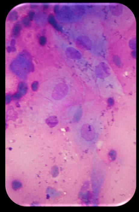
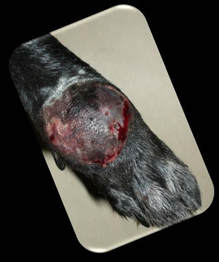
19 Cluster/single+ abundant cytoplasm Epithelial Cell Tumor vacuoles Squamous cell tumor group Epidermal inclusion cyst Squamous papilloma Squamous cell carcinoma Carcinoma SCC 19


20 Cluster/single+ abundant cytoplasm Epithelial Cell Tumor vacuoles Cluster/sheet/row Sebaceous cell tumor group Sebaceous adenoma Sebaceous epithelioma 20


21 Cluster/single+ abundant cytoplasm vacuoles glands Glandular cell tumor group Epithelial Cell Tumor Adenoma Adenocarcinoma Site of mass: mammary gland 21


22 Cluster/single+ abundant cytoplasm vacuoles glands Glandular cell tumor group Epithelial Cell Tumor Adenoma Adenocarcinoma Site of mass: Perianal gland 22
23 Conclusion: Epithelial Cell Tumor Sheet/ Solitary /Cluster Tumor involving superficial squamous epithelial cells = Benign - squamous epithelium (eg; papilloma) = Squamous cell carcinoma = Basal cells group = Epidermal basal cells (eg ; basal cell tumor, trichoblastoma, basal cell carcinoma) = benign hair follicular cells (eg; trichoepithelioma, pilomatricoma) Sebaceous cell group: -adenoma,-epithelioma,-carcinoma Glandular Pattern Perianal gland adenoma/adenocarcinoma Apocrine gland adenoma/adenocarcinoma Tumor of the subcutaneous glandular tissues (salivary, mammary, thyroid etc) Tumor of internal organs; pancreas, adrenal gl., lung etc. Un-differentiated carcinoma 23
24 Spindle Cell Tumor (Mesenchymal Origin) + Melanoma - Vacuoles Lipoma/-sarcoma Granules (brown, black, green) - - Secretion (Mucin) Extracellular matrix Myxoma/-sarcoma Chondrosarcoma, Osteosarcoma Fibroma/-sarcoma Hemangioma/-sarcoma Hemangiopericytoma + Giant cells + Whorls Neurofibrosarcoma Malignant Fibrous Histiocytoma + Giant cells 24

")

25 Spindle Cell Tumor (Mesenchymal Origin) Granules (brown, black, green) + Melanoma 25

")

 -")
26 Spindle Cell Tumor (Mesenchymal Origin) Granules (brown, black, green) - Vacuoles Lipoma/-sarcoma 26


27 Spindle Cell Tumor (Mesenchymal Origin) Granules (brown, black, green) - Extracellular matrix Chondrosarcoma, Osteosarcoma + Giant cells Osteosarcoma Chondrosarcoma 27
")
 - Extracellular")

28 Spindle Cell Tumor (Mesenchymal Origin) Granules (brown, black, green) - Extracellular matrix Fibroma/-sarcoma Hemangioma/-sarcoma Hemangiopericytoma + Whorls Fibrosarcoma 28

 Granules")
 -")

29 Spindle Cell Tumor (Mesenchymal Origin) Granules (brown, black, green) - Extracellular matrix Fibroma/-sarcoma Hemangioma/-sarcoma Hemangiopericytoma + Whorls Hemangiosarcoma 29

 Granules")
 -")

30 Spindle Cell Tumor (Mesenchymal Origin) Granules (brown, black, green) - Extracellular matrix Fibroma/-sarcoma Hemangioma/-sarcoma Hemangiopericytoma + Whorls Hemangiopericytoma 30
31 Conclusion: Spindle Cell Tumor Granulation tissue? Malignant melanoma Lipoma/Liposarcoma Osteoma/osteosarcoma Chondrosarcoma Myxoma & myxosarcoma Fibroma/Fibrosarcoma Hemangiopericytoma Hemangioma/Hemangiosarcoma melanin fat bone matrix, giant cells chondroid matrix mucin production spindle cell whorl endothelial, RBC Neurofibroma/neurofibrosarcoma Malignant fibrous histiocytoma giant cells, macrophage Undifferentiated sarcoma = Malignant mesenchymal cell tumor 31

 (-)")


32 Round Cell Tumor Granules (+) Granules (-) Red / purple Black Mastocytoma Melanocytoma 32 DIP QUIK Toluidine blue
")


33 Round Cell Tumor Granules (+) Granules (-) Red / purple Black Mastocytoma Melanocytoma Melanocytoma 33
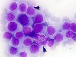
34 Round Cell Tumor < 50 % with plasma cells, neutrophils Granules (+) Dense nuclear chromatin Scant cytoplasm Lymphoid cell = Hyperplasia / Reactive Lymph node >50%, blast cells, multiple nucleoli = lymphoma Granule (-) Large size Histiocytic lymphoma Coarse nuclear chromatin Deep blue cytoplasm Vacuole(+/-) + - TVT Histiocyte, Basal cell Abundant cytoplasm With giant cells Medium size Moderate amount of cytoplasm Malignant histiocytosis Histiocytoma Eccentric nuclei (young dog) Cell-rich FNA Small size Scant cytoplasm Ribbon arrangement Basal cell tumor (middle-aged dog) 34

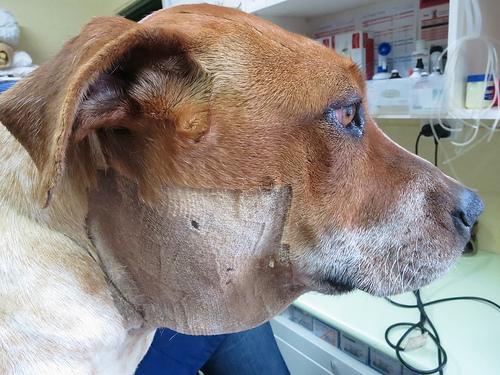
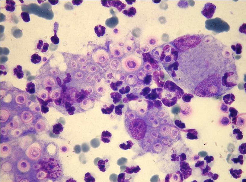
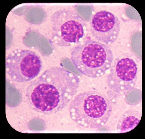
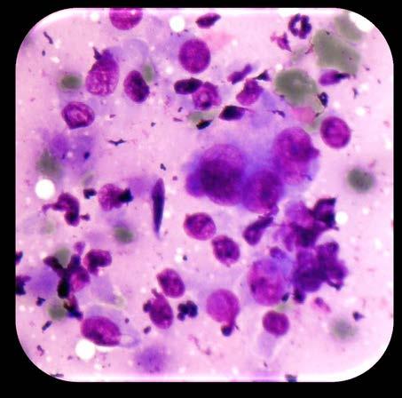
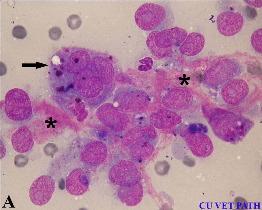
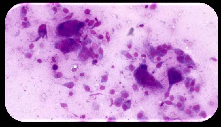
35 Mixed breed dog, 12 Y Lymphoma 35
 Cytology Lymphocytes (main) Plasma cells (found) Low lymphoblasts (normal) Low neutrophils Lymphoglandular bodies")
36 Lymphoma Lymphoglandular bodies Abnormal mitosis Ruptured cells Reactive lymph node Clinical signs gross lymphadenopathy (swollen LN) Cytology Lymphocytes (main) Plasma cells (found) Low lymphoblasts (normal) Low neutrophils Lymphoglandular bodies +/- 36

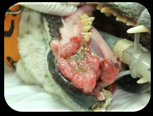
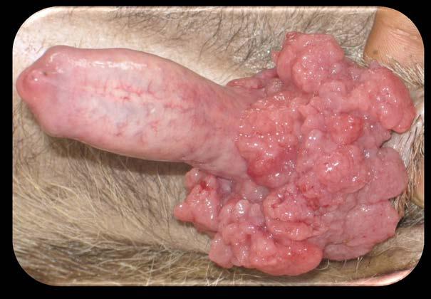
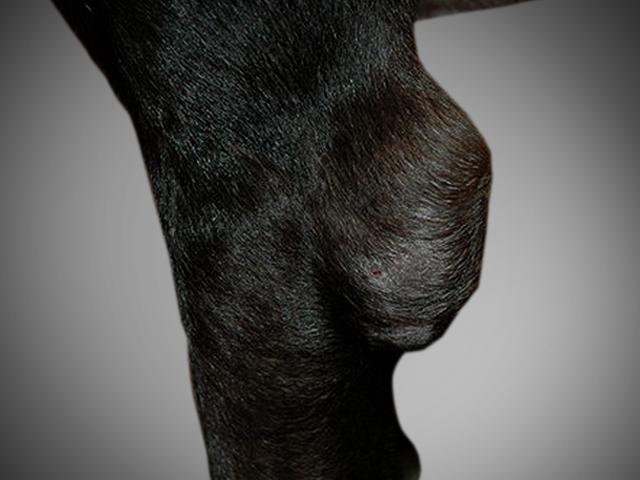
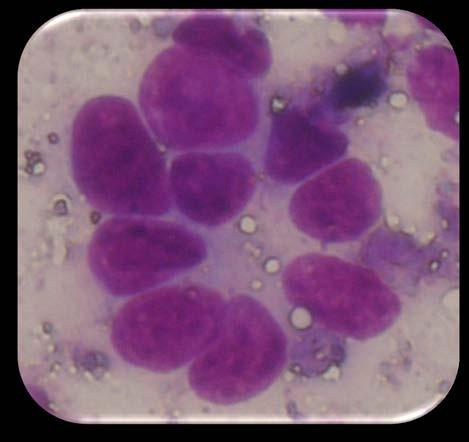
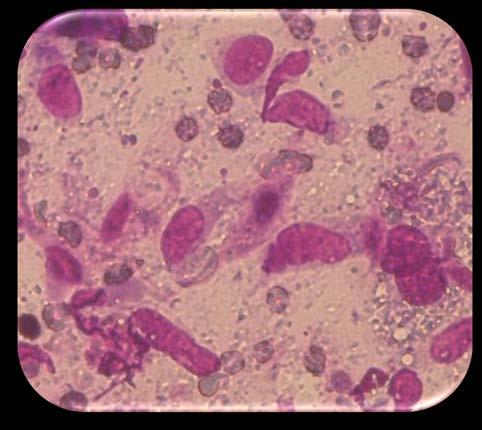
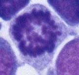
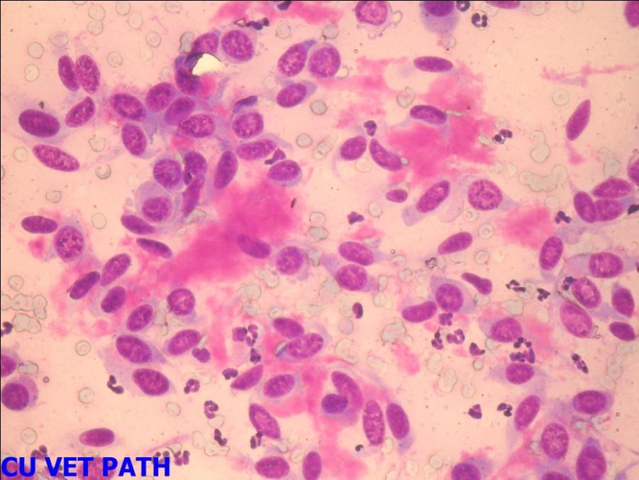
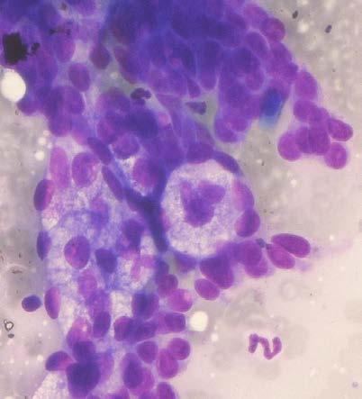
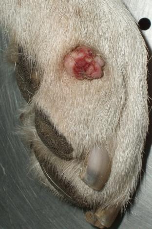
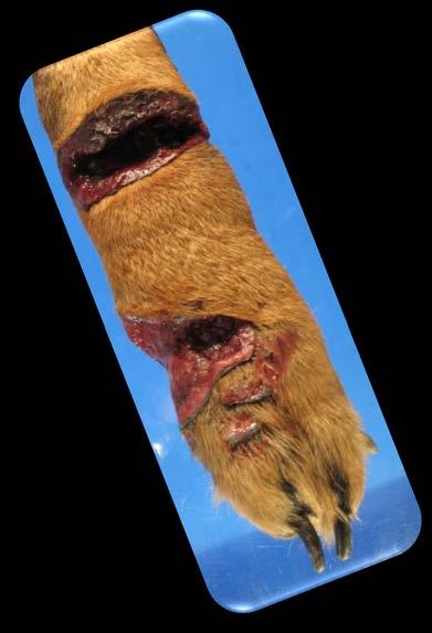
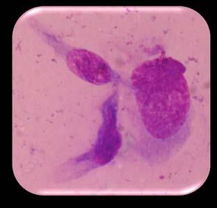
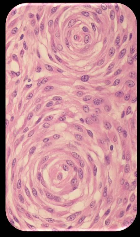
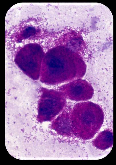
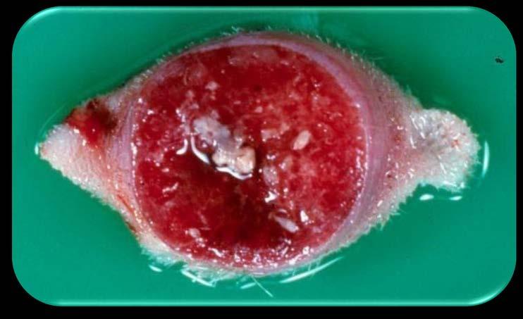
37 TVT (VG) / ETVT 37
 /")
38 38 TVT (VG) / ETVT 38
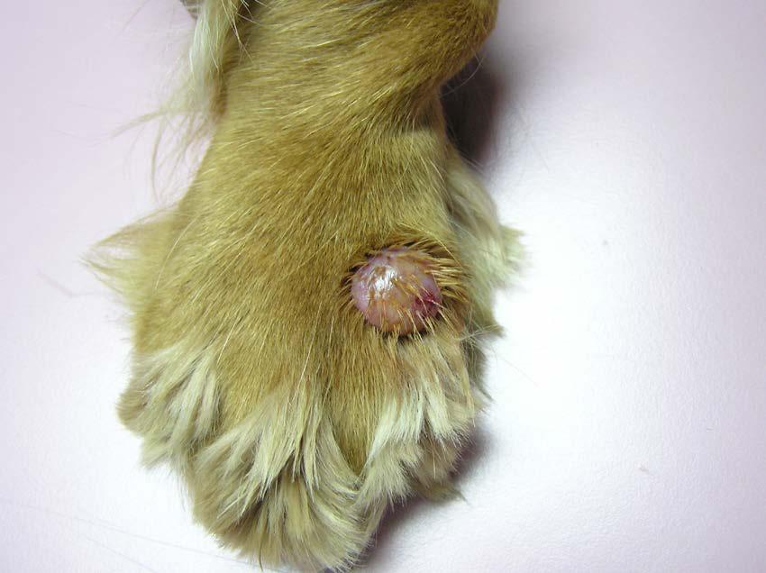
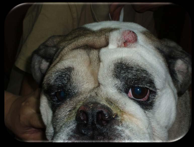
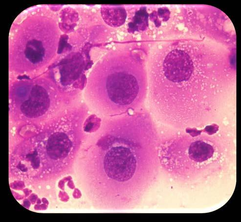
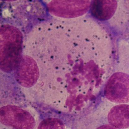
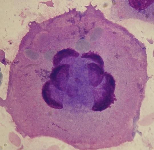
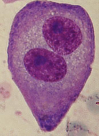
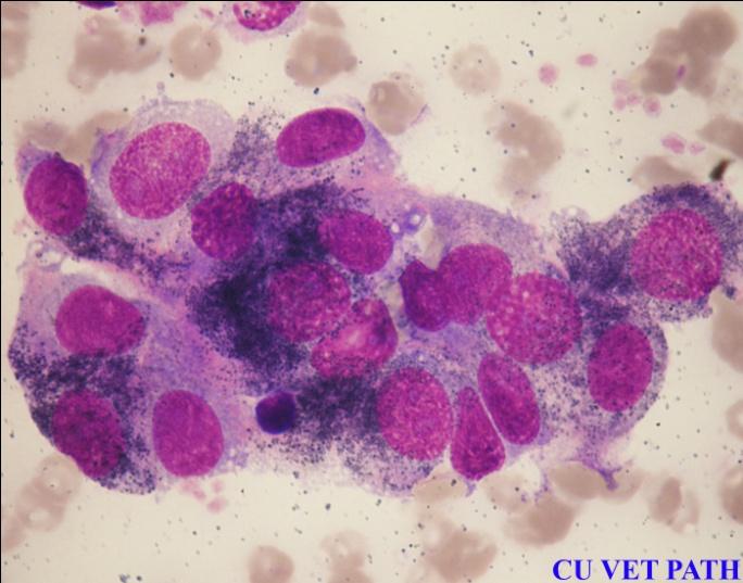
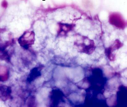
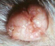
39 Histiocytoma 39

40 Histiocytoma Histiocytoma
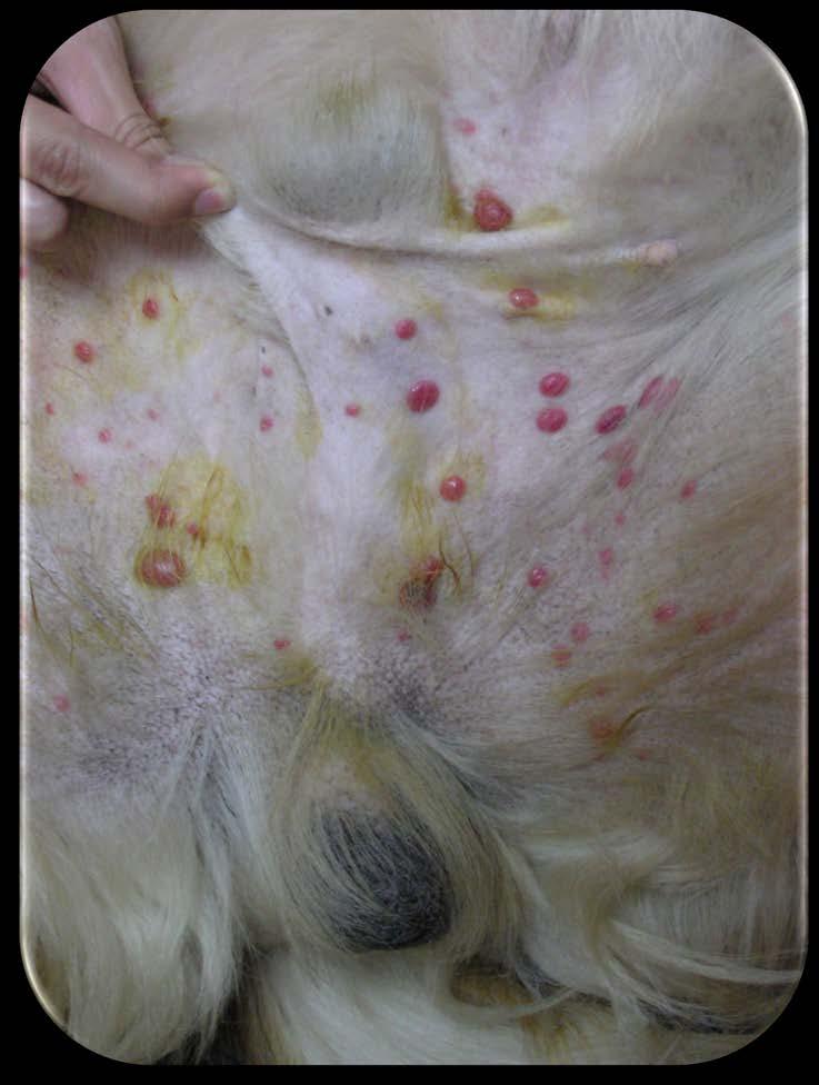
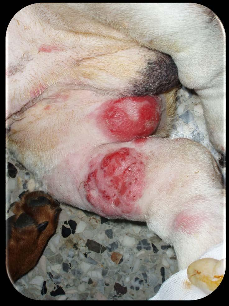
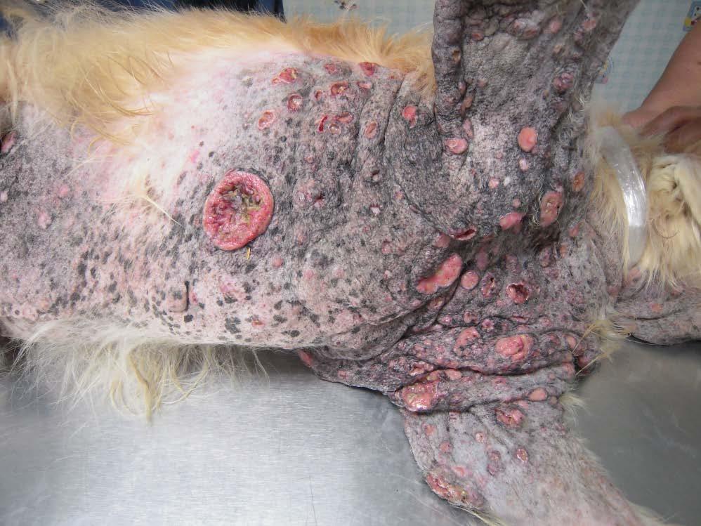
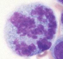
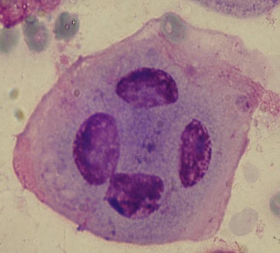
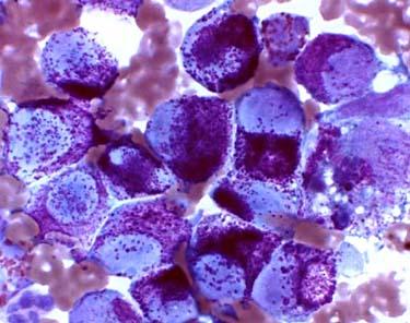
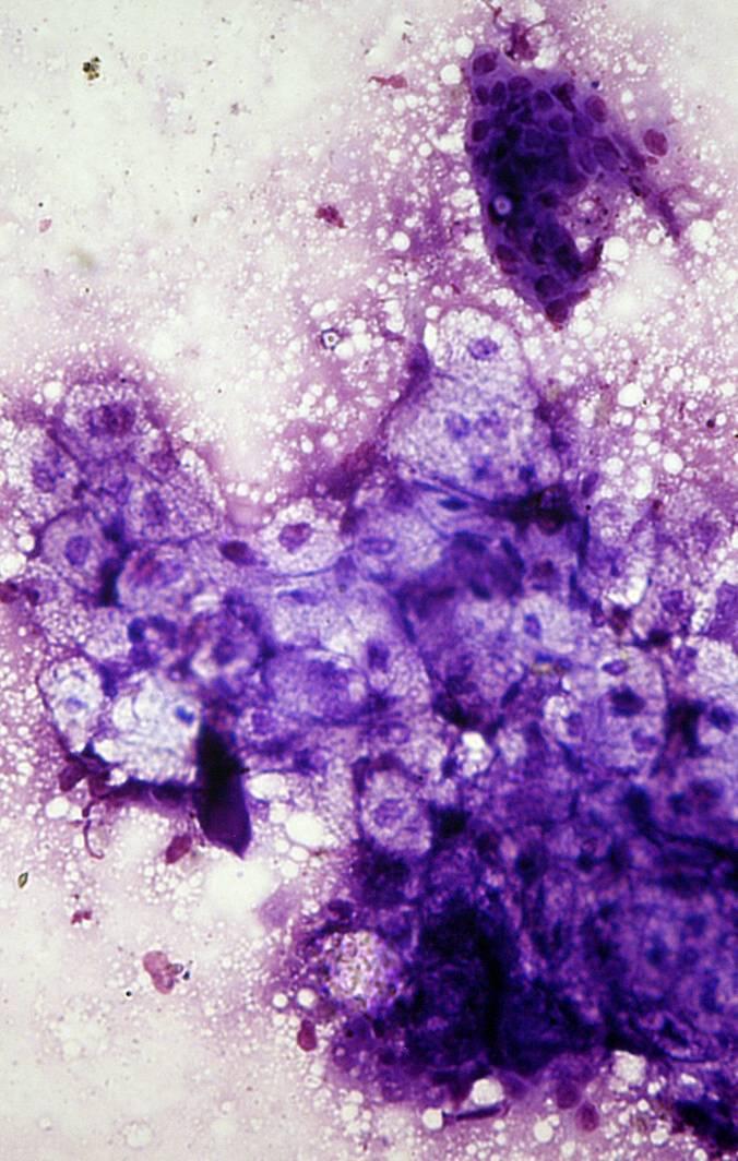
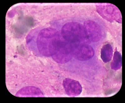
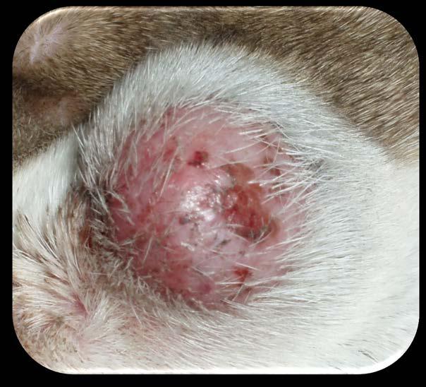
41 Malignant Histiocytosis 41
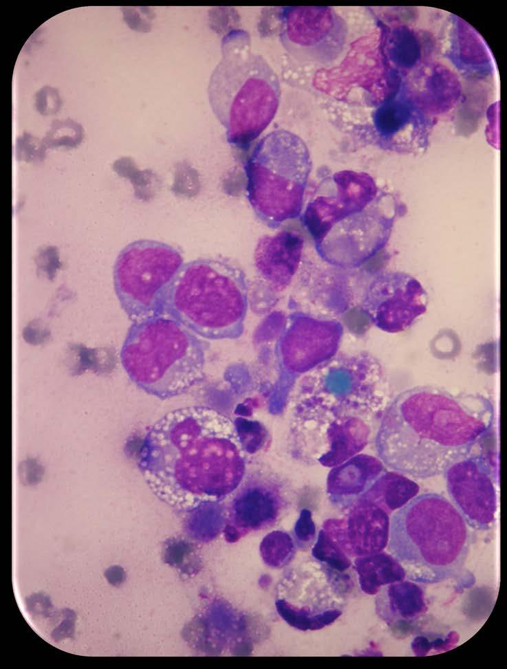
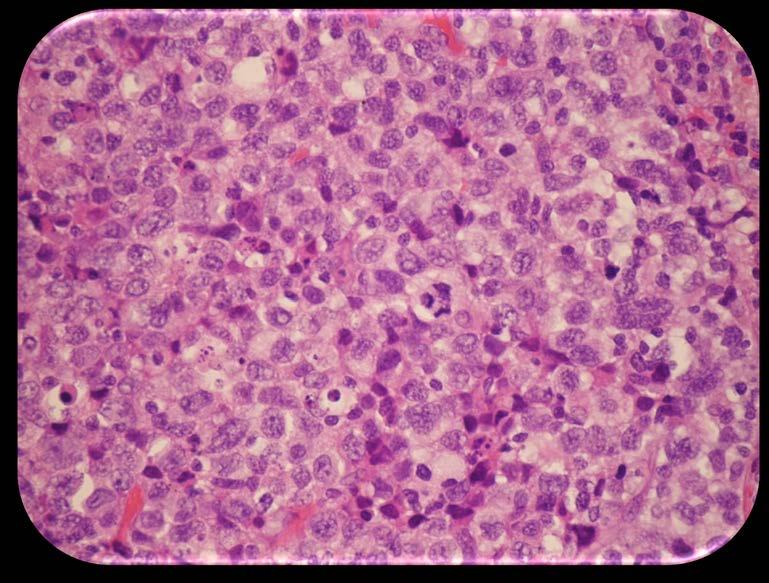
42 Malignant Histiocytosis 42


43 43
Submission of samples. Cytology of Lumps and Bumps. Evaluation of samples. Use caution interpreting. Criteria of malignancy.
 Submission of samples Cytology of Lumps and Bumps Paul Avery VMD, PhD, DACVP paul.avery@colostate.edu Air dry only No wet fixation using formalin or ethanol Stain 1-2 on-site to evaluate quality Send all
Almost any suspected tumor can be aspirated easily and safely. Some masses are more risky to aspirate including:
 DOES THIS PATIENT HAVE CANCER? USING IN-HOUSE CYTOLOGY TO HELP YOU MAKE THIS DIAGNOSIS. Joyce Obradovich, DVM, Diplomate, ACVIM (Oncology) Animal Cancer & Imaging Center, Canton, Michigan Almost every
Table of Contents. Preface xi. Acknowledgments xiii. Part I Overview of the Diagnostic Process 1. 1 Overview of Grading and Staging 3
 Table of Contents Preface xi Acknowledgments xiii Part I Overview of the Diagnostic Process 1 1 Overview of Grading and Staging 3 Identification of the process 3 Identification of tumor types 5 Grading
Proceeding of the SEVC Southern European Veterinary Conference
 www.ivis.org Proceeding of the SEVC Southern European Veterinary Conference Oct. 17-19, 2008 Barcelona, Spain http://www.sevc.info Reprinted in the IVIS website with the permission of the SEVC www.ivis.org
Cytology in Veterinary Practice: Sample Collection, Slide Preparation and Interpretation Guidelines
 Cytology in Veterinary Practice: Sample Collection, Slide Preparation and Interpretation Guidelines ASPIRATION OF A MASS A. Rick Alleman, DVM, PhD, DABVP, DACVP Lighthouse Veterinary Consultants, LLC Gainesville,
Cytology of NeoPlasia
 PEEr reviewed Cytology of NeoPlasia An Essential Component of Diagnosis Anne Barger, DVM, MS, Diplomate ACVP Cytology is a quick, easy, and inexpensive diagnostic tool. It is commonly used for the diagnosis
Proudly Presents: CLINICAL PATHOLOGY
 Chicago Veterinary Medical Association Shaping the Future of Veterinary Medicine - Promoting the Human-Animal Bond Proudly Presents: CLINICAL PATHOLOGY With: ANNE BARGER DVM, MS, DACVP April 13, 2016 Chicago
Salivary Gland Cytology
 Salivary Gland Cytology Diagnostic challenges and potential pitfalls Tarik M. Elsheikh, MD Professor and Medical Director Anatomic Pathology Cleveland Clinic FNA Salivary Gland Lesions Indications Distinguish
Clinical Cytology of Companion Animals: Part 2. Cytology of subcutaneous swellings, skin tumours and skin lesions
 GENERAL COMMISSIONED PAPER Clinical Cytology of Companion Animals: Part 2. Cytology of subcutaneous swellings, skin tumours and skin lesions E. Teske (1) INTRODUCTION Subcutaneous swellings, skin tumours,
E. Staging a WHO (World Health Organization) b TNM (Tumor Node Metastasis)
 b TNM (Tumor Node Metastasis)") I KNOWLEDGE LIST ONCOLOGY Knowledge of neoplasms in animals (dog, cat, pocket pets, horses, cows) A. Clinical signs, History, Physical Exam (PE) B. Work up 1. Diagnostic Imaging a Bone Scan b Thyroid Scan
I KNOWLEDGE LIST ONCOLOGY Knowledge of neoplasms in animals (dog, cat, pocket pets, horses, cows) A. Clinical signs, History, Physical Exam (PE) B. Work up 1. Diagnostic Imaging a Bone Scan b Thyroid Scan
Salivary Glands 3/7/2017
 Salivary Glands 3/7/2017 Goals and objectives Focus on the entities unique to H&N Common board type facts Information for your future practice Salivary Glands Salivary Glands Major gland. Paratid. Submandibular.
Cytology of Neoplasms that Occur on the Limbs Rick Alleman, DVM, PhD, DABVP, DACVP
 Cytology of Neoplasms that Occur on the Limbs Rick Alleman, DVM, PhD, DABVP, DACVP I. Introduction The purpose of this material is to provide information that may be useful in the identification of tumors
EPIDEMIOLOGICAL AND MORPHOLOGICAL CHARACTERISTICS OF CUTANEOUS ROUND CELL TUMORS DIAGNOSED USING ASPIRATIVE CYTOLOGY IN DOGS
 Scientific Works. Series C. Veterinary Medicine. Vol. LXIII (1) ISSN 2065-1295; ISSN 2343-9394 (CD-ROM); ISSN 2067-3663 (Online); ISSN-L 2065-1295 Abstract EPIDEMIOLOGICAL AND MORPHOLOGICAL CHARACTERISTICS
Grading of Bone Tumors
 Grading of Bone Tumors Joon Hyuk Choi, M.D. Department of Pathology College of Medicine, Yeungnam University Introduction to grading system of bone tumor used at Mayo Clinic WHO Histologic Classification
Case year female. Routine Pap smear
 Case 1 57 year female Routine Pap smear Diagnosis? 1. Atypical glandular cells of unknown significance (AGUS) 2. Endocervical AIS 3. Endocervical adenocarcinoma 4. Endometrial adenocarcinoma 5. Adenocarcinoma
Stating the obvious. The Whats, Wheres, and Hows of Treating Companion Animal Cancer. Lethargic. Steven Suter VMD PhD ACVIM (Oncology)
") The Whats, Wheres, and Hows of Treating Companion Animal Cancer Steven Suter VMD PhD ACVIM (Oncology) Stating the obvious Animals with cancer usually don t come to you with a diagnosis or a treatment plan.
The Whats, Wheres, and Hows of Treating Companion Animal Cancer Steven Suter VMD PhD ACVIM (Oncology) Stating the obvious Animals with cancer usually don t come to you with a diagnosis or a treatment plan.
XX. Tumours of the nasal cavity *
 XX. Tumours of the nasal cavity * H. STONZI 1 & B. HAUSER2 Tumours of the nasal cavity are rare in domestic animals, most cases occurring in the dog. Epithelial tumours are the most common type in carnivores
FNA OF SALIVARY GLANDS: A PRACTICAL APPROACH
 FNA OF SALIVARY GLANDS: A PRACTICAL APPROACH FNA of Salivary Glands: Challenges Wide range of neoplastic and non-neoplastic lesions Cytological overlap between the different benign and malignant tumors
Outline 11/2/2017. Pancreatic EUS-FNA general aspects. Cytomorphologic features of solid neoplasms/lesions of the pancreas
 ENDOSCOPIC ULTRASOUND GUIDED-FINE NEEDLE ASPIRATION CYTOLOGY OF PANCREAS Khalid Amin M.D. Assistant Professor Department of Laboratory Medicine and Pathology University of Minnesota Outline Pancreatic
Oncocytic-Appearing Salivary Gland Tumors. Oncocytic, Cystic, Mucinous, and High Grade Salivary Gland Tumors SALIVARY GLAND FNA: PART II
 William C. Faquin, MD, PhD Professor of Pathology Harvard Medical School Director of Head and Neck Pathology Massachusetts Eye and Ear Massachusetts General Hospital SALIVARY GLAND FNA: PART II Oncocytic,
LGM International, Inc.
 Liqui-PREP TM Cytology Atlas Preface The following pictures are examples with descriptions of cytology slides processed with the Liqui-PREP TM System.. The descriptions are reviewed by Pathologists. It
Pathology of the Integumentary System. Lecture 5 Endocrine / Miscellaneous / Neoplasia (web) Paul Hanna Fall 2017
 Paul Hanna Fall 2017") Pathology of the Integumentary System Lecture 5 Endocrine / Miscellaneous / Neoplasia (web) Paul Hanna Fall 2017 ENDOCRINE SKIN DISEASE hormones act on skin & other organs by modifying existing physiological
Pathology of the Integumentary System Lecture 5 Endocrine / Miscellaneous / Neoplasia (web) Paul Hanna Fall 2017 ENDOCRINE SKIN DISEASE hormones act on skin & other organs by modifying existing physiological
FNA of Thyroid. Toward a Uniform Terminology With Management Guidelines. NCI NCI Thyroid FNA State of the Science Conference
 FNA of Thyroid NCI NCI Thyroid FNA State of the Science Conference Toward a Uniform Terminology With Management Guidelines Thyroid Thyroid FNA Cytomorphology NCI Thyroid FNA State of the Science Conference
Charalampos Attipa, Rachel Hampel, Simon L. Priestnall, Kate English Pathobiology and Population Sciences, Royal Veterinary College, Hatfield, UK
 SUBCUTANEOUS MASS OVER THE SCAPULA IN A CAT Charalampos Attipa, Rachel Hampel, Simon L. Priestnall, Kate English Pathobiology and Population Sciences, Royal Veterinary College, Hatfield, UK Signalment:
PRELIMINARY CYTOLOGIC DIAGNOSIS: Suspicious for Acinic Cell Carcinoma. Cell Block: Immunohistochemical Studies CYTOLOGIC DIAGNOSIS:
 1 PRELIMINARY CYTOLOGIC DIAGNOSIS: Suspicious for Acinic Cell Carcinoma. Cell Block: Immunohistochemical Studies GCDFP-15 S-100 CYTOLOGIC DIAGNOSIS: Consistent with mammary analogue secretory carcinoma.
Objectives. Salivary Gland FNA: The Milan System. Role of Salivary Gland FNA 04/26/2018
 Salivary Gland FNA: The Milan System Dr. Jennifer Brainard Section Head Cytopathology Cleveland Clinic Objectives Introduce the Milan System for reporting salivary gland cytopathology Define cytologic
NEOPLASIA-I CANCER. Nam Deuk Kim, Ph.D.
 NEOPLASIA-I CANCER Nam Deuk Kim, Ph.D. 1 2 Tumor in the hieroglyphics of the Edwin Smith papyrus (1,600 B.C., Breasted s translation 1930) 3 War on Cancer (National Cancer Act, 1971) 4 Cancer Acts in Korea
Atlas of Eyelid and Conjunctival Tumors
 Atlas of Eyelid and Conjunctival Tumors Jerry A. Shields, M.D. Director, Ocular Oncology Service Wills Eye Hospital Professor of Ophthalmology Thomas Jefferson University Philadelphia, Pennsylvania Carol
A neoplasm is defined as "an abnormal tissue proliferation, which exceeds that of adjacent normal tissue. This proliferation continues even after
 NEOPLASIA Neoplasia is a very important topic in pathology because neoplasms are both common and serious diseases. A neoplasm literally means a new growth, and this term is used interchangeably with a
CYTOLOGY OF THE LIVER
 CYTOLOGY OF THE LIVER Maxey L. Wellman, DVM, PhD, DACVP (Clinical Pathology) Professor, Department of Veterinary Biosciences, College of Veterinary Medicine, The Ohio State University, Columbus, OH, USA
Prepared By Jocelyn Palao and Layla Faqih
 Prepared By Jocelyn Palao and Layla Faqih The structure of the suspected atypical cell should always be compared to the structure of other similar, benign, cells which are present in the smears. The diagnosis
From Morphology to Molecular Pathology: A Practical Approach for Cytopathologists Part 1-Cytomorphology. Songlin Zhang, MD, PhD LSUHSC-Shreveport
 From Morphology to Molecular Pathology: A Practical Approach for Cytopathologists Part 1-Cytomorphology Songlin Zhang, MD, PhD LSUHSC-Shreveport I have no Conflict of Interest. FNA on Lymphoproliferative
Presentation material is for education purposes only. All rights reserved URMC Radiology Page 1 of 98
 Presentation material is for education purposes only. All rights reserved. 2011 URMC Radiology Page 1 of 98 Radiology / Pathology Conference February 2011 Brooke Koltz, Cytopathology Resident Presentation
2015 Descriptive Vet Path Course. Histo Exam #3 KEY
 2015 Descriptive Vet Path Course Histo Exam #3 KEY Test 3, Slide 1 Tissue from a guinea pig. MORPHOLOGIC DIAGNOSIS: Heart: Multifocally and randomly (1 pt), within the left and right ventricular myocardium
Salivary gland tumor cytologic and histologic correlation: Algorithmic and risk stratification based approaches
 Salivary gland tumor cytologic and histologic correlation: Algorithmic and risk stratification based approaches Christopher C. Griffith, MD, PhD Raja R. Seethala, MD 1. Salivary gland tumor cytology: A
Cytology of Inflammatory Cutaneous lesions in the Dog and Cat
 Cytology of Inflammatory Cutaneous lesions in the Dog and Cat Rick L. Cowell, DVM, MS, MRCVS, Diplomate ACVP Clinical Pathologist IDEXX Laboratories Inc A. Cytologic Patterns of Inflammation: 1. Neutrophilic
Thyroid master class. Thyroid Fine needle aspiration cytology and liquid-based techniques: Hologic and Becton Dickinson
 Thyroid master class Thyroid Fine needle aspiration cytology and liquid-based techniques: Hologic and Becton Dickinson Principle of LBC Collection of cells in liquid medium Immediate fixation Processor-prepared
number Done by Corrected by Doctor Maha Shomaf
 number 16 Done by Waseem Abo-Obeida Corrected by Zeina Assaf Doctor Maha Shomaf MALIGNANT NEOPLASMS The four fundamental features by which benign and malignant tumors can be distinguished are: 1- differentiation
Gross appearance of nodular hyperplasia in material obtained from suprapubic prostatectomy. Note the multinodular appearance and the admixture of
 Tiền liệt tuyến Tiền liệt tuyến Gross appearance of nodular hyperplasia in material obtained from suprapubic prostatectomy. Note the multinodular appearance and the admixture of solid and microcystic areas.
ARIZONA SOCIETY OF PATHOLOGISTS 13 TH APRIL 2013 HEAD AND NECK CYTOPATHOLOGY. F ZAHRA ALY, MD, PhD
 ARIZONA SOCIETY OF PATHOLOGISTS 13 TH APRIL 2013 HEAD AND NECK CYTOPATHOLOGY F ZAHRA ALY, MD, PhD The main areas sites amenable for cytopathology include lymph nodes, thyroid, major salivary glands especially
Ascitic Fluid and Use of Immunocytochemistry. Mercè Jordà, University of Miami
 Ascitic Fluid and Use of Immunocytochemistry Mercè Jordà, University of Miami Is It Malignant? Yes? No Ascitic Fluid Cytomorphologic Useful Findings Tight clusters with smooth borders Cellular and nuclear
Contents. Basic Ultrasound Principles and Terminology. Ultrasound Nodule Characteristics
 Contents Basic Ultrasound Principles and Terminology Basic Ultrasound Principles... 1 Ultrasound System... 2 Linear Transducer for Superficial Images and Ultrasound-Guided FNA... 3 Scanning Planes... 4
EDUCATIONAL COMMENTARY DIFFERENTIATING IMMATURE PERIPHERAL BLOOD CELLS
 Educational commentary is provided through our affiliation with the American Society for Clinical Pathology (ASCP). To obtain FREE CME/CMLE credits click on Continuing Education on the left side of the
HISTOPATHOLOGY. Shannon Martinson
 HISTOPATHOLOGY Shannon Martinson March 2013 Case #1 History: 8 year old beagle Neck pain for the past couple of weeks Paresis, followed by paralysis developed over the past few days Gross Description courtesy
, , 2011 HODGKIN LYMPHOMA
 European Federation of Cytology Societies 4tu Annual Tutorial in Cytopathology Trieste, June 6-10, 2011 HODGKIN LYMPHOMA Classification The World Health Organization Classification of Lymphomas (2001)
Spontaneous Neoplastic Lesions in the Crl:CD-1 (ICR)BR Mouse. March, 2000
BR Mouse. March, 2000") Spontaneous Neoplastic Lesions in the Crl:CD-1 (ICR)BR Mouse March, 2000 Information Prepared by Mary L. A. Giknis, Ph.D. Charles B. Clifford, D.V.M., Ph.D. CHARLES RIVER LABORATORIES TABLE OF CONTENTS
Spontaneous Neoplastic Lesions in the Crl:CD-1 (ICR)BR Mouse March, 2000 Information Prepared by Mary L. A. Giknis, Ph.D. Charles B. Clifford, D.V.M., Ph.D. CHARLES RIVER LABORATORIES TABLE OF CONTENTS
Descriptive Cytology. Agenda. Overview. Limitations. Advantages 6/3/2014
 Descriptive Cytology Brian W. Smith, DVM, DACVP JPC, Veterinary Pathology Service Silver Spring, Maryland Shannon Lacy Jeremy Bearss Sarah Hale Don Meuten Edward Stevens Bruce Williams Descriptive Veterinary
A case of giant cell tumour of soft parts in a horse Francesco Cian 1, Sarah Whiteoak 2, Jennifer Stewart 1
 A case of giant cell tumour of soft parts in a horse Francesco Cian 1, Sarah Whiteoak 2, Jennifer Stewart 1 1 Animal Health Trust, Newmarket, UK 2 608 Equine and Farm Vets, Rowington, UK Signalment: Horse,
SESSION 1: GENERAL (BASIC) PATHOLOGY CONCEPTS Thursday, October 16, :30am - 11:30am FACULTY COPY
 PATHOLOGY CONCEPTS Thursday, October 16, :30am - 11:30am FACULTY COPY") SESSION 1: GENERAL (BASIC) PATHOLOGY CONCEPTS Thursday, October 16, 2008 9:30am - 11:30am FACULTY COPY GOAL: Describe the basic morphologic (structural) changes which occur in various pathologic conditions.
SESSION 1: GENERAL (BASIC) PATHOLOGY CONCEPTS Thursday, October 16, 2008 9:30am - 11:30am FACULTY COPY GOAL: Describe the basic morphologic (structural) changes which occur in various pathologic conditions.
Spontaneous Neoplastic Lesions in the CrI:CD-1(ICR) Mouse in Control Groups from 18 Month to 2 year Studies. March, 2005
 Mouse in Control Groups from 18 Month to 2 year Studies. March, 2005") Spontaneous Neoplastic Lesions in the CrI:CD-1(ICR) Mouse in Control Groups from 18 Month to 2 year Studies March, 2005 Information Prepared by Mary L.A. Giknis Ph.D Charles B. Clifford D.V.M, Ph.D 1063
Spontaneous Neoplastic Lesions in the CrI:CD-1(ICR) Mouse in Control Groups from 18 Month to 2 year Studies March, 2005 Information Prepared by Mary L.A. Giknis Ph.D Charles B. Clifford D.V.M, Ph.D 1063
Special slide seminar
 Special slide seminar Tomáš Rozkoš The Fingerland Department of Pathology Charles University Medical Faculty and Faculty Hospital in Hradec Králové Czech Republic Case history, 33 years old resistance
4/17/2015. Case 1. A 37 year old man with a 2.2 cm solitary left thyroid mass.
 Case 1 A 37 year old man with a 2.2 cm solitary left thyroid mass. Case 1 Case 1 1 Case 1: Diagnosis? A. Benign B. Atypia of undetermined significance/follicular lesion of undetermined significance C.
Neoplasia literally means "new growth.
 NEOPLASIA Neoplasia literally means "new growth. A neoplasm, defined as "an abnormal mass of tissue the growth of which exceeds and is uncoordinated with that of the normal tissues and persists in the
PREVALENCE OF CANINE EPITHELIAL, MELANOCYTIC AND MESENCHYMAL TUMOURS OF THE SKIN AND SOFT TISSUES: A 10-YEAR STUDY
 Bulgarian Journal of Veterinary Medicine (2011), 14, No 3, 171 178 PREVALENCE OF CANINE EPITHELIAL, MELANOCYTIC AND MESENCHYMAL TUMOURS OF THE SKIN AND SOFT TISSUES: A 10-YEAR STUDY Summary R. SIMEONOV
Cytyc Corporation - Case Presentation Archive - March 2002
 FirstCyte Ductal Lavage History: 68 Year Old Female Gail Index: Unknown Clinical History: Negative Mammogram in 1995 6 yrs. later presents with bloody nipple discharge Subsequent suspicious mammogram Suspicious
PLEOMORPHIC ADENOMA ( BENIGN MIXED TUMOR )
") ( BENIGN MIXED TUMOR ) Grossly, the tumor is freely movable, solid, sometimes lobulated and occasionally cystic. If recurrent, multinodular masses are common. Histologically, within a fibrous capsule,
( BENIGN MIXED TUMOR ) Grossly, the tumor is freely movable, solid, sometimes lobulated and occasionally cystic. If recurrent, multinodular masses are common. Histologically, within a fibrous capsule,
Case 1. Slide 1 History: 65 year old male presents with bilateral pleural effusions, a 40 pack year smoking history and peripheral and hilar lung
 Case 1. Slide 1 History: 65 year old male presents with bilateral pleural effusions, a 40 pack year smoking history and peripheral and hilar lung masses. Specimen shown is from a tap of the pleural effusion.
Histology Review Can you identify the Cell Structures? Can you identify the Stain? Can you identify the Cell type?
 Histology Review Can you identify the Cell Structures? Can you identify the Stain? Can you identify the Cell type? 2.01 Border of Epithelia (fluorescence) M A: lamina basalis B: epithelium C: other tissues
Sheet #7. Dr. Heba Kalbouneh. Dr. Heba Kalbouneh. Dr. Heba Kalbouneh
 Sheet #7 Dr. Heba Kalbouneh Dr. Heba Kalbouneh Dr. Heba Kalbouneh Connective tissue The differences between epithelial and connective tissue - Epithelial cells are tightly packed (no or minimal spaces
2008 Gross Ocular Pathology. Gross Pathology 2
 2008 Gross Ocular Pathology Gross Pathology 2 08rd1281 Feline T-Cell Lymphoma 08rd1300 Canine Iridociliary Adenoma Foam Cell Variant 08rd1331 Feline Feline Iridociliary Adenoma 08rd1340 Canine Retinal
Pancreatitis: A Potential Pitfall in Endoscopic Ultrasound Guided Pancreatic FNA
 Pancreatitis: A Potential Pitfall in Endoscopic Ultrasound Guided Pancreatic FNA Jack Yang, MD Department of Pathology, Medical University of South Carolina Objectives Understand the indication of EUS
Objectives. Atypical Glandular Cells. Atypical Endocervical Cells. Reactive Endocervical Cells
 2013 California Society of Pathologists 66 th Annual Meeting San Francisco, CA Atypical Glandular Cells to Early Invasive Adenocarcinoma: Cervical Cytology and Histology Christina S. Kong, MD Associate
LYMPH GLAND. By : Group 1
 LYMPH GLAND By : Group 1 ANATOMY LYMPH NODE Lymphatic Organs Red bone marrow Thymus gland Lymph nodes Lymph nodules Spleen Primary organs Secondary organs Lymph Nodes Firm, smooth-surfaced, bean-shaped
Abid Irshad, MD Director Breast Imaging. Medical University of South Carolina Charleston
 Abid Irshad, MD Director Breast Imaging Medical University of South Carolina Charleston Cases Financial disclosure: I or my family have no financial interest related to the material discussed in this presentation
Effusion Cytology: Diagnostic Challenges
 Effusion Cytology: Diagnostic Challenges Tarik M. Elsheikh, MD Professor and Medical Director, Anatomic Pathology Cleveland Clinic Outside Consult Case 45 year old woman, presented with nausea, dyspnea,
Principles of Surgical Oncology. Winnie Achilles Tierklinik Hollabrunn Lastenstrasse Hollabrunn
 Principles of Surgical Oncology Winnie Achilles Tierklinik Hollabrunn Lastenstrasse 2 2020 Hollabrunn boexi@gmx.de The first surgery provides the best chance for a cure in an animal with a tumor Clinical
Disorders of Cell Growth & Neoplasia. Histopathology Lab
 Disorders of Cell Growth & Neoplasia Histopathology Lab Paul Hanna April 2010 Case #84 Clinical History: 5 yr-old, West Highland White terrier. skin mass from axillary region. has been present for the
Charles Halsey, DVM, PhD, DACVP Pfizer, Inc. IHC Resources
 Charles Halsey, DVM, PhD, DACVP Pfizer, Inc. IHC Resources 1 IHC Identification Targets Specimens Controls 2 Tissue controls Trouble Spots 3 The Key to Description IHC Description 4 Intermediate Filaments
DETERMINATION OF A LYMPHOID PROCESS
 Chapter 2 Applications of Touch Preparation Cytology to Intraoperative Consultations: Lymph Nodes and Extranodal Tissues for Evaluation of Hematolymphoid Disorders INTRODUCTION As discussed in Chap. 1,
Neoplasia 2018 Lecture 2. Dr Heyam Awad MD, FRCPath
 Neoplasia 2018 Lecture 2 Dr Heyam Awad MD, FRCPath ILOS 1. List the differences between benign and malignant tumors. 2. Recognize the histological features of malignancy. 3. Define dysplasia and understand
Review of the AP Part II Practical Examination. Dr David Clift Co Chief Examiner
 Review of the AP Part II Practical Examination Dr David Clift Co Chief Examiner General Remarks The part II practical examination involved 15 cases which were presented with sufficient clinical data to
CASE REPORT SOLITARY SEBACEOUS NEVUS OF JADASSOHN COMPLICATED BY SQUAMOUS CELL CARCINOMA AND BASAL CELL CARCINOMA
 CASE REPORT Dennis H. Kraus, MD, Section Editor SOLITARY SEBACEOUS NEVUS OF JADASSOHN COMPLICATED BY SQUAMOUS CELL CARCINOMA AND BASAL CELL CARCINOMA Ahmad Ridzwan Arshad, FRCS, 1 Wan S. Azman, MS, 1 Ayadurai
C.L. Davis Foundation Descriptive Veterinary Pathology Course
 C.L. Davis Foundation 2015 Descriptive Veterinary Pathology Course IHC Resources IHC Identification Targets Antibodies Antibodies 1 Antibodies Specimens Antigen Retrieval Unmasks antigen epitopes Methods
Pathology of the Hematopoietic System GROSS/HISTO LAB
 Pathology of the Hematopoietic System GROSS/HISTO LAB Paul Hanna (thanks to Dr s Aburto, Martinson & Fenton) Fall 2014 Slide 1 Spleen from a Beaver Give a morphologic diagnosis and possible etiology &
Cytologic Evaluation of the Enlarged Neck Node: FNAC Utility in Metastatic Neck Disease
 ISPUB.COM The Internet Journal of Pathology Volume 6 Number 2 Cytologic Evaluation of the Enlarged Neck Node: FNAC Utility in Metastatic Neck Disease I Bagwan, S Kane, R Chinoy Citation I Bagwan, S Kane,
Educational Cases EQA November T.J. Palmer Raigmore Hospital Inverness
 Educational Cases EQA November 2013 T.J. Palmer Raigmore Hospital Inverness Case 2 Clinical Details Dob 11 February 1951 PMH: 1964 Extraction of 45 aet 13 yr 1966 Cyst between 44 and 46 enucleated 1973
CINtec p16 INK4a Staining Atlas
 CINtec p16 INK4a Staining Atlas Rating Rating Positive The rating positive will be assigned if the p16 INK4a -stained slide shows a continuous staining of cells of the basal and parabasal cell layers of
A Look at the Lymphoid System Rose Raskin, DVM, PhD, DACVP Purdue University West Lafayette, IN
 A Look at the Lymphoid System Rose Raskin, DVM, PhD, DACVP Purdue University West Lafayette, IN The lymphoid organs commonly biopsied include the peripheral and internal lymph nodes, spleen, and occasionally
By Dr. Mohamed Saad Daoud
 By Dr. Mohamed Saad Daoud Part I Introduction Types of White Blood Cells Genesis of the White Blood Cells Life Span of the White Blood Cells Dr. Mohamed Saad Daoud 2 Leucocytes Introduction: Infectious
Medullary Thyroid Carcinoma. This case was provided by Treant Hospital, Bethesda, Hoogeveen, The Netherlands
 Medullary Thyroid Carcinoma This case was provided by Treant Hospital, Bethesda, Hoogeveen, The Netherlands ADS-01504 Rev. 001 2016 Hologic, Inc. All rights reserved. Overview Medullary Thyroid Carcinoma
QUALITY ASSURANCE PROGRAM CYTOLOGY CYCLE 01/2018 (TRIAL)
") [Pick the Date] FINAL REPORT QUALITY ASSURANCE PROGRAM CYTOLOGY CYCLE 01/2018 (TRIAL) NOTES FROM THE COORDINATOR 1. For this cycle 01/2018, a total of 32 pen drives had been circulated. Twenty-eight institutions
[Pick the Date] FINAL REPORT QUALITY ASSURANCE PROGRAM CYTOLOGY CYCLE 01/2018 (TRIAL) NOTES FROM THE COORDINATOR 1. For this cycle 01/2018, a total of 32 pen drives had been circulated. Twenty-eight institutions
Disclosures. Parathyroid Pathology. Objectives. The normal parathyroid 11/10/2012
 Disclosures Parathyroid Pathology I have nothing to disclose Annemieke van Zante MD/PhD Assistant Professor of Clinical Pathology Associate Chief of Cytopathology Objectives 1. Review the pathologic features
Histopathology: Cervical HPV and neoplasia
 Histopathology: Cervical HPV and neoplasia These presentations are to help you identify basic histopathological features. They do not contain the additional factual information that you need to learn about
Benign and malignant epithelial lesions: Seborrheic keratosis: A common benign pigmented epidermal tumor occur in middle-aged or older persons more
 Benign and malignant epithelial lesions: Seborrheic keratosis: A common benign pigmented epidermal tumor occur in middle-aged or older persons more common on the trunk; but extremities, head and neck are
Respiratory Tract Cytology
 Respiratory Tract Cytology 40 th European Congress of Cytology Liverpool, UK Momin T. Siddiqui M.D. Professor of Pathology and Laboratory Medicine Director of Cytopathology Emory University Hospital, Atlanta,
Histopathology: skin pathology
 Histopathology: skin pathology These presentations are to help you identify, and to test yourself on identifying, basic histopathological features. They do not contain the additional factual information
Histopathology: chronic inflammation
 Histopathology: chronic inflammation These presentations are to help you identify, and to test yourself on identifying, basic histopathological features. They do not contain the additional factual information
EQA Circulation 43 Educational Cases
 EQA Circulation 43 Educational Cases E1-E2 Monica Agarwal Monklands Hospital E1 38 yrs male Submandibular gland tumour E1 Formal excision following diagnosis of poorly differentiated carcinoma on core
Ben Witt, MD University of Utah/ARUP Laboratories Assistant Professor of Anatomic Pathology
 Ben Witt, MD University of Utah/ARUP Laboratories Assistant Professor of Anatomic Pathology Review some of the more common cytodiagnoses of the Head and Neck Establish an approach to some of the diagnostic
40th European Congress of Cytology Liverpool, UK, 2-5 th October 2016
 40th European Congress of Cytology Liverpool, UK, 2-5 th October 2016 EUS FNA of abdominal organs: An approach to reporting and triage for ancillary testing Date and time: Sunday 2 nd October 2016 15.00-16.30
Spindle Cell Lesions Of The Breast. Emad Rakha Professor of Breast Pathology and Consultant Pathologist
 Spindle Cell Lesions Of The Breast Emad Rakha Professor of Breast Pathology and Consultant Pathologist * SCLs comprise a wide spectrum of diseases, ranging from reactive processes to aggressive malignant
NEOPLASIA. 3. Which of the following tumour is benign a. Chondrosarcoma b. Osteochondroma c. Chondroblastoma d. Ewing s tumour e.
 NEOPLASIA 1. malignant neoplasms a. are independent of hormonal influence b. are always composed of homogenous cell lines c. arise from differentiated cells by a process of anaplasia d. display abnormal
Research Article Transmucosal Fine Needle Aspiration of Oral and Pharyngeal Lesions
 International Scholarly Research Network ISRN Pathology Volume 2011, Article ID 267145, 7 pages doi:10.5402/2011/267145 Research Article Transmucosal Fine Needle Aspiration of Oral and Pharyngeal Lesions
Clinical Pathology. Mary Anna Thrall, DVM, MS, DACVP. Sponsored by
 Clinical Pathology by Mary Anna Thrall, DVM, MS, DACVP September 10, 2008 Sponsored by 1 CYTOLOGY: WHEN TO SEND YOUR SAMPLES TO A PATHOLOGIST, AND WHEN TO DO IT YOURSELF Mary Anna Thrall Colorado State
SHN-1 Human Digestive Panel Test results
 SHN-1 Human Digestive Panel Test results HN-30 tongue HN-24 salivary gland HN-12 larynx HN-28 esophagus HN-29 stomach HN-20 pancreas HN-13 liver HN-14 gall bladder HN-27-1 duodenum HN-27-2 ileum HN-27-3
Selected Pseudomalignant Soft Tissue Tumors of the Skin and Subcutis
 Selected Pseudomalignant Soft Tissue Tumors of the Skin and Subcutis Andrew L. Folpe, M.D. Professor of Laboratory Medicine and Pathology Mayo Clinic, Rochester, MN folpe.andrew@mayo.edu 2016 MFMER slide-1
BOSNIAN-TURKISH CYTOPATHOLOGY SCHOOL June 18-19, 2016 Sarajevo. Case Discussions. 60 year old woman Routine gynecologic control LBC
 BOSNIAN-TURKISH CYTOPATHOLOGY SCHOOL June 18-19, 2016 Sarajevo Case Discussions Prof Dr Sıtkı Tuzlalı Tuzlalı Pathology Laboratory 60 year old woman Routine gynecologic control LBC 1 2 Endometrial thickening
Problem diagnoses. Current issues in Anatomic pathology. Problem Diagnoses in Tumors of the Oral Cavity 5/29/2009
 Current issues in Anatomic pathology Problem Diagnoses in Tumors of the Oral Cavity Richard Jordan DDS PhD FRCPath Professor of Oral Pathology & Pathology Director, UCSF Oral Pathology Diagnostic Laboratory
CONNECTIVE TISSUE (C.T.)
") CONNECTIVE TISSUE (C.T.) Objectives: By the end of this lecture, the student should be able to: 1. Enumerate the general characteristics of C.T. 2. Classify C.T into C.T. proper and special types of C.T.
CONNECTIVE TISSUE (C.T.) Objectives: By the end of this lecture, the student should be able to: 1. Enumerate the general characteristics of C.T. 2. Classify C.T into C.T. proper and special types of C.T.
Salivary gland cytology. Salivary gland cytology. Triage helps the clinician. Salivary gland tumors. Diagnostic difficulties
 Salivary gland cytology Salivary Gland Cytology Pınar Fırat, MD Professor of Pathology İ.U. İstanbul Faculty of Medicine Çapa, İstanbul It is a reliable diagnostic test However, definitive subclassification