Prof. Dr. NAGUI M. ABDELWAHAB,M.D.; MARYSE Y. AWADALLAH, M.D. AYA M. BASSAM, Ms.C.
|
|
|
- Eustace Riley
- 5 years ago
- Views:
Transcription


1 Role of Whole-body Diffusion MR in Detection of Metastatic lesions Prof. Dr. NAGUI M. ABDELWAHAB,M.D.; MARYSE Y. AWADALLAH, M.D. AYA M. BASSAM, Ms.C.

2
3 Cancer is a potentially life-threatening disease, and the development of skeletal or organ metastases is often not detected until clinical symptoms present. That is why radiological imaging such as plain film X- ray, Tc99m bone scintigraphy, computed tomography (CT), positron emission tomography (PET), magnetic resonance imaging (MRI), are the cornerstone for :- Detection and staging of metastatic lesions. Classifying their type, site and extent. Skeletal scintigraphy is the standard procedure for visualizing bone metastases, (50-70% of distant metstases).
4 DWI is a powerful imaging tool that provides unique information related to tumor cellularity, integrity of the cellular membrane, as well as the movement and functional status of the microenvironment of water in tissue. The technique can be applied widely for tumor detection, tumor characterization and monitoring of tumor response. Until recently, most sensitive radiological procedures were only limited to local coverage and could not interrogate the full body.
5 Diffusion-weighted sequence (DWI) of the entire body is a new promising technique feasible to evaluate multifocal disease. Technological advances and the development of the concept of diffusion-weighted whole-body imaging with background body signal suppression (DWIBS) have opened the path for routine clinical whole-body DWI, which allows detection and characterization of both oncological and non-oncological lesions throughout the entire body.
6 Several studies stated that; whole body magnetic resonance imaging (WB-MRI) has shown better results than skeletal scintigraphy. Practical implementation of whole body DWI, using the DWIBS concept, is relatively easy, since it can be performed on most modern MRI scanners and does not require any contrast agent administration. Furthermore, compared to SPECT/CT and PET CT, MRI scanners are more widely available and whole body DWI is less expensive.
7
8 To evaluate the value of WB DWI in detection of metastatic lesions, using bone scintigraphy for comparison.
9
10 Fifteen patients (5 males and 10 females), referred to the Radiology department of Kasr Al-Ainy Hospitals from the outpatient clinic of the Clinical Oncology department, their ages ranged between 34 to70 years, with known primary malignant tumor (confirmed histologically) underwent :- Whole body MRI including Diffusion weighted Whole body Imaging with Back ground body signal Suppression (DWIBS), Skeletal scintigraphy. Any other modality to detect extra-osseous metastases as computed tomography and ultrasound.
11 WB DWI Qualitative analysis was performed directly from the reformatted Malignant lesions images generally in the coronal exhibit considerably plane. Also, greater the source signal images intensities of DWI and were variability revised when on their required. profile than The benign lesions ones. were only categorized according to the subjectively WB T1-weighted, rated T2-weighted signal pattern, and signal STIR images intensity were and also morphology evaluated to without combine taking information, into account to accurately the apparent detect diffusion pathology coefficients, and rule out which artifacts were from not quantified. the diffusion-weighted sequence series.
12 The skeletal scintigrams were also analyzed; the increased uptake pattern of the identified lesions was rated according to the clinical experience. No quantitative measurements were considered.
13
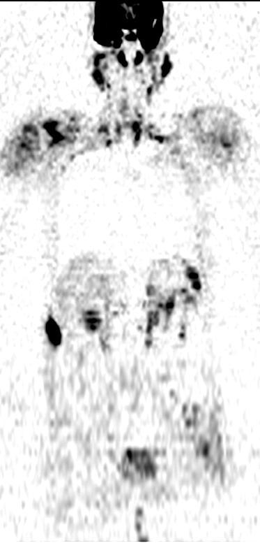
 Prostatic carcinoma(13.")
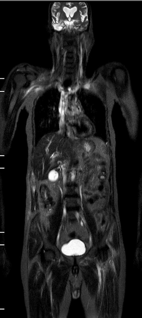
 Bronchogenic carcinoma(6.")
14 RESULTS Breast carcinoma (53.3%) Prostatic carcinoma(13.3%) Bladder carcinoma(13.3%) Bronchogenic carcinoma(6.7%) Thyroid lymphoma(6.7%) Endometrial carcinoma(6.7%)
15
16 RESULTS Also, it showed the bony lesions detected by bone scan in the following sites: vertebra, sternum, femur, acetabulum, humerus, scapula, tibia, shoulder, pelvic bone, greater trochanter, clavicle, coracoid process & sacrum.

17 On the other hand, one rib lesion, lesions in skull & lateral condyle of femur were missed.
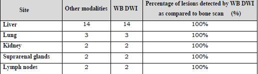
18 RESULTS The sensitivity of WB DWI in detecting metastatic bony lesions was 100% with positive predictive value = 94.7%. The sensitivity of WB DWI in detecting extraosseous lesions was 100% with positive predictive value = 100%.
19
20 Case 1 History and clinical data Female patient 33 years old who gave history of left modified radical mastectomy 3 years ago for breast carcinoma. Pathology: Invasive ductal carcinoma. Abdominal ultrasound: Multiple hepatic focal lesions.

21 CASES Case 1 Bone Scan Multiple metastatic osseous lesions involving sternum, head of left femur, bilateral acetabulum, DV8, LV2, 3, 4 & left 8th rib.


22 CASES Case 1 WB MRI A B C D E Bone metastases: Metastatic osseous lesion of sternum, bilateral acetabulum, head of left femur, DV8, LV2, 3, 4 & left 8th rib. Soft tissue metastases: Multiple liver metastases.


23 CASES Case 1 A WB MRI B The source axial images with the ADC=1.1 (malignant lesions).
24 CASES Case 2 History and clinical data 64 years old female patient with history of total thyroidectomy 1 year ago for thyroid malignancy. Pathology: Thyroid lymphoma. CT chest, abdomen & pelvis: Multiple metastatic lesions in the liver, both kidneys and right axillary LN.

25 Bone Scan Case 2 Multiple infiltrative osseous lesions at sacrum, DV2, 7 & 8 and posterior segment of left 7th rib.

 E Multiple")


26 CASES Case 2 WB MRI A B C D The source axial image (ADC=1.0) E Multiple metastatic lesions in sacrum, DV 2, 7 & 8, left 7th rib, right axillary lymph node, liver & bilaterally enlarged kidneys with multiple infiltrations.
27 CASES Case 3 History and clinical data 70 years old male patient with history of prostatic carcinoma. This patient was referred to us for follow up. CT chest, abdomen & pelvis: Right apical lung and bilateral suprarenal glands metastases.


28 Bone Scan Case 3 Multiple osseous lesions at right scapula, sternum, DV 10, right 9th & 10th ribs & its costovertebral junction and left greater trochanter.

29 CASES Case 3 WB MRI The primary malignant lesion is seen (prostatic carcinoma), in addition to multiple metastatic lesions in the right scapula, left humerus, sternum, DV 10 and its costovertebral junction & left 9th rib, &, left greater trochanter, AS WELL AS bilateral suprarenal glands metastases & right apical lung metastases & right 12 th rib.


30 A CASES WB MRI B C Case 3 D
31 CASES Case 3 Bone Scan
32 DISCUSSION In our study whole body diffusion MRI was capable to detect all extraosseous lesions (23 lesions) and most of bony lesions (107 lesions). The missed lesions were 4 lesions (two lesions in the skull, one in the rib & one in the lateral condyle of the femur).
33 DISCUSSION The limitation in detecting rib lesions is due to artifacts that are related to pulsation and breathing in the thorax, which make examination of the ribs, sternum and scapula more difficult. The reason for missed lesions in the skull is unclear, but may be induced by the interference of high signal in brain. Red marrow in patients (less than 40 years) causes high SI in DWI this may explain the missed lateral condyle metastatic lesion in our 38 years old female pateint by WB DWI.
34 LIMITATIONS The sample size was not large enough for powerful conclusion. We were unable to perform a biopsy of all skeletal metastases determined in routine examinations. The lack of a true gold standard. The standard of reference we chose was, however, the most effective method to determine lesions.
35 Recommendations The question of whether besides signal enhancement, quantitative assessment using the ADC values as well would further improve the results, possibly with threshold values between malignant and benign metastatic lesions, should be addressed in future studies. Larger studies using this WB DWI sequence should be performed. Comparing with other whole-body techniques such as PET/CT is recommended.

36 TAKE HOME MESSAGE WB-MRI WB-MRI that included DWI holds great promise, and has shown utility in the identification of both bony and visceral metastases. However, more optimization is required for WB-DWI to become a routine screening tool, and large-scale studies are needed to fully gauge its impact in oncology.

37
Role of Whole-Body Diffusion-Weighted MR in Assessment of Neoplastic Leisons
 Med J Cairo Univ, Vol 81, No 2, March: 213-220, 2013 wwwmedicaljournalofcairouniversitycom Role of Whole-Body Diffusion-Weighted MR in Assessment of Neoplastic Leisons NAGUI M ABDELWAHAB, MD; MARYSE Y
Index. Surg Oncol Clin N Am 16 (2007) Note: Page numbers of article titles are in boldface type.
 Note: Page numbers of article titles are in boldface type.") Surg Oncol Clin N Am 16 (2007) 465 469 Index Note: Page numbers of article titles are in boldface type. A Adjuvant therapy, preoperative for gastric cancer, staging and, 339 B Breast cancer, metabolic
Surg Oncol Clin N Am 16 (2007) 465 469 Index Note: Page numbers of article titles are in boldface type. A Adjuvant therapy, preoperative for gastric cancer, staging and, 339 B Breast cancer, metabolic
Prostate Case Scenario 1
 Prostate Case Scenario 1 H&P 5/12/16: A 57-year-old Hispanic male presents with frequency of micturition, urinary urgency, and hesitancy associated with a weak stream. Over the past several weeks, he has
Bone PET/MRI : Diagnostic yield in bone metastases and malignant primitive bone tumors
 Bone PET/MRI : Diagnostic yield in bone metastases and malignant primitive bone tumors Lars Stegger, Benjamin Noto Department of Nuclear Medicine University Hospital Münster, Germany Content From PET to
A 64 y.o. man presents to the hospital with persistent cough and hemoptysis. Fernando Mut Montevideo - Uruguay
 A 64 y.o. man presents to the hospital with persistent cough and hemoptysis Fernando Mut Montevideo - Uruguay Teaching case Bone # 1 A 64 y.o. man presents to the hospital with persistent cough and hemoptysis.
F NaF PET/CT in the Evaluation of Skeletal Malignancy
 F NaF PET/CT in the Evaluation of Skeletal Malignancy Andrei Iagaru, MD September 26, 2013 School of of Medicine Ø Introduction Ø F NaF PET/CT in Primary Bone Cancers Ø F NaF PET/CT in Bone Metastases
Imaging in gastric cancer
 Imaging in gastric cancer Gastric cancer remains a deadly disease because of late diagnosis. Adenocarcinoma represents 90% of malignant tumors. Diagnosis is based on endoscopic examination with biopsies.
Whole Body MRI. Dr. Nina Tunariu. Prostate Cancer recurrence, progression and restaging
 Whole Body MRI Prostate Cancer recurrence, progression and restaging Dr. Nina Tunariu Consultant Radiology Drug Development Unit and Prostate Targeted Therapies Group 12-13 Janeiro 2018 Evolving Treatment
HEALTHFIRST 2011 RADIOLOGY PROGRAM CODE LIST
 HEALTHFIRST 2011 RADIOLOGY PROGRAM CODE LIST Outpatient Radiology utilization call Carecore at 1-877-773-6964 Modality CPT CODE Description CT SCANS 70450 CT HEAD/BRAIN W/O CONTRAST CT SCANS 70460 CT HEAD/BRAIN
Case Report Solitary Osteolytic Skull Metastasis in a Case of Unknown Primary Being latter Diagnosed as Carcinoma of Gall Bladder
 Cronicon OPEN ACCESS CANCER Case Report Solitary Osteolytic Skull Metastasis in a Case of Unknown Primary Being latter Diagnosed as Carcinoma of Gall Kartik Mittal 1, Rajaram Sharma 1, Amit Dey 1, Meet
PET-MRI in malignant bone tumours. Lars Stegger Department of Nuclear Medicine University Hospital Münster, Germany
 PET-MRI in malignant bone tumours Lars Stegger Department of Nuclear Medicine University Hospital Münster, Germany Content From PET to PET/MRI General considerations Bone metastases Primary bone tumours
1/25/13 Right partial nephrectomy followed by completion right radical nephrectomy.
 History and Physical Case Scenario 1 45 year old white male presents with complaints of nausea, weight loss, and back pain. A CT of the chest, abdomen and pelvis was done on 12/8/12 that revealed a 12
Dr Sneha Shah Tata Memorial Hospital, Mumbai.
 Dr Sneha Shah Tata Memorial Hospital, Mumbai. Topics covered Lymphomas including Burkitts Pediatric solid tumors (non CNS) Musculoskeletal Ewings & osteosarcoma. Neuroblastomas Nasopharyngeal carcinomas
CPT CODES. Ph: (307) Fax: (307) CATSCAN IV Contrast: 87.00
 Fax: (307) CATSCAN IV Contrast: 87.00") Ph: (307) 382-4282 Fax: (307) 382-4291 CPT CODES CATSCAN IV Contrast: 87.00 74150 Abdomen w/o contrast $ 809.00 74160 Abdomen w/ contrast $1175.00 w/ contrast: $1262.00 74170 Abdomen w_w/o contrast $1324.00
Ph: (307) 382-4282 Fax: (307) 382-4291 CPT CODES CATSCAN IV Contrast: 87.00 74150 Abdomen w/o contrast $ 809.00 74160 Abdomen w/ contrast $1175.00 w/ contrast: $1262.00 74170 Abdomen w_w/o contrast $1324.00
Case Reports: Tumor Detection by Diffusion-Weighted MRI and ADC-Mapping with Correlation to PET/CT Results
 Case Reports: Tumor Detection by Diffusion-Weighted MRI and ADC-Mapping with Correlation to PET/CT Results Matthias Philipp Lichy, M.D.; Philip Aschoff, M.D.; Christina Pfannenberg, M.D.; Schlemmer Heinz-Peter,
Staging Colorectal Cancer
 Staging Colorectal Cancer CT is recommended as the initial staging scan for colorectal cancer to assess local extent of the disease and to look for metastases to the liver and/or lung Further imaging for
FieldStrength. Leuven research is finetuning. whole body staging
 FieldStrength Publication for the Philips MRI Community Issue 40 May 2010 Leuven research is finetuning 3.0T DWIBS for whole body staging The University Hospital of Leuven is researching 3.0T whole body
Imaging of bone metastases
 Imaging of bone metastases Antoine Feydy Service de Radiologie B Hôpital Cochin APHP Université Paris Descartes antoine.feydy@aphp.fr MEXICO 2016 INTRODUCTION Diagnostic Imaging Imaging Modalities Strengths,
Case Scenario 1. 4/19/13 Bone Scan: No scintigraphic findings to suggest skeletal metastases.
 Case Scenario 1 3/8/13 H&P 68 YR W/M presents w/elevated PSA. Patient is a non-smoker, current alcohol use. Physical Exam: On digital rectal exam the sphincter tone is normal and there is a 1 cm nodule
Disclosure. Acknowledgement. What is the Best Workup for Rectal Cancer Staging: US/MRI/PET? Rectal cancer imaging. None
 What is the Best Workup for Rectal Cancer Staging: US/MRI/PET? Zhen Jane Wang, MD Assistant Professor in Residence UC SF Department of Radiology Disclosure None Acknowledgement Hueylan Chern, MD, Department
primary (CUP) syndrome
 syndrome") Role of whole body magnetic resonance imaging with diffusion weighted imaging sequences (DWIBS) vs computed tomography-positron emission tomography (CTPET) in evaluation of patients with carcinoma of unknown
Role of whole body magnetic resonance imaging with diffusion weighted imaging sequences (DWIBS) vs computed tomography-positron emission tomography (CTPET) in evaluation of patients with carcinoma of unknown
CLINICAL PRESENTATION AND RADIOLOGY QUIZ QUESTION
 Donald L. Renfrew, MD Radiology Associates of the Fox Valley, 333 N. Commercial Street, Suite 100, Neenah, WI 54956 11/17/2012 Radiology Quiz of the Week # 99 Page 1 CLINICAL PRESENTATION AND RADIOLOGY
Institution INSTRUCTIONS (I6) 1. This form is to be completed by a DESIGNATED STUDY NUCLEAR MEDICINE SPECIALIST
 1. This form is to be completed by a DESIGNATED STUDY NUCLEAR MEDICINE SPECIALIST") I6 ACRIN 6660 Whole Body MRI in the Evaluation of Pediatric Malignancies Conventional Scintigraphy Imaging Form If this is a revised or corrected form, indicate by checking box and fax to 215-717 - 0936.
I6 ACRIN 6660 Whole Body MRI in the Evaluation of Pediatric Malignancies Conventional Scintigraphy Imaging Form If this is a revised or corrected form, indicate by checking box and fax to 215-717 - 0936.
Cervical Cancer: 2018 FIGO Staging
 Cervical Cancer: 2018 FIGO Staging Jonathan S. Berek, MD, MMS Laurie Kraus Lacob Professor Stanford University School of Medicine Director, Stanford Women s Cancer Center Senior Scientific Advisor, Stanford
PET/CT Frequently Asked Questions
 PET/CT Frequently Asked Questions General Q: Is FDG PET specific for cancer? A: No, it is a marker of metabolism. In general, any disease that causes increased metabolism can result in increased FDG uptake
Using PET/CT in Prostate Cancer
 Using PET/CT in Prostate Cancer Legal Disclaimer These materials were prepared in good faith by MITA as a service to the profession and are believed to be reliable based on current scientific literature.
Radiological / Imaging Services Fee Schedule Provider Specialty 093
 CODE MOD Description 70250 TC RADIOLOGIC EXAM SKULL $18.30 $18.30 7/1/2012 71010 TC RADIOLOGIC EXAM, CHEST $11.41 $11.41 7/1/2012 71020 TC RADILOGICAL EXAM CHEST TWO VIEWS FRONTAL/LATERAL $15.76 $15.76
Institution INSTRUCTIONS (M3) - All other imaging studies, including CT, CONVENTIONAL MRI, PET SCINTIGRAPHY, etc... AND
 - All other imaging studies, including CT, CONVENTIONAL MRI, PET SCINTIGRAPHY, etc... AND") M3 ACRIN 6660 Whole Body MRI in the Evaluation of Pediatric Malignancies Whole Body MRI Imaging Form No. Revised/corrected form, check box and fax to 215-717-0936 INSTRUCTIONS (M3) 1. This form is to be
M3 ACRIN 6660 Whole Body MRI in the Evaluation of Pediatric Malignancies Whole Body MRI Imaging Form No. Revised/corrected form, check box and fax to 215-717-0936 INSTRUCTIONS (M3) 1. This form is to be
ORIGINAL ARTICLE ABSTRACT
 ORIGINAL ARTICLE 99m Tc MDP Bone Scan in Lung Cancer: Predilection Sites for Metastasis Shamim M F Begum, Zeenat Jabin, Rahima Perveen, Nasreen Sultana, Laila S Banu National Institute of Nuclear Medicine
Update on RECIST and Staging of Common Pediatric Tumors Ethan A. Smith, MD
 Update on RECIST and Staging of Common Pediatric Tumors Ethan A. Smith, MD Section of Pediatric Radiology C.S. Mott Children s Hospital University of Michigan ethans@med.umich.edu Disclosures No relevant
Whole body Diffusion Weighted MRI (WB-DWI) in the assessment and treatment response of multiple myeloma (MM).
 in the assessment and treatment response of multiple myeloma (MM).") Whole body Diffusion Weighted MRI (WB-DWI) in the assessment and treatment response of multiple myeloma (MM). Poster No.: C-1467 Congress: ECR 01 Type: Educational Exhibit Authors: A. Mahatma, A. Gogbashian,
Whole body Diffusion Weighted MRI (WB-DWI) in the assessment and treatment response of multiple myeloma (MM). Poster No.: C-1467 Congress: ECR 01 Type: Educational Exhibit Authors: A. Mahatma, A. Gogbashian,
HIP RADIOLOGY PROGRAM CODE LISTS
 EFFECTIVE OCTOBER 1, 2012 70336 MAGNETIC RESONANCE IMAGING TMJ 70450 COMPUTED TOMOGRAPHY HEAD/BRAIN WITHOUT 70460 COMPUTED TOMOGRAPHY HEAD/BRAIN WITH 70470 COMPUTED TOMOGRAPHY HEAD/BRAIN WITHOUT AND WITH
objectives Pitfalls and Pearls in PET/CT imaging Kevin Robinson, DO Assistant Professor Department of Radiology Michigan State University
 objectives Pitfalls and Pearls in PET/CT imaging Kevin Robinson, DO Assistant Professor Department of Radiology Michigan State University To determine the regions of physiologic activity To understand
Lung. 10/24/13 Chest X-ray: 2.9 cm mass like density in the inferior lingular segment worrisome for neoplasm. Malignancy cannot be excluded.
 Lung Case Scenario 1 A 54 year white male presents with a recent abnormal CT of the chest. The patient has a history of melanoma, kidney, and prostate cancers. 10/24/13 Chest X-ray: 2.9 cm mass like density
MRI XR, CT, NM. Principal Modality (2): Case Report # 2. Date accepted: 15 March 2013
: Case Report # 2. Date accepted: 15 March 2013") Radiological Category: Musculoskeletal Principal Modality (1): Principal Modality (2): MRI XR, CT, NM Case Report # 2 Submitted by: Hannah Safia Elamir, D.O. Faculty reviewer: Naga R. Chinapuvvula, M.D.
Radiological Category: Musculoskeletal Principal Modality (1): Principal Modality (2): MRI XR, CT, NM Case Report # 2 Submitted by: Hannah Safia Elamir, D.O. Faculty reviewer: Naga R. Chinapuvvula, M.D.
Principles of nuclear metabolic imaging. Prof. Dr. Alex Maes AZ Groeninge Kortrijk and KULeuven Belgium
 Principles of nuclear metabolic imaging Prof. Dr. Alex Maes AZ Groeninge Kortrijk and KULeuven Belgium I. Molecular imaging probes A. Introduction - Chemical disturbances will precede anatomical abnormalities
Ryan Niederkohr, M.D. Slides are not to be reproduced without permission of author
 Ryan Niederkohr, M.D. CMS: PET/CT CPT CODES 78814 Limited Area (e.g., head/neck only; chest only) 78815 78816 Regional (skull base to mid-thighs) True Whole Body (skull vertex to feet) SELECTING FIELD
PET imaging of cancer metabolism is commonly performed with F18
 PCRI Insights, August 2012, Vol. 15: No. 3 Carbon-11-Acetate PET/CT Imaging in Prostate Cancer Fabio Almeida, M.D. Medical Director, Arizona Molecular Imaging Center - Phoenix PET imaging of cancer metabolism
POSITRON EMISSION TOMOGRAPHY (PET)
") Status Active Medical and Behavioral Health Policy Section: Radiology Policy Number: V-27 Effective Date: 08/27/2014 Blue Cross and Blue Shield of Minnesota medical policies do not imply that members should
Status Active Medical and Behavioral Health Policy Section: Radiology Policy Number: V-27 Effective Date: 08/27/2014 Blue Cross and Blue Shield of Minnesota medical policies do not imply that members should
Research Article Prevalence of Clinically Significant Extraosseous Findings on Unenhanced CT Portions of 18 F-Fluoride PET/CT Bone Scans
 The Scientific World Journal Volume 2012, Article ID 979867, 5 pages doi:10.1100/2012/979867 The cientificworldjournal Research Article Prevalence of Clinically Significant Extraosseous Findings on Unenhanced
Cigna - Prior Authorization Procedure List: Radiology & Cardiology
 Cigna - Prior Authorization Procedure List: Radiology & Cardiology Product Category CPT Code CPT Code Description Radiology MR 70336 MRI Temporomandibular Joint(s), (TMJ) Radiology CT 70450 CT Head or
Clinical indications for positron emission tomography
 Clinical indications for positron emission tomography Oncology applications Brain and spinal cord Parotid Suspected tumour recurrence when anatomical imaging is difficult or equivocal and management will
Case Scenario 1: Breast
 Case Scenario 1: Breast A 63 year old white female presents with a large mass in her left breast. 4/15/13 Mammogram/US: 1. Left breast mammographic and sonographic at 3:00 measuring 7.1 cm highly suggestive
1 Introduction. 2 Materials and methods. LI Na 1 LI Yaming 1,* YANG Chunming 2 LI Xuena 1 YIN Yafu 1 ZHOU Jiumao 1
 Nuclear Science and Techniques 20 (2009) 354 358 18 F-FDG PET/CT in diagnosis of skeletal metastases LI Na 1 LI Yaming 1,* YANG Chunming 2 LI Xuena 1 YIN Yafu 1 ZHOU Jiumao 1 1 Department of Nuclear Medicine,
Role of positron emission mammography (PEM) for assessment of axillary lymph node status in patients with breast cancer
 for assessment of axillary lymph node status in patients with breast cancer") Role of positron emission mammography (PEM) for assessment of axillary lymph node status in patients with breast cancer Poster No.: C-1260 Congress: ECR 2011 Type: Scientific Paper Authors: K. M. Kulkarni,
Role of positron emission mammography (PEM) for assessment of axillary lymph node status in patients with breast cancer Poster No.: C-1260 Congress: ECR 2011 Type: Scientific Paper Authors: K. M. Kulkarni,
PET IMAGING (POSITRON EMISSION TOMOGRAPY) FACT SHEET
 FACT SHEET") Positron Emission Tomography (PET) When calling Anthem (1-800-533-1120) or using the Point of Care authorization system for a Health Service Review, the following clinical information may be needed to
Positron Emission Tomography (PET) When calling Anthem (1-800-533-1120) or using the Point of Care authorization system for a Health Service Review, the following clinical information may be needed to
High Resolution T2-Weighted Dixon Based Whole Body Magnetic Resonance Imaging in Detecting Bone Metastasis; Initial Results
 Acta Medica Anatolia Volume 4 Issue 4 2016 High Resolution T2-Weighted Dixon Based Whole Body Magnetic Resonance Imaging in Detecting Bone Metastasis; Initial Results Ali Özgen 1, Nalan Alan Selçuk 2,
Cigna - Prior Authorization Procedure List: Radiology & Cardiology
 Cigna - Prior Authorization Procedure List: Radiology & Cardiology Category CPT Code CPT Code Description 93451 Right heart catheterization 93452 Left heart catheterization 93453 Combined right and left
05/02/ CPT Preauthorization Groupings Effective May 2, Computerized Tomography (CT) Abdomen 6. CPT Description SEGR CT01
 Abdomen 6. CPT Description SEGR CT01") Computerized Tomography (CT) 6 & 101 5 Upper Extremity 11 Lower Extremity 12 Head 3 Orbit 1 Sinus 2 Neck 4 7 Cervical Spine 8 Thoracic Spine 9 Lumbar Spine 10 Colon 13 CPT Preauthorization Groupings CPT
Computerized Tomography (CT) 6 & 101 5 Upper Extremity 11 Lower Extremity 12 Head 3 Orbit 1 Sinus 2 Neck 4 7 Cervical Spine 8 Thoracic Spine 9 Lumbar Spine 10 Colon 13 CPT Preauthorization Groupings CPT
FDG-PET/CT for cancer management
 195 REVIEW FDG-PET/CT for cancer management Hideki Otsuka, Naomi Morita, Kyo Yamashita, and Hiromu Nishitani Department of Radiology, Institute of Health Biosciences, The University of Tokushima, Graduate
CLINICAL PRESENTATION AND RADIOLOGY QUIZ QUESTION
 Donald L. Renfrew, MD Radiology Associates of the Fox Valley, 333 N. Commercial Street, Suite 100, Neenah, WI 54956 4/30/2011 Radiology Quiz of the Week # 18 Page 1 CLINICAL PRESENTATION AND RADIOLOGY
Breast Cancer Diagnosis, Treatment and Follow-up
 Breast Cancer Diagnosis, Treatment and Follow-up What is breast cancer? Each of the body s organs, including the breast, is made up of many types of cells. Normally, healthy cells grow and divide to produce
University of Groningen
 University of Groningen Can FDG-PET/CT replace blind bone marrow biopsy of the posterior iliac crest in Ewing sarcoma? Kasalak, Omer; Glaudemans, Andor W.J.M.; Overbosch, Jelle; Jutte, Paul C.; Kwee, Thomas
Emerging Referral Patterns for Whole-Body Diffusion Weighted Imaging (WB-DWI) in an Oncology Center
 in an Oncology Center") Emerging Referral Patterns for Whole-Body Diffusion Weighted Imaging (WB-DWI) in an Oncology Center Poster No.: C-1296 Congress: ECR 2014 Type: Scientific Exhibit Authors: G. Petralia 1, G. Conte 1, S.
Emerging Referral Patterns for Whole-Body Diffusion Weighted Imaging (WB-DWI) in an Oncology Center Poster No.: C-1296 Congress: ECR 2014 Type: Scientific Exhibit Authors: G. Petralia 1, G. Conte 1, S.
Tumor Board Discussions: Case 1
 Tumor Board Discussions: Case 1 David S. Ettinger, MD The Alex Grass Professor of Oncology Johns Hopkins University School of Medicine Baltimore, Maryland Case #1 50-year-old Asian female, never smoker
Case #1: 75 y/o Male (treated and followed by prostate cancer oncology specialist ).
.") SOLID TUMORS WORKSHOP Cases for review Prostate Cancer Case #1: 75 y/o Male (treated and followed by prostate cancer oncology specialist ). January 2009 PSA 4.4, 20% free; August 2009 PSA 5.2; Sept 2009
SOLID TUMORS WORKSHOP Cases for review Prostate Cancer Case #1: 75 y/o Male (treated and followed by prostate cancer oncology specialist ). January 2009 PSA 4.4, 20% free; August 2009 PSA 5.2; Sept 2009
Nuclear medicine in oncology. 1. Diagnosis 2. Therapy
 Nuclear medicine in oncology 1. Diagnosis 2. Therapy Diagnosis - Conventional methods - Nonspecific radiopharmaceuticals cumulating in tumours - Specific radiopharmaceuticals (receptor- and immunoscintigraphy)
بسم هللا الرحمن الرحيم. Prof soha Talaat
 بسم هللا الرحمن الرحيم Ovarian tumors The leading indication for gynecologic surgery. Preoperative characterization of complex solid and cystic adnexal masses is crucial for informing patients about possible
SEER Summary Stage Still Here!
 SEER Summary Stage Still Here! CCRA NORTHERN REGION STAGING SYMPOSIUM SEPTEMBER 20, 2017 SEER Summary Stage Timeframe: includes all information available through completion of surgery(ies) in the first
SELF-ASSESSMENT MODULE REFERENCE SPR 2018 Oncologic Imaging Course Adrenal Tumors November 10, :00 12:10 p.m.
 SELF-ASSESSMENT MODULE REFERENCE SPR 2018 Oncologic Imaging Course Adrenal Tumors November 10, 2018 10:00 12:10 p.m. Staging Susan E. Sharp, MD 1. In the International Neuroblastoma Risk Group Staging
High Tech Imaging Quick Reference Guide
 High Tech Imaging Quick Reference Guide 1 High Tech Imaging Authorizations may now be requested through our secure provider portal, BlueAccess. Getting Started Step 1: Log into BlueAccess from www.bcbst.com
Cancer Cases Treated and Results
 Cancer Cases Treated and Results Below are some of the cases, from more than 30 cases we have treated so far with good results. When reading the PET/CT scans, the picture on the left is before treatment,
Lugano classification: Role of PET-CT in lymphoma follow-up
 CAR Educational Exhibit: ID 084 Lugano classification: Role of PET-CT in lymphoma follow-up Charles Nhan 4 Kevin Lian MD Charlotte J. Yong-Hing MD FRCPC Pete Tonseth 3 MD FRCPC Department of Diagnostic
Recommendations for cross-sectional imaging in cancer management, Second edition
 www.rcr.ac.uk Recommendations for cross-sectional imaging in cancer management, Second edition Musculoskeletal tumours Faculty of Clinical Radiology www.rcr.ac.uk Contents Primary bone tumours 3 Clinical
AIM 2014 CPT Radiology & Cardiac Codes Requiring Review
 AIM 2014 CPT Radiology & Cardiac Codes Requiring Review Modality Body Part CT Head 1 70480 CT orbit, sella or posterior fossa; w/o contrast 1 CT Head 1 70481 CT orbit, sella or posterior fossa; with CT
Optimized. clinical pathway. propels high utilization of PET/MR at Pitié-Salpêtrière Hospital
 Optimized propels high utilization of PET/MR at Pitié-Salpêtrière Hospital clinical pathway As one of Europe s largest teaching hospitals, Pitié-Salpêtrière Hospital is renowned for its innovative research
Osteopoikilosis: A Sign Mimicking Skeletal Metastases in a Cancer Patient
 Case Report Middle East Journal of Cancer 2011; 2(1): 37-41 Osteopoikilosis: A Sign Mimicking Skeletal Metastases in a Cancer Patient Sepideh Sefidbakht *, Yaghoub Ashouri-Taziani **, Sareh Hoseini **,
Icd 10 code for abnormal ct scan of chest
 Buscar... Icd 10 code for abnormal ct scan of chest This extremely helpful guide, called the Fatty Liver Diet Guide is an ebook that deals with every aspect and ramification of being diagnosed with fatty
Fast and easy diagnostic imaging from head to toe
 Publication for the Philips MRI Community ISSUE 50 2014 / 1 Fast and easy diagnostic imaging from head to toe Ingenia 1.5T with dstream provides speed and convenience, IntelliSpace Portal provides flexibility
Role of MRI Diffusion in Assessment of Malignant Mediastinal Masses
 Med. J. Cairo Univ., Vol. 85, No. 4, June: 1497-1504, 2017 www.medicaljournalofcairouniversity.net Role of MRI Diffusion in Assessment of Malignant Mediastinal Masses YOUSSRIAH Y. SABRI, M.D.*; TAKEYA
Cancer Program Report 2014
 Cancer Program Report 2014 Queen of the Valley Hospital St Joseph Health Queen of the Valley Hospital - 2014 Site Table Site Total Class Sex Group Cases Analytic NonAn M F 0 I II ALL SITES 661 494 167
Molecular Imaging and Cancer
 Molecular Imaging and Cancer Cancer causes one in every four deaths in the United States, second only to heart disease. According to the U.S. Department of Health and Human Services, more than 512,000
Louisa Fleure. Advanced Prostate Cancer Clinical Nurse Specialist. Guys and St Thomas NHS Trust
 Louisa Fleure Advanced Prostate Cancer Clinical Nurse Specialist Guys and St Thomas NHS Trust The classification of advanced prostate cancer The incidence of patients presenting with, or developing advanced
FDG-PET value in deep endometriosis
 Gynecol Surg (2011) 8:305 309 DOI 10.1007/s10397-010-0652-6 ORIGINAL ARTICLE FDG-PET value in deep endometriosis A. Setubal & S. Maia & C. Lowenthal & Z. Sidiropoulou Received: 3 December 2010 / Accepted:
Newcastle HPB MDM updated radiology imaging protocol recommendations. Author Dr John Scott. Consultant Radiologist Freeman Hospital
 Newcastle HPB MDM updated radiology imaging protocol recommendations Author Dr John Scott. Consultant Radiologist Freeman Hospital This document is intended as a guide to aid radiologists and clinicians
Abdominal applications of DWI
 Postgraduate course, SPR San Antonio (Texas), May 14-15, 2013 Abdominal applications of DWI Rutger A.J. Nievelstein Wilhelmina Children s s Hospital, Utrecht (NL) Outline What is DWI? How to perform? Challenges
Case Scenario 1. Pathology report Specimen from mediastinoscopy Final Diagnosis : Metastatic small cell carcinoma with residual lymphatic tissue
 Case Scenario 1 Oncology Consult: Patient is a 51-year-old male with history of T4N3 squamous cell carcinoma of tonsil status post concurrent chemoradiation finished in October two years ago. He was hospitalized
Zurich, January 19, 2018
 Brain metastases as first presentation of malignancy: Immediate management, differential diagnosis; prevalence of primaries and suggested work-up Symposium on Brain Metastasis Cancer Center Zurich Zurich,
AMERICAN IMAGING MANAGEMENT
 2012 CPT Codes Computerized Tomography (CT) CPT Description Abdomen 74150 CT abdomen; w/o 74160 CT abdomen; with 74170 CT abdomen; w/o followed by Chest 71250 CT thorax; w/o 71260 CT thorax; with 71270
AMERICAN IMAGING MANAGEMENT
 2010 BCBS of Georgia CPT Codes With Grouper Numbers Computerized Tomography (CT) CPT Description Abdomen 74150 CT abdomen; w/o contrast 6 74160 CT abdomen; with contrast 74170 CT abdomen; w/o contrast
Jeffrey C. Weinreb, MD, FACR Yale School of Medicine Yale-New Haven Hospital
 Jeffrey C. Weinreb, MD, FACR Yale School of Medicine Yale-New Haven Hospital jeffrey.weinreb@yale.edu 1991 1997 Whole body MRI: multistation approach x z Isocenter: Table Move: Multiple Steps Whole body
Louisa Fleure. Advanced Prostate Cancer Clinical Nurse Specialist. Guys and St Thomas NHS Trust
 Louisa Fleure Advanced Prostate Cancer Clinical Nurse Specialist Guys and St Thomas NHS Trust The classification of advanced prostate cancer The incidence of patients presenting with, or developing advanced
Solitary Skull Metastasis as Initial Manifestation of Hepatocellular Carcinoma A Case Report
 Solitary Skull Metastasis as Initial Manifestation of Hepatocellular Carcinoma A Case Report Ellyda MN a and Mohd Shafie A b a Department of Radiology, International Islamic University Malaysia, Kuantan,
FDG PET/CT STAGING OF LUNG CANCER. Dr Shakher Ramdave
 FDG PET/CT STAGING OF LUNG CANCER Dr Shakher Ramdave FDG PET/CT STAGING OF LUNG CANCER FDG PET/CT is used in all patients with lung cancer who are considered for curative treatment to exclude occult disease.
Role of MRI Diffusion in Assessment of Mediastinal Lymphadenopathy
 Med. J. Cairo Univ., Vol. 85, No. 3, June: 925-931, 2017 www.medicaljournalofcairouniversity.net Role of MRI Diffusion in Assessment of Mediastinal Lymphadenopathy YOUSSRIAH Y. SABRI, M.D.*; MARIAN FAYEK,
ADI Procedure Codes. August 2016 Revised April 2017 Page 1 of 7 ADI Procedure Codes
 Code Description 70450 CT Head without contrast 70460 CT Head with contrast 70470 CT Head with & without contrast 70480 CT Orbit, et al without contrast 70481 CT Orbit, et al with contrast 70482 CT Orbit,
Learning Objectives. 1. Identify which patients meet criteria for annual lung cancer screening
 Disclosure I, Taylor Rowlett, DO NOT have a financial interest /arrangement or affiliation with one or more organizations that could be perceived as a real or apparent conflict of interest in the context
Whole body F-18 sodium fluoride PET/CT in the detection of bone metastases in patients with known malignancies: A pictorial review
 Whole body F-18 sodium fluoride PET/CT in the detection of bone metastases in patients with known malignancies: A pictorial review Poster No.: C-1196 Congress: ECR 2014 Type: Educational Exhibit Authors:
Unusual Osteoblastic Secondary Lesion as Predominant Metastatic Disease Spread in Two Cases of Uterine Leiomyosarcoma
 49 Unusual Osteoblastic Secondary Lesion as Predominant Metastatic Disease Spread in Two Cases of Uterine Leiomyosarcoma Loredana Miglietta a Maria Angela Parodi b Luciano Canobbio b Luca Anselmi c a Medical
Bone and CT Scans Are Complementary for Diagnoses of Bone Metastases in Breast Cancer When PET Scans Findings Are Equivocal: A Case Report
 Bone and CT Scans Are Complementary for Diagnoses of Bone Metastases in Breast Cancer When Scans Findings Are Equivocal: A Case Report Yuk-Wah Tsang 1, Jyh-Gang Leu 2, Yen-Kung Chen 3, Kwan-Hwa Chi 1,4
Los Angeles Radiological Society 62 nd Annual Midwinter Radiology Conference January 31, 2010
 Los Angeles Radiological Society 62 nd Annual Midwinter Radiology Conference January 31, 2010 Self Assessment Module on Nuclear Medicine and PET/CT Case Review FDG PET/CT IN LYMPHOMA AND MELANOMA Submitted
screening; including image post processing CT, heart; without contrast material; with Requires authorization
 0042T Cerebral perfusion analysis using CT; with ; including of parametric maps with determination of cerebral blood flow, cerebral blood volume, and mean transit time 74263 Computed tomographic (CT) colonography,
Quiz. b. 4 High grade c. 9 Unknown
 Quiz 1. 10/11/12 CT scan abdomen/pelvis: Metastatic liver disease with probable primary colon malignancy. 10/17/12 Colonoscopy with polypectomy: Adenocarcinoma of sigmoid colon measuring at least 6 mm
The role of multimodality imaging in Multiple Myeloma: Past, Present and Future
 The role of multimodality imaging in Multiple Myeloma: Past, Present and Future Poster No.: C-1661 Congress: ECR 2015 Type: Educational Exhibit Authors: J. Niza, R. Gil, P. Pereira, C. Oliveira ; Setúbal/PT,
Cancer of Unknown Primary (CUP)
") Cancer of Unknown Primary (CUP) Pathways and Guidelines V1.0 London Cancer September 2013 The following pathways and guidelines document has been compiled by the London Cancer CUP technical subgroup and
Cancer of Unknown Primary (CUP) Pathways and Guidelines V1.0 London Cancer September 2013 The following pathways and guidelines document has been compiled by the London Cancer CUP technical subgroup and
CT Chest. Verification of an opacity seen on the straight chest X ray
 CT Chest Indications: To assess equivocal plain x-ray findings Staging of lung neoplasm Merastatic workup of extra thoraces malignancies Diagnosis of diffuse lung diseases with HRCT Assessment of bronchietasis
Computed Tomography of Normal Adrenal Glands in Indian Population
 IOSR Journal of Dental and Medical Sciences (IOSR-JDMS) e-issn: 2279-0853, p-issn: 2279-0861.Volume 17, Issue 01 Ver. V January. (2018), PP 26-30 www.iosrjournals.org Computed Tomography of Normal Adrenal
Medical Diagnostic Imaging
 Medical Diagnostic Imaging Laboratories Medical Diagnostic Imaging Lab Name Location Person in Charge Programs Served Courses Served Patient Care and Management (2) Introduction to MDI Radiographic Technique
RADIOLOGIC AND IMAGING SCIENCE (RIS)
") Kent State University Catalog 2017-2018 1 RADIOLOGIC AND IMAGING SCIENCE (RIS) RIS 34001 INTRODUCTION TO DIAGNOSTIC MEDICAL SONOGRAPHY 1 Credit Provides an introduction to diagnostic medical sonography.
Kent State University Catalog 2017-2018 1 RADIOLOGIC AND IMAGING SCIENCE (RIS) RIS 34001 INTRODUCTION TO DIAGNOSTIC MEDICAL SONOGRAPHY 1 Credit Provides an introduction to diagnostic medical sonography.
Multi-Organ Distant Metastases in Follicular Thyroid Cancer- Rare Case Report
 Multi-Organ Distant Metastases in Follicular Thyroid Cancer- Rare Case Report Dr. Mohammed Raza 1, Dr. Sindhuri K 2, Dr. Dinesh Reddy Y 3 1 Professor, Department of Surgery, JSS University, Mysore, India