Chest Radiology Interpretation: Findings of Tuberculosis
|
|
|
- Dale Osborne
- 5 years ago
- Views:
Transcription


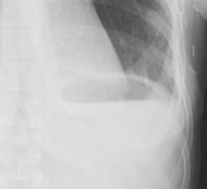
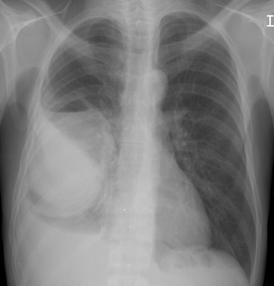
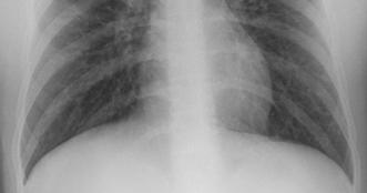
1 Chest Radiology Interpretation: Findings of Tuberculosis Get out your laptops, smart phones or other devices pollev.com/chestradiology Case #1 1


2 Plombage Pneumonia Cancer 2
3 Reading the TB CXR Be systematic! Start centrally and work outwards Normal or abnormal If abnormal, consider technique as cause Describe the finding(s) Consider the significance of the finding(s) Mediastinum Hila Lungs Pleura Bones Mediastinum 3

4 Normal Abnormal Lymphoma Normal Abnormal Metastatic disease (unknown primary) 4


5 AO PA Normal Abnormal Lung Cancer Heart <55% thoracic diameter Technique important Larger in: AP film Poor inspiration Rotation Children True enlargement Chamber enlargement Pericardial effusion Mass Artifactual cardiomegaly 5


6 End stage rheumatic heart disease Pericarditis 6

7 Hila Normal Sarcoidosis Abnormal 7


8 Normal Abnormal Pulmonary Hypertension Lungs Pleura & Diaphragms 8
9 Pleura & Diaphragms 9
10 10

11 Lung Pleura Lung Pleura Lung Pleura 11


12 Lung Pleura TB Empyema Don t forget about the bones 12

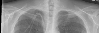
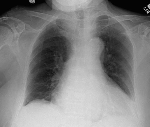
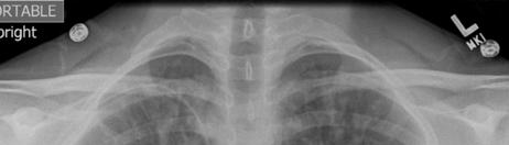
13 Case #1 Case #2 13
 1st rib 3rd rib 2nd rib")
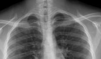
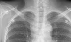
14 Inspiration: ( 10 posterior ribs) 1st rib 3rd rib 2nd rib 14


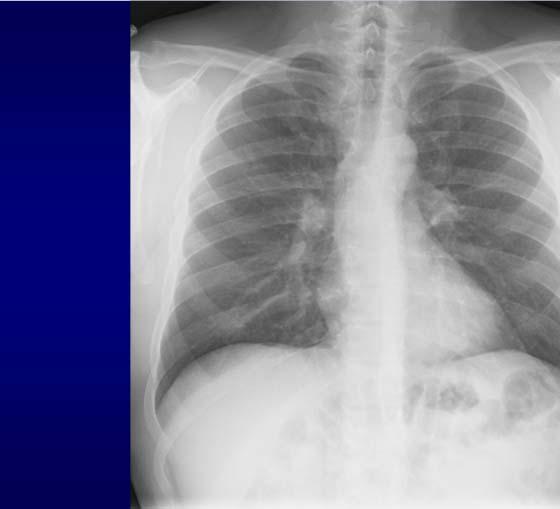
15 1st 2nd 3rd 4th 5th 6th 7th 8th 9th 10th Poor inspiration Good inspiration 15

16 Rotation Rotation 16
17 Intervertebral Disks Penetration Over-penetrated 17
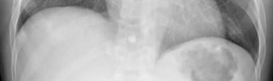
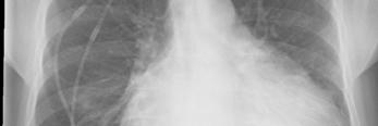
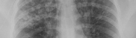
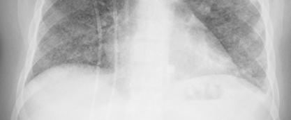
18 Case #3 Categories of lung opacities 1. Nodule(s) or mass(es) 2. Alveolar, airspace, consolidation 3. Interstitial (diffuse lines or nodules) 4. Airways (circular or tubular) 18


19 Nodule 3cm, Mass > 3 cm 3.4 cm 2.7 cm Consolidation Confluent opacity Fluffy around the periphery Air bronchograms ARDS 19
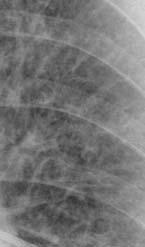


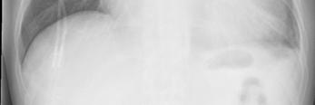
20 Interstitial disease Normal Nodular Reticular Miliary TB Idiopathic pulmonary fibrosis 20


21 Airways disease Circular Tubular 21
 diagnosis?")

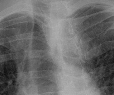
22 Tuberculosis Case 3 Questions Could this be TB? (the answer is always Is TB the most likely yes!) diagnosis? If so, what form of TB does the radiology suggest? Is active disease likely or unlikely? What are possible alternative diseases to produce the radiographic pattern? 22
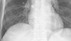
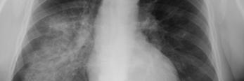
23 Key points TB patterns overlap with each other TB patterns overlap with other diseases If there is an abnormality, it could be due to TB You must know the classic TB patterns But, if it doesn t fit into a typical TB pattern, it is unlikely to be TB It s all about likelihood! Clinical-radiographic correlation Case #3 Reactivaton TB- radiology Location Apical/posterior segments upper lobes Superior segment lower lobes Isolated anterior disease very unusual Presence of cavities Pleural disease Volume loss/scarring early in disease Diff dx: fungal, bacterial infections 23
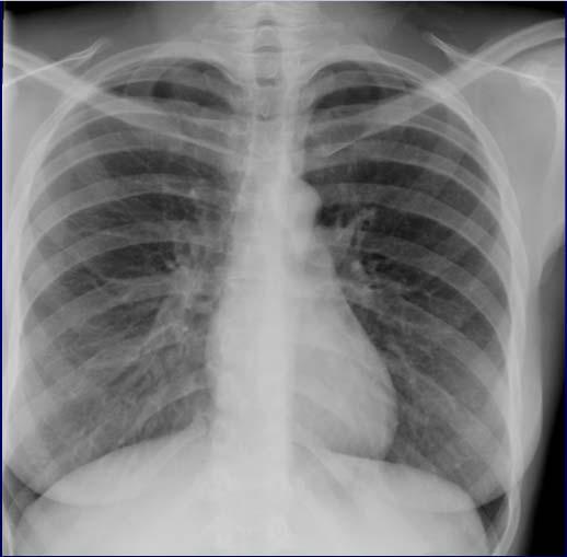
24 Chest Radiology Interpretation: Findings of Tuberculosis (Part 2) Is this likely TB? 24
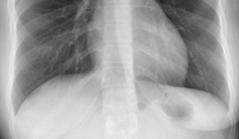
25 Lobar anatomy LUL LLL Left Lung Lobar anatomy RUL RML RLL Right Lung Lobar anatomy RUL RML RLL Right Lung 25


26 RUL Pneumonia Lobar anatomy RUL RML RLL Right Lung Lobar anatomy RUL RML RLL Right Lung 26
27 Silhouette sign A B A B Silhouette sign A B AB 27
28 Lobar anatomy RUL RML RLL Right Lung Diaphragm RLL pneumonia RLL Obscured Diaphragm Clear Heart Border? Which lobe is involved 28
29 Lobar anatomy RUL RML RLL Right Lung RML pneumonia RML Clear Diaphragm Obscured Heart Border? pneumonia 29
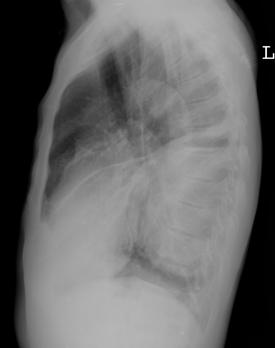
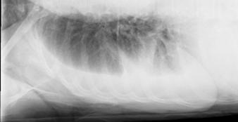
30 ? pneumonia Superior Anterior Posterior Inferior Lateral View of the Chest Heart 30
31 Lateral View of the Chest Spine Lateral View of the Chest Diaphragm Lateral View of the Chest Diaphragm 31


32 Normal LLL Pneumonia Normal Pleural effusion Normal Nodule 32

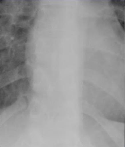
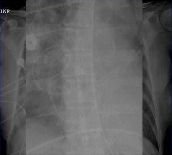
33 Normal Pott s disease Case #4 33

34 Abnormal Normal Prior reactivation tuberculosis Upper lobe scarring Volume loss Retraction of hila superiorly Band-like (linear) opacities Architectural distortion Asymmetric > symmetric Bronchiectasis Cystic changes Diff dx: fungal, sarcoid, pneumoconioses Prior TB 34
35 Warning signs Consolidation outside areas of fibrosis Consolidation with cavitation Lower lobe abnormalities Non-calcified nodules (ill-defined) Change from prior CXR Reactivation TB 35

36 Case #5 Solitary nodule/mass- the top 5 Granuloma Hamartoma Solitary metastasis Bronchogenic carcinoma Lots of others 36
37 So you see a nodule on CXR 1. Look for old films 2. Is diffuse calcification present? 3. Get a CT scan When to get a CT scan? Questionable CXR findings Further characterization of CXR findings Concern for cancer 37


38 Role of CT scan for nodules 1. Attempt to prove they are definitively benign Benign pattern of calcification (diffuse, central, ring-like, popcorn) Fat 2 years of stability Features of benign nodules include: Benign patterns of calcification Presence of fat Long term stability Diffuse Central Hamartoma Initial CT Ring-like Popcorn 24 month follow-up Hamartoma. 38
 Fat 2 years of stability 2.")

39 Irregular calcification: adenocarcinoma Role of CT scan for nodules 1. Attempt to prove they are definitively benign Benign pattern of calcification (diffuse, central, ring-like, popcorn) Fat 2 years of stability 2. Determine likelihood of nodule being benign or malignant Low likelihood -> CT follow-up High likelihood -> immediate action (e.g. biopsy) Suspicious features of nodules include: Large size Spiculated borders Growth Initial CT The size threshold above which malignancy is likely demonstrates geographic variability, depending upon the prevalence of endemic granulomatous infection. Follow-up 39


40 Size and likelihood of cancer 81% 0% 1% 15% Swensen. Radiology 2005; 235: 259 Follow-up recommendations Nodule size Low-risk patient High-risk patients 4 mm No follow-up 12 months >4-6 mm 12 months 6-12 months months 6-8 mm 6-12 months months >8 mm 3 months 9 months 24 months 3-6 months 9-12 months 24 months 3 months 9 months 24 months Fleischner Guidelines. Radiology 2005; 237: 395. Old tuberculosis 40


41 Bronchogenic carcinoma Case #6 Ghon focus Case #6 41
42 Ranke complex Case #6 Case #7 42

 Primary tuberculosis Primary")
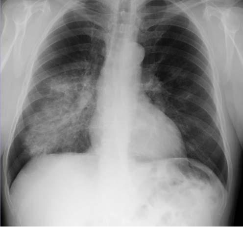
43 Primary tuberculosis Difficult radiologic diagnosis Mimics other diseases Findings Nonspecific consolidation Nodule Lymphadenopathy Cavitation unusual LAD more common than with 2 TB (particularly kids + HIV) Primary tuberculosis Primary tuberculosis 43
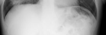
44 Case #8 Miliary pattern CXR Miliary tuberculosis Fungal infection (histo, cocci, blasto) Metastases Sarcoidosis 44
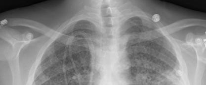
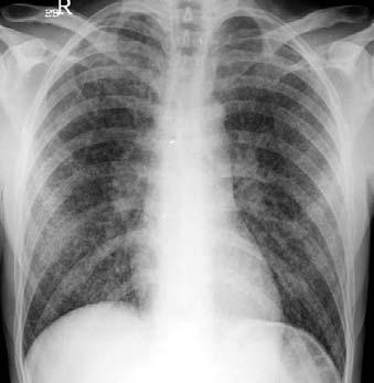

45 Miliary tuberculosis Miliary TB Sarcoidosis 45

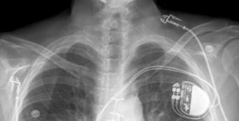
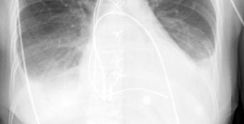
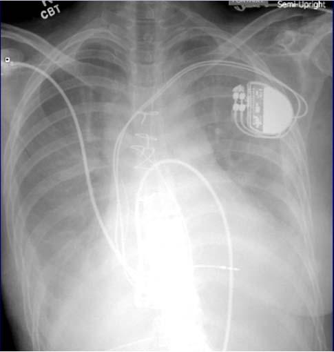
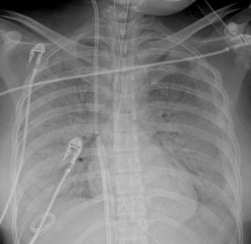
46 Metastases Case #10 Pleural + pericardial disease Primary or secondary May be only manifestation in 1 TB Empyema more common in secondary Adults >> kids 46


47 Suspected pleural effusion Case #11 47


48 Case #11 Lymphoma Leukemia Germ cell tumor Bacterial mediastinitis Fungal infection Tuberculosis 48

49 Lymphadenopathy with TB Kids >> adults Primary >> secondary Asymmetric (right > left) Most common locations Hilar Right paratracheal Necrosis very common TB lymphadenitis Case #12 49


50 thymus heart <65% thoracic diameter Conclusions Be systematic when reading CXR Typical TB patterns Mimics of TB Get a CT scan when appropriate 50
TB Radiology for Nurses Garold O. Minns, MD
 TB Nurse Case Management Salina, Kansas March 31-April 1, 2010 TB Radiology for Nurses Garold O. Minns, MD April 1, 2010 TB Radiology for Nurses Highway Patrol Training Center Salina, KS April 1, 2010
An Introduction to Radiology for TB Nurses
 An Introduction to Radiology for TB Nurses Garold O. Minns, MD September 14, 2017 TB Nurse Case Management September 12 14, 2017 EXCELLENCE EXPERTISE INNOVATION Garold O. Minns, MD has the following disclosures
TB Intensive Houston, Texas
 TB Intensive Houston, Texas October 15-17, 17 2013 Diagnosis of TB: Radiology Rosa M Estrada-Y-Martin, MD MSc FCCP October 16, 2013 Rosa M Estrada-Y-Martin, MD MSc FCCP, has the following disclosures to
10/17/2016. Nuts and Bolts of Thoracic Radiology. Objectives. Techniques
 Nuts and Bolts of Thoracic Radiology October 20, 2016 Carleen Risaliti Objectives Understand the basics of chest radiograph Develop a system for interpreting chest radiographs Correctly identify thoracic
PULMONARY TUBERCULOSIS RADIOLOGY
 PULMONARY TUBERCULOSIS RADIOLOGY RADIOLOGICAL MODALITIES Medical radiophotography Radiography Fluoroscopy Linear (conventional) tomography Computed tomography Pulmonary angiography, bronchography Ultrasonography,
Chest XRay interpretation INTERPRETATIONS Identifications: Name & Date Technical evaluation Basic Interpretations
 Chest XRay interpretation INTERPRETATIONS Identifications: Name & Date Technical evaluation Basic Interpretations TECHNICAL EVALUATION 1. Projection: AP/PA view To differentiate between AP & PA films,
UERMMMC Department of Radiology. Basic Chest Radiology
 UERMMMC Department of Radiology Basic Chest Radiology PHYSICS DENSITIES BONE SOFT TISSUES WATER FAT AIR TELEROENTGENOGRAM Criteria for an Ideal Chest Radiograph 1. Upright 2. Posteroanterior View 3. Full
Chest X-ray Interpretation
 Chest X-ray Interpretation Introduction Routinely obtained Pulmonary specialist consultation Inherent physical exam limitations Chest x-ray limitations Physical exam and chest x-ray provide compliment
Radiological Aspects of Pulmonary Tuberculosis in Immunocompetent Hosts
 Nov 2003 Radiological Aspects of Pulmonary Tuberculosis in Immunocompetent Hosts Josh Rempell, Harvard Medical School Year III Tuberculosis: the captain of all (wo)men of death Overall, one third of the
Lecture 3. Inflammatory Processes
 Lecture 3 Inflammatory Processes Process: Increased vascular permeability Water and cellular infiltrations Results: Abscess, ulceration, cavitation Penetration, perforation and fistula formation Scarring,
Bronchial syndrome. Atelectasis Draining bronchus Bronchiectasis
 Bronchial syndrome Atelectasis Draining bronchus Bronchiectasis Etienne Leroy Terquem Pierre L Her SPI / ISP Soutien Pneumologique International / International Support for Pulmonology Atelectasis Consequence
Diagnosis of TB: Radiology David Finlay, MD
 TB Intensive Tyler, Texas June 2-4, 2010 Diagnosis of TB: Radiology David Finlay, MD June 3, 2010 2stages stages- Tuberculosis 1. primary infection 2. reactivation, or post primary disease 2 1 Primary
Let s Talk TB A Series on Tuberculosis, A Disease That Affects Over 2 Million Indians Every Year
 A Series on Tuberculosis, A Disease That Affects Over 2 Million Indians Every Year Barry Rabinovitch, MD, FRCP(C) Author Madhukar Pai, MD, PhD co-author and Series Editor Barry Rabinovitch is an assistant
Chest X-ray (CXR) Interpretation Brent Burbridge, MD, FRCPC
 Interpretation Brent Burbridge, MD, FRCPC") Chest X-ray (CXR) Interpretation Brent Burbridge, MD, FRCPC An approach to reviewing a chest x-ray will create a foundation that will facilitate the detection of abnormalities. You should create your own
Chest X-ray (CXR) Interpretation Brent Burbridge, MD, FRCPC An approach to reviewing a chest x-ray will create a foundation that will facilitate the detection of abnormalities. You should create your own
Tuberculosis: The Essentials
 Tuberculosis: The Essentials Kendra L. Fisher, MD, PhD THORACIC TUBERCULOSIS: THE BARE ESSENTIALS Kendra Fisher MD, FRCP (C) Department of Radiology Loma Linda University Medical Center TUBERCULOSIS ()
Chief Complain. For chemotherapy
 Chief Complain For chemotherapy Present Illness 93.12 Progressive weakness of R t arm for 1 year X-ray: peneative lesion over right proximal humorous Bone scan: multiple increased intake Biopsy of distal
Chest X rays and Case Studies. No disclosures. Outline 5/31/2018. Carlo Manalo, M.D. Department of Radiology Loma Linda University Children s Hospital
 Chest X rays and Case Studies Carlo Manalo, M.D. Department of Radiology Loma Linda University Children s Hospital No disclosures. Outline Importance of history Densities delineated on radiography An approach
Chest imaging (Case-based teaching) 胸腔病例教學 謝叔強 財團法人恩主公醫院主治醫師萬芳醫院放射科兼任主治醫師.
 胸腔病例教學 謝叔強 財團法人恩主公醫院主治醫師萬芳醫院放射科兼任主治醫師.") Chest imaging (Case-based teaching) 胸腔病例教學 謝叔強 財團法人恩主公醫院主治醫師萬芳醫院放射科兼任主治醫師 e510019@gmail.com Check List(1) 1. Check patient data, position, technical quality and normal anatomy. 2. Review systematically
Chest imaging (Case-based teaching) 胸腔病例教學 謝叔強 財團法人恩主公醫院主治醫師萬芳醫院放射科兼任主治醫師 e510019@gmail.com Check List(1) 1. Check patient data, position, technical quality and normal anatomy. 2. Review systematically
Introduction to Radiology for TB Nurses
 Introduction to Radiology for TB Nurses Juzar Ali, MD; FRCP(C); FCCP May 4, 2018 Essential Skills for the TB Nurse Case Manager Little Rock, AR May 3 4, 2017 Juzar Ali, MD; FRCP(C); FCCP has the following
Interpretation of Chest Radiographs Paul Christensen, MD 10/21/09. Diagnostic Evaluation. Medical Evaluation & CXR Interpretation.
 Diagnostic Evaluation Medical Evaluation & CXR Interpretation University of Michigan TB Consultant Washtenaw County Medical history Physical examination Testing for TB exposure (previously covered) Radiologic
X-Rays. Kunal D Patel Research Fellow IMM
 X-Rays Kunal D Patel Research Fellow IMM The 12-Steps } 1: Name 2: Date 3: Old films 4: What type of view(s) 5: Penetration } Pre-read 6: Inspiration 7: Rotation Quality Control 8: Angulation 9: Soft tissues
David E. Griffith, MD has the following disclosures to make:
 Diagnosis of TB: Radiology David E. Griffith, MD March 13, 2015 TB for Pulmonologist March 13, 2015 Phoenix, AZ EXCELLENCE EXPERTISE INNOVATION David E. Griffith, MD has the following disclosures to make:
100 Chest X Rays for Study Group. by Dr. Suneet Khurana
 100 Chest X Rays for Study Group by Dr. Suneet Khurana Approach to - Chest X Ray (shadow of the viscera on a photographic plate) Gas appears Black Fat appears Dark Grey Water Appears as Light Grey Bone
Approach to Pulmonary Nodules
 Approach to Pulmonary Nodules Edwin Jackson, Jr., DO Assistant Professor-Clinical Director, James Early Detection Clinic Department of Internal Medicine Division of Pulmonary, Allergy, Critical Care and
Pulmonary Sarcoidosis - Radiological Evaluation
 Original Research Article Pulmonary Sarcoidosis - Radiological Evaluation Jayesh Shah 1, Darshan Shah 2*, C. Raychaudhuri 3 1 Associate Professor, 2 1 st Year Resident, 3 Professor and HOD Radiology Department,
Web Chapter 3. Image Gallery: Lesion detection on low dose chest CT
 Web Chapter 3 Image Gallery: Lesion detection on low dose chest CT Sarabjeet Singh, MD Mannudeep K. Kalra, MD *Eugene J. Mark, MD *James Stone, MD James H. Thrall, MD Department of Radiology and *Department
I9 COMPLETION INSTRUCTIONS
 The I9 Form is completed for each screening exam at T0, T1, and T2. At T0 (baseline), the I9 documents comparison review of the baseline screen (C2 Form) with any historical images available. At T1 and
Pediatric TB Intensive Houston, Texas
 Pediatric TB Intensive Houston, Texas November 13, 2009 Radiographic Manifestations of Pediatric TB Susan D. John, MD, FACR November 13, 2009 Radiologic Presentation of Childhood TB Susan D. John, MD,
Undergraduate Teaching
 Prof. James F Meaney Undergraduate Teaching Chest X-Ray Understanding the normal anatomical by reference to cross sectional imaging Radiology? It s FUN! Cryptic puzzle Sudoku (Minecraft?) It s completely
How to Analyse Difficult Chest CT
 How to Analyse Difficult Chest CT Complex diseases are:- - Large lesion - Unusual or atypical pattern - Multiple discordant findings Diffuse diseases are:- - Numerous findings in both sides 3 basic steps
Key messages. CXR interpretation in TB/HIV setting. Training course
 Key messages CXR interpretation in TB/HIV setting Training course Normal CXR Front view and lateral view Good notions of technical conditions to obtain a good CXR Good knowledge of criteria for quality
A Case of Pediatric Plasma Cell Granuloma
 August 2001 A Case of Pediatric Plasma Cell Granuloma Nii Tetteh, Harvard Medical School Year IV Our Patient 8 year old male with history of recurrent left lower lobe and lingular pneumonias since 1994.
Pictorial essay of unusual radiologic manifestations of pulmonary and airway metastasis at initial presentation of lung cancer
 Pictorial essay of unusual radiologic manifestations of pulmonary and airway metastasis at initial presentation of lung cancer Poster No.: C-2297 Congress: ECR 2012 Type: Educational Exhibit Authors: Y.
FDG PET/CT in Lung Cancer Read with the experts. Homer A. Macapinlac, M.D.
 FDG PET/CT in Lung Cancer Read with the experts Homer A. Macapinlac, M.D. Patient with suspected lung cancer presents with left sided chest pain T3 What is the T stage of this patient? A) T2a B) T2b C)
Pediatric TB Intensive Houston, Texas October 14, 2013
 Pediatric TB Intensive Houston, Texas October 14, 2013 Radiologic Presentation of Childhood TB Susan D. John, MD, FACR October 14, 2013 Disclosures I have no disclosures or conflicts of interest to report
Radiology of the respiratory disease
 Radiology of the respiratory disease [ Color index: Important Notes Extra ] [ Editing file Feedback Share your notes Shared notes ] Resources: - 435 Slides - 434 Team - 435 Notes Done by: - Mai Alageel
The Dr. Jae Yang Lecture: An Overview of the Radiographic Picture of TB
 The Dr. Jae Yang Lecture: An Overview of the Radiographic Picture of TB Harvey H. Wong, MD FRCPC MScCH Assistant Professor Department of Medicine Division of Respirology University of Toronto Financial
Interesting Cases. Pulmonary
 Interesting Cases Pulmonary 54M with prior history of COPD, hep B/C, and possible history of TB presented with acute on chronic dyspnea, and productive cough Hazy opacity overlying the left hemithorax
Man, 65 years old, heavy smoker, cough, dyspnea and weight loss. AFB negative in sputum. Is TB possible?
 Chapter 10 Man, 65 years old, heavy smoker, cough, dyspnea and weight loss. AFB negative in sputum. Is TB possible? The opacity of the left upper lobe is not cavited and looks like a tissular mass, not
Signs in Chest Radiology
 Signs in Chest Radiology Jonathan H. Chung, MD Disclosures No pertinent disclosures Jonathan H. Chung, MD Assistant Professor Institute t of fadvanced d Biomedical Imaging National Jewish Health Denver,
Do you want to be an excellent Radiologist? - Focus on the thoracic aorta on lateral chest image!!!
 The lateral chest radiograph: Challenging area around the thoracic aorta!!! Do you want to be an excellent Radiologist? - Focus on the thoracic aorta on lateral chest image!!! Dong Yoon Han 1, So Youn
Objectives. What is a Chest X Ray? CXR Workshop. Definition (diagnostic tool/internal PE) Types. Cost
 Types. Cost") Objectives CAPA 2011 Christy Wilson, PA C Georgia Lung Associates Identify the radiographic landmarks on a chest radiograph Recognize identifiers of poor quality on the chest radiograph Outline an approach
Objectives CAPA 2011 Christy Wilson, PA C Georgia Lung Associates Identify the radiographic landmarks on a chest radiograph Recognize identifiers of poor quality on the chest radiograph Outline an approach
4/16/2017. Learning Objectives. Interpretation of the Chest Radiograph. Components. Production of the Radiograph. Density & Appearance
 Interpretation of the Arthur Jones, EdD, RRT Learning Objectives Identify technical defects in chest radiographs Identify common radiographic abnormalities This Presentation is Approved for 1 CRCE Credit
Boy 8 months TPRC. 21 Sep 06 CXR. Flat and. CLE findings. BPD findings a. Left opacity
 CLE in BPD lung Boy 8 months 17 Sep 06 21 Sep 06 CXR Flat and low position of the diaphragm ICD insertion, right; ET tube slightly shift to the left RUL atelectasis RML hyperinflation, herniating across
Manage TB Dr. A. Chitrakumar Madras Medical College and RGGGH Institute of Thoracic Medicine, Chennai
 Manage TB Dr. A. Chitrakumar Madras Medical College and RGGGH Institute of Thoracic Medicine, Chennai Lecture 16 Radiology in diagnosis of Tuberculosis Session 01 So, welcome to the session Radiology in
Typical and atypical findings of pulmonary sarcoidosis at high resolution CT
 Typical and atypical findings of pulmonary sarcoidosis at high resolution CT Poster No.: C-0169 Congress: ECR 2013 Type: Educational Exhibit Authors: L. Raposo Rodríguez, C. Mejía, B. Escobar Mallada,
Pulmonary Nodules & Masses
 Pulmonary Nodules & Masses A Diagnostic Approach Heber MacMahon The University of Chicago Department of Radiology Disclosure Information Consultant for Riverain Technology Minor equity in Hologic Royalties
X-Rays. Prepared by Prof.Dr. Magda Hassab Allah Assist.lecturer Marwa Al Hady
 X-Rays Prepared by Prof.Dr. Magda Hassab Allah Assist.lecturer Marwa Al Hady CHEST X-RAYS Normal Chest X-ray Comments on chest X ray includes examination of 1- Bony cage (ribs,clavicles &vertebral column
Right infrahilar nodule
 Right infrahilar nodule Search Infrahilar nodule Nov 9, 2015.. CT chest showed a right infrahilar mass 3.5 2.5 cm along with multiple bilateral lung nodules of size 9 to 11 mm. Bronchoscopy. Jun 13, 2015.
CT findings of high-attenuation pulmonary abnormalities
 Insights Imaging (2010) 1:287 292 DOI 10.1007/s13244-010-0039-2 PICTORIAL REVIEW CT findings of high-attenuation pulmonary abnormalities Naim Ceylan & Selen Bayraktaroglu & Recep Savaş & Hudaver Alper
Sectional Anatomy Quiz II
 Sectional Anatomy II Rashid Hashmi Rural Clinical School, University of New South Wales, Wagga Wagga, New South Wales, Australia A R T I C L E I N F O Article type: Article history: Received: 3 Aug 2017
Learning Radiology: Recognizing the Basics. Text with Student Consult Online Access Code
 Learning Radiology: Recognizing the Basics. Text with Student Consult Online Access Code Herring, W ISBN-13: 9780323074445 Table of Contents 1. Recognizing Anything The "colorful" world of radiology A
C2 COMPLETION INSTRUCTIONS
 The C2 Form is completed for each screening exam at T0, T1, and T2. The C2 Form is to be completed by each of the following ACRIN-NLST study staff: the research associate (study coordinator), CT technologist,
Use of Integrated PET CT in the Clinical Staging of Non Small Cell Lung Cancer
 November 2010 Use of Integrated PET CT in the Clinical Staging of Non Small Cell Lung Cancer Laura Myers, Harvard Medical School, Year III Clinical Presentation 79yo woman with cough productive of green
Thoracic Sarcoidosis Imaging Updated: Jul 19, 2013
 Thoracic Sarcoidosis Imaging Updated: Jul 19, 2013 Overview Radiography Computed Tomography Magnetic Resonance Imaging Nuclear Imaging Show All Multimedia Library References Overview For patients with
TB Intensive San Antonio, Texas November 29-December 2, 2011
 TB Intensive San Antonio, Texas November 29-December 2, 2011 Diagnosis of TB: Radiology Michael McCarthy, MD, FACR November 30, 2011 Michael McCarthy, MD, FACR has the following disclosures to make: No
Thoracic Imaging: A Case of Metastatic Adenocarcinoma of Unknown Primary
 January 28, 2009 Thoracic Imaging: A Case of Metastatic Adenocarcinoma of Unknown Primary Kristina Mirabeau-Beale, Harvard Medical School Year III Gillian Lieberman, MD Agenda Introduce Patient RS Discuss
Tanya Van, M.D. has the following disclosures to make:
 Imaging of Pulmonary Mycobacterial TB Infection Tanya Van, M.D. April6, 2016 TB Intensive April 5 8, 2016 San Antonio, TX EXCELLENCE EXPERTISE INNOVATION Tanya Van, M.D. has the following disclosures to
Respiratory Interactive Session. Elaine Borg
 Respiratory Interactive Session Elaine Borg Case 1 Respiratory Cytology 55 year old gentleman Anterior mediastinal mass EBUS FNA Case 1 Respiratory Cytology 55 year old gentleman with anterior mediastinal
Guide to thoracic imaging
 FOCUS Guide to thoracic imaging Sarah Skinner Background Thoracic imaging is commonly ordered in general practice. Guidelines exist for ordering thoracic imaging but few are specific for general practice.
Rodney C Richie MD FACP FCCP DBIM Texas Life and EMSI
 Rodney C Richie MD FACP FCCP DBIM Texas Life and EMSI Pulmonary Nodules Well-circumscribed, radiographic opacities measuring 3 cm in diameter Surrounded by aerated lung Not associated with atelectesis
Dilemma of Thoracic Tuberculosis Vs. Sarcoidosis in TB Endemic Areas: An Imaging Approach
 Dilemma of Thoracic Tuberculosis Vs. Sarcoidosis in TB Endemic Areas: An Imaging Approach A. S. Bhalla, A. Das, A. GOYAL, P. NARANJE, R. GULERIA, G. C. KHILNANI ALL INDIA INSTITUTE OF MEDICAL SCIENCES
The Spectrum of Management of Pulmonary Ground Glass Nodules
 The Spectrum of Management of Pulmonary Ground Glass Nodules Stanley S Siegelman CT Society 10/26/2011 No financial disclosures. Noguchi M et al. Cancer 75: 2844-2852, 1995. 236 surgically resected peripheral
INTRATHORACIC TUBERCULOSIS IN CHILDREN
 DIAGNOSTIC ATLAS OF INTRATHORACIC TUBERCULOSIS IN CHILDREN A Guide For Low Income Countries 2003 International Union Against Tuberculosis and Lung Disease DIAGNOSTIC ATLAS OF INTRATHORACIC TUBERCULOSIS
8/14/2017. Objective: correlate radiographic findings of common lung diseases to actual lung pathologic features
 What is that lung disease? Pulmonary Patterns & Correlated Pathology Dr. Russell Tucker, DACVR Objective: correlate radiographic findings of common lung diseases to actual lung pathologic features Improved
Case 1: Question. 1.1 What is the main pattern of this HRCT? 1. Intralobular line 2. Groundglass opacity 3. Perilymphatic nodule
 HRCT WORK SHOP Case 1 Case 1: Question 1.1 What is the main pattern of this HRCT? 1. Intralobular line 2. Groundglass opacity 3. Perilymphatic nodule Case 1: Question 1.2 What is the diagnosis? 1. Hypersensitivity
Radiology Pathology Conference
 Radiology Pathology Conference Sharlin Johnykutty,, MD, Cytopathology Fellow Sara Majewski, MD, Radiology Resident Friday, August 28, 2009 Presentation material is for education purposes only. All rights
IS IT A GOOD PA CHEST X-RAY?
 1 CHEST IMAGING 1. PLAIN X-RAYS THE CHEST FILM A Chest X-ray (CXR) is normally taken erect and PA (posterior anterior) at a distance of 5 or 6 feet (150 or 200cm). The anterior chest wall is against the
Evaluation of the chest Part II.
 Evaluation of the chest Part II. Nagy Endre SZEGEDI TUDOMÁNYEGYETEM ÁOK, RADIOLÓGIAI KLINIKA, SZEGED ANATOMY parenchyma: alveoloacinar system, pulmonary arteries and veins interstitium: connective tissues
OBJECTIVES. Solitary Solid Spiculated Nodule. What would you do next? Case Based Discussion: State of the Art Management of Lung Nodules.
 Organ Imaging : September 25 2015 OBJECTIVES Case Based Discussion: State of the Art Management of Lung Nodules Dr. Elsie T. Nguyen Dr. Kazuhiro Yasufuku 1. To review guidelines for follow up and management
Eun-Young Kang, M.D., Jae Wook Lee, M.D., Ji Yung Choo, M.D., Hwan Seok Yong, M.D., Ki Yeol Lee, M.D., Yu-Whan Oh, M.D.
 Eun-Young Kang, M.D., Jae Wook Lee, M.D., Ji Yung Choo, M.D., Hwan Seok Yong, M.D., Ki Yeol Lee, M.D., Yu-Whan Oh, M.D. Department of Radiology, Korea University Guro Hospital, College of Medicine, Korea
Atypical radiologic appearances of pulmonary tuberculosis in non-hiv adult patients
 Atypical radiologic appearances of pulmonary tuberculosis in non-hiv adult patients Poster No.: R-0200 Congress: RANZCR-AOCR 2012 Type: Educational Exhibit Authors: K. N. Jeon, K. Bae, M. J. Park Keywords:
ACUTE PULMNARY INFECTIONS: UNDERSTANDING THE CHEST RADIOGRAPH. Leonard E. Swischuk, M.D. University of Texas Medical Branch
 ACUTE PULMNARY INFECTIONS: UNDERSTANDING THE CHEST RADIOGRAPH Leonard E. Swischuk, M.D. University of Texas Medical Branch AUTHOR HAS NOTHING TO DECLARE LEARNING OBJETIVES Understand the pathophysiology
Lung Tumor Cases: Common Problems and Helpful Hints
 Lung Tumor Cases: Common Problems and Helpful Hints Brandon T. Larsen, MD, PhD Senior Associate Consultant Department of Laboratory Medicine and Pathology Mayo Clinic Arizona Arizona Society of Pathologists
Pediatric TB Radiology: It s Not Black and White Part 2
 Experiencing technical difficulties? Please call Adobe Connect for technical assistance at 1-800-422-3623 Pediatric TB Radiology: It s Not Black and White Part 2 June 18, 2018 A National Webinar June 18,
Differential Diagnosis in Conventional Radiology
 Differential Diagnosis in Conventional Radiology Bearbeitet von Francis A. Burgener, Martti Kormano, Tomi Pudas Neuausgabe 2007. Buch. 872 S. Hardcover ISBN 978 3 13 656103 4 Format (B x L): 21 x 29,7
Extraordinary Patterns of Tuberculosis
 Extraordinary Patterns of Tuberculosis E. Kadakovska Infectology Center of Latvia, Clinic of Tuberculosis and Lung Diseases, Diagnostics and Radiology Department 1 Target Importance of recognizing of tuberculosis
Introduction to Chest Radiography
 Introduction to Chest Radiography RSTH 366: DIAGNOSTIC TECHNIQUES Alan Alipoon BS, RCP, RRT Instructor Department of Cardiopulmonary Sciences 1 Introduction Discovered in 1895 by Wilhelm Roentgen Terminology
Bronchogenic Carcinoma
 A 55-year-old construction worker has smoked 2 packs of ciggarettes daily for the past 25 years. He notes swelling in his upper extremity & face, along with dilated veins in this region. What is the most
CHEST & ABDOMINAL X-RAYS MALIKA IBRAHIM CORE MEDICAL TRAINEE BLACKPOOL VICTORIA HOSPITAL DATA INTERPRETATION COURSE FEB 20, 2017
 CHEST & ABDOMINAL X-RAYS MALIKA IBRAHIM CORE MEDICAL TRAINEE BLACKPOOL VICTORIA HOSPITAL DATA INTERPRETATION COURSE FEB 20, 2017 1. Sample x-rays 2. Basic chest x-ray interpretation skills 3. Chest x-ray
Tuberculosis - clinical forms. Dr. A.Torossian,, M.D., Ph. D. Department of Respiratory Diseases
 Tuberculosis - clinical forms Dr. A.Torossian,, M.D., Ph. D. Department of Respiratory Diseases 1 TB DISEASE Primary Post-primary (Secondary) Common primary forms Primary complex Tuberculosis of the intrathoracic
Acute and Chronic Lung Disease
 KATHOLIEKE UNIVERSITEIT LEUVEN Faculty of Medicine Acute and Chronic Lung Disease W De Wever, JA Verschakelen Department of Radiology, University Hospitals Leuven, Belgium Clinical utility of HRCT To detect
CALGARY ZONE PULMONARY REFERRAL QUICK REFERENCE
 CALGARY ZONE PULMONARY REFERRAL QUICK REFERENCE EMERGENCY (Patient needs to be seen immediately) Hemoptysis (Active & 2 TBSP per day) Hypoxemia (if resting O2 SAT 85%) Pulmonary embolism (Acute - known
B-I-2 CARDIAC AND VASCULAR RADIOLOGY
 (YEARS 1 3) CURRICULUM FOR RADIOLOGY 13 B-I-2 CARDIAC AND VASCULAR RADIOLOGY KNOWLEDGE To describe the normal anatomy of the heart and vessels including the lymphatic system as demonstrated by radiographs,
Case 1 : Question. 1.1 What is the intralobular distribution? 1. Centrilobular 2. Perilymphatic 3. Random
 Interesting case Case 1 Case 1 : Question 1.1 What is the intralobular distribution? 1. Centrilobular 2. Perilymphatic 3. Random Case 1: Answer 1.1 What is the intralobular distribution? 1. Centrilobular
CT Signs of Solitary Pulmonary Lesions: Revisited
 CT Signs of Solitary Pulmonary Lesions: Revisited Poster No.: C-1764 Congress: ECR 2015 Type: Educational Exhibit Authors: H. Hayashi, K. Ashizawa, Y. Ogihara, A. Nishida, T. Tanaka, 1 1 2 1 1 1 1 1 2
Lung. 10/24/13 Chest X-ray: 2.9 cm mass like density in the inferior lingular segment worrisome for neoplasm. Malignancy cannot be excluded.
 Lung Case Scenario 1 A 54 year white male presents with a recent abnormal CT of the chest. The patient has a history of melanoma, kidney, and prostate cancers. 10/24/13 Chest X-ray: 2.9 cm mass like density
X-rays. Dr Will Dooley
 X-rays Dr Will Dooley Plan Chest X-Rays Abdominal X-Rays Exam approach Presentation skills EMQ EMQ- answers Chest X-Ray - Systematic Approach D R Details RIP Image Quality +/- OBVIOUS ABNORMALITY A B C
SCBT-MR 2015 Incidentaloma on Chest CT
 SCBT-MR 2015 Incidentaloma on Chest CT Reginald F. Munden MD, DMD, MBA I have no conflicts of interest to report Incidentaloma Pulmonary Nodule Mediastinal Lymph Node Coronary Artery Calcium Incidental
HRCT in Diffuse Interstitial Lung Disease Steps in High Resolution CT Diagnosis. Where are the lymphatics? Anatomic distribution
 Steps in High Resolution CT Diagnosis Pattern of abnormality Distribution of disease Associated findings Clinical history Tomás Franquet MD What is the diagnosis? Hospital de Sant Pau. Barcelona Secondary
Imaging in breast cancer. Mammography and Ultrasound Donya Farrokh.MD Radiologist Mashhad University of Medical Since
 Imaging in breast cancer Mammography and Ultrasound Donya Farrokh.MD Radiologist Mashhad University of Medical Since A mammogram report is a key component of the breast cancer diagnostic process. A mammogram
The use of advanced imaging in the diagnosis of TB. Pierre Goussard, Robert Gie Tygerberg Children`s Hospital and University of Stellenbosch
 The use of advanced imaging in the diagnosis of TB Pierre Goussard, Robert Gie Tygerberg Children`s Hospital and University of Stellenbosch Imaging Bronchoscopy Tracheo-bronchograms Chest CT-scan Ultrasound
FDG PET/CT STAGING OF LUNG CANCER. Dr Shakher Ramdave
 FDG PET/CT STAGING OF LUNG CANCER Dr Shakher Ramdave FDG PET/CT STAGING OF LUNG CANCER FDG PET/CT is used in all patients with lung cancer who are considered for curative treatment to exclude occult disease.
Interstitial syndrome
 Interstitial syndrome Ground-glass attenuation Miliary and nodular images linear images Etienne Leroy Terquem Pierre L Her SPI / ISP Soutien Pneumologique International / International Support for Pulmonology
Resident Case Review CHEST. Daria Manos CAR 2016
 Resident Case Review CHEST CAR 2016 Daria Manos Disclosure Speakers bureau, Roche CAR 2016 Daria Manos 1. Recognize common and critical chest radiograph and computed tomography signs and use these clues
Mediastinal Tumors: Imaging
 Mediastinal Tumors: Imaging References Imaging in Oncology, Husband and Reznek Computed Tomography and Magnetic Resonance of the thorax, Naidich, Zerhouni, Siegelman, Mediastinal compartments Anterior:
Radiological Features of Mycobacterium tuberculosis TUBERCULE BACILLUS TUBERCULE BACILLUS DIAGNOSIS. Guy Richards. PATHOGENESIS of TUBERCULOSIS
 Radiological Features of Guy Richards Department of critical care Charlotte Maxeke Johannesburg Academic Hospital University of the Witwatersrand, Johannesburg, South Africa TUBERCULE BACILLUS Discovery
Financial disclosure COMMON DIAGNOSES IN HRCT. High Res Chest HRCT. HRCT Pre test. I have no financial relationships to disclose. Anatomy Nomenclature
 Financial disclosure I have no financial relationships to disclose. Douglas Johnson D.O. Cardiothoracic Imaging Gaston Radiology COMMON DIAGNOSES IN HRCT High Res Chest Anatomy Nomenclature HRCT Sampling