Emerging Techniques in Breast Imaging: Contrast-Enhanced Mammography and Fast MRI
|
|
|
- Ralf Allen
- 5 years ago
- Views:
Transcription


1 Emerging Techniques in Breast Imaging: Contrast-Enhanced Mammography and Fast MRI Lilian Wang, M.D. Breast Imaging Section Department of Radiology Northwestern Medicine
2 Overview Rationale for new imaging techniques Contrast enhanced mammography (CEM) Fast (abbreviated) MRI (AB-MR) Current data
3 Screening Mammography Widely available Low cost Only modality shown to reduce breast cancer mortality 30% reduction
4 Limitations of Mammography Less sensitive in women with dense breasts Mammographic sensitivity 62% in extremely dense vs. 88% in fatty breasts Interval cancers Recall rate and false positive biopsies Overdiagnosis (~1-10% est. from RCT)
5 Alternative Modalities Tomosynthesis Ultrasound Molecular breast imaging MRI Contrast-enhanced mammography (CEM)
6 Alternative Modalities Tomosynthesis Ultrasound Molecular breast imaging MRI Contrast-enhanced mammography (CEM) vascular or functional based imaging
7 CONTRAST ENHANCED MAMMOGRAPHY (CEM)
8 Contrast-Enhanced Mammography Relies on abnormal blood flow related to neovascularity IV injection of iodinated contrast Performed using adaptation to standard DM unit Dual energy or temporal technique
9 CEM Temporal Technique Temporal subtraction technique Baseline high-energy image with breast mildly compressed Multiple images of single view of one breast over 5-7 minutes Generate subtractions Advantage: Kinetic analysis Disadvantage Only 1 view of 1 breast Motion artifact
10 CEM: Temporal Technique Cancers (SN) Benign (FP) Missed CA (FN) Jong et al Diekmann et al /10 (80%) 5/12 1 IDC, 1 DCIS 14/14/ (100%) Dromain et al /22 (73%) 4 IDC ( cm), 2 not included on film


11 Diekmann et al., Eur J Radiol 2011.
12 CEM: Temporal Technique Prospective study CEM vs 2D FFDM 70 pts, 80 lesions on MG, US, or MR 30 cancers, 50 benign Improves diagnostic accuracy Sensitivity increased from 0.43 FFDM to 0.62 with CEM Significant improvement in AUC Diekmann et al., Eur J Radiol 2011.
13 CEM: Temporal Technique Limitations Patient motion Kinetics do not differentiate between benign and malignant lesions Single view only
14 CEM: Dual Energy Two images performed in CC, MLO: - a low-energy image (below the k edge of iodine, same as conventional MG) - a high-energy image (above the k edge). Uses algorithm to suppress background breast to highlight iodine-enhanced areas.

15 CC view 2 min after injection MLO view 4 min after injection Dromain et al. Eur Radiol 2011; 21:
16 CEM: Dual Energy vs Temporal Dual energy: Less motion Can be prone to rim artifact from radiation scatter No kinetic info


17 Dromain et al. Breast Cancer Research 2012, 14:R94.


18 Dromain et al. Breast Cancer Research 2012, 14:R94.
19 CEM finds more cancer than FFDM. Study CEM: Cancer Detection # CA % seen CEM % seen DM Dromain (2011) 80 92% 78% Jochelson (2013) 26 96% 85% Fallenberg (2014) % 78% Cheung (2014) 89 93% 72% Lobbes (2014) % 97% Lalji (2016) % 93%
20 CEM: Cancer Detection Identification of multifocal carcinoma Dromain et al., Eur Radiol multifocal cancer cases - 100% seen with CEM - 70% seen with FFDM Jochelson et al., Radiology additional ipsilateral cancers - 56% (14/25) seen with CEM - 88% (22/25) seen with MR - 25% (4/25) seen with DM

21 DM CEM MRI
22 CEM vs. MRI: Cancer Detection Study CEM MRI DM Jochelson Radiology % (50/52) 92% (50/52) 81% (42/52) Fallenberg Eur Radiol % (80/80) 97.4% (78/80)* Fallenberg Eur Radiol % (80/80) 97% (77/79) 83% (66/80) Lee-Felker Radiology % (66/70) 99% (69/70) * MR missed 8 mm IDC w 12 mm DCIS, 16 mm ILC
23 CEM vs. MRI: Lesion Specificity Jochelson et al. Radiology 2013 Of all enhancing lesions seen, those which were true cancers were: - CEM 30/30 = 100% - MRI 30/37 = 81% Of 52 pts, FP findings in 4% (2/52) CEM vs. 25% (13/52) for MRI. An abnormality seen on CEM was significantly more likely to be cancerous than one seen on MRI (97% vs. 85%, p<.01).
24 CEM vs. MRI: Lesion Specificity Lee-Welker et al., Radiology women with 120 breast lesions CEM and MRI had similar sensitivity - 66/70 (94%) for CEM vs 69/70 (99%) MRI CEM and MRI had equal sensitivity for detecting additional disease - CEM 11/11 (100%) and MRI 10/11 (91%) CEM had significantly higher PPV than MR - 66/71 (93%) vs 69/115 (60%) CEM had fewer false positives than MR - 5 vs 45 CEM and MRI have similar sensitivity, but CEM has greater specificity
25 CEM: False positives Fibroadenomas Papillomas Hamartomas Intramamary lymph nodes Fat necrosis

26 CEM: False Positives PASH From Jochelson et al. Radiol Clin N America, 2014.
27 CEM: False negatives Lesion not included on mammographic field of view ILC (infiltrative nature) Thibault et al., 4/6 false negatives DCIS Subtle or no enhancement Mucinous carcinomas - No enhancement or eclipse sign: misinterpreted as cyst

28 Background Parenchymal Enhancement MRI CEM
29 CEM: Palpable Abnormality Improved performance with CEM vs. lowenergy images alone AUC 0.93 vs 0.83 Improved sensitivity 84% to 95% Improved specificity 63% to 81% Tennant et al., Clin Radiol 2016.
30 CEM: Screening 611 women with intermediate breast cancer risk and dense breasts Sensitivity Specificity PPV NPV DM 52.4% 90.5% 16.4% 98.2% CEM 90.5% 76.1% 11.9% 99.6% CEM significantly more sensitive than standard DM for detecting cancer role for supplemental screening Sorin et al. AJR 2018.
31 CEM: Limitations Additional radiation dose ~43-81%, but well within dose recommendations for mammography Contrast reactions to iodinated contrast (occur <1%, mostly mild) Interpretation can be affected by BPE No CEM bx capability
32 CEM: Summary Benefits: - Good diagnostic accuracy: more sensitive than DM and fewer FP than MRI - Reproducible without operator dependency - Fast imaging technique - Direct correlation with conventional DM - Uses current DM system w/ software and hardware adaptations - Less expensive, faster, and more comfortable than MRI Limitations: - Radiation - Need for IV - Iodine allergy - Artifact near edge of images (scattered radiation) - Artifact from patient motion - No dedicated biopsy system - Interpretation may be limited by BPE
33 CEM Potential applications: Screening intermediate risk patients EOD evaluation if no access to MR Diagnostic setting: palpable abnormality, etc.
34 ABBREVIATED BREAST MRI
35 Breast MRI Most sensitive imaging modality for breast cancer detection Currently recommended as supplemental screening for women with >20% lifetime risk of breast cancer BRCA mutation, history of chest irradiation before age 30, Li Fraumeni, etc.
36 MRI for Breast Cancer Screening Not limited by breast density No ionizing radiation Most sensitive test for breast cancer detection PPV similar to mammography Preferentially detects higher grade lesions
37 Compare efficacy of MRI vs. MG in high-risk women 1909 women screened, incl. 358 carriers germ-line mutations 51 cancers identified (44 invasive, 6 DCIS, 1 lymphoma) Sensitivity Specificity Clinical breast exam 17.9% 98.1% Mammography 33.3% 95.0% MRI 79.5% 89.8% MRI significantly better than MG in detecting tumors, but MG had higher sensitivity for DCIS (83% vs. 17%, p=0.22). Kriege et al., N Engl J Med 2004;351:
38 MG US MG+US MRI MG+MRI Sensitivity 32.6% 39.5% 48.8% 90.7% 93.0% Specificity 96.8% 90.5% 89.0% 97.2% 96.1% PPV 23.7% 11.3% 11.9% 50.0% 42.1% 19 cancers seen only on MRI 5 DCIS and 14 invasive Kuhl et al., J Clin Oncol 2005, 23:
39 Comparison of Diagnostic Yield of Different Imaging Modalities Regarding Prognostically Relevant Tumor Features Detected by MG Detected by US Detected by MG + US Detected by MRI Only MRI Detected* # Primary invasive CA Node + inv. CA (%) 4/10 (40) 5/12 (42) 5/16 (31) 5/31 (16) 0/14 (0) Mean Size inv. (mm) # Minimal cancers (%) 5/25 (20) 3/25 (12) 6/25 (24) 23/25 (92) 18/25 (72) # Intraductal Ca (%) 3/9 (33) 0/9 (0) 3/9 (33) 8/9 (89) 5/9 (56) *not detected with MG+US Kuhl et al., J Clin Oncol 2005, 23:
 4/10 (40) 5/12 (42) 5/16 (31) 5/31 (16) 0/14 (0) Mean Size inv. (mm) 13.2 15.1 13.9 12.4 9.")
40 Detected by MG Detected by US Detected by MG+US Detected by MRI Only MRI Detected* # Primary invasive CA Node + inv. CA (%) 4/10 (40) 5/12 (42) 5/16 (31) 5/31 (16) 0/14 (0) Mean Size inv. (mm) # Minimal cancers (%) 5/25 (20) 3/25 (12) 6/25 (24) 23/25 (92) 18/25 (72) # Intraductal Ca (%) 3/9 (33) 0/9 (0) 3/9 (33) 8/9 (89) 5/9 (56) *not detected with MG+US Node + rate 16% vs % published for MG screening Rate of interval CA 2% vs % with MG surveillance Kuhl et al., J Clin Oncol 2005, 23:
41 Kuhl et al., J Clin Oncol 2010, 28:

42 Kuhl et al., J Clin Oncol 2010, 28:


43 Berg et al., JAMA. 2012;307(13): ACRIN 6666
44 Supplemental Screening: Cancer Yield US: 3-4/1,000 screened MRI: 10-24/1,000 screened Why not screen everyone with MRI?
45 Limitations of MR for Screening Average Risk Women Cost Time IV contrast Availability Perceived low specificity
46 Abbreviated MRI (AB-MR) Total scan time <10 minutes Localizer scan 1 pre- and 1 post-contrast gradient echo (GRE) axial acquisition; in-plane resolution of 1 mm or less Slice thickness of 3 mm or less (+/-) Axial T2 weighted sequences with inplane resolution matching GRE sequences and <3 mm slice thickness
47 Benefits AB-MR Low cost: $300-$500 Quick: <10 minutes PPV similar to MG: 20-30% % increase in cancer detection Potential to preferentially detect higher grade lesions
48 Benefits AB-MR Shorter acquisition time Shorter interpretation time à higher volume of patient throughput à more cost-effective while maintaining high diagnostic accuracy
49 Limitations of AB-MR Loss of kinetic information Sensitivity for morphologically benign appearing cancers demonstrating washout may be lower without kinetics (rare)
50 443 women with negative FFDM 427 with dense or extremely dense breasts also had negative WBUS Mild to moderate risk Radiologists reviewed: MIP and FAST images for significant enhancement complete AP (MIP, FAST images, and nonsubtracted source images) Regular full diagnostic protocol Kuhl et al., J Clin Oncol 2014.

51 AB-MR Acquisition time: AP: 3 minutes Full protocol: 17 minutes Kuhl et al., J Clin Oncol 2014.
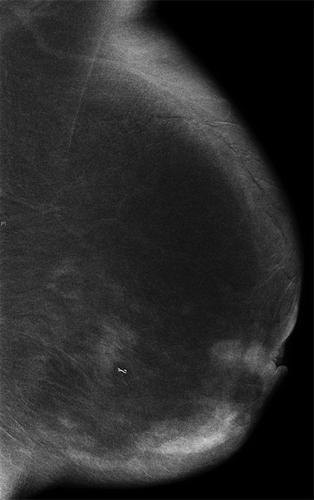
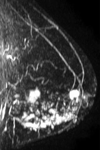
 MIP Kuhl et al.")
52 Pre-contrast Post-contrast FAST (subtraction) MIP Kuhl et al., J Clin Oncol 2014.
53 AB-MR 11 cancers detected (18.2/1000) 7 invasive, 4 DCIS Small T1, node negative cancers Predominantly high-grade tumors US negative in 11 cancers Kuhl et al., J Clin Oncol 2014.


54 Intermediate grade IDC Kuhl et al., J Clin Oncol 2014.
55 AB-MR Sensitivity MIP only: 90.9% (10/11 cancers) 1 st post contrast: 100% (11/11) Specificity 1 st post: 94.3% Full protocol 93.9% Interpretation time MIP only: 3 seconds 1 st post: 28 seconds NPV MIP only: 99.8% Kuhl et al., J Clin Oncol 2014.
56 AB-MR MIP image interpretation time of 3 s NPV 99.8% reading time <30 seconds for complete AP diagnostic accuracy equivalent to FDP with cancer yield of 18.2/1000 à batch read AB-MR for general screening like screening mammography? Kuhl et al., J Clin Oncol 2014.
57 AB-MR Study of 100 consecutive MRs in patients with biopsy proven unicentric breast CA - 79 invasive, 21 DCIS Four readers AP: 1 st post, 1 st subtraction, MIP Compared AP to FDP (13 pre-contrast, post-contrast, and post-processed sequ) Mango et al., Eur J Radiol 2015
58 AB-MR Sensitivity: 1 st post-contrast: 96% 1 st post-contrast subtraction: 96% MIP: 93% Mean interpretation time: 44 s ( s) Acquisition time: AP: min FDP: min Mango et al., Eur J Radiol 2015
59 AB-MR Elimination of sequences from FDP may limit interpretation: T1 non-fat saturated images for fat necrosis, lymph nodes T2 weighted images to enable benign assessment Lack of kinetic information?increase unnecessary recalls
60 Grimm et al., Acad Radiol 2015 AB-MR Compared two AP to standard FDP 48 MRIs (24 normal, 12 benign, 12 CA) Protocol T2 Pre 1 st post 2 nd post 3 rd /4 th post Non- FS T1 AP1 x x x AP2 x x x x FDP x x x x x x
61 Grimm et al., Acad Radiol 2015 AB-MR No significant difference in sensitivity: AP 1: 86% AP2: 89% FDP: 95% No significant difference in specificity: AP 1: 52% AP2: 45% FDP: 52% Lesion kinetics did not affect specificity Similar interpretation times: AP1: 2.98 minutes FDP: 2.95 minutes
62 AB-MR: Protocols Moschetta et al.: STIR, T2W, pre-, single post Dogan et al.: T2W, single pre- and post Choi et al.: fat-suppressed T2, single preand post, subtracted MIPs Petrillo et al: single pre- and post T1W Romeo et al: pre- and three post Studies show comparable diagnostic accuracy to full protocol
63
64 ECOG-ACRIN 4112 All patients undergo DBT and AB-MR on the same day at year 0 and year 1 Randomization as to which test is performed first After year 1, patients return to standard screening and are followed for 3 years
65 ECOG-ACRIN 4112 Primary endpoint: Compare rates of detection of invasive cancers between initial AB-MR and DBT. Secondary endpoints: Compare PPV of biopsies, callback rates, and short-term follow-up rates after AB-MR and DBT on initial and 1 year follow-up Estimate and compare sensitivity and specificity of AB-MR and DBT Compare patient-reported short-term quality of life related to diagnostic testing with AB-MR and DBT Comparative cost analysis of both exams
66 AB-MR: Goal Expand access to MRI Allow women with dense breasts to have a faster, more sensitive, and more accurate option to screening US AB-MR every 2-3 years may prove to be a better stand alone test than mammography and US every year
67 AB-MR: Future Directions Standardize protocol across sites Determine financial cost: cost-effective enough to be first-line exam? Establish screening interval?multihead scanners to increase patient throughput Improving specificity: DWI, Ultrafast, CAD Non-contrast MR imaging
68 Summary Reviewed rationale for development of new imaging techniques Discussed CEM and AB-MR Applications Current data Future directions

69 Thank You
Contrast-Enhanced Digital Mammography
 2015 ARRS Breast Symposium Contrast-Enhanced Digital Mammography John Lewin, M.D. Diversified Radiology of Colorado CEDM - Outline History Technique Literature Review / Cases Clinical Status Inexpensive,
Current Status of Supplementary Screening With Breast Ultrasound
 Current Status of Supplementary Screening With Breast Ultrasound Stephen A. Feig, M.D., FACR Fong and Jean Tsai Professor of Women s Imaging Department of Radiologic Sciences University of California,
Screening with Abbreviated Breast MRI (AB-MR)
") Screening with Abbreviated Breast MRI (AB-MR) Christopher Comstock, MD, FACR, FSBI Department of Radiology Memorial Sloan-Kettering Cancer Center New York, NY Outline History of our approach to screening
Screening with Abbreviated Breast MRI (AB-MR) Christopher Comstock, MD, FACR, FSBI Department of Radiology Memorial Sloan-Kettering Cancer Center New York, NY Outline History of our approach to screening
Outline. Abbreviated MRI and the Dense Breast. 2D Analog Screen Mammography. RCTs of Mammographic Screening
 Abbreviated MRI and the Dense Breast Christopher Comstock, MD FACR Breast Imaging Section Department of Radiology Memorial Sloan-Kettering Cancer Center New York, NY Outline History of mammographic screening
Standard Breast Imaging Modalities. Lilian Wang, M.D. Breast Imaging Section Department of Radiology Northwestern Medicine
 Standard Breast Imaging Modalities Lilian Wang, M.D. Breast Imaging Section Department of Radiology Northwestern Medicine Overview Standard breast imaging modalities Mammography Ultrasound MRI Imaging
What s New in Breast Imaging. Jennifer A. Harvey, M.D., FACR Professor of Radiology University of Virginia
 What s New in Breast Imaging Jennifer A. Harvey, M.D., FACR Professor of Radiology University of Virginia Disclosure Hologic, Inc. Shareholder and research agreement. Volpara Solutions, Ltd. Shareholder
Pitfalls and Limitations of Breast MRI. Susan Orel Roth, MD Professor of Radiology University of Pennsylvania
 Pitfalls and Limitations of Breast MRI Susan Orel Roth, MD Professor of Radiology University of Pennsylvania Objectives Review the etiologies of false negative breast MRI examinations Discuss the limitations
Screening Options in Dense Breasts. Donna Plecha, M.D. Co-Director UHCMC Breast Centers Associate Professor of Radiology Director of Breast Imaging
 Screening Options in Dense Breasts Donna Plecha, M.D. Co-Director UHCMC Breast Centers Associate Professor of Radiology Director of Breast Imaging Dense Breasted Women Decreased sensitivity of mammography
Financial Disclosures
 Financial Disclosures 3D Mammography: The Latest Developments in the Breast Imaging Arena I have no financial disclosures Dr. Katharine Lampen-Sachar Breast and Body Radiologist Radiology Associates of
EARLY DETECTION: MAMMOGRAPHY AND SONOGRAPHY
 EARLY DETECTION: MAMMOGRAPHY AND SONOGRAPHY Elizabeth A. Rafferty, M.D. Avon Comprehensive Breast Center Massachusetts General Hospital Harvard Medical School Breast Cancer Screening Early detection of
Successful Breast MRI Program : The ingredients
 Successful Breast MRI Program : The ingredients Dr. Smriti Hari Associate Professor Deptt. Of Radiology All India Institute of Medical Sciences New Delhi How to perform Breast MRI Breast MRI descriptors
Contrast-enhanced Breast MRI RSSA 2013
 Contrast-enhanced Breast MRI RSSA 2013 Prof. dr. Maurice van den Bosch University Medical Center Utrecht, the Netherlands Index 1) Breast cancer 2) Why MRI of the breast 3) Technique 4) Interpretation
Supplemental Screening for Dense Breasts. Reagan Leverett, MD, MS
 Supplemental Screening for Dense Breasts Reagan Leverett, MD, MS Outline Anatomy and Density Risk of dense breasts Theory of Supplemental Screening Options for supplemental screening Tomosynthesis Ultrasound
BREAST MRI. Elizabeth A. Rafferty, M.D. Avon Comprehensive Breast Center Massachusetts General Hospital Harvard Medical School
 BREAST MRI Elizabeth A. Rafferty, M.D. Avon Comprehensive Breast Center Massachusetts General Hospital Harvard Medical School BREAST MRI Any assessment of the breast parenchyma requires the administration
High Risk Screening: A Multimodality Approach
 High Risk Screening: A Multimodality Approach John Lewin, M.D., FACR, FSBI The Women s Imaging Center Denver, Colorado Disclosures Consultant to Hologic Previously received research funds from Hologic
EARLY DETECTION: MAMMOGRAPHY AND SONOGRAPHY
 EARLY DETECTION: MAMMOGRAPHY AND SONOGRAPHY Elizabeth A. Rafferty, M.D. Avon Comprehensive Breast Center Massachusetts General Hospital Harvard Medical School Breast Cancer Screening Early detection of
Breast Cancer Screening and Diagnosis
 Breast Cancer Screening and Diagnosis Priya Thomas, MD Assistant Professor Clinical Cancer Prevention and Breast Medical Oncology University of Texas MD Anderson Cancer Center Disclosures Dr. Thomas has
Mammographic imaging of nonpalpable breast lesions. Malai Muttarak, MD Department of Radiology Chiang Mai University Chiang Mai, Thailand
 Mammographic imaging of nonpalpable breast lesions Malai Muttarak, MD Department of Radiology Chiang Mai University Chiang Mai, Thailand Introduction Contents Mammographic signs of nonpalpable breast cancer
BREAST MRI. Elizabeth A. Rafferty, M.D. Avon Comprehensive Breast Center Massachusetts General Hospital Harvard Medical School
 BREAST MRI Elizabeth A. Rafferty, M.D. Avon Comprehensive Breast Center Massachusetts General Hospital Harvard Medical School BREAST MRI Any assessment of the breast parenchyma requires the administration
New Imaging Modalities for better Screening and Diagnosis
 New Imaging Modalities for better Screening and Diagnosis Miri Sklair-Levy, MD Department of Diagnostic Imaging Sheba Medical Center, Sackler School of Medicine, Tel Aviv University Department of Diagnostic
Breast density: imaging, risks and recommendations
 Breast density: imaging, risks and recommendations Maureen Baxter, MD Radiologist Director of Ruth J. Spear Breast Center Providence St. Vincent Medical Center Alison Conlin, MD/MPH Medical Oncologist
Tomosynthesis and breast imaging update. Dr Michael J Michell Consultant Radiologist King's College Hospital NHS Foundation Trust
 Tomosynthesis and breast imaging update Dr Michael J Michell Consultant Radiologist King's College Hospital NHS Foundation Trust Breast imaging new technology BREAST CANCER FLT PET shows different grades
BREAST DENSITY WHAT IS IT? WHY IS IT IMPORTANT? & What IOWA SF250 Means to Patients and Providers
 BREAST DENSITY WHAT IS IT? WHY IS IT IMPORTANT? & What IOWA SF250 Means to Patients and Providers Arnold Honick, MD Radiology Consultants of Iowa, PLC ahonick@rciowa.com BREAST DENSITY LEGISLATION Nancy
Updates in Mammography. Dr. Yang Faridah A. Aziz Department of Biomedical Imaging University Malaya Medical Centre
 Updates in Mammography Dr. Yang Faridah A. Aziz Department of Biomedical Imaging University Malaya Medical Centre Updates in Mammography Breast Imaging Dr. Yang Faridah A. Aziz Department of Biomedical
Contrast Enhanced Spectral Mammography (CESM) in Cancer Staging
 in Cancer Staging") Contrast Enhanced Spectral Mammography (CESM) in Cancer Staging Carrie Rochman,MD Assistant Professor,Breast Imaging UVA Dept of Radiology and Medical Imaging Disclosures Research Agreement and Funding;
Contrast Enhanced Spectral Mammography (CESM) in Cancer Staging Carrie Rochman,MD Assistant Professor,Breast Imaging UVA Dept of Radiology and Medical Imaging Disclosures Research Agreement and Funding;
Breast MRI Update. Jeffrey C. Weinreb, MD, FACR Yale University School of Medicine
 Breast MRI Update Jeffrey C. Weinreb, MD, FACR jeffrey.weinreb@yale.edu Yale University School of Medicine I disclose the following financial relationships with relevant commercial interests: Bracco Bayer
Index. C Calcifications fat necrosis 1, 61 fat necrosis 4, 69 nipple/peri-areolar involvement 1, 165
 A ADH. See Atypical ductal hyperplasia (ADH) American College of Radiology (ACR), BI-RADS background parenchymal enhancement, 8, 9, 81, 82 fibroglandular tissue guidelines, 6 American Joint Committee on
Breast MRI: Friend or Foe?
 Breast MRI: Friend or Foe? UCSF Postgraduate Course May 18, 2013 Cheryl Ewing, MD Clinical Professor of Surgery UCSF Department of Surgery APPLEGATE HAS DOUBLE MASTECTOMY IN CANCER SCARE DIAGNOSED WITH
Breast Imaging! Ravi Adhikary, MD!
 Breast Imaging! Ravi Adhikary, MD! ACS Estimated Cancers Statistics 2014! Breast! New Cases in Women! 232,670 (+67,570 in situ)! Deaths in Women! 40,000! Colon! 48,380! 24,040! Cervical! 12,360! 4,020!
Challenges to Delivery of High Quality Mammography
 Challenges to Delivery of High Quality Mammography Overview of Current Challenges Barbara Monsees, Washington University Geographic Access, Equity and Impact on Quality Tracy Onega, Dartmouth Medical School
Does elastography change the indication to biopsy? IBDC
 Does elastography change the indication to biopsy? A LEXANDRA A THANASIOU, M D DEPARTMENT OF RADIOLOGY CURIE INSTITUTE PARIS, FRANCE IBDC Ultrasound Detected Cancers Physician-performed ultrasound increases
Armed Forces Institute of Pathology.
 Armed Forces Institute of Pathology www.radpath.com Armed Forces Institute of Pathology Breast Disease www.radpath.org Armed Forces Institute of Pathology Interpretation of Breast MRI Leonard M. Glassman
Contrast Enhanced Spectral Mammography (CESM) Updates
 Updates") Contrast Enhanced Spectral Mammography (CESM) Updates Georgeta Mihai, PhD, DABR Medical Physicist, BIDMC, Boston Assistant Professor, Harvard Medical School, Boston Disclosures None Acknowledgments: Da
Contrast Enhanced Spectral Mammography (CESM) Updates Georgeta Mihai, PhD, DABR Medical Physicist, BIDMC, Boston Assistant Professor, Harvard Medical School, Boston Disclosures None Acknowledgments: Da
Update of Digital Breast Tomosynthesis. Susan Orel Roth, MD
 Update of Digital Breast Tomosynthesis Susan Orel Roth, MD NCI estimates that : Why DBT? Approximately 20% of breast cancers are missed at mammography screening Average recall rates approximately 10%
ROLE OF MRI IN SCREENING, DIAGNOSIS AND MANAGEMENT OF BREAST CANCER. B.Zandi Professor of Radiology
 ROLE OF MRI IN SCREENING, DIAGNOSIS AND MANAGEMENT OF BREAST CANCER B.Zandi Professor of Radiology Introduction In the USA, Breast Cancer is : The Most Common Non-Skin Cancer The Second Leading cause of
Outline. Digital Breast Tomosynthesis: Update and Pearls for Implementation. Tomosynthesis Dataset: 2D/3D (Hologic Combo Acquisition)
") Outline Digital Breast Tomosynthesis (DBT) the new standard of care Digital Breast Tomosynthesis: Update and Pearls for Implementation Emily F. Conant, M.D. Professor, Chief of Breast Imaging Department
Outline Digital Breast Tomosynthesis (DBT) the new standard of care Digital Breast Tomosynthesis: Update and Pearls for Implementation Emily F. Conant, M.D. Professor, Chief of Breast Imaging Department
Angela Gilliam, MD University of Colorado Surgical Grand Rounds November 3, 2008
 Angela Gilliam, MD University of Colorado Surgical Grand Rounds November 3, 2008 Breast Cancer Most common cancer in American women 180,000 new cases per year Second most common cause of cancer death 44,000
Assessment of extent of disease: digital breast tomosynthesis (DBT) versus full-field digital mammography (FFDM)
 versus full-field digital mammography (FFDM)") Assessment of extent of disease: digital breast tomosynthesis (DBT) versus full-field digital mammography (FFDM) Poster No.: C-1237 Congress: ECR 2012 Type: Scientific Paper Authors: N. Seo 1, H. H. Kim
Assessment of extent of disease: digital breast tomosynthesis (DBT) versus full-field digital mammography (FFDM) Poster No.: C-1237 Congress: ECR 2012 Type: Scientific Paper Authors: N. Seo 1, H. H. Kim
BREAST IMAGING and NEW IMAGING MODALITIES- A Surgeons view
 BREAST IMAGING and NEW IMAGING MODALITIES- A Surgeons view DR CHANTEL THORNTON SPECIALIST BREAST CANCER SURGEON BMSc (hons) MBBS (hons) FRACS Epworth Hospital, Richmond- Agora Centre for Women s Health
CURRENTLY FDA APPROVED ARE FULL FIELD DIGITAL MAMMOGRAPHY SYSTEMS AND FILM SCREEN STILL BEING USED AT SOME INSTITUTIONS
 ABBY DUROJAYE,M.D CURRENTLY FDA APPROVED ARE FULL FIELD DIGITAL MAMMOGRAPHY SYSTEMS AND FILM SCREEN STILL BEING USED AT SOME INSTITUTIONS BOTH HAVE BEEN SHOWN TO BE EFFECTIVE TOOLS EARLY DETECTION OF BREAST
SCREENING FOR BREAST CANCER BREAST IMAGING
 SCREENING FOR BREAST CANCER BREAST IMAGING Liane Philpotts, MD, FSBI, FACR Professor, Radiology and Biomedical Imaging Division Chief, Breast Imaging Dec. 5, 2017 Warner, E. NEJM 2011 Screening for
Breast MRI: Friend or Foe?
 Breast : Friend or Foe? APPLEGATE HAS DOUBLE MASTECTOMY IN CANCER SCARE DIAGNOSED WITH CANCER IN ONE BREAST Comments: 0 ASSOCIATED PRESS 8/19/2008 UCSF Postgraduate Course March 19, 2009 E. Shelley Hwang
Since its introduction in 2000, digital mammography has become
 Review Article Smith A, PhD email : Andrew.smith@hologic.com Since its introduction in 2000, digital mammography has become an accepted standard of care in breast cancer screening and has paved the way
Contrast-enhanced spectral mammography (CESM) in a large scale breast cancer screening program. Preliminary clinical experience.
 in a large scale breast cancer screening program. Preliminary clinical experience.") Contrast-enhanced spectral mammography (CESM) in a large scale breast cancer screening program. Preliminary clinical experience. Poster No.: C-1651 Congress: ECR 2012 Type: Educational Exhibit Authors:
Contrast-enhanced spectral mammography (CESM) in a large scale breast cancer screening program. Preliminary clinical experience. Poster No.: C-1651 Congress: ECR 2012 Type: Educational Exhibit Authors:
Melissa Hartman, DO Women s Health Orlando VA Medical Center
 Melissa Hartman, DO Women s Health Orlando VA Medical Center Most common non-skin cancer and Second deadliest cancer in women Majority are diagnosed by abnormal screening study An approach to breast cancer
#46: DIGITAL TOMOSYNTHESIS: What is the Data Really Showing? TERMS (AKA) WHAT IS TOMOSYNTHESIS? 3/3/2014. Digital breast tomosynthesis =
 WHAT IS TOMOSYNTHESIS? 3/3/2014. Digital breast tomosynthesis =") #46: DIGITAL TOMOSYNTHESIS: What is the Data Really Showing? January K. Lopez, MD Hoag Breast Care Center Newport Beach, CA Disclosures: None TERMS (AKA) Digital breast tomosynthesis = DBT Tomo 3D Full
#46: DIGITAL TOMOSYNTHESIS: What is the Data Really Showing? January K. Lopez, MD Hoag Breast Care Center Newport Beach, CA Disclosures: None TERMS (AKA) Digital breast tomosynthesis = DBT Tomo 3D Full
MR sin plass i brystkreftdiagnostikk, dagens anbefalinger og fremtidsperspektiver
 MR sin plass i brystkreftdiagnostikk, dagens anbefalinger og fremtidsperspektiver Kathinka Kurz, MD, PhD, seksjonsoverlege SUS, kathinka.dehli.kurz@sus.no Technique - Subtraction Without contrast agent
The Future of Breast MRI Improving Outcomes
 The Future of Breast MRI Improving Outcomes Connie Lehman MD PhD Professor of Radiology Harvard Medical School Director of Breast Imaging Massachusetts General Hospital Opportunities New technology provides
Digital Breast Tomosynthesis Ready for Routine Screening?
 Digital Breast Tomosynthesis Ready for Routine Screening? Sophia Zackrisson MD, PhD, Assoc Prof of Radiology Skåne University Healthcare, Lund University, Sweden 1 Mammography screening 20% reduced breast
Contrast-Enhanced Spectral Mammography
 Contrast-Enhanced Spectral Mammography Illuminating Breast Cancer Detection SenoBright HD TM gehealthcare.com/senobright Mammography is the most reliable imaging technique for breasts, but limitations
Breast Cancer Screening and High Risk
 Breast Cancer Screening and High Risk Mary Freyvogel, DO Breast Surgeon Clinical Assistant Professor of Surgery University Hospitals Case Medical Center St. John Medical Center / Elyria Medical Center
Update in Breast Cancer Screening
 Disclosure information: Update in Breast Cancer Screening Karla Kerlikowske, MDDis Update in Breast Cancer Screening Grant/Research support from: National Cancer Institute and Grail - and - Karla Kerlikowske,
Breast Cancer Imaging
 Breast Cancer Imaging I. Policy University Health Alliance (UHA) will cover breast imaging when such services meet the medical criteria guidelines (subject to limitations and exclusions) indicated below.
MANAGEMENT OF DENSE BREASTS. Nichole K Ingalls, MD, MPH NW Surgical Specialists September 25, 2015
 MANAGEMENT OF DENSE BREASTS Nichole K Ingalls, MD, MPH NW Surgical Specialists September 25, 2015 No financial disclosures National Cancer Institute National Cancer Institute Increased Cancer Risk... DENSITY
TOMOSYNTHESIS: WORTH ALL THE HYPE?
 X-Ray Associates of New Mexico, P.C. TOMOSYNTHESIS: WORTH ALL THE HYPE? MICHAEL N. LINVER, MD, FACR MAMMOGRAPHY: THE GOOD, THE PRETTY GOOD, & THE NOT SO GOOD MAMMOGRAPHY: THE GOOD, THE PRETTY GOOD, & THE
Ruud Pijnappel Professor of Radiology, UMC Utrecht. Chair Dutch Expert Centre for Screening Board EUSOBI
 Ruud Pijnappel Professor of Radiology, UMC Utrecht Best practice in Breast Imaging: what s new and what women need to know and Update on the Second Implementation Report of the 2003 Council Recommendation
Contrast enhanced spectral mammography: A literature review
 Contrast enhanced spectral mammography: A literature review Poster No.: R-0172 Congress: RANZCR-AOCR 2012 Type: Authors: Keywords: DOI: Educational Exhibit S. Buzynski, D. Taylor; Perth/AU Breast, Digital
Here are examples of bilateral analog mammograms from the same patient including CC and MLO projections.
 Good afternoon. It s my pleasure to be discussing Diagnostic Breast Imaging over the next half hour. I m Wei Yang, Professor of Diagnostic Radiology and Chief, the Section of Breast Imaging as well as
Digital breast tomosynthesis (DBT) occult breast cancers: clinical, radiological and histopathological features.
 occult breast cancers: clinical, radiological and histopathological features.") Digital breast tomosynthesis (DBT) occult breast cancers: clinical, radiological and histopathological features. Poster No.: C-1707 Congress: ECR 2015 Type: Scientific Exhibit Authors: V. Vinci 1, A. Iqbal
Digital breast tomosynthesis (DBT) occult breast cancers: clinical, radiological and histopathological features. Poster No.: C-1707 Congress: ECR 2015 Type: Scientific Exhibit Authors: V. Vinci 1, A. Iqbal
MRI BI-RADS: How to make it out?
 MRI BI-RADS: How to make it out? Poster No.: C-1850 Congress: ECR 2016 Type: Educational Exhibit Authors: M. Ben Ammar, A. Ben Miled, O. Ghdes, S. Harguem, A. Gaja, N. Mnif; Tunis/TN Keywords: Breast,
MEDICAL IMAGING AND BREAST DISEASE HOW CAN WE HELP YOU?
 MEDICAL IMAGING AND BREAST DISEASE HOW CAN WE HELP YOU? Barbara M. Preston, M.D. SCREENING MAMMOGRAPHY AVERAGE RISK PATIENTS KAISER RECOMMENDATION: ALL WOMEN (INCLUDING TRANSGENDER FEMALES) Every 1-21
FDA Executive Summary
 Meeting of the Radiological Devices Advisory Panel On October 24, 22, the panel will discuss, make recommendations, and vote on a premarket approval application supplement (P83/S) to expand the indications
Imaging in breast cancer. Mammography and Ultrasound Donya Farrokh.MD Radiologist Mashhad University of Medical Since
 Imaging in breast cancer Mammography and Ultrasound Donya Farrokh.MD Radiologist Mashhad University of Medical Since A mammogram report is a key component of the breast cancer diagnostic process. A mammogram
Contrast-Enhanced Breast Tomosynthesis: Combining the Best of Both Worlds for Better Breast-Cancer Diagnosis
 Contrast-Enhanced Breast Tomosynthesis: Combining the Best of Both Worlds for Better Breast-Cancer Diagnosis T Wu (twu2@partners.org), E Rafferty, R Moore, D Kopans, Massachusetts General Hospital, Boston,
Screening Mammography: The Controversy, Risk Assessment and Individualized Screening recommendations. Jonathan T. Sims MD, MBA
 Screening Mammography: The Controversy, Risk Assessment and Individualized Screening recommendations. Jonathan T. Sims MD, MBA I have no relevant Financial Disclosures Agenda Discuss the recent studies
Can Digital Breast Tomosynthesis(DBT) Perform Better than Standard Digital Mammography Workup in a Breast Cancer Assessment Clinic?
 Perform Better than Standard Digital Mammography Workup in a Breast Cancer Assessment Clinic?") Can Digital Breast Tomosynthesis(DBT) Perform Better than Standard Digital Mammography Workup in a Breast Cancer Assessment Clinic? Accepted for publication in European Radiology Authors: S Mall, J Noakes,
Can Digital Breast Tomosynthesis(DBT) Perform Better than Standard Digital Mammography Workup in a Breast Cancer Assessment Clinic? Accepted for publication in European Radiology Authors: S Mall, J Noakes,
Fremtidens rolle for tomosyntese
 Dansk Radiologisk Selskab 9.årsmøde Odense 3.januar, 214 Fremtidens rolle for tomosyntese Professor dr. med. Per Skaane Oslo University Hospital Ullevaal Breast Imaging Center Oslo / Norway PERSKA@ous-hf.no
Digital Breast Tomosynthesis in the Diagnostic Environment: A Subjective Side-by-Side Review
 Women s Imaging Original Research Hakim et al. Digital Breast Tomosynthesis Women s Imaging Original Research Christiane M. Hakim 1 Denise M. Chough 1 Marie A. Ganott 1 Jules H. Sumkin 1 Margarita L. Zuley
Emerging Technologies in Breast Imaging
 June 30, 2016 Emerging Technologies in Breast Imaging Jay A. Baker, M.D. Division of Breast Imaging New Technologies in Breast Imaging Full Field Digital Mammography (FFDM) X-ray/Mammography Computer-Aided
Ge elastography cpt codes
 Ge elastography cpt codes Aetna considers digital mammography a medically necessary acceptable alternative to film mammography. Currently, there are no guideline recommendations from leading medical professional
Disclosures. Breast Cancer. Breast Imaging Modalities. Breast Cancer Screening. Breast Cancer 6/4/2014
 : Information for the Primary Care Physician Disclosures No financial relationships with commercial entities producing health care products/services. Roxsann Roberts, MD Section Chief, MRI Erlanger/EmCare
Mammography. What is Mammography?
 Scan for mobile link. Mammography Mammography is a specific type of breast imaging that uses low-dose x-rays to detect cancer early before women experience symptoms when it is most treatable. Tell your
Digital Breast Tomosynthesis from a first idea to clinical routine
 International Master Programm Biomedical Engineering Digital Breast Tomosynthesis from a first idea to clinical routine Historical background 2D imaging of 3D objects has important limitations Jörg Barkhausen
Dr Robin Wilson, The Royal Marsden
 Screening: State of the Art High risk and dense breasts Robin Wilson Smart Breast Screening? 1 in 8 women in the will get breast cancer 8 in 9 will not 55% of breast cancers are not screen detected One
Screening Mammography: Who, what, where, when, why and how?
 Screening Mammography: Who, what, where, when, why and how? Jillian Lloyd, MD, MPH Breast Surgical Oncologist University Surgical Oncology Department of Surgery University of Tennessee Medical Center Disclosures
National Diagnostic Imaging Symposium 2013 SAM - Breast MRI 1
 National Diagnostic Imaging Symposium 2013 December 8-12, 2013 Disney s Yacht Club Resort Lake Buena Vista, Florida Self Assessment Module Questions, Answers and References Day SAM Title - Each SAM title
Current Strategies in the Detection of Breast Cancer. Karla Kerlikowske, M.D. Professor of Medicine & Epidemiology and Biostatistics, UCSF
 Current Strategies in the Detection of Breast Cancer Karla Kerlikowske, M.D. Professor of Medicine & Epidemiology and Biostatistics, UCSF Outline ν Screening Film Mammography ν Film ν Digital ν Screening
Anyone can get breast cancer BREAST MRI BREAST CANCER. The incidence of getting breast cancer is 1:19 in Malaysia
 Anyone can get breast cancer BREAST MRI KATE Datin Dr Fatimah Moosa Sunway Medical Centre DATIN SERI ENDON KYLIE SIZE DOES NOT MAKE A DIFFERENCE BREAST CANCER The incidence of getting breast cancer is
Epworth Healthcare Benign Breast Disease Symposium. Sat Nov 12 th 2016
 Epworth Healthcare Benign Breast Disease Symposium Breast cancer is common Sat Nov 12 th 2016 Benign breast disease is commoner, and anxiety about breast disease commoner still Breast Care Campaign UK
BI-RADS and Breast MRI. Kathy Borovicka, M.D. Thursday February 15, 2018
 BI-RADS and Breast MRI Kathy Borovicka, M.D. Thursday February 15, 2018 Learning Objectives Be familiar with the Breast Imaging Reporting and Data System (BI-RADS) Understand the components of a breast
Dense Breasts. A Breast Cancer Risk Factor and Imaging Challenge
 Dense Breasts A Breast Cancer Risk Factor and Imaging Challenge Renee Pinsky, MD University of Michigan Department of Radiology Division of Breast Imaging No Disclosures QUIZ: ARE YOU DENSE? a. Breast
AB MR Interpretation Overview
 AB MR Interpretation Overview Goal of AB MR interpretation is to maintain high sensitivity and specificity In order to minimize false positives and short term follow ups, it is fundamental to focus only
arxiv: v2 [cs.cv] 8 Mar 2018
![arxiv: v2 [cs.cv] 8 Mar 2018](/thumbs/87/97094636.jpg "arxiv: v2 [cs.cv] 8 Mar 2018") Automated soft tissue lesion detection and segmentation in digital mammography using a u-net deep learning network Timothy de Moor a, Alejandro Rodriguez-Ruiz a, Albert Gubern Mérida a, Ritse Mann a, and
Automated soft tissue lesion detection and segmentation in digital mammography using a u-net deep learning network Timothy de Moor a, Alejandro Rodriguez-Ruiz a, Albert Gubern Mérida a, Ritse Mann a, and
BREAST MRI. VASILIKI FILIPPI RADIOLOGIST CT MRI & PET/CT Departments Hygeia Hospital, Athens, Greece
 BREAST MRI VASILIKI FILIPPI RADIOLOGIST CT MRI & PET/CT Departments Hygeia Hospital, Athens, Greece Breast ΜR Imaging (MRM) Breast MR imaging is an extremely powerful diagnostic tool, that when used in
Update in Breast Cancer Screening
 Disclosure information: Update in Breast Cancer Screening Karla Kerlikowske, MDDis Update in Breast Cancer Screening Grant/Research support from: National Cancer Institute - and - Karla Kerlikowske, MD
Contrast-enhanced spectral mammography: CESM descriptive analysis with pathologic correlation
 Contrast-enhanced spectral mammography: CESM descriptive analysis with pathologic correlation Poster No.: C-0930 Congress: ECR 2015 Type: Scientific Exhibit Authors: F. RUBIO, P. Escobar, C. Jurado; Sevilla/ES
Detection to Prediction: Imaging Markers of Breast Cancer Risk
 Detection to Prediction: Imaging Markers of Breast Cancer Risk Carrie B. Hruska, PhD, DABR Associate Professor of Medical Physics Mayo Clinic, Rochester, MN 2017 MFMER slide-1 Disclosure Per agreement
Leonard M. Glassman MD
 BI-RADS The New BI-RADS Leonard M. Glassman MD FACR Former Chief of Breast Imaging American Institute for Radiologic Pathology Washington Radiology Associates, PC Breast Imaging Reporting and Data System
Screening with New Modalities: Breast Ultrasound
 Screening with New Modalities: Breast Ultrasound Wendie A. Berg, MD, PhD Professor of Radiology Magee-Womens Hospital of UPMC University of Pittsburgh School of Medicine Disclosures No personal financial
Advances in Breast Cancer Diagnosis and Treatment. Heidi Memmel, MD FACS Surgical Director of Caldwell Breast Center September 26, 2015
 Advances in Breast Cancer Diagnosis and Treatment Heidi Memmel, MD FACS Surgical Director of Caldwell Breast Center September 26, 2015 Advances in Breast Cancer Diagnosis and Treatment Recommendations
A comparison of the accuracy of film-screen mammography, full-field digital mammography, and digital breast tomosynthesis
 Clinical Radiology xxx (2012) 1e6 Contents lists available at SciVerse ScienceDirect Clinical Radiology journal homepage: www.clinicalradiologyonline.net A comparison of the accuracy of film-screen mammography,
Breast Tomosynthesis. What is breast tomosynthesis?
 Scan for mobile link. Breast Tomosynthesis Breast tomosynthesis is an advanced form of mammography, a specific type of breast imaging that uses low-dose x-rays to detect cancer early when it is most treatable.
Non-mass Enhancement on Breast MRI. Aditi A. Desai, MD Margaret Ann Mays, MD
 Non-mass Enhancement on Breast MRI Aditi A. Desai, MD Margaret Ann Mays, MD Breast MRI Important screening and diagnostic tool, given its high sensitivity for breast cancer detection Breast MRI - Indications
SenoBright Contrast Enhanced Spectral Mammography Technology. Ann-Katherine Carton Sylvie Saab-Puong Matt Suminski
 SenoBright Contrast Enhanced Spectral Mammography Technology Ann-Katherine Carton Sylvie Saab-Puong Matt Suminski White Paper October 2012 SenoBright Contrast Enhanced Spectral Mammography Technology Ann-Katherine
Monitoring neo-adjuvant chemotherapy: comparison of contrast-enhanced spectral mammography (CESM) and MRI versus breast cancer characteristics
 and MRI versus breast cancer characteristics") Monitoring neo-adjuvant chemotherapy: comparison of contrast-enhanced spectral mammography (CESM) and MRI versus breast cancer characteristics Poster No.: B-1062 Congress: ECR 2016 Type: Scientific Paper
Monitoring neo-adjuvant chemotherapy: comparison of contrast-enhanced spectral mammography (CESM) and MRI versus breast cancer characteristics Poster No.: B-1062 Congress: ECR 2016 Type: Scientific Paper
Proven clinical effectiveness at low radiation dose
 MicroDose Mammography Solutions Proven clinical effectiveness at low radiation dose Several studies provide evidence that Philips MicroDose Mammography* can provide outstanding image quality at 18% to
MRI in breast cancer: diagnosis and intervention. Dr Sue Barter Addenbrookes Hospital, Cambridge UK
 MRI in breast cancer: diagnosis and intervention Dr Sue Barter Addenbrookes Hospital, Cambridge UK Intervention will be discussed in High Risk Screening! Indications UK and Europe: Breast MRI is well established
The latest developments - Automated Breast Volume Scanning. Dr. med. M. Golatta
 The latest developments - Automated Breast Volume Scanning Dr. med. M. Golatta Automated Breast Volume US: Why? o Mammography is limited in dense breasts: high false negative rate o Many of these tumors
Role of positron emission mammography (PEM) for assessment of axillary lymph node status in patients with breast cancer
 for assessment of axillary lymph node status in patients with breast cancer") Role of positron emission mammography (PEM) for assessment of axillary lymph node status in patients with breast cancer Poster No.: C-1260 Congress: ECR 2011 Type: Scientific Paper Authors: K. M. Kulkarni,
Role of positron emission mammography (PEM) for assessment of axillary lymph node status in patients with breast cancer Poster No.: C-1260 Congress: ECR 2011 Type: Scientific Paper Authors: K. M. Kulkarni,
Low Dose Molecular Breast Imaging
 Low Dose Molecular Breast Imaging Dr. M.K. O Connor Conflict of Interest Royalties - Gamma Medica Research funding GE Healthcare Research support MTTI Michael O Connor, Ph.D Dept. of Radiology Mayo Clinic