Neoplasia part I. Dr. Mohsen Dashti. Clinical Medicine & Pathology nd Lecture
|
|
|
- Camron Jennings
- 5 years ago
- Views:
Transcription
1 Neoplasia part I By Dr. Mohsen Dashti Clinical Medicine & Pathology nd Lecture
2 Lecture outline Review of structure & function. Basic definitions. Classification of neoplasms. Morphologic features. Life history & spread of cancer. Grading & staging.
3 Review of structure & function Where is our cell factory & when does it start working? - The cells of the body are derived from one cell. Which is? - The fertilized ovum (the female reproductive cell, which is capable of developing, usually only after fertilization, into a new individual), in which during embryonic development successive cell divisions lead to more and more specialized cell types. So what happens to the less specialized cells? - Simply start to disappear. - So what do we call the specialized cells? - Epithelial cells, connective tissue cells, muscle cells, and nervous tissue cells. - Review the previous lecture for the difference between epithelial and connective tissue cells!!!
4 Review of structure & function So what do they control in the body? 1. Epithelial cells: - They generally control the majority of our body organs. They compose the skin, mouth, pharynx, larynx, esophagus and anus. - They also compose the urinary tract including renal pelvis, ureter, bladder and urethra. - They line the nose, trachea, bronchi, stomach, small intestine, colon and many ducts including bile ducts and breast ducts. - They are also responsible to compose the different glands, thyroid, pancreas and the kidneys.
5 Review of structure & function 2. Connective tissue cells. - They mainly compose cartilage, osteocytes with bone, and endothelial cells with blood vessels. 3. Muscle cells. - They derived from the same composition of connective tissue cells, but differ in their close approximation to each other. - They off course compose the muscles and muscle fibers. 4. Nervous tissue cells. - They derived from the same composition of epithelial cells and formulate neurons and their supporting cells. - They mainly compose the glial cells and Schwann cells responsible for the brain and spinal core, and peripheral nerves, respectively.
6 Review of structure & function Refreshment time what is hyperplasia & neoplasia? - Hyperplasia is a proliferation reaction to a prolonged external stimulus and will usually regress when the stimulus is removed. - Neoplasia is presumed to result from a genetic change producing a single population of new (neoplastic) cells, which can proliferate beyond the degree allowed. - So how do cells undergo hyperplasia or neoplasia? - Remember cell injury??? - The tendency of cells to undergo hyperplasia or neoplasia is roughly related to their involvement in physiologic replacement. What does that mean?
7 Review of structure & function - Some cells are more susceptible to undergo hyperplasia or neoplasia more than others. How, and examples? - Surface epithelial cells undergo continuous replacement, and this process of replacement can be accelerated by mild injury. Therefore, skin for example; can easily undergo hyperplasia or neoplasia compared to the thyroid or pancreas. - Again remember location location location. - Different cell types have different degrees of cell proliferation. - The proliferative capacity of cells also relates to the process of differentiation in which cells mature from a nonspecific cell type into a specialized cell. Example? - Daughter cells of the stem cell pass through several intermediate stages of differentiation until they become mature differentiated white or red blood cells. Along the way the proliferation could undergo hyperplasia or neoplasia if the cells are injured.
8 Basic definitions We now know that both hyperplasia & neoplasia are both characterized by proliferation of cells that increase tissue mass. How do they caused? - Hyperplasia may be caused by a wide variety of stimuli, such as a remote response to inflammation (lymph node hyperplasia), hormone excess or hormone deficiency, hyperplasia of the bone marrow, and chronic irritation (skin). - It should be always remembered that the very basic definition of hyperplasia refers to an increase in cell numbers. So what does an increase in cell size called? - Hypertrophy. Example? - Muscle fiber enlargement as a response to increased work load.

9 Basic definitions
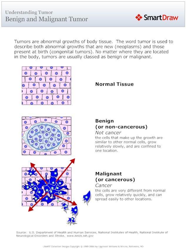
10 Basic definitions - Neoplasia represents an overgrowth of a single cell type in most cases that goes sometimes out of control. - Almost all cells in the body can undergo neoplasia, but some do so much more frequently than others. - As mentioned in previous lectures, neoplasia can develop into?? - Benign or malignant neoplasms. - Benign is localized single mass of cells that remain localized at their site of origin and limited in their growth. - Malignant neoplasms are defined by their potential to invade and metastasize (transplantation of cells into new site) at some point in their life history. - Tumor or cancer are used as synonym for neoplasm.
11 Basic definitions
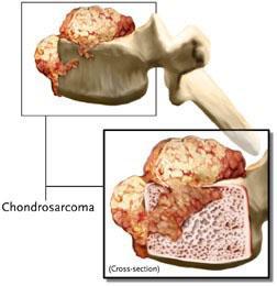
12 Classification of neoplasms Is there a system to name different classification of neoplasms? - Yes & no. what is the most common suffix for cancer? - oma. Most benign neoplasms are named by the cell or tissue they resemble plus the suffix oma indicating neoplasm. Example? - Fibroma, lipoma, adenoma etc. - With malignant neoplasms, either carcinoma or sarcoma is added to the name of the tissue to name the type of neoplasm. Example? - Adenocarcinoma of the breast (breast glands), bronchogenic carcinoma (bronchial epithelium of lung), osteosarcoma (bone), chondrosarcoma (cartilage).
13 Classification of neoplasms Do all neoplasms follow the same naming system? - Not really. There are several exceptions to this naming system. Example? - Hematoma!!! It is in fact a collection of blood, not a neoplasm. - A few additional types of malignant neoplasms of nonepithelial or ambiguous origin are named separately. Example? - Leukemia, and malignant lymphoma.
14 Morphologic features What is morphology? - The branch of biology concerned with the form and structure of organisms. Why is it important to understand the morphologic features of neoplasms? - So differentiation between hyperplasisas, hypertrophies, benign, and malignant neoplasms can be made to select the most appropriate prevention or treatment plan. - Hyperplasias: - They represent a very diverse group of conditions about which it is difficult to generalize. - They are generally characterized by an increase in the normal cellular elements of the organ. Example? - Lymph node hyperplasia, breast hyperplasia common during pregnancy, and enlarged thyroid.

15 Morphologic features
16 Morphologic features - Hyperplasias of epithelial surface cells, such as skin, oral cavity and cervix are particularly important as sites of cancer development. - They develop into lesions that can be visualized grossly and appear more opaque than surrounding surface. - Some surface hyperlasias could be confused with inflammation. In this case pathologists should confirm the condition microscopically and determine whether it is a simple hyperplasia or premalignant hyperplasia. - A premalignant hyperplasia lesion is one in which there is an increased likelihood of cancer compared to adjacent normal tissue.


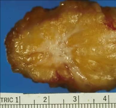
17 - Benign neoplasms. Morphologic features - The morphologic features of benign neoplasms are relatively easy to recognize grossly and microscopically, because they produce a single mass that is discrete from surrounding tissue. - When originating on a body surface, benign neoplasms extend outwardly, producing a polyp. - When originating within solid organs or connective tissue, benign neoplasms compress tissue around them to form a fibrous rim (capsule).
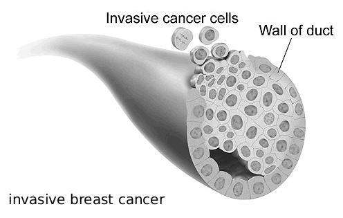
18 Morphologic features - Malignant neoplasms. - Early lesions are hardest to diagnose but are most important. - Two criteria are required for the diagnosis of malignant neoplasm including establishing that the cells are neoplastic and demonstrating invasion. - Invasion and metastasis are the principal criteria used to distinguish benign and malignant neoplasms. - Invasion is a more commonly used criterion for cancer than metastasis. Invasion is characterized by infiltration of cancer cells with poor respect for tissue boundaries. - Invasion is characterized by irregularity of tumor tissue margins, failure of the tumor to separate from surrounding tissue during removal, and when advanced, direct spread beyond the organ of origin.

19 Morphologic features
20 Life history & spread of cancer How long is cancer life history? - They have a very long life history and most occur before there is any lesion that can be called cancer. - Genetic alteration is the basis for development of cancer, but of all the mutations that occur in cells, very few lead to cancer. - So what alterations are needed to develop cancer in cells? a. Initiation: series of alterations to acquire the growth potential referred to as cancer. b. Carcinogens: agents that alter cells and could be physical, chemical or biological. c. Promoter: another agent that furthers the expression of the cancer in that cell and its progeny (outcome).
 acquire metastatic capacity late in tumor - genesis.")
21 Life history & spread of cancer Models of the metastatic process. a, The traditional model of metastasis suggests that only subpopulations of tumor cells (red) acquire metastatic capacity late in tumor - genesis. b, Spontaneous metastasis assays indicate that all tumor cells have the capability to develop a metastasis. c, The 'dynamic heterogeneity' model proposes that the frequency with which metastatic variants arise within the primary tumor determines its metastatic potential. d, The 'clonal dominance' theory proposes that metastatic subclones within a primary tumor can overgrow and dominate the tumor mass itself. e, The 'genometastasis hypothesis' proposes that metastasis occurs through transfection of susceptible cells in distant organs.
22 Grading & staging Is there a difference between grading & staging cancer? - Yes. What is the difference? - Grading refers to the histologic differentiation of cancer in which the degree of resemblance of the cancer to its tissue of origin is evaluated. - Cancer are commonly graded as; well-differentiated, moderately differentiated, poorly differentiated, or undifferentiated. - Staging reflects the degree of local invasion and regional distant metastasis. - Rules for staging involve evaluation for in situ change, localization to the organ of origin, direct spread beyond the organ, and distant metastasis.

23 Enough is enough for today
Neoplasia literally means "new growth.
 NEOPLASIA Neoplasia literally means "new growth. A neoplasm, defined as "an abnormal mass of tissue the growth of which exceeds and is uncoordinated with that of the normal tissues and persists in the
Abdulrahman Alhanbali. Bahaa Najjar. Maha shomaf
 14 Abdulrahman Alhanbali Bahaa Najjar Maha shomaf 1 Neoplasia In this lecture we will talk about neoplasia, its features and the nomenclature of different types of tumors. Neoplasia (neo: new and plasia:
ONCOLOGY. Csaba Bödör. Department of Pathology and Experimental Cancer Research november 19., ÁOK, III.
 ONCOLOGY Csaba Bödör Department of Pathology and Experimental Cancer Research 2018. november 19., ÁOK, III. bodor.csaba1@med.semmelweis-univ.hu ONCOLOGY Characteristics of Benign and Malignant Neoplasms
Epithelial tumors. Dr. F.F. Khuzin, PhD Dr. M.O. Mavlikeev
 Epithelial tumors Dr. F.F. Khuzin, PhD Dr. M.O. Mavlikeev Epithelial tumors Tumors from the epithelium are the most frequent among tumors. There are 2 group features of these tumors: The presence in most
NEOPLASIA-I CANCER. Nam Deuk Kim, Ph.D.
 NEOPLASIA-I CANCER Nam Deuk Kim, Ph.D. 1 2 Tumor in the hieroglyphics of the Edwin Smith papyrus (1,600 B.C., Breasted s translation 1930) 3 War on Cancer (National Cancer Act, 1971) 4 Cancer Acts in Korea
CODING TUMOUR MORPHOLOGY. Otto Visser
 CODING TUMOUR MORPHOLOGY Otto Visser INTRODUCTION The morphology describes the tissue of the tumour closest to normal tissue Well differentiated tumours are closest to normal Undifferentiated tumours show
Biochemistry of Carcinogenesis. Lecture # 35 Alexander N. Koval
 Biochemistry of Carcinogenesis Lecture # 35 Alexander N. Koval What is Cancer? The term "cancer" refers to a group of diseases in which cells grow and spread unrestrained throughout the body. It is difficult
Chapter 3. Neoplasms. Copyright 2015 Cengage Learning.
 Chapter 3 Neoplasms Terminology Related to Neoplasms and Tumors Neoplasm New growth Tumor Swelling or neoplasm Leukemia Malignant disease of bone marrow Hematoma Bruise or contusion Classification of Neoplasms
DUSTURBANCES OF GROWTH. MLS Basic histological diagnosis MLS HIST 422 Semester 8- batch 7 L8 Uz: Musa
 DUSTURBANCES OF GROWTH MLS Basic histological diagnosis MLS HIST 422 Semester 8- batch 7 L8 Uz: Musa Agnesia: means complete absence of an organ (Kidney). Aplasia: s defined in general as "defective development
Introduction to Basic Oncology
 Introduction to Basic Oncology Cancer Cell AHS 102 Med Term Dr. Susie Turner 1/3/13 General Oncology Study of Tumors Neoplasms/Tumors Abnormal growth of new tissue Are either; Benign or Malignant Onc/o
Cancer in Estonia 2014
 Cancer in Estonia 2014 Estonian Cancer Registry (ECR) is a population-based registry that collects data on all cancer cases in Estonia. More information about ECR is available at the webpage of National
BY Mrs. K.SHAILAJA., M. PHARM., LECTURER DEPT OF PHARMACY PRACTICE, SRM COLLEGE OF PHARMACY
 BY Mrs. K.SHAILAJA., M. PHARM., LECTURER DEPT OF PHARMACY PRACTICE, SRM COLLEGE OF PHARMACY Cancer is a group of more than 100 different diseases that are characterized by uncontrolled cellular growth,
A neoplasm is defined as "an abnormal tissue proliferation, which exceeds that of adjacent normal tissue. This proliferation continues even after
 NEOPLASIA Neoplasia is a very important topic in pathology because neoplasms are both common and serious diseases. A neoplasm literally means a new growth, and this term is used interchangeably with a
Neoplasia 2018 Lecture 2. Dr Heyam Awad MD, FRCPath
 Neoplasia 2018 Lecture 2 Dr Heyam Awad MD, FRCPath ILOS 1. List the differences between benign and malignant tumors. 2. Recognize the histological features of malignancy. 3. Define dysplasia and understand
CODING PRIMARY SITE. Nadya Dimitrova
 CODING PRIMARY SITE Nadya Dimitrova OUTLINE What is coding and why do we need it? ICD-10 and ICD-O ICD-O-3 Topography coding rules ICD-O-3 online WHAT IS CODING AND WHY DO WE NEED IT? Coding: to assign
Overview of Anatomy & Physiology
 Overview of Anatomy & Physiology Anatomy the study of the structure of body parts and their relationships to one another Gross or macroscopic Microscopic Developmental Physiology the study of the function
Tumour Structure and Nomenclature. Paul Edwards. Department of Pathology and Cancer Research UK Cambridge Institute, University of Cambridge
 Tumour Structure and Nomenclature Paul Edwards Department of Pathology and Cancer Research UK Cambridge Institute, University of Cambridge Malignant Metastasis Core idea of cancer Normal Cell Slightly
7/4/2018. Key Objectives. A and P 2401 Lecture 2 TWO MECHANISMS USED TO MAINTAIN HOMEOSTASIS. Negative Feedback Examples. Review of Homeostasis
 Key Objectives Review of Homeostasis Negative Feedback Mechanisms Positive Feedback Mechanisms Body Systems and Function A and P 2401 Lecture 2 HOMEOSTASIS TWO MECHANISMS USED TO MAINTAIN HOMEOSTASIS The
Body Systems Overview
 Body Systems Overview Body Systems work together: If you damage one system, you may damage several for example, smoking irritates the lungs and destroys the cells of the immune system Levels of Organization
Diseases of the vulva
 Diseases of the vulva 1. Bartholin Cyst - Infection of the Bartholin gland produces an acute inflammation within the gland (adenitis) and may result in an abscess. Bartholin duct cysts - Are relatively
Basic Tissue Types and Functions
 Tissues Histology Basic Tissue Types and Functions 1) Epithelial tissue covering 2) Connective tissue support 3) Muscle tissue movement 4) Nervous tissue control Epithelial Tissue 1) Covers a body surface
Overview of Anatomy and Physiology
 1 The Human Body: An Orientation Overview of Anatomy and Physiology Anatomy the study of the structure of body parts and their relationships to one another Gross or macroscopic Microscopic Developmental
Disorders of Cell Growth & Neoplasia. Histopathology Lab
 Disorders of Cell Growth & Neoplasia Histopathology Lab Paul Hanna April 2010 Case #84 Clinical History: 5 yr-old, West Highland White terrier. skin mass from axillary region. has been present for the
CANCER = Malignant Tumor = Malignant Neoplasm
 CANCER = Malignant Tumor = Malignant Neoplasm A tissue growth: Not necessary for body s development or repair Invading healthy tissues Spreading to other sites of the body (metastasizing) Lethal because
ANNUAL CANCER REGISTRY REPORT-2005
 ANNUAL CANCER REGISTRY REPORT-25 CANCER STATISTICS Distribution of neoplasms Of a total of 3,115 new neoplasms diagnosed or treated at the Hospital from January 25 to December, 25, 1,473 were seen in males
Truman Medical Center-Hospital Hill Cancer Registry 2014 Statistical Summary Incidence
 Truman Medical Center-Hospital Hill Cancer Registry 2014 Statistical Summary Incidence In 2014, there were 452 new cancer cases diagnosed and or treated at Truman Medical Center- Hospital Hill and an additional
performed to help sway the clinician in what the appropriate diagnosis is, which can substantially alter the treatment of management.
 Hello, I am Maura Polansky at the University of Texas MD Anderson Cancer Center. I am a Physician Assistant in the Department of Gastrointestinal Medical Oncology and the Program Director for Physician
Format Of ICD-O Terms In Numerical List Each topographic and morphologic term appears only once The first listed term in Bold Type is the Preferred Te
 Florida Cancer Data System International Classification of Diseases for Oncology ICD-O-3 1 Basic Concepts Primary Site/Topography Histology/Morphology Behavior Grade/Immunophenotype 2 ICD-O 3 Structure/Format
Dr Rodney Itaki Lecturer Anatomical Pathology Discipline. University of Papua New Guinea School of Medicine & Health Sciences Division of Pathology
 Neoplasia Dr Rodney Itaki Lecturer Anatomical Pathology Discipline University of Papua New Guinea School of Medicine & Health Sciences Division of Pathology General Considerations Overview: Neoplasia uncontrolled,
Unit I Problem 9 Histology: Basic Tissues of The Body
 Unit I Problem 9 Histology: Basic Tissues of The Body - What is the difference between cytology and histology? Cytology: it is the study of the structure and functions of cells and their contents. Histology:
- A cancer is an uncontrolled, independent proliferation of robust, healthy cells.
 1 Cancer A. What is it? - A cancer is an uncontrolled, independent proliferation of robust, healthy cells. * In some the rate is fast; in others, slow; but in all cancers the cells never stop dividing.
2011 to 2015 New Cancer Incidence Truman Medical Center - Hospital Hill
 Number of New Cancers Truman Medical Center Hospital Hill Cancer Registry 2015 Statistical Summary Incidence In 2015, Truman Medical Center diagnosed and/or treated 406 new cancer cases. Four patients
Introduction to ICD-O-3 coding rules
 Introduction to ICD-O-3 coding rules Weena Laddachayaporn, MD National Cancer Institute, Bangkok, Thailand ICD-O-3 The International Classification of Diseases for Oncology Is a coding system for primary
1.Acute and Chronic Cervicitis - At the onset of menarche, the production of estrogens by the ovary stimulates maturation of the cervical and vaginal
 Diseases of cervix I. Inflammations 1.Acute and Chronic Cervicitis - At the onset of menarche, the production of estrogens by the ovary stimulates maturation of the cervical and vaginal squamous mucosa
number Done by Corrected by Doctor Maha shomaf
 number 17 Done by Ahmad rawajbeh Corrected by أسامة الخضر Doctor Maha shomaf 0 P a g e In this lecture, we are going to: complete the differentiation between benign and malignant tumors. - -start to study
The European Commission s science and knowledge service. Joint Research Centre
 The European Commission s science and knowledge service Joint Research Centre Coding Primary Site and Tumour Morphology JRC-ENCR training course Copenhagen, 25 September 2018 Nadya Dimitrova Outline What
- is a common disease - 1 person in 3 can expect to contract cancer at some stage in their life -1 person in 5 can expect to die from it
 MBB157 Dr D Mangnall The Molecular Basis of Disease CANCER Lecture 1 One of the simpler (and better) definitions of cancer comes from the American Cancer Society, who define cancer as; 'Cancer is a group
155.2 Malignant neoplasm of liver not specified as primary or secondary. C22.9 Malignant neoplasm of liver, not specified as primary or secondary
 ICD-9 TO ICD-10 Reference ICD-9 150.9 Malignant neoplasm of esophagus unspecified site C15.9 Malignant neoplasm of esophagus, unspecified 151.9 Malignant neoplasm of stomach unspecified site C16.9 Malignant
Comprehensive cancer cover
 Retirement Investments Insurance Health Comprehensive cancer cover Life Insurance+ with critical illness and Critical Illness+ Cancer is one of the biggest fears for the British public This is why our
NEOPLASIA. 3. Which of the following tumour is benign a. Chondrosarcoma b. Osteochondroma c. Chondroblastoma d. Ewing s tumour e.
 NEOPLASIA 1. malignant neoplasms a. are independent of hormonal influence b. are always composed of homogenous cell lines c. arise from differentiated cells by a process of anaplasia d. display abnormal
25 TH ICRO DEHRADUN STAGING OF GENITOURINARY MALIGNANCIES
 25 TH ICRO DEHRADUN STAGING OF GENITOURINARY MALIGNANCIES SPEAKER DR DEEPAK ABROL CLINICAL ONCOLOGIST JAND K HEALTH SERVICES CONSULTANT ONCOLOGIST MAHARISHI DAYANAND HOSPITAL AND MEDICAL RESEARCH CENTER
Radiation Oncology Study Guide
 Radiation Oncology Study Guide For the Initial CertificationQualifying (Computer-Based) Examination General and Radiation Oncology This examination is designed to assess your understanding of the entire
the urinary system pathology Dr. Fairoz A Eltorgman
 the urinary system pathology Dr. Fairoz A Eltorgman Tumors of the renal pelvis & kidney Benign tumors of the renal pelvis: Hemangioma Leiomyoma Malignant tumors: Transitional cell carcinoma Squamous cell
FOLLICULARITY in LYMPHOMA
 FOLLICULARITY in LYMPHOMA Reactive Follicular Hyperplasia Follicular Hyperplasia irregular follicles Follicular Hyperplasia dark and light zones Light Zone Dark Zone Follicular hyperplasia MIB1 Follicular
number Done by Corrected by Doctor Maha Shomaf
 number 16 Done by Waseem Abo-Obeida Corrected by Zeina Assaf Doctor Maha Shomaf MALIGNANT NEOPLASMS The four fundamental features by which benign and malignant tumors can be distinguished are: 1- differentiation
Cancer A Superficial Introduction
 Cancer A Superficial Introduction Gabor Fichtinger, Queen s University Cancer some definitions Medical term: malignant neoplasm Class of diseases in which a group of cells display: uncontrolled growth
Comprehensive cancer cover
 Retirement Investments Insurance Health Comprehensive cancer cover Life Insurance+ with critical illness and Critical Illness+ Cancer is one of the biggest fears for the British public This is why our
American Cancer Society Estimated Cancer Deaths by Sex and Age (years), 2013
, 2013") American Cancer Society Estimated Cancer Deaths by Sex and Age (years), 2013 All ages Younger than 45 45 and Older Younger than 65 65 and Older All sites, men 306,920 9,370 297,550 95,980 210,940 All sites,
American Cancer Society Estimated Cancer Deaths by Sex and Age (years), 2013 All ages Younger than 45 45 and Older Younger than 65 65 and Older All sites, men 306,920 9,370 297,550 95,980 210,940 All sites,
Icd 10 code lung ca with mets to bone
 Icd 10 code lung ca with mets to bone 2018 ICD-10 -CM Diagnosis Code C79.9.. C79.9 is a billable/specific ICD - 10 -CM code that can be used to indicate a diagnosis. Metastasis from malignant tumor of
THE BURDEN OF CANCER IN NEBRASKA: RECENT INCIDENCE AND MORTALITY DATA
 THE BURDEN OF CANCER IN NEBRASKA: RECENT INCIDENCE AND MORTALITY DATA Presented by: Bryan Rettig, MS Nebraska Dept. of Health & Human Services Division of Public Health May 31, 2017 Nebraska Cancer Registry
Greater Manchester and Cheshire HPB Unit Guidelines for the Assessment & Management of Hepatobiliary and Pancreatic Disease Chapter 14
 Greater Manchester and Cheshire HPB Unit Guidelines for the Assessment & Management of Hepatobiliary and Pancreatic Disease Chapter 14 Contents 14. Neuroendocrine Tumours 161 14.1. Diagnostic algorithm
They cells can not function death.
 Jenna Hellack Jan 2001 Tissues What do you think happens when the cells use up their food and oxygen before there is time to replenish it? They cells can not function death. Blood Cell Cancer cell Plant
CANCER Uncontrolled Cell Division
 CANCER Uncontrolled Cell Division What is cancer? Why does it occur? Where does it occur? Benign vs. Malignant? Types of Cancer (3 main groups) There are over 200 different types of cancer 1) Carcinomas
Aberrant cell Growth. Younas Masih New Life College of Nursing Karachi. 3/4/2016 Younas Masih ( NLCON)
") Aberrant cell Growth Younas Masih New Life College of Nursing Karachi 1 Objectives By the end of this session the learners will be able to, Define the characteristics of the normal cell Describe the characteristics
Aberrant cell Growth Younas Masih New Life College of Nursing Karachi 1 Objectives By the end of this session the learners will be able to, Define the characteristics of the normal cell Describe the characteristics
SMOKING AND CANCER RISK
 SMOKING AND CANCER RISK The effects of smoking on health were documented in a landmark report by the Surgeon General in 1964. Since then the devastating effect from smoking on millions of American lives
SHN-1 Human Digestive Panel Test results
 SHN-1 Human Digestive Panel Test results HN-30 tongue HN-24 salivary gland HN-12 larynx HN-28 esophagus HN-29 stomach HN-20 pancreas HN-13 liver HN-14 gall bladder HN-27-1 duodenum HN-27-2 ileum HN-27-3
Outcomes Report: Accountability Measures and Quality Improvements
 Outcomes Report: Accountability Measures and Quality Improvements The FH Memorial Medical Center s Cancer Committee ensures that patients with cancer are treated according to the nationally accepted measures.
Human Body. The student knows that the human body is made of systems with structure and functions that are related.
 Human Body The student knows that the human body is made of systems with structure and functions that are related. Cells Cells are the smallest living structures. All living things are made up of one or
S2 File. Clinical Classifications Software (CCS). The CCS is a
. The CCS is a") S2 File. Clinical Classifications Software (CCS). The CCS is a diagnosis categorization scheme based on the ICD-9-CM that aggregates all diagnosis codes into 262 mutually exclusive, clinically homogeneous
S2 File. Clinical Classifications Software (CCS). The CCS is a diagnosis categorization scheme based on the ICD-9-CM that aggregates all diagnosis codes into 262 mutually exclusive, clinically homogeneous
Epidemiology in Texas 2006 Annual Report. Cancer
 Epidemiology in Texas 2006 Annual Report Cancer Epidemiology in Texas 2006 Annual Report Page 94 Cancer Incidence and Mortality in Texas, 2000-2004 The Texas Department of State Health Services Texas Cancer
Icd-10 code for small cell lung cancer
 P ford residence southampton, ny Icd-10 code for small cell lung cancer Non-small cell lung carcinomas are usually adenocarcinomas, squamous cell carcinomas, or large cell carcinomas. Metastatic carcinomas
Health Reference Series. Seventh Edition. Cancer
 Health Reference Series Seventh Edition Cancer SOURCEBOOK Basic Consumer Health Information about Major Forms and Stages of Cancer, Featuring Facts about Head and Neck Cancers, Lung Cancers, Gastrointestinal
Cells Tissues Organs Organ Systems Organism. Cells: the smallest unit of life.
 Cells Tissues Organs Organ Systems Organism Cells: the smallest unit of life. The Circulatory Systems brings oxygen, nutrients and hormones to cells; fights infections; removes cell wastes; regulates
BREAST PATHOLOGY. Fibrocystic Changes
 BREAST PATHOLOGY Lesions of the breast are very common, and they present as palpable, sometimes painful, nodules or masses. Most of these lesions are benign. Breast cancer is the 2 nd most common cause
Introduction to pathology
 Introduction to pathology By Dr. Mohsen Dashti Clinical Medicine & Pathology 316 1 st Lecture Lecture outilne Pathology. Disease. Cell injury. Manifestations of disease. Structural diseases. Functional
-1- Pathology Department (code: 0605) Final Exam for Third year students Date: Time allowed: 2 hours. Paper II (75 marks).
 Final Exam for Third year students Date: Time allowed: 2 hours. Paper II (75 marks).") -1- BENHA UNIVERSITY FACULTY OF MEDICINE Pathology Department (code: 0605) Final Exam for Third year students Date: 28-5-2011 Time allowed: 2 hours. Paper II (75 marks). Please note that this question
-1- BENHA UNIVERSITY FACULTY OF MEDICINE Pathology Department (code: 0605) Final Exam for Third year students Date: 28-5-2011 Time allowed: 2 hours. Paper II (75 marks). Please note that this question
Neoplasia 2018 Lecture 1. Dr Heyam Awad MD, FRCPath
 Neoplasia 2018 Lecture 1 Dr Heyam Awad MD, FRCPath Dear All Welcome to this part of your course ( introduction to pathology) where we will study neoplasia in detail. Please note that each lecture builds
RCPS Curriculum Pacing Guide Subject: Anatomy and Physiology
 RCPS Curriculum Pacing Guide 2013 2014 Subject: Anatomy and Physiology Week of: SOL # Unit Bloom s Objectives Throughout All units the course During field trip Throughout the course A+P1 Collecting, analyzing,
Cancer Fundamentals. Julie Randolph-Habecker, Ph.D. Director, Experimental Histopathology Shared Resource
 Cancer Fundamentals Julie Randolph-Habecker, Ph.D. Director, Experimental Histopathology Shared Resource Cancer Overview Leading cause of death in US 1.2 million diagnosed each year More common after age
Tissues are: group of similar or identical cells that share a common function. used to build organs
 Tissues: Four classes Epithelium Connective Muscle Nervous Tissues are: group of similar or identical cells that share a common function. used to build organs Overview: Epithelial o Line body cavities
Human Anatomy & Physiology
 Human Anatomy & Physiology Hey I thought those were the same thing! Nope they ain t Anatomy-Where everything is and to what it is connected. Physiology-How all that stuff works to keep you alive! Morphology-How
Cancer Association of South Africa (CANSA)
") Cancer Association of South Africa (CANSA) Fact Sheet on ICD-10 Coding of Neoplasms Introduction The International Statistical Classification of Diseases and Related Health Problems, 10 th Revision (ICD-10)
Cancer Association of South Africa (CANSA) Fact Sheet on ICD-10 Coding of Neoplasms Introduction The International Statistical Classification of Diseases and Related Health Problems, 10 th Revision (ICD-10)
Part I. An Introduction to Cancer
 Part I An Introduction to Cancer 2 Chapter 1 Cancer: Descriptive Overview Cancer is a disease in which cells propagate uncontrollably. These cells can come from many different parts of the body and the
SMOKING AND CANCER RISK
 SMOKING AND CANCER RISK The effects of smoking on health were documented in a landmark report by the Surgeon General in 1964. Since then the devastating effect from smoking on millions of American lives
TUMOR,NEOPLASM. Pathology Department, Zhejiang University School of Medicine,
 TUMOR,NEOPLASM Pathology Department, Zhejiang University School of Medicine, 马丽琴,maliqin198@zju.edu.cn The points in this chapter What is a neoplasm (conception) Morphology of neoplasm Macroscopy of Neoplasm
Pathology of Sarcoma ELEANOR CHEN, MD, PHD, ASSISTANT PROFESSOR DEPARTMENT OF PATHOLOGY UNIVERSITY OF WASHINGTON
 Pathology of Sarcoma ELEANOR CHEN, MD, PHD, ASSISTANT PROFESSOR DEPARTMENT OF PATHOLOGY UNIVERSITY OF WASHINGTON Presentation outline Background and epidemiology of sarcomas Sarcoma classification Sarcoma
Mast Cell Tumors in Dogs
 Mast Cell Tumors in Dogs 803-808-7387 www.gracepets.com These notes are provided to help you understand the diagnosis or possible diagnosis of cancer in your pet. For general information on cancer in pets
Test Bank for Robbins and Cotran Pathologic Basis of Disease 9th Edition by Kumar
 Link full download:https://getbooksolutions.com/download/test-bank-for-robbinsand-cotran-pathologic-basis-of-disease-9th-edition-by-kumar Test Bank for Robbins and Cotran Pathologic Basis of Disease 9th
Note: The cause of testicular neoplasms remains unknown
 - In the 15- to 34-year-old age group, they are the most common tumors of men. - Tumors of the testis are a heterogeneous group of neoplasms that include: I. Germ cell tumors : 95%; all are malignant.
Ocular Neoplasia What s Common? What s New? Richard R Dubielzig
 Ocular Neoplasia What s Common? What s New? Richard R Dubielzig Orbit 288 6% Tumors of the globe make up 3225 out of 6110 total neoplasms = 53%. Tumors of the conjunctiva make up 1192 out of 6110 total
MVST BOD & NST PART IB Thurs. 2 nd & Fri. 3 rd March 2017 Pathology Practical Class 23
 MVST BOD & NST PART IB Thurs. 2 nd & Fri. 3 rd March 2017 Pathology Practical Class 23 Neoplasia I Neoplasia I: Benign and malignant neoplasms in glandular epithelium and mesenchyme 1.0. Aims 1. To understand
Test Bank for Robbins and Cotran Pathologic Basis of Disease 9th Edition by Kumar
 Link full download: http://testbankair.com/download/test-bank-for-robbins-cotran-pathologic-basis-of-disease-9th-edition-bykumar-abbas-and-aster Test Bank for Robbins and Cotran Pathologic Basis of Disease
Epithelial Tissue. Simple Cuboidal Function: secretion and absorption. Simple Squamous
 Epithelial Tissue General Functions: Lines and covers organs Absorbs / secretes substances Gas exchange Protection Special Characteristics: - have an apical surface on top - have a basement membrane below
Cerebral Parenchymal Lesions: I. Metastatic Neoplasms
 Chapter 4 Cerebral Parenchymal Lesions: I. Metastatic Neoplasms After one has reasonably ruled out the possibility of a nonneoplastic diagnosis (see Chap. 3), one is left with considering a diagnosis of
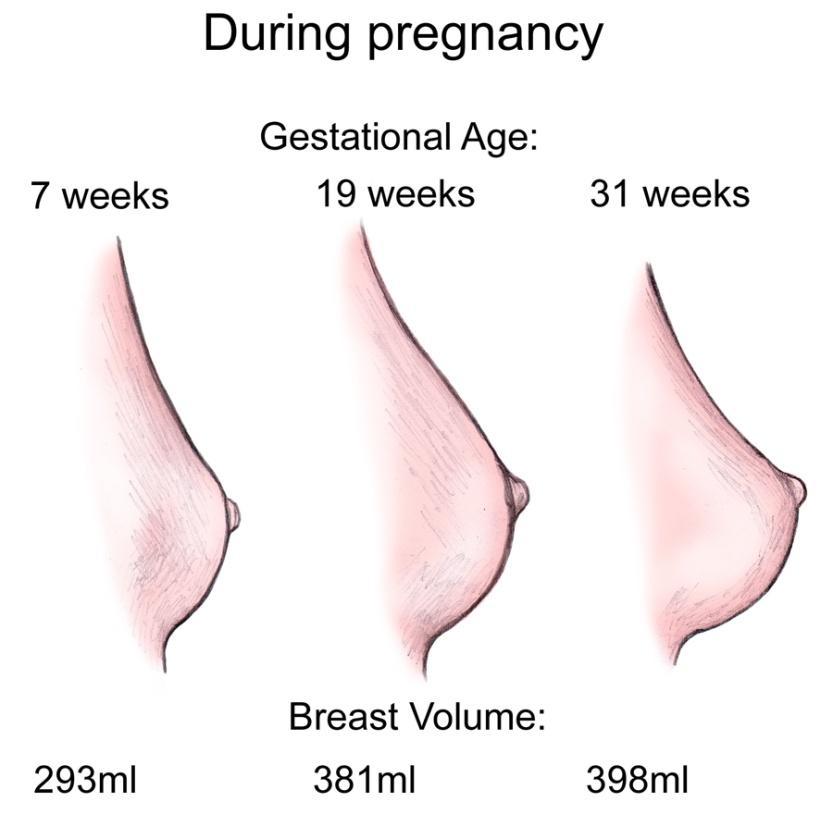
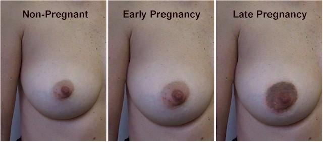
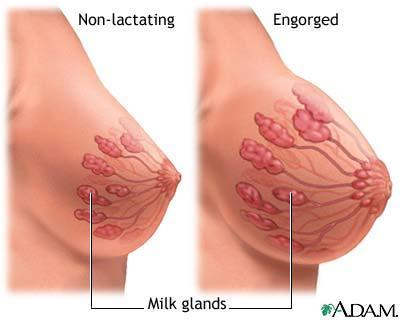
Mousa. Israa Ayed. Abdullah AlZibdeh. 0 P a g e
 1 Mousa Israa Ayed Abdullah AlZibdeh 0 P a g e Breast pathology The basic histological units of the breast are called lobules, which are composed of glandular epithelial cells (luminal cells) resting on
Chapter 9, Part 1: Biology of Cancer and Tumor Spread
 PATHOPHYSIOLOGY Name Chapter 9, Part 1: Biology of Cancer and Tumor Spread I. Cancer Characteristics and Terminology Neoplasm new growth, involves the overgrowth of tissue to form a neoplastic mass (tumor).
DATA STANDARDS AND QUALITY CONTROL MEMORANDUM DSQC #
 DATA STANDARDS AND QUALITY CONTROL MEMORANDUM DSQC #2006-01 CATEGORY: CLARIFICATION SUBJECT: RESCINDMENT - DSQC MEMORANDUM 2002-08 Coding Complex Morphologic Diagnoses (revised 8/02) EFFECTIVE: For Cases
Diseases of the breast (1 of 2)
") Diseases of the breast (1 of 2) Introduction A histology introduction Normal ducts and lobules of the breast are lined by two layers of cells a layer of luminal cells overlying a second layer of myoepithelial
Diseases of the breast (1 of 2) Introduction A histology introduction Normal ducts and lobules of the breast are lined by two layers of cells a layer of luminal cells overlying a second layer of myoepithelial
Chapter 1- An Orientation to the Human Body
 Chapter 1- An Orientation to the Human Body Overview of Anatomy and Physiology: -Anatomy- of body parts and their relationships to one another. -Gross or Macroscopic= large and easily observable -Microscopic=
Table of Contents. Preface xi. Acknowledgments xiii. Part I Overview of the Diagnostic Process 1. 1 Overview of Grading and Staging 3
 Table of Contents Preface xi Acknowledgments xiii Part I Overview of the Diagnostic Process 1 1 Overview of Grading and Staging 3 Identification of the process 3 Identification of tumor types 5 Grading
Microscopic Anatomy Cytology study of the cell Histology study of tissues
 Introduction to Anatomy and Physiology Dr. Gary Mumaugh Overview of Anatomy and Physiology Anatomy the study of the structure of body parts and their relationships to one another o Gross or macroscopic
number Done by Corrected by Doctor مها شوماف
 number 15 Done by Ali Yaghi Corrected by Waseem Alhaj Doctor مها شوماف 1 P a g e Epidemiology Epidemiology is the study of the incidence of a disease. It can give us information about the possible causes
INTRODUCTION TO ANIMALS
 AP BIOLOGY ANIMALS ACTIVITY #1 NAME DATE HOUR INTRODUCTION TO ANIMALS LEVELS OF ORGANIZATION Animals Activity #1 page 1 HOMEOSTASIS: DEFINITION IMPORTANCE MECHANISMS FOR MAINTAINING HOMEOSTASIS: Animals
-The cause of testicular neoplasms remains unknown
 - In the 15- to 34-year-old age group, they are the most common tumors of men. - include: I. Germ cell tumors : (95%); all are malignant. II. Sex cord-stromal tumors: from Sertoli or Leydig cells; usually
The Human Body: An Orientation
 PowerPoint Lecture Slide Presentation by Patty Bostwick-Taylor, Florence-Darlington Technical College The Human Body: An Orientation 1 PART A The Human Body An Orientation Anatomy Study of the structure
Malignant Cardiac Tumors Rad-Path Correlation
 Malignant Cardiac Tumors Rad-Path Correlation Vincent B. Ho, M.D., M.B.A. 1 Jean Jeudy, M.D. 2 Aletta Ann Frazier, M.D. 2 1 Uniformed Services University of the Health Sciences 2 University of Maryland
Greater Baltimore Medical Center Sandra & Malcolm Berman Cancer Institute
 2008 ANNUAL REPORT Greater Baltimore Medical Center Sandra & Malcolm Berman Cancer Institute Cancer Registry Report The Cancer Data Management System/ Cancer Registry collects data on all types of cancer
Organ Systems (ch21-26) Practice Questions. Name:
 Practice Questions. Name:") 1. Which one of the following types of tissue stores fat in the body? A) blood B) cartilage C) bone D) adipose tissue E) fibrous connective tissue 2. Which of the following tissues does not match its function?
1. Which one of the following types of tissue stores fat in the body? A) blood B) cartilage C) bone D) adipose tissue E) fibrous connective tissue 2. Which of the following tissues does not match its function?
Quiz. b. 4 High grade c. 9 Unknown
 Quiz 1. 10/11/12 CT scan abdomen/pelvis: Metastatic liver disease with probable primary colon malignancy. 10/17/12 Colonoscopy with polypectomy: Adenocarcinoma of sigmoid colon measuring at least 6 mm
Icd-10 code for metastatic small cell cancer
 P ford residence southampton, ny Icd-10 code for metastatic small cell cancer A term for diseases in which abnormal cells divide without control and can invade nearby tissues. Malignant cells can also