Interpretation Manual - Gastric or Gastroesophageal Junction Adenocarcinoma. PD-L1 IHC 22C3 pharmdx is FDA-approved for in vitro diagnostic use
|
|
|
- Pauline Simon
- 5 years ago
- Views:
Transcription

1 Interpretation Manual - Gastric or Gastroesophageal Junction Adenocarcinoma PD-L1 IHC 22C3 pharmdx is FDA-approved for in vitro diagnostic use
2 For countries outside of the United States, see the local KEYTRUDA product label for approved indications and expression cutoff values to guide therapy. 2
3 Table of Contents Intended Use 04 Introduction 06 PD-L1 Overview 08 PD-L1 IHC 22C3 pharmdx Overview 10 Kit Configuration (SK006) 11 Technical Considerations 12 Specimen Preparation 12 In-house Control Tissue 12 Optional Additional In-house Control: Tonsil Tissue 13 Tissue Processing 13 PD-L1 IHC 22C3 pharmdx Staining Procedure 14 Technical Checklist 17 Slide Evaluation 18 General Considerations 18 Specimen Adequacy 18 Evaluating Controls 18 Slide Evaluation Flow Chart 22 Combined Positive Score 23 Definition of Combined Positive Score (CPS) 23 CPS Numerator Inclusion and Exclusion Criteria 23 Determining Combined Positive Score 23 Suggested Methods 25 Interpretation of CPS 28 Identifying Patients with Gastric or GEJ Adenocarcinoma for Treatment 29 PD-L1 IHC 22C3 pharmdx Testing Scheme 30 Reporting Results 31 Combined Positive Score Summary and Examples 32 Key Considerations in Scoring PD-L1 IHC 22C3 pharmdx Stained Specimens 32 Image Guide for Interpretation of PD-L1 IHC 22C3 pharmdx Staining in Gastric or GEJ Adenocarcinoma 33 CPS < 1 Case Examples 47 CPS 1 Case Examples 53 Near Cut-off Case Examples (CPS 0 10) 59 Artifacts 62 Troubleshooting Guide 67 Clinical Performance Evaluation 68 References 70 3
4 Intended Use For in vitro diagnostic use. PD-L1 IHC 22C3 pharmdx is a qualitative immunohistochemical assay using Monoclonal Mouse Anti-PD-L1, Clone 22C3 intended for use in the detection of PD-L1 protein in formalin-fixed, paraffin-embedded (FFPE) non-small cell lung cancer (NSCLC) and gastric or gastroesophageal junction (GEJ) adenocarcinoma tissues using EnVision FLEX visualization system on Autostainer Link 48. Non-Small Cell Lung Cancer (NSCLC) PD-L1 protein expression in NSCLC is determined by using Tumor Proportion Score (TPS), which is the percentage of viable tumor cells showing partial or complete membrane staining at any intensity. The specimen should be considered to have PD-L1 expression if TPS 1% and high PD-L1 expression if TPS 50%. PD-L1 IHC 22C3 pharmdx is indicated as an aid in identifying NSCLC patients for treatment with KEYTRUDA (pembrolizumab). See the KEYTRUDA product label for expression cutoff values guiding therapy in specific clinical circumstances. Gastric or Gastroesophageal Junction (GEJ) Adenocarcinoma PD-L1 protein expression in gastric or GEJ adenocarcinoma is determined by using Combined Positive Score (CPS), which is the number of PD-L1 staining cells (tumor cells, lymphocytes, macrophages) divided by the total number of viable tumor cells, multiplied by 100. The specimen should be considered to have PD-L1 expression if CPS 1. PD-L1 IHC 22C3 pharmdx is indicated as an aid in identifying gastric or GEJ adenocarcinoma patients for treatment with KEYTRUDA (pembrolizumab). KEYTRUDA is a registered trademark of Merck Sharp & Dohme Corp., a subsidiary of Merck & Co., Inc. 4
5 5
6 Introduction PD-L1 IHC 22C3 pharmdx is the only companion diagnostic approved by the FDA as an aid in identifying patients with gastric or gastroesophageal junction (GEJ) adenocarcinoma for treatment with KEYTRUDA (pembrolizumab). This Interpretation Manual is provided as a tool to help guide pathologists and laboratory personnel in achieving correct and reproducible results in assessing PD-L1 expression in formalinfixed, paraffin-embedded gastric or GEJ adenocarcinoma specimens. PD-L1 expression evaluation may be used to identify patients for anti-pd-1 immunotherapy. The manual provides detailed scoring guidelines and technical information from the PD-L1 IHC 22C3 pharmdx Instructions for Use (IFU) to ensure high-quality staining and diagnostic assessment. To help familiarize you with the requirements for scoring gastric or GEJ adenocarcinoma stains with PD-L1 IHC 22C3 pharmdx, example cases of various PD-L1 expression levels are provided as reference. These example cases and in-depth recommendations for interpretation of gastric or GEJ adenocarcinoma specimens stained with PD-L1 IHC 22C3 pharmdx can help individual labs achieve reproducible and reliable results. PD-L1 IHC 22C3 pharmdx is considered a qualitative immunohistochemistry assay. PD-L1 expression in gastric or GEJ adenocarcinoma is determined by using Combined Positive Score (CPS), which is the number of PD-L1 staining cells (tumor cells, lymphocytes, macrophages) divided by the total number of viable tumor cells, multiplied by 100. Gastric or GEJ adenocarcinoma tissue specimens that are tested for PD-L1 expression are scored and divided into two groups based on their Combined Positive Score (CPS): CPS < 1: No PD-L1 expression CPS 1: PD-L1 expression For more details on staining and interpretation, please refer to the current version of the IFU provided with PD-L1 IHC 22C3 pharmdx, Code SK006 or visit 6
7 Assay Interpretation The clinical interpretation of any staining, or the absence of staining, must be complemented by the evaluation of proper controls. Evaluation must be made by a qualified pathologist within the context of the patient s clinical history and other diagnostic tests. This product is intended for in vitro diagnostic (IVD) use. Reporting Results To help understand what information should be reported to the treating physician, please refer to the Reporting Results section of this manual on page 26. Photomicrographs The included photomicrographs are of gastric or GEJ adenocarcinoma unless otherwise noted. Note: Photomicrograph magnification levels may appear different than indicated in respective annotations due to adjustment of image size. 7
8 PD-L1 Overview The PD-1/PD-L1 Pathway Controls the Immune Response in Normal Tissue Programmed death-ligand 1 (PD-L1) is a transmembrane protein that binds to the programmed death-1 receptor (PD-1) during immune system modulation. The PD-1 receptor is typically expressed on cytotoxic T-cells and other immune cells, while the PD-L1 ligand is typically expressed on normal cells. Normal cells use the PD-1/PD-L1 interaction as a mechanism of protection against immune recognition by inhibiting the action of T-cells (Figure 1). Inactivation of cytotoxic T-cells downregulates the immune response such that the inactive T-cell is exhausted, ceases to divide, and might eventually die by programmed cell death, or apoptosis. The Tumor Escapes Detection by Utilizing the PD-1/PD-L1 Pathway Many tumor cells are able to upregulate the expression of PD-L1 as a mechanism to evade the body s natural immune response. Activated T-cells recognize the PD-L1 marker on the tumor cell, similar to that of a normal cell, and PD-L1 signaling renders the T-cell inactive (Figure 2). The tumor cell escapes the immune cycle, continues to avoid detection for elimination, and is able to proliferate. Anti-PD-1 Therapy Enables the Immune Response Against Tumors PD-1/PD-L1 interaction between tumor cells and activated T-cells (Figure 3) is a mechanistic pathway used by immunotherapeutic agents. When the tumor cell is unable to interact with the activated T-cell, the immune system remains active, helping to prevent immunosuppression. PD-L1 IHC 22C3 pharmdx Detects PD-L1 in Urothelial Carcinoma Specimens Detection of PD-L1 upregulation in urothelial carcinoma is a biomarker for response to anti-pd-1 therapy. PD-L1 IHC 22C3 pharmdx is the only companion diagnostic used in the KEYTRUDA (pembrolizumab) clinical trial (KEYNOTE-052) to evaluate the relationship between PD-L1 expression and clinical efficacy. KEYTRUDA is a humanized monoclonal PD-1-blocking antibody. 8 Note: Gastric cancer specimens include gastric or gastroesophageal junction adenocarcinoma.


9 PD-L1 expressing cell Inactive cytotoxic T-cell PD-L1 PD-1 Figure 1: Inactivation of T-cells limits damage to normal tissue. Tumor cell Inactive cytotoxic T-cell PD-L1 PD-1 Figure 2: Inactivation of T-cells reduces tumor cell death and elimination. Tumor cell Active cytotoxic T-cell Anti-PD-1 therapy Figure 3: Blocking the PD-1/PD-L1 interaction helps to enable active T-cells and tumor cell death and elimination. 9
10 PD-L1 IHC 22C3 pharmdx Overview What is PD-L1 IHC 22C3 pharmdx? PD-L1 IHC 22C3 pharmdx is the only companion diagnostic indicated as an aid in identifying patients with gastric or gastroesophageal junction (GEJ) adenocarcinoma for treatment with KEYTRUDA (pembrolizumab). PD-L1 IHC 22C3 pharmdx is a qualitative immunohistochemical (IHC) assay intended for use in the detection of PD-L1 protein in formalin-fixed, paraffin-embedded (FFPE) gastric cancer tissue samples using Autostainer Link 48. Components of PD-L1 IHC 22C3 pharmdx PD-L1 IHC 22C3 pharmdx contains optimized reagents to perform an IHC staining procedure using a linker and a chromogen enhancement reagent (Figure 4). Deparaffinization, rehydration, and target retrieval is performed using a 3-in-1 procedure on PT Link. Following peroxidase block, specimens are incubated with monoclonal mouse primary antibody to PD-L1 or the Negative Control Reagent. Specimens are then incubated with a Mouse LINKER, followed by incubation with a ready-to-use Visualization Reagent consisting of secondary antibody molecules and horseradish peroxidase molecules coupled to a dextran polymer backbone. The enzymatic conversion of the subsequently added chromogen results in precipitation of a visible reaction product at the site of the antigen. The color of the chromogenic reaction is modified by a chromogen enhancement reagent. The specimen may then be counterstained and coverslipped. Results are interpreted using a light microscope. Application of primary antibody. Application of Linker. 10 Figure 4: PD-L1 IHC 22C3 pharmdx staining procedure.

11 Kit Configuration PD-L1 IHC 22C3 pharmdx (Code SK006) contains reagents to perform 50 tests in up to 15 individual runs (Figure 5): EnVision FLEX Target Retrieval Solution, Low ph (50 ) 2 Peroxidase-Blocking Reagent Primary antibody: Monoclonal Mouse Anti-PD-L1, Clone 22C3 4 Negative Control Reagent Figure 5: PD-L1 IHC 22C3 pharmdx components. 5 Mouse LINKER 6 Visualization Reagent-HRP 7 DAB+ Substrate Buffer 8 DAB+ Chromogen 9 DAB Enhancer 10 PD-L1 IHC 22C3 pharmdx Control Cell Line Slides* * Dr. AF Gazdar and Dr. JD Minna at NIH are acknowledged for their contribution in developing NCI-H226 (ATCC Number: CRL-5826 ) EnVision FLEX Wash Buffer, (20 ) (Code K8007) and EnVision FLEX Hematoxylin (Code K8008) are required but not included in the kit. Application of Visualization Reagent. Application of DAB+ Substrate Chromogen Solution. Application of DAB Enhancer. 11
12 Technical Considerations Technical problems related to PD-L1 IHC 22C3 pharmdx may arise and can be attributed to two factors: specimen collection and preparation prior to performing the test, and the actual performance of the test itself. Technical problems are generally related to procedural deviations and can be controlled and minimized through training and, where necessary, clarification of the product instructions. Specimen Preparation Specimens must be handled to preserve the tissue for immunohistochemical staining. Determine intact tumor morphology and the presence of sufficient tumor cells for evaluation. Use standard methods of tissue processing for all specimens. In-house Control Tissue Differences in processing and embedding in the user s laboratory may produce significant variability in results. Include positive and negative in-house control tissue in each staining run, in addition to the PD-L1 IHC 22C3 pharmdx Control Cell Line Slide. Select positive and negative control tissue from fresh specimens of the same tumor indication as the patient specimen. Fix, process, and embed the control tissue in the same manner. Control tissues processed differently from the patient specimen validate reagent performance only and do not verify tissue preparation. The ideal positive control tissue provides a complete dynamic representation of weak to moderate staining of tumor cells and tumor-associated mononuclear inflammatory cells (MICs: lymphocytes and macrophages). The negative control tissue should demonstrate a CPS less than 1 (CPS < 1); with no staining in tumor cells and only a few staining immune cells. 12
13 Optional Additional In-house Control: Tonsil Tissue Tonsil stained with PD-L1 should be pre-screened to exhibit strong staining in portions of the crypt epithelium and weak to moderate staining of the follicular macrophages in the germinal centers. PD-L1 expression of the endothelium, fibroblasts, as well as the surface epithelium should be negative. Tissue Processing Formalin-fixed, paraffin-embedded tissues have been validated for use. Block specimens into a thickness of 3 mm or 4 mm, fix in formalin and dehydrate and clear in a series of alcohols and xylene, followed by infiltration with melted paraffin. The paraffin temperature should not exceed 60 C. Feasibility studies on NSCLC tissue samples were performed with fixation in 10% neutral buffered formalin for hours. Fixation times of 3 hours or less should not be used for PD-L1 assessment. The use of PD-L1 IHC 22C3 pharmdx on decalcified tissues or tissues processed with other fixatives has not been validated and is not recommended. Cut tissue specimens into sections of 4 5 µm. After sectioning, tissues should be mounted on Dako FLEX IHC microscope slides (Code K8020) or Fisherbrand Superfrost Plus slides, and then placed in a 58 ± 2 C oven for 1 hour. Store tissue sections in the dark at 2 8 C (preferred) or at room temperature in the dark up to 25 C to preserve antigenicity, and stain within 6 months of sectioning. 13
14 PD-L1 IHC 22C3 pharmdx Staining Procedure The PD-L1 IHC 22C3 pharmdx reagents and instructions have been designed for optimal performance. Further dilution of the reagents, alteration of incubation times, temperatures, or materials may give erroneous results. All of the required steps and incubation times for staining are pre-programmed in the DakoLink software. Reagent Storage Store all components of PD-L1 IHC 22C3 pharmdx, including Control Cell Line Slides, in the dark at 2 8 C when not in use. Reagent Preparation Equilibrate all components to room temperature (20 25 C) prior to immunostaining. Do not use after the expiration date printed on the outside of the package. EnVision FLEX Target Retrieval Solution, Low ph Dilute EnVision FLEX Target Retrieval Solution, Low ph, (50x) 1:50 using distilled or deionized water (reagent-quality water). One 30 ml bottle of concentrate provides 1.5 L of working solution, which is sufficient to fill one PT Link tank. Discard 1x EnVision FLEX Target Retrieval Solution, Low ph after 3 uses or 5 days after dilution. EnVision FLEX Wash Buffer Dilute EnVision FLEX Wash Buffer, (20x) 1:20 using distilled or deionized water (reagent-quality water). Store unused 1x buffer at 2 8 C for no more than one month. Discard if cloudy in appearance. 14
15 DAB+ Substrate-Chromogen Solution Add 1 drop of DAB+ Chromogen per ml of DAB+ Substrate Buffer and mix. Prepared DAB+ Substrate-Chromogen is stable for 5 days if stored in the dark at 2 8 C. Mix the DAB+ Substrate-Chromogen Solution thoroughly prior to use. Any precipitate developing in the solution will not affect staining quality. If using an entire bottle of DAB+ Substrate Buffer, add 9 drops of DAB+ Chromogen. Although the DAB+ Substrate Buffer label states 7.2 ml, this is the usable volume and does not account for the dead volume of DAB+ Substrate Buffer in the bottle. The color of the DAB+ Chromogen may vary from clear to lavender brown. This will not affect the performance of the product. Dilute per the guidelines above. Adding excess DAB+ Chromogen to the DAB+ Substrate Buffer results in deterioration of the positive signal Controls to Assess Staining Quality The following quality controls should be included in each staining run: One PD-L1 IHC 22C3 pharmdx Control Cell Line Slide stained with the primary antibody Positive and negative in-house control tissues stained with the primary antibody Subsequent sections of each patient specimen stained with the Negative Control Reagent Deparaffinization, Rehydration, and Target Retrieval Use PT Link to perform a Deparaffinization, Rehydration, and Target Retrieval 3-in-1 procedure. Set Preheat and Cool to 65 C, and set Heat to 97 C for 20 minutes Fill PT Link tanks with 1.5 L per tank of 1x EnVision FLEX Target Retrieval Solution, Low ph, working solution to cover the tissue sections Preheat the Target Retrieval Solution, Low ph to 65 C Immerse Autostainer racks containing mounted, FFPE tissue sections into the preheated Target Retrieval Solution, Low ph in PT Link tank. Incubate for 20 minutes at 97 C When incubation has been completed and the temperature has cooled to 65 C, remove each Autostainer slide rack with slides from the PT Link tank and immediately place the slides into a tank (e.g., PT Link Rinse Station, Code PT109) containing room temperature 1x EnVision FLEX Wash Buffer working solution Leave Autostainer rack with slides in room temperature 1x EnVision FLEX Wash Buffer for 5 minutes 15
16 Staining and Counterstaining Place the Autostainer rack with slides on the Autostainer Link 48 Ensure slides remain wet with buffer while loading and prior to initiating the run. Dried tissue sections may display increased non-specific staining Select the PD-L1 IHC 22C3 pharmdx protocol. The instrument performs the staining and counterstaining procedures by applying the appropriate reagent, monitoring the incubation time, and rinsing slides between reagents Counterstain slides using EnVision FLEX Hematoxylin, Code K8008 Mounting Use non-aqueous permanent mounting media. To minimize fading, store slides in the dark at room temperature (20 25 C). 16
17 Technical Checklist Use the checklist below to ensure correct usage of PD-L1 IHC 22C3 pharmdx: Customer Name/Institution Name and Title Autostainer Link 48 Serial Number Software Version Yes No Regular preventive maintenance is performed on the Autostainer Link 48 and PT Link? PD-L1 IHC 22C3 pharmdx is used before the expiration date printed on the outside of the box? All PD-L1 IHC 22C3 pharmdx components, including Control Cell Line Slides, are stored in the dark at 2 8 C? All PD-L1 IHC 22C3 pharmdx components, including Control Cell Line Slides, are equilibrated to room temperature (20 25 C) prior to immunostaining? Appropriate positive and negative control tissue from urothelial carcinoma are identified? Tissues are fixed in neutral buffered formalin? Tissues are infiltrated with melted paraffin, at or below 60 C? Tissue sections of 4 5 µm are mounted on Dako FLEX IHC Microscope Slides or Fisherbrand Superfrost Plus charged slides? Specimens are oven-dried at 58 ± 2 C for 1 hour? Specimens are stained within 1 month of sectioning when stored in the dark at 2 8 C (preferred) or at room temperature in the dark up to 25 C? EnVision FLEX Target Retrieval Solution, Low ph is prepared properly? ph of 1 Target Retrieval Solution must be 6.1 ± 0.2. EnVision FLEX Wash Buffer is prepared properly? DAB+ Substrate-Chromogen Solution is prepared properly? Slides are counterstained with EnVision FLEX Hematoxylin? The Deparaffinization, Rehydration, and Target Retrieval 3-in-1 procedure is followed using PT Link? Slides remain wet with buffer while loading and prior to initiating run on Autostainer Link 48? The PD-L1 IHC 22C3 pharmdx protocol is selected on Autostainer Link 48? Do you have all the necessary equipment to perform the PD-L1 IHC 22C3 pharmdx according to protocol? If not, specify what is missing in comments below. Additional observations or comments: 17

18 Slide Evaluation General Considerations PD-L1 IHC 22C3 pharmdx evaluation should be performed by a qualified pathologist using a light microscope of diagnostic quality. Details of the PD-L1 IHC 22C3 pharmdx interpretation guidelines are reviewed on page 30. Before examining the patient specimen for PD-L1 staining, it is important to examine the controls to assess staining quality. PD-L1 interpretation is best assessed by requesting 3 serial tissue sections (H&E, PD-L1 stain, and NCR stain) so that if the H&E is first assessed and is acceptable, IHC staining of the remaining 2 serial sections is likely to be acceptable. Each PD-L1 IHC 22C3 pharmdx is configured with Control Cell Line Slides that should be included in each IHC run. Guidelines on interpreting the Control Cell Line Slide are reviewed to the right. In-house control tissue slides should also be assessed with every IHC run. Specimen Adequacy Confirm the Presence of at Least 100 Viable Tumor Cells A hematoxylin and eosin (H&E) stained section is recommended for the evaluation of specimen adequacy. PD-L1 IHC 22C3 pharmdx and the H&E staining should be performed on serial sections from the same paraffin block of the specimen. A minimum of 100 viable tumor cells must be present in the PD-L1 stained slide for the specimen to be considered adequate for PD-L1 evaluation. Instructions for Patient Specimens With Less Than 100 Viable Tumor Cells Tissue from a deeper level of the block, or potentially another block, could have a sufficient number of viable tumor cells for PD-L1 IHC 22C3 pharmdx testing. Evaluating Controls Figure 6: Each Control Cell Line Slide contains sections of cell pellets with positive and negative PD-L1 expression. PD-L1 IHC 22C3 pharmdx Control Cell Line Slide Examine the PD-L1 IHC 22C3 pharmdx Control Cell Line Slide to determine that reagents are functioning properly. Each slide contains sections of cell pellets with positive and negative PD-L1 expression (Figure 6). Assess the percentage of positive cells and the staining intensity. If any staining of the Control Cell Line Slide is not satisfactory, all results with the patient specimens should be considered invalid. 18
: Cell membrane staining of 70% of cells 2+ average staining intensity Non-specific staining < 1+ intensity Figure 7: Positive cell pellet with")
: The majority of cells should demonstrate no staining.")
19 Evaluate the overall staining intensity using the following guide: 0 Negative 1+ Weak intensity 2+ Moderate intensity 3+ Strong intensity Positive Control Cell Pellet The following staining is acceptable for the PD-L1 positive cell pellet (Figure 7): Cell membrane staining of 70% of cells 2+ average staining intensity Non-specific staining < 1+ intensity Figure 7: Positive cell pellet with acceptable staining of PD-L1 IHC 22C3 pharmdx Control Cell Line Slide (20 magnification). Negative Control Cell Pellet For the PD-L1 negative cell pellet, the following staining is acceptable (Figure 8): The majority of cells should demonstrate no staining. Note: The presence of 10 or fewer cells with distinct cell membrane staining is acceptable Any background staining is less than 1+ staining intensity Figure 8: Negative cell pellet with no staining of PD-L1 IHC 22C3 pharmdx Control Cell Line Slide (20x magnification). 19


20 Positive and Negative In-house Control Tissue (Gastric Cancer) Examine the positive in-house gastric cancer control tissue to determine that the tissues are correctly prepared and reagents are functioning properly. The ideal positive control tissue provides a complete dynamic representation of weak to moderate staining of tumor cells and tumor-associated mononuclear inflammatory cells (MICs) (Figure 9). If staining of positive in-house control tissue is not satisfactory, all results with the patient specimen should be considered invalid. Figure 9: Ideal positive in-house control tissue (10x magnification). The ideal negative control tissue should demonstrate no staining on tumor cells and immune cells (Figure 10). However, because prevalence of PD-L1 expression on immune cells is high, positively staining immune cells are acceptable. Examine the negative in-house control tissue to determine the expected staining. The variety of different cell types present in most tissue sections offers internal negative control sites; this should be verified by the user. If unwanted staining occurs in the in-house control tissues, results with the patient specimen should be considered invalid. Figure 10: Ideal negative in-house control tissue demonstrating lack of staining (10x magnification). Note: Gastric cancer specimens include gastric or gastroesophageal junction adenocarcinoma. 20

 and weak to moderate membrane staining of follicular macrophages")
21 Optional Control Tissue FFPE tonsil may also be used as an optional control specimen. Tonsil stained with PD-L1 should exhibit strong membrane staining in portions of the crypt epithelium and weak to moderate membrane staining of the follicular macrophages in the germinal centers (Figure 11). PD-L1 expression of the endothelium, fibroblasts, and the surface epithelium should be absent. A B Figure 11: Tonsil stained with PD-L1 primary antibody exhibiting strong membrane staining in portions of the crypt epithelium (A) and weak to moderate membrane staining of follicular macrophages in the germinal centers (B) (10x magnification). Do not use in-house control tissue as an aid in interpretation of patient results. Negative Control Reagent (NCR) Examine the slides stained with the NCR to identify non-specific background staining that may interfere with PD-L1 staining interpretation, making the specimen non-evaluable. Satisfactory performance is indicated by the absence of staining (Figure 12). Figure 12: Ideal negative in-house control tissue stained with Negative Control Reagent (10x magnification). Negative Control Reagent stained slides indicate non-specific background staining and allow better interpretation of patient specimens stained with the primary antibody. 21

 Is H&E slide adequate?")


22 Slide Evaluation Flowchart Tissue Block 3 serial sections are cut/prepared Sections of 4 5 µm thickness are mounted on glass microscope slides One section is stained with H&E (H&E Patient Specimen) Is H&E slide adequate? (intact, well-preserved, gastric or GEJ Adenocarcinoma) Yes Control Cell Line Slide adequate? No Repeat staining run Yes Positive control tissue adequate? No Repeat staining run Yes Negative control tissue adequate? No Repeat staining run Yes Patient specimen stained with Negative Control Reagent adequate? No Repeat staining run Yes Patient specimen stained with primary antibody exhibiting 100 viable tumor cells? No Repeat staining run with a deeper cut in the block or a new patient specimen Scored by Pathologist Provide case report Figure 13: Recommended order of slide evaluation. 22
23 Combined Positive Score Definition of Combined Positive Score (CPS) PD-L1 expression in gastric or GEJ adenocarcinoma is determined by using Combined Positive Score (CPS), which is the number of PD-L1 staining cells (tumor cells, lymphocytes, macrophages*) divided by the total viable tumor cells, multiplied by 100. Although the result of the calculation can exceed 100, the maximum score is defined as CPS 100. CPS is defined accordingly: * Macrophages and histiocytes are considered the same cells # PD-L1 staining cells (tumor cells, CPS = lymphocytes, macrophages) 100 Total # viable tumor cells CPS Numerator Inclusion and Exclusion Criteria Any convincing partial or complete linear membrane staining ( 1+) of viable tumor cells that is perceived as distinct from cytoplasmic staining is considered PD-L1 staining and should be included in scoring. Any convincing membrane and/or cytoplasmic staining ( 1+) of lymphocytes and macrophages (mononuclear inflammatory cells, MICs) within tumor nests and/or adjacent supporting stroma is considered PD-L1 staining and should be included in scoring. Only MICs directly associated with the response against the tumor are scored. See Table 1 on page 24 for additional CPS numerator inclusion/exclusion criteria. Determining Combined Positive Score At low magnifications (4x, 10x), examine all well-preserved tumor areas. Evaluate overall areas of PD-L1 staining and non-staining tumor cells, keeping in mind that partial membrane staining or 1+ membrane staining may be difficult to see at low magnifications. Ensure there are at least 100 viable tumor cells in the sample A minimum of 100 viable tumor cells must be present in the PD-L1 stained slide (biopsy and resection) for the specimen to be considered adequate for evaluation If patient specimens include more than one biopsy (ie. 3-5 endoscopic biopsies) on a slide, all tissue on the slide needs to be evaluated to generate a single CPS for determining the PD-L1 expression level. Each biopsy should not be reported independently For specimens with less than 100 viable tumor cells, tissue from a deeper level of the block or potentially another block could have a sufficient number of tumor cells for evaluation of PD-L1 expression 23
24 At higher magnification (20x), evaluate PD-L1 expression and calculate CPS: Determine the total number of viable tumor cells, both PD-L1 staining and non-staining (CPS denominator) Determine the number of PD-L1 staining cells (tumor cells, lymphocytes, macrophages) (CPS numerator; see Table 1 for additional CPS numerator inclusion/exclusion criteria) Calculate CPS Evaluation of membrane staining at 20x magnification is recommended Table 1: CPS numerator inclusion/exclusion criteria Tissue Elements Included in the Numerator Excluded from the Numerator Tumor Cells Immune Cells Convincing partial or complete linear membrane staining (at any intensity) of viable invasive gastric or GEJ adenocarcinoma tumor cells Membrane and/or cytoplasmic* staining (at any intensity) of mononuclear inflammatory cells (MICs) within tumor nests and adjacent supporting stroma : Lymphocytes (including lymphocyte aggregates) Macrophages Only MICs directly associated with the response to the tumor are scored Non-staining tumor cells Tumor cells with only cytoplasmic staining Adenoma, dysplasia, and carcinoma in situ Non-staining MICs MICs associated with adenoma, dysplasia, and carcinoma in situ MICs (including lymphoid aggregates) associated with ulcers, chronic gastritis, and other processes not associated with the tumor MICs associated with normal structures Neutrophils, eosinophils, and plasma cells Other Cells Not included Normal cells Stromal cells (including fibroblasts) Necrotic cells and/or cellular debris * In MICs, membrane and cytoplasmic staining are often indistinguishable due to high nuclear to cytoplasmic ratio. Therefore, membrane and/or cytoplasmic staining of MICs is included in the CPS numerator Adjacent MICs are defined as being within the same 20 field as the tumor. However, MICs that are NOT directly associated with the response to the tumor should be excluded Macrophages and histiocytes are considered the same cells The CPS denominator includes all viable invasive tumor cells (PD-L1 staining and non-staining). Dysplasia, carcinoma in situ, and all other cells are excluded. 24
25 Suggested Methods Agilent recommends that scoring be performed within the context of the pathologist s past experience and best judgment in interpreting IHC stains. We offer three different examples of techniques that may be used when determining the respective Combined Positive Scores (CPS) of various staining patterns. The entire IHC slide should be reviewed to determine which of the following example techniques may be used. Example 1: Calculation of Combined Positive Score in a Small Tumor Area With Staining At lower magnifications (4, 10 ): Evaluate the tumor At higher magnification (20 ): Confirm there is no area for convincing staining as described in Determining staining in areas that appeared void of staining at Combined Positive Score on page 23. lower magnifications. Evaluate the area of staining to estimate the number of PD-L1 staining cells (tumor cells, Assessment: 10% of area with staining, 90% of area lymphocytes, macrophages). Also estimate the total without staining number of viable tumor cells (PD-L1 staining and non-staining tumor cells). All tumor Assessment: There are approximately 100 viable tumor cells and about 80 PD-L1 staining cells (per the CPS numerator) 90% unstained 10% stained Calculate the Combined Positive Score of the entire tumor area: Assessment: CPS of area with staining: # PD-L1 staining cells* ~80 PD-L1 staining cells CPS = 100 = Total # viable tumor cells 100 tumor cells CPS of entire tumor area: 10% 80 = ~CPS = 80 PD-L1 staining tumor cell PD-L1 non-staining tumor cell PD-L1 staining mononuclear inflammatory cell (MIC) PD-L1 non-staining mononuclear inflammatory cell (MIC) Clinical Interpretation: PD-L1 expression * Including tumor cells, lymphocytes, macrophages Figure 14: Example of tumor with small staining area. 25
26 Example 2: Calculation of Combined Positive Score in a Heterogeneous Tumor Area At lower magnifications (4, 10 ): Visually divide the tumor At higher magnification (20 ): Observe each region and area into regions with equal numbers of tumor cells. estimate the total number of viable tumor cells and PD-L1 staining cells (tumor cells, lymphocytes, macrophages). Calculate the Combined Positive Score for each region. Assessment: The four sections have ~80, ~30, ~50, and ~100 PD-L1 staining cells (tumor cells, lymphocytes, macrophages). Each section has a total of 100 tumor cells (including PD-L1 staining cells). The CPS for each section: ~CPS 80, ~CPS 30, ~CPS 50, and ~CPS 100 ~CPS 80 ~CPS 30 ~CPS 50 ~CPS 100 PD-L1 staining tumor cell PD-L1 non-staining tumor cell PD-L1 staining mononuclear inflammatory cell Calculate the Combined Positive Score of the entire tumor area: Assessment: Combined Positive Score: ( ) / 4 = ~CPS 65 Clinical Interpretation: PD-L1 expression # PD-L1 staining cells (tumor cells, CPS = lymphocytes, macrophages) Total # viable tumor cells 100 Figure 15: Example with heterogeneous tumor area. 26
27 Example 3: Calculation of Combined Positive Score for a Near Cut-off Specimen (CPS 0 10) At lower magnifications (4, 10 ): Evaluate the specimen At higher magnification (20 ): Confirm that there is no for convincing staining as described in Determining staining in areas that appeared void of staining at lower Combined Positive Score on page 23. magnifications. Evaluate all staining areas and estimate the total number of PD-L1 staining cells (tumor cells, lymphocytes, macrophages). Then re-evaluate the entire specimen (staining and non-staining areas) and estimate the total number of viable tumor cells (PD-L1 staining and non-staining tumor cells). Calculate the Combined Positive Score. Assessment: Four areas of the tumor specimen have convincing staining. There are 8 PD-L1 staining cells (tumor cells, lymphocytes, macrophages) in the four staining areas. There are approximately 200 viable tumor cells present in the entire specimen PD-L1 staining tumor cell PD-L1 non-staining tumor cell PD-L1 staining mononuclear inflammatory cell Calculate the Combined Positive Score of the entire tumor area: Assessment: Combined Positive Score: # PD-L1 staining cells* CPS = 100 = Total # viable tumor cells 8 PD-L1 staining cells 200 tumor cells 100 = CPS 4 Clinical Interpretation: PD-L1 expression * Including tumor cells, lymphocytes, macrophages Figure 16: Example of near cut-off specimen (CPS 0 10). 27


28 Interpretation of CPS The Combined Positive Score describes the PD-L1 expression of the specimen. See the table below for scoring guideline examples. Table 2: CPS and PD-L1 expression CPS Expression Level Image (20 ) < 1 No PD-L1 Expression 1 PD-L1 Expression 28
29 Identifying Patients With Urothelial Carcinoma for Treatment PD-L1 IHC 22C3 pharmdx is the only companion diagnostic indicated as an aid in identifying patients with gastric or GEJ adenocarcinoma for treatment with KEYTRUDA (pembrolizumab). Clinical Validation of PD-L1 IHC 22C3 pharmdx in Previously Treated* Patients with Gastric or GEJ Adenocarcinoma The clinical validity of PD-L1 IHC 22C3 pharmdx in evaluating PD-L1 expression (CPS 1) in previously treated patients* with gastric or GEJ adenocarcinoma is based on the KEYTRUDA KEYNOTE-059 study sponsored by Merck Sharp & Dohme Corp. Specimens from previously treated patients with gastric or GEJ adenocarcinoma were tested for PD-L1 expression using PD-L1 IHC 22C3 pharmdx. Fifty-eight percent of enrolled patients had tumors that expressed PD-L1 with a Combined Positive Score (CPS) of greater than or equal to 1 (CPS 1). Clinical efficacy of KEYTRUDA treatment in patients is presented in the Clinical Performance Evaluation section on page 68. * At least 2 prior systemic treatments for advanced disease Table 3: PD-L1 Prevalence a in Patients with Gastric or GEJ Adenocarcinoma b Enrolled in KEYNOTE-059 c PD-L1 Expression CPS < 1 CPS 1 Prevalence (n) 42.0% (109) 58.0% (148) a. Merck & Co., data on file. b. Patients enrolled in KEYNOTE-059 Cohort 1. c. International phase 2 study of pembrolizumab in patients with recurrent or metastatic gastric or GEJ adenocarcinoma who have experienced disease progression after two lines of prior therapy. ClinicalTrials.gov number NCT Table 4: Tumor PD-L1 Expression by Specimen Type Tumor Tissue PD-L1 Expression (CPS 1), n (%) No PD-L1 Expression (CPS < 1), n (%) Overall Study, n=257 (%) 148 (58) 109 (42) Archival Tissue, n= (49) 85 (51) Newly Obtained Tissue, n=90 66 (73) 24 (27) In the context of clinical trial KN059, a newly obtained biopsy was defined as a specimen obtained up to 6 weeks (42 days) prior to initiation of treatment on Day 1 (Cycle 1) with KEYTRUDA and with no additional anti-cancer treatment having been given after the specimen was obtained. Specimens that were > 42 days were classified as archival. NOTE: If PD-L1 expression is not detected in an archival gastric or GEJ adenocarcinoma specimen, evaluate the feasibility of obtaining an additional tumor biopsy for PD-L1 testing. 29
30 PD-L1 IHC 22C3 pharmdx Interpretation Manual Cervical Cancer PD-L1 IHC 22C3 pharmdx Testing Scheme Use the following flow chart to help you understand which patients are indicated for treatment with KEYTRUDA based on their CPS and treatment history. Gastric or GEJ adenocarcinoma patient specimen Use PD-L1 IHC 22C3 pharmdx to determine PD-L1 expression Pathologist reports numerical CPS and CPS category CPS < 1 No PD-L1 expression CPS 1 PD-L1 expression Oncologist determines treatment Previously treated* patients with gastric or GEJ adenocarcinoma are indicated for treatment with KEYTRUDA if CPS 1 *At least 2 prior systemic treatments for advanced disease Figure 17: Testing algorithm for PD-L1 IHC 22C3 pharmdx. 30
31 PD-L1 IHC 22C3 pharmdx Interpretation Manual Cervical Cancer Reporting Results Suggested information to include when reporting results with PD-L1 IHC 22C3 pharmdx. PD-L1 IHC 22C3 pharmdx Summary of Sample Tested Date of Run: PD-L1 IHC 22C3 pharmdx Lot: Staining Run Log ID: Specimen ID: Patient Identifiers: Type of Service: IHC Stain with Manual Interpretation Other: PD-L1 Included in Gastric or GEJ Adenocarcinoma Comprehensive Panel: Yes: No: Type of Tissue: Gastric Adenocarcinoma: Gastroesophageal Junction Adenocarcinoma: Other: Number of Biopsies in Tissue Block*: * If patient specimens include more than one biopsy (ie. 3 5 endoscopic biopsies) on a slide, all tissues on the slide need to be evaluated to generate a single CPS for determining the PD-L1 expression level. Each biopsy should not be reported independently. PD-L1 Testing Results Control Cell Line Slide Results: Pass: Fail: Adequate Tumor Cells Present ( 100 cells): PD-L1 IHC 22C3 pharmdx Result to Treating Physician Combined Positive Score: CPS 1 (PD-L1 expression): CPS < 1 (No PD-L1 expression): Comments to Treating Physician: KEYTRUDA (pembrolizumab) is indicated for the treatment of patients with recurrent locally advanced or metastatic gastric or gastroesophageal junction adenocarcinoma with disease progression on or after two or more prior lines of therapy whose tumors have PD-L1 expression [Combined Positive Score (CPS) greater than or equal to 1] as determined by an FDAapproved test. See KEYTRUDA prescribing information for details In the context of clinical trial KN059, a newly obtained biopsy was defined as a specimen obtained up to 6 weeks (42 days) prior to initiation of treatment on Day 1 (Cycle 1) with KEYTRUDA and with no additional anti-cancer treatment having been given after the specimen was obtained. Specimens that were > 42 days were classified as archival NOTE: If PD-L1 expression is not detected in an archival gastric or GEJ adenocarcinoma specimen, evaluate the feasibility of obtaining an additional tumor biopsy for PD-L1 testing. 31
32 PD-L1 IHC 22C3 pharmdx Interpretation Manual Cervical Cancer Combined Positive Score Summary and Examples Key Considerations in Scoring PD-L1 IHC 22C3 pharmdx Stained Specimens By definition, PD-L1 staining cells in gastric or GEJ adenocarcinoma are: Tumor cells with convincing partial or complete linear membrane staining (at any intensity) that is perceived distinct from cytoplasmic staining Lymphocytes and macrophages (mononuclear inflammatory cells, MICs) within the tumor nests and/or adjacent supporting stroma with convincing membrane and/or cytoplasmic staining (at any intensity). MICs must be directly associated with the response against the tumor PD-L1 expression status in gastric or GEJ adenocarcinoma is determined by Combined Positive Score (CPS), which is the number of PD-L1 staining cells (tumor cells, lymphocytes, macrophages) divided by the total number of viable tumor cells, multiplied by 100. # PD-L1 staining cells (tumor cells, CPS = lymphocytes, macrophages) Total # viable tumor cells 100 This section will define and illustrate all scoring inclusions and exclusions for accurate determination of Combined Positive Score. All images are gastric or GEJ adenocarcinoma unless otherwise noted in figure caption. 32


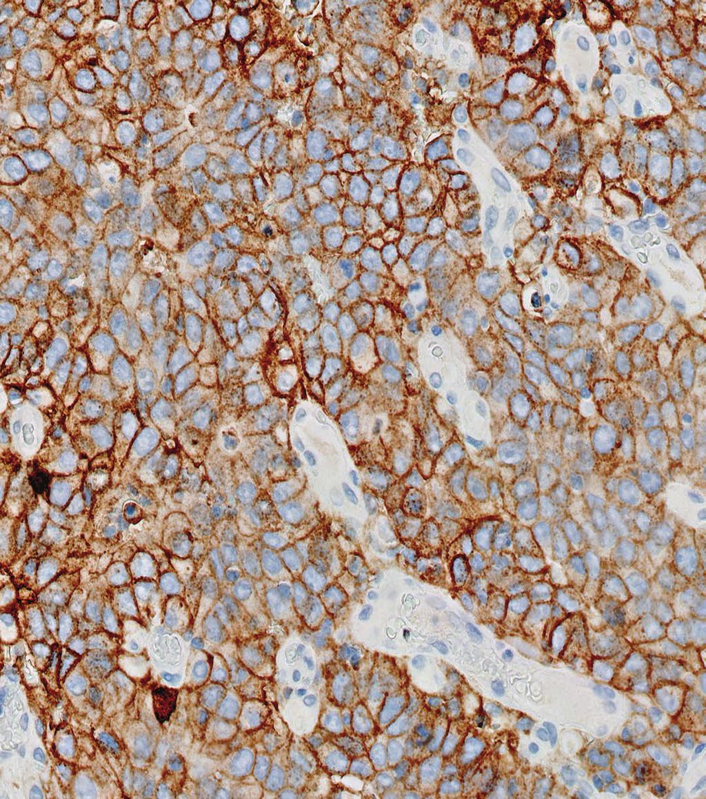
33 Image Guide for Interpretation of PD-L1 IHC 22C3 pharmdx Staining in Gastric or GEJ Adenocarcinoma PD-L1 Staining Cells Included in the CPS Numerator Tumor cells, lymphocytes, and macrophages exhibiting appropriate PD-L1 expression are defined as PD-L1 staining cells. All PD-L1 staining cells are included in the CPS numerator for determination of the Combined Positive Score (see Table 1 on page 24 for additional CPS numerator inclusion/exclusion criteria). Below are common staining characteristics of PD-L1 staining cells that must be included in the CPS numerator. All images are gastric or GEJ adenocarcinoma unless otherwise noted in figure caption. Linear Membrane Staining Tumor cells and tumor-associated mononuclear inflammatory cells (MICs: lymphocytes, macrophages) exhibiting convincing partial and/or complete linear membrane staining are considered PD-L1 staining cells. Convincing linear membrane staining can be present at any intensity and must be convincing at a 20x magnification. Figure 18a: PD-L1 primary antibody exhibiting linear membrane staining of tumor cells (20x magnification). A Key point Convincing linear membrane staining of tumor cells and convincing linear membrane and/or cytoplasmic staining of tumor-associated lymphocytes and macrophages should be included in the CPS numerator Figure 18b: PD-L1 primary antibody exhibiting linear membrane staining of tumor-associated mononuclear inflammatory cells (A: lymphocytes, B: macrophages) (20x magnification). Note: Tumor-associated MICs are present within the tumor nests and/or adjacent supporting stroma and are directly associated with the response against the tumor. B 33

34 Partial and Complete Linear Membrane Staining Tumor cells and tumor-associated mononuclear inflammatory cells (MICs: lymphocytes, macrophages) can exhibit partial and/or complete linear membrane staining. Any partial or complete linear membrane staining observed at any intensity and observed at a 20x magnification must be included in the CPS numerator. Note: Tumor-associated MICs are present within the tumor nests and/or adjacent supporting stroma and are directly associated with the response against the tumor. A B Figure 19: PD-L1 primary antibody exhibiting partial (A) and complete (B) linear membrane staining of tumor cells (20x magnification). Key point Convincing partial and/or complete staining of tumor cells and tumor-associated lymphocytes and macrophages should be included in the CPS numerator 34

 (20x magnification).")
35 Weak Linear Membrane Staining Tumor cells and tumor-associated mononuclear inflammatory cells (MICs: lymphocytes, macrophages) must exhibit convincing staining at any intensity, including weak 1+ intensity, at a 20x magnification. Note: Tumor-associated MICs are present within the tumor nests and/or adjacent supporting stroma and are directly associated with the response against the tumor. Figure 20a: PD-L1 primary antibody exhibiting weak but perceptible and convincing membrane staining of tumor cells (arrows) (20x magnification). Figure 20b: PD-L1 primary antibody exhibiting weak but perceptible and convincing membrane staining of tumor-associated mononuclear inflammatory cells (arrows) (20x magnification). Key point Weak 1+ convincing staining of tumor cells and tumorassociated lymphocytes and macrophages should be included in the CPS numerator 35

36 Linear Membrane and Cytoplasmic Staining: Tumor Cells Tumor cells with both convincing linear membrane staining ( 1+ intensity) and cytoplasmic staining at 20x magnification should be included in the CPS numerator. Tumor cells exhibiting only cytoplasmic staining are excluded from the CPS numerator, as this is considered non-specific staining. Figure 21: PD-L1 primary antibody exhibiting linear membrane staining distinct from cytoplasmic staining (arrows) (20x magnification). Key point Tumor cells exhibiting convincing linear membrane staining that is distinct from cytoplasmic staining is included in the CPS numerator 36
 are considered PD-L1 staining cells and should be included in the CPS numerator.")

37 Membrane and Cytoplasmic Staining: Tumor-associated Mononuclear Inflammatory Cells (MICs) Tumor-associated lymphocytes and macrophages (mononuclear inflammatory cells, MICs) exhibiting convincing membrane and/or cytoplasmic staining at a 20x magnification ( 1+ intensity) are considered PD-L1 staining cells and should be included in the CPS numerator. Tumor-associated MICs are present within the tumor nests and/or adjacent supporting stroma and are directly associated with the response against the tumor. Note: PD-L1 staining lymphocytes often have indistinguishable membrane and cytoplasmic staining due to a high nuclear to cytoplasmic ratio; PD-L1 staining macrophages often have distinct membrane staining and low cytoplasmic staining. All PD-L1 staining tumor-associated MICs should be included in the CPS numerator. Figure 22a: PD-L1 primary antibody exhibiting linear membrane and/or cytoplasmic staining of tumor-associated mononuclear inflammatory cells (20x magnification). Figure 22b: PD-L1 primary antibody exhibiting linear membrane and/or cytoplasmic staining of tumor-associated lymphocytes (20x magnification). 37

38 Figure 22c: PD-L1 primary antibody exhibiting linear membrane staining of tumor-associated macrophages (arrows) (20x magnification). Key point Tumor-associated lymphocytes and macrophages with convincing membrane and/or cytoplasmic staining should be included in the CPS numerator 38


39 Lattice and Interface Staining Patterns PD-L1 staining tumor cells and tumor-associated mononuclear inflammatory cells can exhibit two distinct staining patterns: lattice and interface. Gastric or GEJ adenocarcinoma specimens may exhibit one or both patterns. The lattice pattern of PD-L1 staining cells may be partial or complete, and present within tumor nests and/or adjacent supporting stroma. Staining can be present at one or several intensities. Specimens with PD-L1 staining at the leading edge and/or margin of the tumor nests exhibit the interface pattern. PD-L1 staining can also be present in the adjacent supporting stroma. PD-L1 staining can be present at one or several intensities. Figure 23a: PD-L1 primary antibody exhibiting the lattice pattern (20x magnification). Figure 23b: PD-L1 primary antibody exhibiting the interface pattern (arrows) (10x magnification). 39

 (20x magnification).")
40 Heterogeneous Staining Intensities Convincing staining of tumor cells (linear membrane) and tumor-associated lymphocytes and macrophages (membrane and/or cytoplasmic) is often heterogeneous, with various staining intensities present. At 20x magnification, any convincing staining of tumor cells and tumor-associated lymphocytes and macrophages at any intensity should be included in the CPS numerator. C A B Figure 24a: PD-L1 primary antibody exhibiting heterogeneous staining intensities of tumor cells (A: 1+ intensity, B: 2+ intensity, C: 3+ intensity) (20x magnification). C A B Figure 24b: PD-L1 primary antibody exhibiting heterogeneous staining intensities of tumor-associated mononuclear inflammatory cells (A: 1+ intensity, B: 2+ intensity, C: 3+ intensity) (20x magnification). Key point Convincing staining of tumor cells and tumor-associated lymphocytes and macrophages at all intensities should be included in the CPS numerator 40
 observed at a 20x magnification should be included in the CPS numerator.")
41 Granular Staining Tumor cells can exhibit a granular membrane staining pattern where membrane and cytoplasmic staining are indistinguishable. Only convincing staining of tumor cells ( 1+ intensity) observed at a 20x magnification should be included in the CPS numerator. Figure 25: PD-L1 primary antibody exhibiting a granular membrane staining pattern (arrows) (20x magnification). Key point Granular staining of tumor cells must exhibit a convincing linear membrane pattern to be included in the CPS numerator Diffuse Signet Ring Cell Carcinoma Signet ring cells are commonly present in diffuse-type gastric or GEJ adenocarcinoma. The tumor cell nuclei are small, crescent-shaped, and present at the periphery of the cells. Signet ring cells exhibiting any convincing partial and/or complete membrane staining at a 20x magnification ( 1+ intensity) are considered PD-L1 staining cells and should be included in the CPS numerator. Signet ring cells exhibiting only cytoplasmic staining should be considered nonstaining, as this is considered non-specific staining. Key point Convincing partial and/or complete membrane staining of signet ring cells should be included in the CPS numerator 41
.")
42 Cells Excluded from CPS Only tumor cells exhibiting PD-L1 membrane staining and MICs exhibiting PD-L1 membrane and/or cytoplasmic staining should be included in the CPS numerator. Below are other cells that can exhibit PD-L1 expression but should be excluded from the CPS calculation (CPS numerator or denominator). Tumor Cells with Only Cytoplasmic Staining Tumor cells exhibiting only cytoplasmic staining are not to be included in the CPS numerator, as this is considered non-specific staining. They should, however, still be included in the CPS denominator. Figure 26: PD-L1 primary antibody exhibiting only cytoplasmic staining (arrows) (20x magnification). Key point Tumor cells exhibiting only cytoplasmic staining should not be included in the CPS numerator 42

43 Other Immune Cells Excluded from CPS Various types of immune cells can exhibit PD-L1 staining, but only tumorassociated lymphocytes and macrophages should be included in the CPS calculation. Neutrophils, eosinophils, and plasma cells should be excluded from the CPS calculation. Key point Neutrophils, eosinophils, and plasma cells should be excluded from the CPS calculations Non-tumor-associated MICs Mononuclear inflammatory cells (MICs: lymphocytes and macrophages) commonly exhibit PD-L1 staining in gastric or GEJ adenocarcinoma specimens. Only PD-L1 staining MICs that are tumor-associated (present within tumor nests and/or adjacent supportive stroma; directly associated with the response against the tumor) should be included in the CPS calculation. PD-L1 staining MICs that are not tumor-associated must be excluded from the CPS calculation. Examples of non-tumor-associated MICs include those associated with adenoma, dysplasia, carcinoma in situ, ulceration, chronic gastritis, normal cells, and other processes not associated with the tumor. Figure 27a: PD-L1 staining of mononuclear inflammatory cells associated with normal gastric glands (10x magnification). 43
.")
.")
 (10x magnification).")
44 Figure 27b: PD-L1 staining mononuclear inflammatory cells contributing to gastritis (10x magnification). Figure 27c: PD-L1 staining mononuclear inflammatory cells associated with no tumor cells (10x magnification). Figure 27d: PD-L1 staining mononuclear inflammatory cells associated with dysplasia (arrows) (10x magnification). 44 Key point Non-tumor-associated MICs should be excluded from the CPS calculation

.")
45 Other Cell Types Excluded from CPS Various other tissue elements can exhibit PD-L1 staining, but only PD-L1 staining tumor cells and tumor-associated lymphocytes and macrophages should be included in the CPS numerator. Normal cells (including ganglion cells), stromal cells (including fibroblasts), and endocrine cells should be excluded from the CPS calculation. Figure 28a: PD-L1 staining of benign gastric glands (20x magnification). Figure 28b: PD-L1 staining of fibroblasts (arrows) (20x magnification). 45
.")

46 Figure 28c: PD-L1 staining of ganglion cells (arrows) (20x magnification). Figure 28d: PD-L1 staining of endocrine cells (20x magnification). Key point PD-L1 staining normal cells, stromal cells, and endocrine cells should be excluded from the CPS calculation 46


47 CPS < 1 Case Examples Case 1: CPS < 1 Figure 29a: 10x magnification. Figure 29b: 20x magnification. Figure 29a 29b: PD-L1 antibody exhibiting CPS < 1 at 20 magnification. 47


48 Case 2: CPS < 1 Figure 30a: 10x magnification. Figure 30b: 20x magnification. Figure 30a 30b: PD-L1 antibody exhibiting CPS < 1 at 20x magnification. 48


49 Case 3: CPS < 1 Figure 31a: 10x magnification. Figure 31b: 20x magnification. Figure 31a 31b: PD-L1 antibody exhibiting CPS < 1 with non-specific staining at 20x magnification. 49


50 Case 4: CPS < 1 Figure 32a: 10x magnification. Figure 32b: 20x magnification. Figure 32a 32b: PD-L1 antibody exhibiting CPS < 1 at 20x magnification. 50


51 Case 5: CPS < 1 Figure 33a: 10x magnification. Figure 33b: 20x magnification. Figure 33a 33b: PD-L1 antibody exhibiting CPS < 1 at 20x magnification. 51


52 Case 6: CPS < 1 Figure 34a: 10x magnification. Figure 34b: 20x magnification. Figure 34a 34b: PD-L1 antibody exhibiting CPS < 1 at 20x magnification. 52


53 CPS 1 Case Examples Case 7: CPS 1 Figure 35a: 10x magnification. Figure 35b: 20x magnification. Figure 35a 35b: PD-L1 antibody exhibiting CPS 1 at 20x magnification. 53


54 Case 8: CPS 1 Figure 36a: 10x magnification. Figure 36b: 20x magnification. Figure 36a 36b: PD-L1 antibody exhibiting CPS 1 at 20x magnification. 54


55 Case 9: CPS 1 Figure 37a: 10x magnification. Figure 37b: 20x magnification. Figure 37a 37b: PD-L1 antibody exhibiting CPS 1 at 20x magnification. 55


56 Case 10: CPS 1 Figure 38a: 10x magnification. Figure 38b: 20x magnification. Figure 38a 38b: PD-L1 antibody exhibiting CPS 1 at 20x magnification. 56


57 Case 11: CPS 1 Figure 39a: 10x magnification. Figure 39b: 20x magnification. Figure 39a 39b: PD-L1 antibody exhibiting CPS 1 at 20x magnification. 57


58 Case 12: CPS 1 Figure 40a: 10x magnification. Figure 40b: 20x magnification. Figure 40a 40b: PD-L1 antibody exhibiting CPS 1 at 20x magnification. 58
 Figure 41: PD-L1 antibody exhibiting CPS 2 at 20x")
 Figure 42: PD-L1 antibody")
59 Near Cut-off Case Examples (CPS 0 10) Challenging Case 1: Near Cut-off (CPS 0 10) Figure 41: PD-L1 antibody exhibiting CPS 2 at 20x magnification. Challenging Case 2: Near Cut-off (CPS 0 10) Figure 42: PD-L1 antibody exhibiting CPS 2 at 20x magnification with weak PD-L1 staining. 59

 Figure 44: PD-L1")
60 Challenging Case 3: Near Cut-off (CPS 0 10) Figure 43: PD-L1 antibody exhibiting CPS < 1 and non-specific staining at 20x magnification. Challenging Case 4: Near Cut-off (CPS 0 10) Figure 44: PD-L1 antibody exhibiting CPS 2 and non-specific staining at 20x magnification. 60


61 Challenging Case 5: Near Cut-off (CPS 0 10) Figure 45: PD-L1 antibody exhibiting CPS < 1 at 20x magnification. Challenging Case 6: Near Cut-off (CPS 0 10) Figure 46: PD-L1 antibody exhibiting CPS 1 at 20x magnification. 61

62 Artifacts The following pages provide examples of artifacts you may see when staining with PD-L1 IHC 22C3 pharmdx. Non-specific Background Staining Background staining is defined as diffuse, non-specific staining of a specimen. It is caused by several factors. These factors include, but are not limited to, preanalytic fixation and processing of the specimen, incomplete removal of paraffin from sections, and incomplete rinsing of slides during staining. The use of fixatives other than neutral buffered formalin may be a source of background staining. Background staining with PD-L1 IHC 22C3 pharmdx is rare. Possible Causes of Background Improper drying of slides; ensure slides remain wet with buffer while loading onto Autostainer Link 48 and prior to initiating run Improper deparaffinization procedure Incomplete rinsing of reagents from slides The non-specific background staining of the NCR-stained test specimen is useful in determining the level of background staining in the positive test specimen. All specimens must have 1+ non-specific background staining. Key point All specimens must have 1+non-specific background staining 62 Figure 47: Negative Control Reagent (NCR) exhibiting acceptable non-specific background staining (20x magnification).

63 Edge Artifact Commonly, edge artifacts are linked to the pre-analytic handling of the tissue. Inadequate processing of thick tissue samples may mimic edge artifact by rendering the central portion of the tissue suboptimally fixed relative to the peripheral areas. In these circumstances, the immunoreactivity based on the suboptimal central portion may be mistakenly interpreted as non-staining as optimal fixation is only present at the periphery Increased staining is observed around the periphery of the tissue specimen, known as the edge artifact Edge artifacts can be due to drying of the tissue specimen prior to fixation or during the staining procedure If the positive reaction is only at the edge of the tissue section (i.e., a few cell layers of staining at the periphery and ending abruptly with penetration into the centrally located tumor), scoring at the edge of the tissue specimen should be avoided Key point Scoring of the edge of a specimen should be avoided if staining is inconsistent with the rest of the specimen Figure 48: PD-L1 primary antibody exhibiting edge artifact staining; edge staining should be excluded from the scoring (5x magnification). 63

64 Crush Artifact Crush artifact is closely related to edge artifact. The compression of the tissues along the edges of the specimen can produce a linear staining that has to be interpreted as an artifact. Inadvertent crushing of the tissue occasionally occurs during sectioning, resulting in morphologically distorted cellular architecture When compared to surrounding cells, stronger staining may be observed in crushed cells. Crushed cells typically demonstrate condensed nuclei. Crushed cells should be avoided in scoring Key point Scoring of crush artifact should be avoided if staining is inconsistent with entire specimen Figure 49: PD-L1 primary antibody exhibiting crush artifact (20x magnification). 64

65 Necrosis Necrosis can be described as morphological changes indicative of cell death with undefined cellular detail. Necrosis is often present in gastric or GEJ adenocarcinoma specimens and should be excluded from scoring. Key point Scoring of necrotic areas should be excluded from the CPS calculation Figure 50: PD-L1 primary antibody exhibiting staining of necrosis; necrosis should not be scored (20x magnification). 65
. 66")
66 Poor Fixation Standardization of fixation is very important when using PD-L1 IHC 22C3 pharmdx. Suboptimal fixation of tissues may give erroneous results. Key point Proper fixation is important for accurate diagnosis Figure 51: PD-L1 primary antibody exhibiting poor tissue fixation (10x magnification). 66
PD-L1 IHC 22C3 pharmdx. SK tests for use with Autostainer Link 48 Table of Contents
 PD-L1 IHC 22C3 pharmdx SK006 50 tests for use with Autostainer Link 48 Table of Contents 1 Intended Use... 2 2 Summary and Explanation... 2 2.1 NSCLC... 2 2.2 Gastric or Gastroesophageal Junction (GEJ)
Urothelial Carcinoma (UC)
") EDUCATION PD-L1 IHC 28-8 pharmdx Interpretation Manual Urothelial Carcinoma (UC) For In Vitro Diagnostic Use Table of Contents Introduction...5 Intended Use in Urothelial Carcinoma...5 How to Use the
EDUCATION PD-L1 IHC 28-8 pharmdx Interpretation Manual Urothelial Carcinoma (UC) For In Vitro Diagnostic Use Table of Contents Introduction...5 Intended Use in Urothelial Carcinoma...5 How to Use the
Urothelial Carcinoma (UC)
") EDUCATION PD-L1 IHC 28-8 pharmdx Interpretation Manual Urothelial Carcinoma (UC) PD-L1 IHC 28-8 pharmdx is FDA-approved for In Vitro Diagnostic Use Table of Contents Introduction...5 Intended Use in Urothelial
EDUCATION PD-L1 IHC 28-8 pharmdx Interpretation Manual Urothelial Carcinoma (UC) PD-L1 IHC 28-8 pharmdx is FDA-approved for In Vitro Diagnostic Use Table of Contents Introduction...5 Intended Use in Urothelial
Buffered solution containing hydrogen peroxide, detergent and mol/l sodium azide.
 PD-L1 IHC 22C3 pharmdx SK006 50 tests for use with Autostainer Link 48 Intended Use For in vitro diagnostic use. PD-L1 IHC 22C3 pharmdx is a qualitative immunohistochemical assay using Monoclonal Mouse
Assessment Run C1 2017
 Assessment Run C1 2017 PD-L1 The first assessment in this new NordiQC Companion module C1 focused on the accuracy of the PD-L1 IHC assays performed by the participating laboratories to identify patients
Results you can trust
 PRODUCT I NF OR MAT ION pharmdx Results you can trust The first and only FDA-approved PD-L1 test to assess the magnitude of treatment effect on progression-free survival in melanoma patients from OPDIVO
PD-L1 expression as detected by PD-L1 IHC 28-8 pharmdx in non-squamous NSCLC may be associated with enhanced survival from OPDIVO (nivolumab).
.") PD-L1 IHC 28-8 pharmdx SK005 50 tests for use with Autostainer Link 48 Intended Use For in vitro diagnostic use. PD-L1 IHC 28-8 pharmdx is a qualitative immunohistochemical assay using Monoclonal Rabbit
PD-L1 IHC 28-8 pharmdx SK005 50 tests for use with Autostainer Link 48 Intended Use For in vitro diagnostic use. PD-L1 IHC 28-8 pharmdx is a qualitative immunohistochemical assay using Monoclonal Rabbit
Immunotherapy in NSCLC Pathologist role
 Immunotherapy in NSCLC Pathologist role Pimpin Incharoen, M.D. Assistant Professor, Thoracic Pathology Department of Pathology, Ramathibodi Hospital Genetic alterations in NSCLC Khono et al, Trans Lung
HER2 CISH pharmdx TM Kit Interpretation Guide Breast Cancer
 P A T H O L O G Y HER2 CISH pharmdx TM Kit Interpretation Guide Breast Cancer FROM CERTAINTY COMES TRUST For in vitro diagnostic use HER2 CISH pharmdx Kit HER2 CISH pharmdx Kit is intended for dual-color
HercepTest for the Dako Autostainer Code K5207
 HercepTest for the Dako Autostainer Code K5207 9th edition For immunocytochemical staining. The kit is for 50 tests (100 slides). (126659-002) P04088US_02_K520721-5/2016.05 p. 1/54 Contents Page Intended
HercepTest TM Code K5204
 HercepTest TM Code K5204 9th edition For immunocytochemical staining. The kit is for 35 tests (70 slides). (126559-002) P04086US_02_K520421-5/US/2016.05 p. 1/55 Contents Intended Use... 4 Summary and Explanation
Assessment Run C3 2018
 Assessment Run C3 2018 PD-L1 Amended version May 14 th 2018 The third assessment in NordiQC Companion module C3 focused on the accuracy of the PD-L1 IHC assays performed by the participating laboratories
GLOBAL REGISTRATION STRATEGIES:
 GLOBAL REGISTRATION STRATEGIES: Therapeutic Product (TP) and the Companion Diagnostic (CDx) Erin Pedalino Regulatory Affairs International MSD Unique Role as CDx Regulatory Liaison at a Therapeutic Product
VENTANA PD-L1 (SP263) Assay Staining in Urothelial Carcinoma Interpretation Guide
 Assay Staining in Urothelial Carcinoma Interpretation Guide") VENTANA PD-L1 (SP263) Assay Staining in Urothelial Carcinoma Interpretation Guide 2 VENTANA PD-L1 (SP263) Assay in Urothelial Carcinoma Interpretation Guide Table of Contents Introduction 4 Intended Use
VENTANA PD-L1 (SP263) Assay Staining in Urothelial Carcinoma Interpretation Guide 2 VENTANA PD-L1 (SP263) Assay in Urothelial Carcinoma Interpretation Guide Table of Contents Introduction 4 Intended Use
Three Hours Thirty Minutes
 INTERPRETATION HER2 IQFISH pharmdx TM Interpretation Guide Three Hours Thirty Minutes it s about time Breast carcinoma (FFPE) stained with HER2 IQFISH pharmdx Gastric cancer (FFPE) stained with HER2 IQFISH
VENTANA MMR IHC Panel Interpretation Guide for Staining of Colorectal Tissue
 VENTANA MMR IHC Panel Interpretation Guide for Staining of Colorectal Tissue VENTANA anti-mlh1 (M1) Mouse Monoclonal Primary Antibody VENTANA anti-pms2 (A16-4) Mouse Monoclonal Primary Antibody VENTANA
VENTANA PD-L1 (SP142) Assay Guiding immunotherapy in NSCLC
 Assay Guiding immunotherapy in NSCLC") VENTANA (SP142) Assay Guiding immunotherapy in NSCLC Hiker s path: VENTANA (SP142) Assay on non-small cell lung cancer tissue Location: Point Conception, CA VENTANA (SP142) Assay Assess NSCLC patient benefit
VENTANA (SP142) Assay Guiding immunotherapy in NSCLC Hiker s path: VENTANA (SP142) Assay on non-small cell lung cancer tissue Location: Point Conception, CA VENTANA (SP142) Assay Assess NSCLC patient benefit
Dako IT S ABOUT TIME. Interpretation Guide. Agilent Pathology Solutions. ALK, ROS1 and RET IQFISH probes (Dako Omnis) MET IQFISH probe (Dako Omnis)
 MET IQFISH probe (Dako Omnis)") INTERPRETATION Dako Agilent Pathology Solutions IQFISH Interpretation Guide ALK, ROS1 and RET IQFISH probes (Dako Omnis) MET IQFISH probe (Dako Omnis) IT S ABOUT TIME For In Vitro Diagnostic Use ALK, ROS1,
INTERPRETATION Dako Agilent Pathology Solutions IQFISH Interpretation Guide ALK, ROS1 and RET IQFISH probes (Dako Omnis) MET IQFISH probe (Dako Omnis) IT S ABOUT TIME For In Vitro Diagnostic Use ALK, ROS1,
IT S ABOUT TIME. IQFISH pharmdx Interpretation Guide THREEHOURSTHIRTYMINUTES. HER2 IQFISH pharmdxtm. TOP2A IQFISH pharmdxtm
 I N T E R P R E TAT I O N IQFISH pharmdx Interpretation Guide TM HER2 IQFISH pharmdxtm TOP2A IQFISH pharmdxtm Breast carcinoma (FFPE) stained with HER2 IQFISH pharmdx Breast carcinoma (FFPE) stained with
Single and Multiplex Immunohistochemistry
 Single and Multiplex Immunohistochemistry Steve Westra, BS Reagent Product Specialist Leica Biosystems IHC Theory Polyclonal vs Monoclonal Polyclonal reagents Detect a multitude of epitopes Batch to batch
VENTANA PD-L1 (SP142) Assay Guiding immunotherapy
 Assay Guiding immunotherapy") VENTANA PD-L1 (SP142) Assay Guiding immunotherapy Hiker s path: VENTANA PD-L1 (SP142) Assay on urothelial carcinoma tissue Location: Point Conception, CA VENTANA PD-L1 (SP142) Assay Identify patients most
VENTANA PD-L1 (SP142) Assay Guiding immunotherapy Hiker s path: VENTANA PD-L1 (SP142) Assay on urothelial carcinoma tissue Location: Point Conception, CA VENTANA PD-L1 (SP142) Assay Identify patients most
Estrogen receptor (ER)
") Material The slide to be stained for ER comprised: Assessment Run B26 2018 Estrogen receptor (ER) No. Tissue ER-positivity* ER-intensity* 1. Uterine cervix 80-90% Moderate to strong 2. Tonsil 1-5% Weak
Material The slide to be stained for ER comprised: Assessment Run B26 2018 Estrogen receptor (ER) No. Tissue ER-positivity* ER-intensity* 1. Uterine cervix 80-90% Moderate to strong 2. Tonsil 1-5% Weak
Novocastra Liquid Mouse Monoclonal Antibody Myeloperoxidase
 Novocastra Liquid Mouse Monoclonal Antibody Myeloperoxidase Product Code: NCL-L-MYELO Leica Biosystems Newcastle Ltd Balliol Business Park West Benton Lane Newcastle Upon Tyne NE12 8EW United Kingdom (
Anti-PD-L1 antibody [28-8] ab205921
![Anti-PD-L1 antibody [28-8] ab205921](/thumbs/84/91196262.jpg "Anti-PD-L1 antibody [28-8] ab205921") Anti-PD-L1 antibody [28-8] ab205921 2 Abreviews 16 References 15 Images Overview Product name Anti-PD-L1 antibody [28-8] Description Tested applications Species reactivity Immunogen Rabbit monoclonal [28-8]
Anti-PD-L1 antibody [28-8] ab205921 2 Abreviews 16 References 15 Images Overview Product name Anti-PD-L1 antibody [28-8] Description Tested applications Species reactivity Immunogen Rabbit monoclonal [28-8]
Digital Pathology and CAP Guidelines
 Digital Pathology and CAP Guidelines Frequently asked questions The VENTANA family of digital pathology products empowers you with the convenience of a comprehensive image and workflow solution. When used
Table of Contents. 1. Overview. 2. Interpretation Guide. 3. Staining Gallery Cases Negative for CINtec PLUS
 Staining Atlas Table of Contents 1. Overview 1.1 Introduction 1.2 Role of p16 INK4a 1.3 Role of Ki-67 1.4 Molecular Pathogenesis 1.5 p16 INK4a Expression in Cervical Dysplasia 1.6 The Concept of CINtec
VENTANA PD-L1 (SP142) Assay
 Assay") VENTANA (SP142) Assay Guiding immunotherapy Hiker s path: VENTANA (SP142) Assay on urothelial carcinoma tissue Location: Point Conception, CA VENTANA (SP142) Assay Assess UC patient benefit from TECENTRIQ
VENTANA (SP142) Assay Guiding immunotherapy Hiker s path: VENTANA (SP142) Assay on urothelial carcinoma tissue Location: Point Conception, CA VENTANA (SP142) Assay Assess UC patient benefit from TECENTRIQ
HER2 FISH pharmdx TM Interpretation Guide - Breast Cancer
 P A T H O L O G Y HER2 FISH pharmdx TM Interpretation Guide - Breast Cancer For In Vitro Diagnostic Use FDA approved as an aid in the assessment of patients for whom Herceptin TM (trastuzumab) treatment
LIST OF ORGANS FOR HISTOPATHOLOGICAL ANALYSIS:!! Neural!!!!!!Respiratory:! Brain : Cerebrum,!!! Lungs and trachea! Olfactory, Cerebellum!!!!Other:!
 LIST OF ORGANS FOR HISTOPATHOLOGICAL ANALYSIS:!! Neural!!!!!!Respiratory:! Brain : Cerebrum,!!! Lungs and trachea! Olfactory, Cerebellum!!!!Other:! Spinal cord and peripheral nerves! Eyes, Inner ear, nasal
Interpretation guide. Abnormal cytology can t hide anymore
 Interpretation guide Abnormal cytology can t hide anymore Unique dual-biomarker technology makes you certain about the presence of transforming HPV infection. The science that creates certainty. Table
Instructions for Use. APO-AB Annexin V-Biotin Apoptosis Detection Kit 100 tests
 3URGXFW,QIRUPDWLRQ Sigma TACS Annexin V Apoptosis Detection Kits Instructions for Use APO-AB Annexin V-Biotin Apoptosis Detection Kit 100 tests For Research Use Only. Not for use in diagnostic procedures.
CINtec PLUS Cytology. Interpretation training
 CINtec PLUS Cytology Interpretation training Objectives After reviewing this learning module, you will have a basic understanding of how to interpret CINtec PLUS Cytology, including: The mechanism of action
Development of a Companion Diagnostic for Pembrolizumab in Non Small Cell Lung Cancer Using Immunohistochemistry for Programmed Death Ligand-1
 Development of a Companion Diagnostic for Pembrolizumab in Non Small Cell Lung Cancer Using Immunohistochemistry for Programmed Death Ligand-1 Marisa Dolled-Filhart, PhD; Charlotte Roach, BS; Grant Toland,
Brief Formalin Fixation and Rapid Tissue Processing Do Not Affect the Sensitivity of ER Immunohistochemistry of Breast Core Biopsies
 Brief Formalin Fixation and Rapid Tissue Processing Do Not Affect the Sensitivity of ER Immunohistochemistry of Breast Core Biopsies Victoria Sujoy, MD, Mehrdad Nadji, MD, and Azorides R. Morales, MD From
Integrazione in Anatomia Patologica dei markers predittivi nel NSCLC: l esperienza di PD-L1
 Integrazione in Anatomia Patologica dei markers predittivi nel NSCLC: l esperienza di PD-L1 Dipartimento di Sanità Pubblica Università degli Studi di Napoli Federico II Dott. Antonino Iaccarino PDL1-Mechanism
Pepsin Solution ready-to-use
 SIE HABEN DIE VISION, WIR HABEN DIE SUBSTANZ. Pepsin Solution Single component Pepsin Solution: only one component refrigerator stable Pepsin is a commonly used digestive enzyme for immunohistochemical
IHC Polymer. BioGenex Website. Presented for: Presented by: Date: BioGenex Tech Support 2016
 IHC Polymer Presented for: Presented by: Date: BioGenex Website BioGenex Tech Support 2016 Immunohistochemistry (IHC) Quick Guide# Super SensitiveTM Polymer-HRP kits. Baking An-gen Retrieval DeWax Overnight,
Assessment Run GATA3
 Assessment Run 44 2015 GATA3 Material The slide to be stained for GATA3 comprised: 1. Tonsil 2. Kidney, 3. Urothelial carcinoma, 4. Breast ductal carcinoma, 5. Colon adenocarcinoma All tissues were fixed
Atlas of Stains. Special Stains on Artisan Link Pro
 Atlas of Stains Special Stains on Artisan Link Pro Intended use Routinely processed samples (paraffin-embedded) may be used. The preferred fixative is neutral buffered formalin. The clinical interpretation
Novocastra Liquid Mouse Monoclonal Antibody MyoD1 (Rhabdomyosarcoma Marker)
") Novocastra Liquid Mouse Monoclonal Antibody MyoD1 (Rhabdomyosarcoma Marker) Product Code: NCL-L-MyoD1 Leica Biosystems Newcastle Ltd Balliol Business Park West Benton Lane Newcastle Upon Tyne NE12 8EW
Novocastra Liquid Mouse Monoclonal Antibody MyoD1 (Rhabdomyosarcoma Marker) Product Code: NCL-L-MyoD1 Leica Biosystems Newcastle Ltd Balliol Business Park West Benton Lane Newcastle Upon Tyne NE12 8EW
Supplementary Online Content
 Supplementary Online Content Rimm DL, Han G, Taube JM, et al. A prospective, multi-institutional, pathologistbased assessment of 4 immunohistochemistry assays for PD-L1 expression in non small cell lung
Optimal algorithm for HER2 testing
 Optimal algorithm for HER2 testing The revised definition of IHC 2+ (equivocal) is invasive breast cancer with Weak to moderate complete membrane staining observed in >10% of tumor cells. (see Figure 1
Estrogen receptor (ER)
") Material The slide to be stained for ER comprised: Assessment B25 208 Estrogen receptor (ER) No. Tissue ER-positivity* ER-intensity*. Uterine cervix 80-90% Moderate to strong 2. Tonsil < 2-5% Weak to strong
Material The slide to be stained for ER comprised: Assessment B25 208 Estrogen receptor (ER) No. Tissue ER-positivity* ER-intensity*. Uterine cervix 80-90% Moderate to strong 2. Tonsil < 2-5% Weak to strong
Next-Generation Immunohistochemistry: Multiplex tissue imaging with mass cytometry
 Nat Met, April 2014 Nat Med, April 2014 Next-Generation Immunohistochemistry: Multiplex tissue imaging with mass cytometry Journal Club Timo Böge Overview Introduction Conventional Immunohistochemistry
Supplemental figure 1. PDGFRα is expressed dominantly by stromal cells surrounding mammary ducts and alveoli. A) IHC staining of PDGFRα in
 IHC staining of PDGFRα in") Supplemental figure 1. PDGFRα is expressed dominantly by stromal cells surrounding mammary ducts and alveoli. A) IHC staining of PDGFRα in nulliparous (left panel) and InvD6 mouse mammary glands (right
Supplemental figure 1. PDGFRα is expressed dominantly by stromal cells surrounding mammary ducts and alveoli. A) IHC staining of PDGFRα in nulliparous (left panel) and InvD6 mouse mammary glands (right
APPENDIX 1 ETHICAL CLEARANCE
 APPENDIX 1 ETHICAL CLEARANCE 75 APPENDIX 2 76 PROCEDURE FOR PREPARING OF LIVER HISTOLOGY SLIDES Overview: Histology involves the use of a set of techniques to examine the morphology, architecture and composition
HBeAg and HBeAg Ab ELISA Kit
 HBeAg and HBeAg Ab ELISA Kit Catalog Number KA0290 96 assays Version: 17 Intended for research use only www.abnova.com Table of Contents Introduction... 3 Intended Use... 3 Principle of the Assay... 3
Assessment Run B HER2 IHC
 Assessment Run B26 208 HER2 IHC Material The slide to be stained for HER2 comprised the following 5 materials: IHC: HER2 Score* (0, +, 2+, 3+) FISH: HER2 gene/chr 7 ratio**. Breast carcinoma, no. 2+..3
Milestones in Her 2 Testing
 Human Epidermal Growth Factor Receptor 2 (HER2) Testing - Validation, Application and Correlation Her2 is encoded by the C-erbB2 gene and is one of four oncoproteins belonging to the Human Epidermal Growth
Product Introduction
 Product Introduction Product Codes: HCL026, HCL027 and HCL028 Contents Introduction to HER2 2 HER2 immunohistochemistry 3 Cell lines as controls 5 HER2 Analyte Control DR IHC 7 HER2 Analyte Control DR
SMH (Myosin, smooth muscle heavy chain)
") Material The slide to be stained for SMH comprised: Assessment Run 50 2017 SMH (Myosin, smooth muscle heavy chain) 1.Tonsil, 2. Esophagus, 3. Breast hyperplasia, 4. Breast ductal carcinoma in situ (DCIS),
Material The slide to be stained for SMH comprised: Assessment Run 50 2017 SMH (Myosin, smooth muscle heavy chain) 1.Tonsil, 2. Esophagus, 3. Breast hyperplasia, 4. Breast ductal carcinoma in situ (DCIS),
Novocastra Liquid Mouse Monoclonal Antibody Glial Fibrillary Acidic Protein
 Novocastra Liquid Mouse Monoclonal Antibody Glial Fibrillary Acidic Protein Product Code: NCL-L-GFAP-GA5 Leica Biosystems Newcastle Ltd Balliol Business Park West Benton Lane Newcastle Upon Tyne NE12 8EW
Cotinine (Mouse/Rat) ELISA Kit
 ELISA Kit") Cotinine (Mouse/Rat) ELISA Kit Catalog Number KA2264 96 assays Version: 03 Intended for research use only www.abnova.com Table of Contents Introduction... 3 Intended Use... 3 Background... 3 Principle
Cotinine (Mouse/Rat) ELISA Kit Catalog Number KA2264 96 assays Version: 03 Intended for research use only www.abnova.com Table of Contents Introduction... 3 Intended Use... 3 Background... 3 Principle
H. pylori Antigen ELISA Kit
 H. pylori Antigen ELISA Kit Catalog Number KA3142 96 assays Version: 04 Intended for research use only www.abnova.com Table of Contents Introduction... 3 Intended Use... 3 Background... 3 Principle of
Assessment performed on Friday, September 18, 2015, at Vancouver General Hospital
 Assessors report for ciqc Run 49: ATRX (June 2015) Assessors: S Yip and J Won (recorder) Assessment performed on Friday, September 18, 2015, at Vancouver General Hospital Background The combined application
Novocastra Liquid Mouse Monoclonal Antibody Carcinoembryonic Antigen (CD66e)
") Novocastra Liquid Mouse Monoclonal Antibody Carcinoembryonic Antigen (CD66e) Product Code: NCL-L-CEA-2 Leica Biosystems Newcastle Ltd Balliol Business Park West Benton Lane Newcastle Upon Tyne NE12 8EW
Novocastra Liquid Mouse Monoclonal Antibody Carcinoembryonic Antigen (CD66e) Product Code: NCL-L-CEA-2 Leica Biosystems Newcastle Ltd Balliol Business Park West Benton Lane Newcastle Upon Tyne NE12 8EW
H.Pylori IgG
 DIAGNOSTIC AUTOMATION, INC. 21250 Califa Street, Suite 102 and116, Woodland Hills, CA 91367 Tel: (818) 591-3030 Fax: (818) 591-8383 onestep@rapidtest.com technicalsupport@rapidtest.com www.rapidtest.com
H.Pylori IgG Cat # 1503Z
 DIAGNOSTIC AUTOMATION, INC. 23961 Craftsman Road, Suite D/E/F, Calabasas, CA 91302 Tel: (818) 591-3030 Fax: (818) 591-8383 onestep@rapidtest.com technicalsupport@rapidtest.com www.rapidtest.com See external
Assessment Run B HER2 IHC
 Assessment Run B24 2017 HER2 IHC Material The slide to be stained for HER2 comprised the following 5 materials: IHC: HER2 Score* (0, 1+, 2+, 3+) FISH: HER2 gene/chr 17 ratio** 1. Breast carcinoma, no.
HER2 IQFISH pharmdx. Code K th edition
 HER2 IQFISH pharmdx Code K5731 11th edition HER2 IQFISH pharmdx is a direct fluorescence in situ hybridization (FISH) assay designed to quantitatively determine HER2 gene amplification in formalin-fixed,
02006B 1 vial 02006B 1 vial Store at -20 C. Lyophilized recombinant IL-2
 For detection and measurement of human interleukin 2 Catalog #02006 Catalog #02007 2 Plates 10 Plates Product Description The Human Interleukin 2 (IL-2) ELISA Kit is designed for the quantitative detection
SIV p27 Antigen ELISA Catalog Number:
 INTENDED USE The RETRO-TEK SIV p27 Antigen ELISA is for research use only and is not intended for in vitro diagnostic use. The RETRO-TEK SIV p27 Antigen ELISA is an enzyme linked immunoassay used to detect
CIHRT Exhibit P-1830 Page 1
 Message CIHRT Exhibit P-1830 Page 1 Page 1 of 1 Wpc1 From: Sent: To: Cc: Subject: Mendes, Maria Friday, April 20, 2007 3:43 PM Mullen, Dr. Brendan Kuruzar, Gordana; Ni, Ruoyu; D Mello, Vince ER/PR Newfoundland
Malaria Pf/pan antigen Rapid Test
 Malaria Pf/pan antigen Rapid Test Cat. No.:IVDTS003 Pkg.Size:10T/50T Intended use The Malaria Pf/pan antigen Rapid Test is a self-performing, qualitative, sandwich immunoassay, utilizing whole blood for
Novocastra Liquid Mouse Monoclonal Antibody CD71
 Novocastra Liquid Mouse Monoclonal Antibody CD71 Product Code: NCL-L-CD71-309 Leica Biosystems Newcastle Ltd Balliol Business Park West Benton Lane Newcastle Upon Tyne NE12 8EW United Kingdom ( +44 191
FD Rapid MultiStain Kit
 Quality & Excellence since 1996 FD Rapid MultiStain Kit An effective histological staining system with multiple functions designed for easy use in all types of neuroscience laboratories User Manual PK
H. pylori IgM. Cat # H. pylori IgM ELISA. ELISA: Enzyme Linked Immunosorbent Assay. ELISA - Indirect; Antigen Coated Plate
 DIAGNOSTIC AUTOMATION, INC. 23961 Craftsman Road, Suite D/E/F, Calabasas, CA 91302 Tel: (818) 591-3030 Fax: (818) 591-8383 onestep@rapidtest.com technicalsupport@rapidtest.com www.rapidtest.com H. pylori
Novocastra Liquid Mouse Monoclonal Antibody Multi-Cytokeratin (4/5/6/8/10/13/18)
") Novocastra Liquid Mouse Monoclonal Antibody Multi-Cytokeratin (4/5/6/8/10/13/18) Product Code: NCL-L-C11 Leica Biosystems Newcastle Ltd Balliol Business Park West Benton Lane Newcastle Upon Tyne NE12 8EW
Novocastra Liquid Mouse Monoclonal Antibody Multi-Cytokeratin (4/5/6/8/10/13/18) Product Code: NCL-L-C11 Leica Biosystems Newcastle Ltd Balliol Business Park West Benton Lane Newcastle Upon Tyne NE12 8EW
Human Thyroglobulin EIA
 Human Thyroglobulin EIA For the quantitative determination of human thyroglobulin (htg) in plasma and serum. For Research Use Only. Not For Use In Diagnostic Procedures. Catalog Number: 30-7510 Size: 96
ZytoFast CMV Probe (Biotin-labeled)
") ZytoFast CMV Probe (Biotin-labeled) T-1013-400 40 (0.4 ml) For the detection of Cytomegalovirus (CMV) DNA by chromogenic in situ hybridization (CISH).... For Research Use Only. Not for use in diagnostic
ZytoFast CMV Probe (Biotin-labeled) T-1013-400 40 (0.4 ml) For the detection of Cytomegalovirus (CMV) DNA by chromogenic in situ hybridization (CISH).... For Research Use Only. Not for use in diagnostic
White paper Comparative analysis of H&E stain quality between the VENTANA HE 600 system and a traditional linear stainer
 White paper Comparative analysis of H&E stain quality between the VENTANA HE 600 and a traditional linear stainer 1 11/5/2015 8:55:39 AM Introduction Most tissue-based diagnostic decisions in anatomic
Assessment Run
 Assessment Run 50 2017 S100 Material The slide to be stained for S100 comprised: 1. Appendix, 2. Tonsil, 3. Schwannoma, 4-5. Malignant melanoma, 6. Colon adenocarcinoma. All tissues were fixed in 10% neutral
Predictive Biomarkers for Pembrolizumab. Eric H. Rubin, M.D.
 Predictive Biomarkers for Pembrolizumab Eric H. Rubin, M.D. PD-1 and PD-L1/L2 Pathway PD-1 is an immune checkpoint receptor Binding of PD-1 by its ligands PD-L1 or PD-L2 leads to downregulation of T-cell
Quantitative Image Analysis of HER2 Immunohistochemistry for Breast Cancer
 Quantitative Image Analysis of HER2 Immunohistochemistry for Breast Cancer Guideline from the College of American Pathologists Early Online Release Publication: Archives of Pathology & Laboratory Medicine
Novocastra Liquid Mouse Monoclonal Antibody p57 Protein (Kip2)
") Novocastra Liquid Mouse Monoclonal Antibody p57 Protein (Kip2) Product Code: NCL-L-p57 Leica Biosystems Newcastle Ltd Balliol Business Park West Benton Lane Newcastle Upon Tyne NE12 8EW United Kingdom
Novocastra Liquid Mouse Monoclonal Antibody p57 Protein (Kip2) Product Code: NCL-L-p57 Leica Biosystems Newcastle Ltd Balliol Business Park West Benton Lane Newcastle Upon Tyne NE12 8EW United Kingdom
1.0 Purpose - This procedure specifies the method for conducting analysis for semen and sperm in forensic casework.
 Procedure for Semen and Sperm Analysis 1.0 Purpose - This procedure specifies the method for conducting analysis for semen and sperm in forensic casework. 2.0 Scope - This procedure applies to those Forensic
Programmed death ligand-1 (PD-L1) protein expression
 protein expression") Original Articles Clinical Utility of the Combined Positive Score for Programmed Death Ligand-1 Expression and the Approval of Pembrolizumab for Treatment of Gastric Cancer Karina Kulangara, PhD; Nancy
Original Articles Clinical Utility of the Combined Positive Score for Programmed Death Ligand-1 Expression and the Approval of Pembrolizumab for Treatment of Gastric Cancer Karina Kulangara, PhD; Nancy
SUPPLEMENTARY MATERIAL. Sample preparation for light microscopy
 SUPPLEMENTARY MATERIAL Sample preparation for light microscopy To characterize the granulocytes and melanomacrophage centers, cross sections were prepared for light microscopy, as described in Material
Cell Culture. The human thyroid follicular carcinoma cell lines FTC-238, FTC-236 and FTC-
 Supplemental material and methods Reagents. Hydralazine was purchased from Sigma-Aldrich. Cell Culture. The human thyroid follicular carcinoma cell lines FTC-238, FTC-236 and FTC- 133, human thyroid medullary
(A) PCR primers (arrows) designed to distinguish wild type (P1+P2), targeted (P1+P2) and excised (P1+P3)14-
 PCR primers (arrows) designed to distinguish wild type (P1+P2), targeted (P1+P2) and excised (P1+P3)14-") 1 Supplemental Figure Legends Figure S1. Mammary tumors of ErbB2 KI mice with 14-3-3σ ablation have elevated ErbB2 transcript levels and cell proliferation (A) PCR primers (arrows) designed to distinguish
1 Supplemental Figure Legends Figure S1. Mammary tumors of ErbB2 KI mice with 14-3-3σ ablation have elevated ErbB2 transcript levels and cell proliferation (A) PCR primers (arrows) designed to distinguish
Corning BioCoat Matrigel Invasion Chamber
 Corning BioCoat Matrigel Invasion Chamber Catalog No. 354480, 354481 Guidelines for Use Discovery Labware, Inc., Two Oak Park, Bedford, MA 01730, Tel: 1.978.442.2200 (U.S.) CLSTechServ@Corning.com www.corning.com/lifesciences
MARSHALLTOWN MEDICAL & SURGICAL CENTER Marshalltown, Iowa
 MARSHALLTOWN MEDICAL & SURGICAL CENTER Marshalltown, Iowa CARE OF PATIENT POLICY & PROCEDURE Policy Number: 4:10 Subject: Policy: Glucose Monitoring (Accuchek) Nursing department staff and laboratory staff
To enable high quality in the diagnosis of cancer patients,
 RESEARCH ARTICLE Tissue Thickness Effects on Immunohistochemical Staining of Markers of Cancer Adrienne S. McCampbell, PhD, Varun Raghunathan, PhD, May Tom-Moy, PhD, Richard K. Workman, PhD, Rick Haven,
Vernieuwing en diagnostiek bij NSCLC: Immunotherapy: PD-L1 analyse: waar staan we
 9e avondsymposium: "Nieuwe ontwikkelingen in de behandeling van NSCLC" 9 november 2016, UMCG Vernieuwing en diagnostiek bij NSCLC: Immunotherapy: PD-L1 analyse: waar staan we Wim Timens Professor and Chair
Instant Quality FISH. The name says it all.
 PRODUCT INFORMATION HER2 IQFISH pharmdx Instant Quality FISH Instant Quality FISH. The name says it all. HER2 IQFISH pharmdx IQ: Instant Quality every time. HER2 IQFISH pharmdx stains of a HER2 non-amplified
NordiQC External Quality Assurance in Immunohistochemistry
 NordiQC External Quality Assurance in Immunohistochemistry Mogens Vyberg Professor of Clinical Pathology Director of NordiQC Aalborg University Hospital, Aalborg, Denmark AALBORG (~ 200.000 inhabitants)
Polymer Technology Systems, Inc. CardioChek PA Comparison Study
 Polymer Technology Systems, Inc. CardioChek PA Comparison Study Evaluation Protocol: Accuracy Precision Clinical Correlation PTS Panels Lipid Panel Test Strips For Use in Comparisons to a Reference Laboratory
Estrogen receptor (ER)
") Assessment Run B7 204 Estrogen receptor (ER) Material The slide to be stained for ER comprised: No. Tissue ER-positivity* ER-intensity*. Uterine cervix 80-90% Moderate to strong 2. Breast carcinoma 0%
Assessment Run B7 204 Estrogen receptor (ER) Material The slide to be stained for ER comprised: No. Tissue ER-positivity* ER-intensity*. Uterine cervix 80-90% Moderate to strong 2. Breast carcinoma 0%
IMMUNOHISTOCHEMICAL EXPRESSION OF TISSUE INHIBITOR OF METALLOPROTEINASE-1 (TIMP-1) IN INVASIVE BREAST CARCINOMA
 IN INVASIVE BREAST CARCINOMA") & IMMUNOHISTOCHEMICAL EXPRESSION OF TISSUE INHIBITOR OF METALLOPROTEINASE-1 (TIMP-1) IN INVASIVE BREAST CARCINOMA Suada Kuskunović*, Svjetlana Radović, Mirsad Dorić, Ajna Hukić, Mirsad Babić, Ivana Tomić,
& IMMUNOHISTOCHEMICAL EXPRESSION OF TISSUE INHIBITOR OF METALLOPROTEINASE-1 (TIMP-1) IN INVASIVE BREAST CARCINOMA Suada Kuskunović*, Svjetlana Radović, Mirsad Dorić, Ajna Hukić, Mirsad Babić, Ivana Tomić,
EPIGENTEK. EpiQuik Global Histone H3 Acetylation Assay Kit. Base Catalog # P-4008 PLEASE READ THIS ENTIRE USER GUIDE BEFORE USE
 EpiQuik Global Histone H3 Acetylation Assay Kit Base Catalog # PLEASE READ THIS ENTIRE USER GUIDE BEFORE USE The EpiQuik Global Histone H3 Acetylation Assay Kit is suitable for specifically measuring global
HRP cytochemistry. Division of Radiooncology, Deutsches Krebsforschungszentrum, Heidelberg, Germany
 HRP cytochemistry WOLF D. KUHLMANN, M.D. Division of Radiooncology, Deutsches Krebsforschungszentrum, 69120 Heidelberg, Germany A range of substrates is available for the cytochemical staining of peroxidase
H. pylori IgM CLIA kit
 H. pylori IgM CLIA kit Cat. No.:DEEL0251 Pkg.Size:96 tests Intended use Helicobacter pylori IgM Chemiluminescence ELISA is intended for use in evaluating the serologic status to H. pylori infection in
Mapping Basal Cell and Squamous Carcinoma By 10 Min CK5 Direct Immunohistochemistry on Frozen Section Skin Tissues during Mohs Micrographic Surgery
 Mapping Basal Cell and Squamous Carcinoma By 10 Min CK5 Direct Immunohistochemistry on Frozen Section Skin Tissues during Mohs Micrographic Surgery Robert Glinert, MD and Song Q. Zhao, MD, Ph.D., MPH Department
H.pylori IgA Cat #
 DIAGNOSTIC AUTOMATION, INC. 23961 Craftsman Road, Suite D/E/F, Calabasas, CA 91302 Tel: (818) 591-3030 Fax: (818) 591-8383 onestep@rapidtest.com technicalsupport@rapidtest.com www.rapidtest.com See external
Dr. dr. Primariadewi R, SpPA(K)
") Curriculum Vitae Dr. dr. Primariadewi R, SpPA(K) Education : Medical Doctor from UKRIDA Doctoral Degree from Faculty of Medicine University of Indonesia Pathologist Specialist and Consultant from Faculty
Curriculum Vitae Dr. dr. Primariadewi R, SpPA(K) Education : Medical Doctor from UKRIDA Doctoral Degree from Faculty of Medicine University of Indonesia Pathologist Specialist and Consultant from Faculty
Human Thyroid-Peroxidase Antibody, TPO-Ab ELISA Kit
 Human Thyroid-Peroxidase Antibody, TPO-Ab ELISA Kit Catalog No: E0442h 96 Tests Operating instruction www.eiaab.com FOR RESEARCH USE ONLY; NOT FOR THERAPEUTIC OR DIAGNOSTIC APPLICATIONS! PLEASE READ THROUGH
What in the world is Histotechnology? Karen Stiffler, MA, HTL Program Director for Histotechnology
 What in the world is Histotechnology? Karen Stiffler, MA, HTL Program Director for Histotechnology The Basics of Histology Histology: the study of body tissues "histo" is from the Greek "histos" meaning
Annexin V APC Assay Kit
 Annexin V APC Assay Kit Item No. 601410 www.caymanchem.com Customer Service 800.364.9897 Technical Support 888.526.5351 1180 E. Ellsworth Rd Ann Arbor, MI USA TABLE OF CONTENTS GENERAL INFORMATION 3 Materials