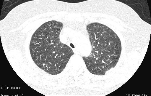
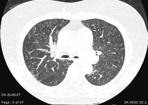
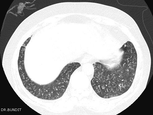
Case 1 : Question. 1.1 What is the intralobular distribution? 1. Centrilobular 2. Perilymphatic 3. Random
|
|
|
- Noel Woods
- 5 years ago
- Views:
Transcription
1 Interesting case
2 Case 1
3
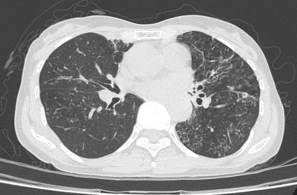
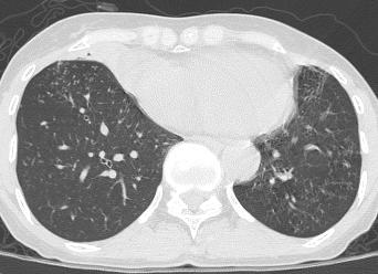
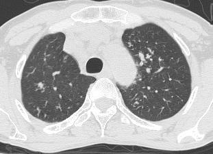
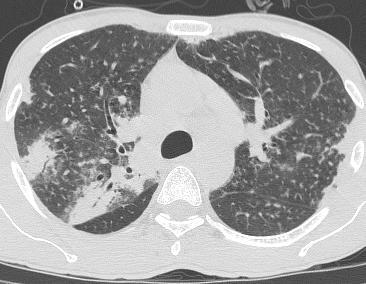
4 Case 1 : Question 1.1 What is the intralobular distribution? 1. Centrilobular 2. Perilymphatic 3. Random
5 Case 1: Answer 1.1 What is the intralobular distribution? 1. Centrilobular 2. Perilymphatic 3. Random
6 Perilymphatic Centrilobular Random

7 Perilymphatic Centrilobular Random
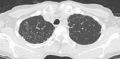
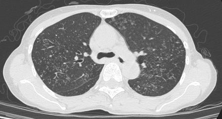
8 1. Centrilobular
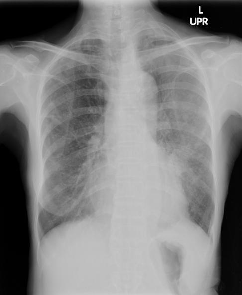
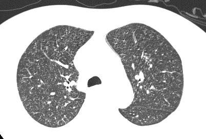
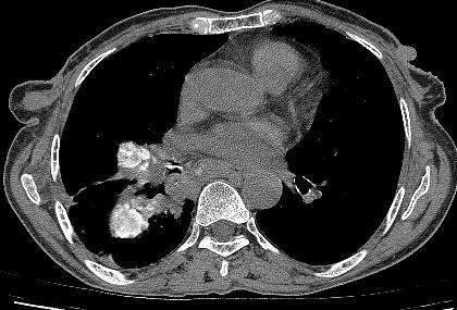
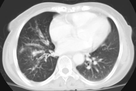
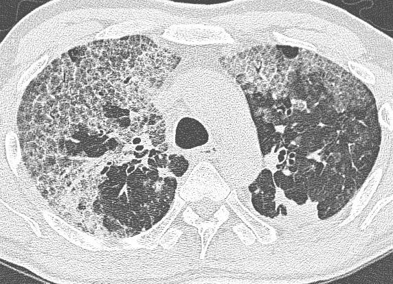
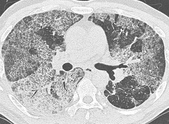
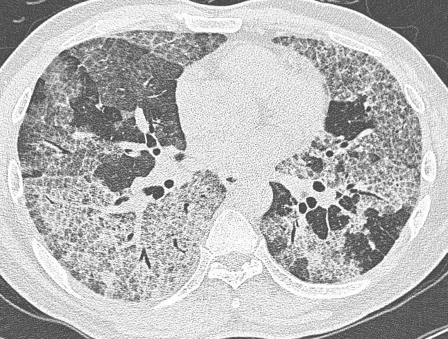
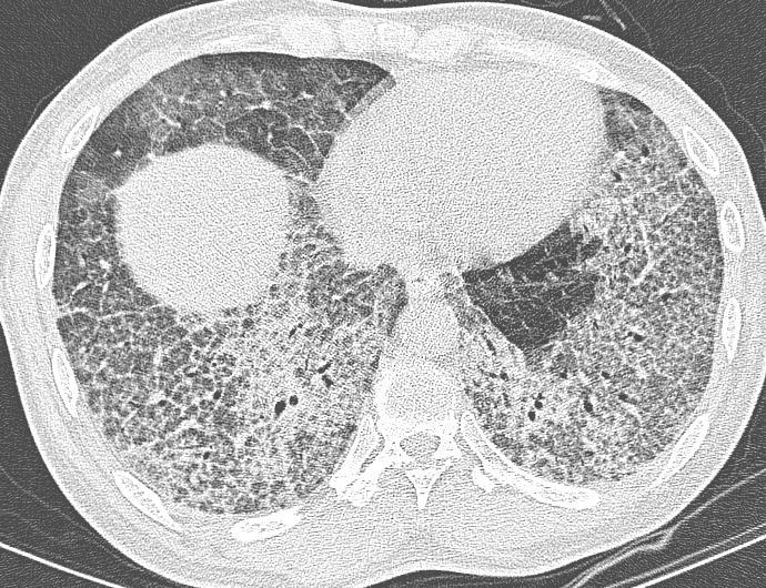
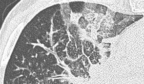
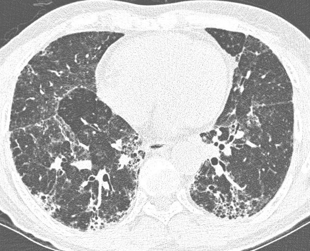
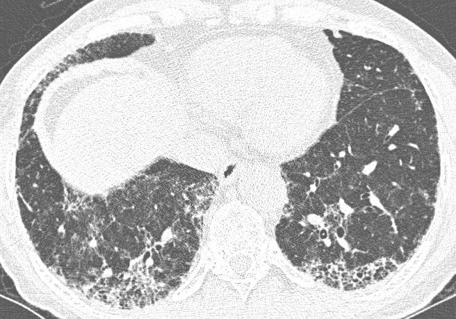
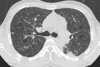
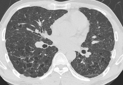
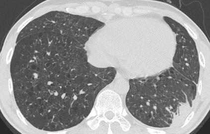
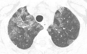
9 Case 1 : Question 1.2 What is the diagnosis? 1. Silicosis 2. Bronchiolitis 3. Lymphangitic carcinomatosis 4. Miliary tuberculosis
10 Case 1: Answer 1.2 What is the diagnosis? 1. Silicosis 2. Bronchiolitis 3. Lymphangitic carcinomatosis 4. Miliary tuberculosis
11 Common presentation 1. Silicosis Centrilobular well defined nodule/ Perilymphahtic nodule 2. Bronchiolitis Centrilobular ill-defined nodule/ Tree in bud 3. Lymphangitic carcinomatosis Smooth-nodular thickened interlobular septum/ Perilymphatic nodule 4. Miliary tuberculosis Random well defined nodule
12 Case 1 Lung volume Pattern Distribution Normal Craniocaudal Lower Axial Associated finding Intralobular Diagnosis : Bronchiolitis Histology : Bronchiolitis Centrilobular nodule/ Bronchial dilatation Diffuse Centrilobular Pulmonary-arterial hypertension
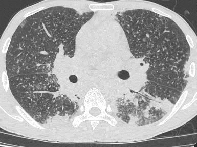
13 Case 2
14
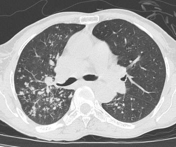
15 Case 2 : Question 2.1 What is the intralobular distribution? 1. Centrilobular 2. Perilymphatic 3. Random
16 Case 2: Answer 2.1 What is the intralobular distribution? 1. Centrilobular 2. Perilymphatic 3. Random
17 1. Centrilobular
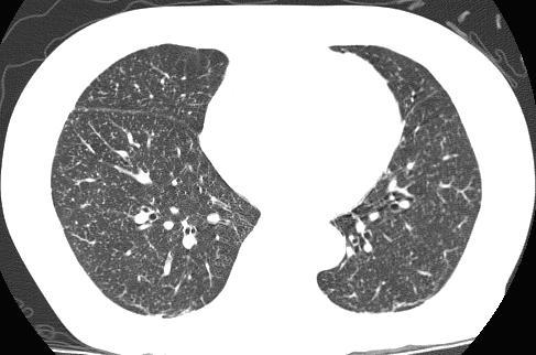
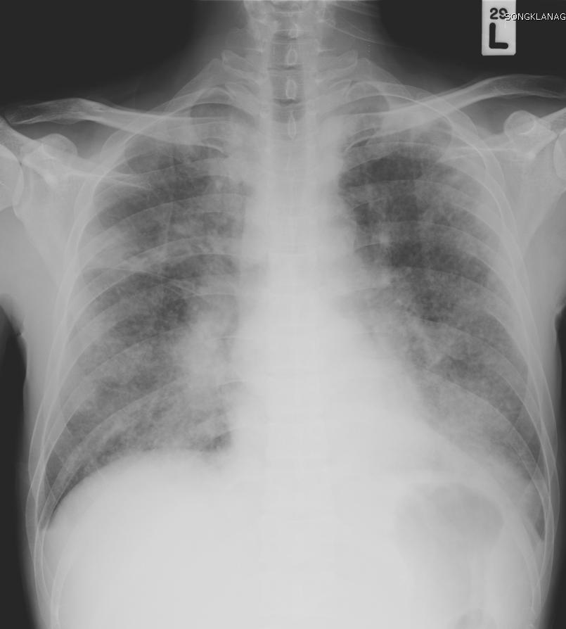
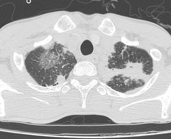
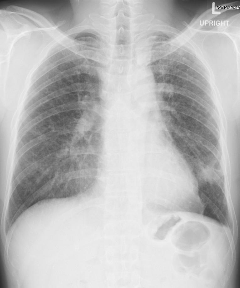
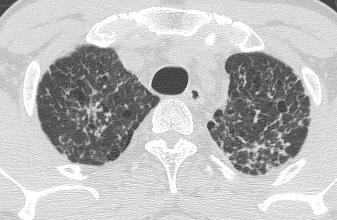
18 Case 2 : Question 2.2 What is the diagnosis? 1. Bronchial spreading tuberculosis 2. Miliary metastasis 3. Lymphatic carcinomatosis
19 Case 2: Answer 2.2 What is the diagnosis? 1. Bronchial spreading tuberculosis 2. Miliary metastasis 3. Lymphatic carcinomatosis
20 Common presentation 1. Bronchial spreading tuberculosis Centrilobular well defined nodule 2. Miliary tuberculosis Random well defined nodule 3. Lymphatic carcinomatosis Smooth-nodular thickened interlobular septum/ Perilymphatic nodule
21 เฉลย 1. Bronchial spreading tuberculosis - Sputum AFB 08/12/2006 : AFB positive 2+ - Sputum Culture 08/12/2006 : Mycobacterium tuberculosis - Histopathology : Peribronchial inflammation
22 Case 2 Lung volume Increase Pattern Centrilobular nodule Craniocaudal Upper Distribution Axial Diffuse Intralobular Centrilobular Associated finding Bronchiectasis Diagnosis: Pulmonary tuberculosis Histology : Peribronchial inflammation
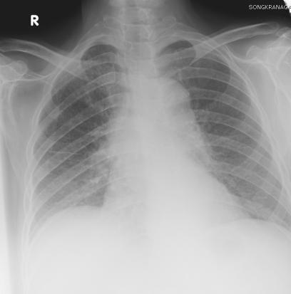
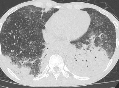
23 Case 3
24
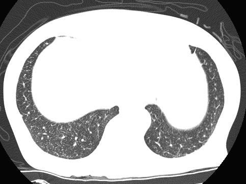
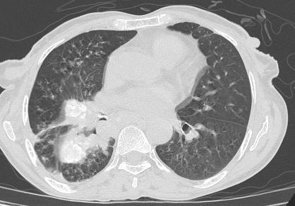
25 Case 3 : Question 3.1 What is the intralobular distribution? 1. Centrilobular 2. Perilymphatic 3. Random
26 Case 3: Answer 3.1 What is the intralobular distribution? 1. Centrilobular 2. Perilymphatic 3. Random
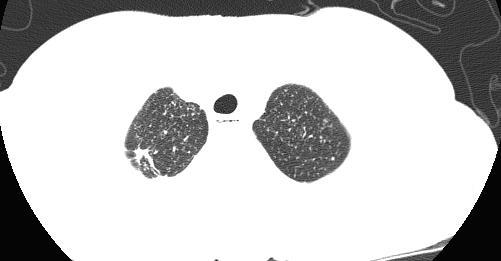
27 3. Random
28 Case 3: Question 3.2 What is the diagnosis? 1. Bronchial spreading tuberculosis 2. Miliary tuberculosis 3. Lymphatic carcinomatosis
29 Case 3: Answer 3.2 What is the diagnosis? 1. Bronchial spreading tuberculosis 2. Miliary tuberculosis 3. Lymphatic carcinomatosis
30 Common presentation 1. Bronchial spreading tuberculosis Centrilobular well defined nodule 2. Miliary tuberculosis Random well defined nodule 3. Lymphatic carcinomatosis Smooth-nodular thickened interlobular septum/ Perilymphatic nodule
31 2. Miliary tuberculosis - Sputum AFB 17/06/2013 : AFB positive 1+ - Sputum Culture 17/06/2013 : Negative - Histopathology : Caseous granulomatous inflammation
32 Case 3 Lung volume Normal Pattern Nodule Craniocaudal Diffuse Distribution Axial Diffuse Intralobular Random Associated finding - Diagnosis: Miliary tuberculosis Histology : Caseous granulomatous inflamation
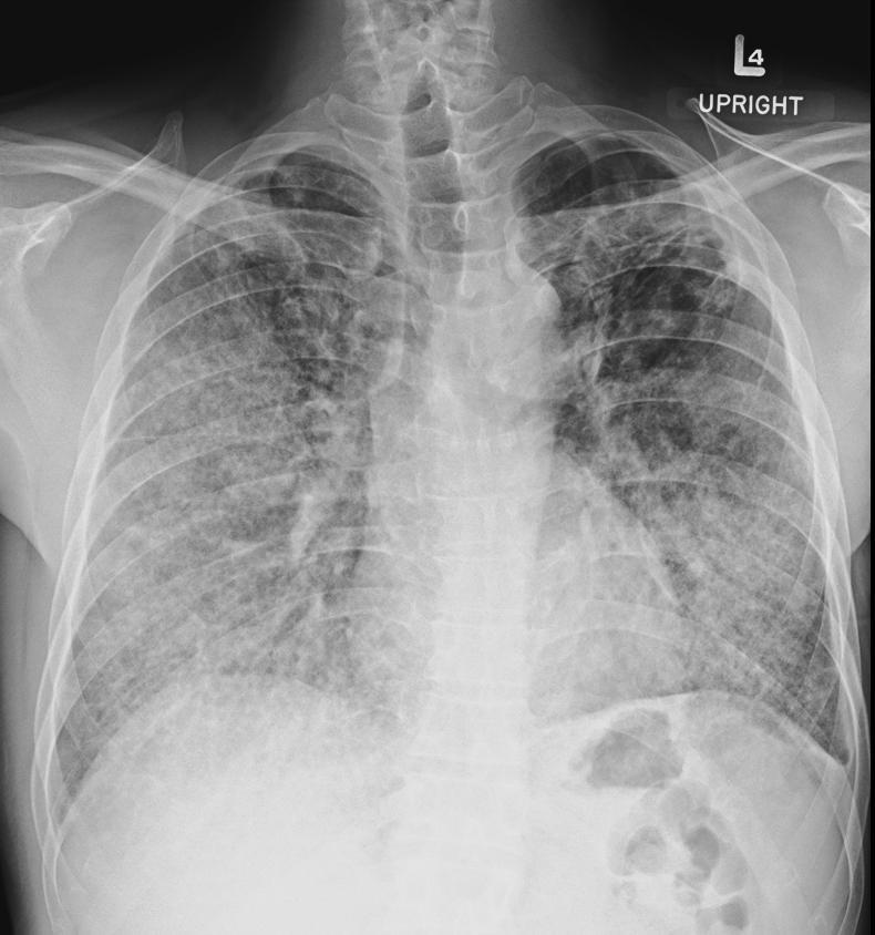
33 Case 4
34
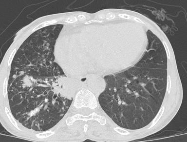
35 Case 4: Question 4.1 What is the intralobular distribution? 1. Centrilobular 2. Perilymphatic 3. Random
36 Case 4: Answer 4.1 What is intralobular distribution? 1. Centrilobular 2. Perilymphatic 3. Random
37 1. Centrilobular
38 Case 4: Question 4.2 What is the diagnosis? 1. Bacterial bronchiolitis 2. Silicosis 3. Bronchial spreading tuberculosis
39 Case 4: Answer 4.2 What is the diagnosis? 1. Bacterial bronchiolitis 2. Silicosis 3. Bronchial spreading tuberculosis
40 Common presentation 1. Bacterial bronchiolitis Centrilobular poorly defined nodule 2. Silicosis Centrilobular well defined nodule/ Perilymphatic 3. Bronchial spreading tuberculosis Centrilobular well defined nodule
41 เฉลย 3. Bronchial spreading tuberculosis - Sputum AFB 16/01/2006 : Negative - Sputum Culture 16/01/2006 : Negative - Histopathology : Necrosis and fibrosis


42 Pre treatment 2549 Post treatment 2552
43 Case 4 Lung volume Pattern Distribution Normal Craniocaudal Lower Axial Associated finding Intralobular Diagnosis: Tuberculosis Centrilobular nodule/ Consolidation Diffuse Histology : Necrosis and fibrosis Centrilobular Lymph node enlargement
44 Case 5
45
46 Case 5: Question 5.1 What is the intralobular distribution? 1. Centrilobular 2. Perilymphatic 3. Random
47 Case 5: Answer 5.1 What is the intralobular distribution? 1. Centrilobular 2. Perilymphatic 3. Random
48 1. Centrilobular


49 Histology Granulomatous inflammation suggestive tuberculosis. Few eosinophils are scathered at submucosal.(afb negative)
50 Clinical discussion -A known case of Tb ileum status post complete treat - ment - Sputum AFB 02/03/2006 : Negative - Sputum Culture 15/06/200 6 : Negative - Histology: Granulomatous inflammation (AFB negative)
51 Confidence in diagnosis Group B: Disease accurately identified with short Ddx Silicosis, Coal worker pneumoconiosis Sarcoidosis, Beryliosis COP, Chronic eosinophilic pneumonia DIP, Hypersensitivity pneumonitis NSIP RB-ILD

52 Category1 No invasive procedure needed if clinical and CT feature are typical Category2 BAL Category3 Transbronchial Bx Category4 Surgical lung Bx UIP Lymphangiomyo -matosis Langerhan's histiocytosis HP Pneumoconiosis Collagen vascular disease PAP Infection Sarcoidosis Lymphagitic carcinoma Lymphoperi -ferative disorders Malignancy Nonspecific CT appearance without a clinical explanation Typical CT appearance of condition with atypical features Typical clinical features of a condition with atypical CT Pg 140
53 Case 5 Lung volume Normal Pattern Centrilobular nodule Craniocaudal Lower Distribution Axial Diffuse Intralobular Centrilobular Associated finding Lymph node enlargement Diagnosis: Hypersensitivity Histology : Granulomatous inflammation
54 Case 6
55
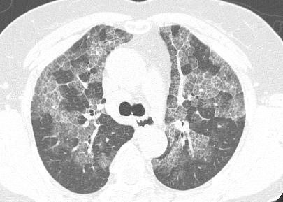
56 Case6 : Question 6.1 What is the main pattern of this HRCT? 1. Reticulation 2. Groundglass opacity 3. 1 and 2

57 Case 6: Answer 6.1 What is the main pattern of this HRCT? 1. Reticulation 2. Groundglass opacity 3. 1 and 2
58 Case 6 : Question 6.2 What kind of the lines is in majority? 1. Interlobular line 2. Intralobular line

59 Case 6: Answer 6.2 What kind of the lines is in majority? 1. Interlobular line 2. Intralobular line
60 Case 6 : Question 6.3 What is the intralobular distribution? 1. Panlobular 2. Perilobular 3. Centrilobular
61 Case 6: Answer 6.3 What is the intralobular distribution? 1. Panlobular 2. Perilobular 3. Centrilobular
62 Case 6: Question 6.4 What is the diagnosis? 1. Pulmonary alveolar proteinosis 2. Hypersensitivity pneumonitis 3. Lymphangitic carcinomatosis
63 Case 6 : Answer 6.4 What is the diagnosis? 1. Pulmonary alveolar proteinosis 2. Hypersensitivity pneumonitis 3. Lymphangitic carcinomatosis


64 Case 6 PAP Lymphangitic carcinomatosis


65 Histology Consistent with pulmonary alveolar proteinosis (PAS positive)
66 Confidence in diagnosis Group A: Confident CT diagnosis IPF Lymphangitic spreading: Bx Alveolar proteinosis: BAL, Bx Asbestosis Lung edema/ Left heart failure page 54
67 Case 6 Category1 No invasive procedure needed if clinical and CT feature are typical Category2 BAL Category3 Transbronchial Bx Category4 Surgical lung Bx UIP Lymphangiomyomatosis Langerhan's histiocytosis HP Pneumoconiosis Collagen vascular disease PAP Infection Sarcoidosis Lymphagitic carcinoma Lymphoperiferative disorders Malignancy Nonspecific CT appearance without a clinical explanation Typical CT appearance of condition with atypical features Typical clinical features of a condition with atypical CT
68 Case 6 Lung volume Normal Pattern GGO/ intralobular line Craniocaudal Diffuse Distribution Axial Diffuse Intralobular Panlobular Associated finding - Diagnosis Histology : PAP : PAP
69 Case 7
70
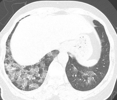
71 Case 7 : Question 7.1 What is the main pattern of this HRCT? 1. Groundglass opacity 2. Honeycombing 3. Consolidation 4. All of the above
72 Case 7 : Answer 7.1 What is the main pattern of this HRCT? 1. Groundglass opacity 2. Honeycombing 3. Consolidation 4. All of the above
73 Case 7 : Question 7.2 What is the craniocaudal distribution? 1. Upper 2. Diffuse 3. Lower

74 Case 7 : Answer 7.2 What is the craniocaudal distribution? 1. Upper 2. Diffuse 3. Lower
75 Case 7 : Question 7.3 What is the axial distribution? 1. Peripheral 2. Diffuse 3. Central

76 Case 7 : Answer 7.3 What is the axial distribution? 1. Peripheral 2. Diffuse 3. Central
77 Case 7 : Question 7.4 Are this HRCT findings typical of UIP? 1. Yes 2. No
78 Case 7 : Answer 7.4 Are this HRCT findings typical of UIP? 1. Yes 2. No
79 HRCT Criteria for UIP 1.Peripheral basal reticulation 2.Honeycombing 3.Abscence of atypical findings Consolidate GGO Centrilobular nodules
80 Category1 No invasive procedure needed if clinical and CT feature are typical Category2 BAL Category3 Transbronchial Bx Category4 Surgical lung Bx UIP Lymphangiomyomatosis Langerhan's histiocytosis HP Pneumoconiosis Collagen vascular disease PAP Infection Sarcoidosis Lymphagitic carcinoma Lymphoperiferative disorders Malignancy Nonspecific CT appearance without a clinical explanation Typical CT appearance of condition with atypical features Typical clinical features of a condition with atypical CT
81 Lung volume Pattern Case 7 Clinical diagnosis: Chronic hypersensitivity Histology : None Normal Craniocaudal Diffuse Distribution Axial Peripheral Intralobular Associated finding - GGO/ reticulation/honeycombing Centrilobular
82 Case 8
83
84 Case 8 : Question 8.1 What about the lung volume? 1. Increased 2. Normal 3. Decreased
85 Case 8 : Answer 8.1 What is lung volume? 1. Increased 2. Normal 3. Decreased
86 Case 8 : Question 8.2 What is the main pattern of this HRCT? 1. Cyst 2. Reticulation 3. Septal thickening
87 Case 8 : Answer 8.2 What is the main pattern of this HRCT? 1. Cyst 2. Reticulation 3. Septal thickening
88 Case 8 : Question 8.3 What is the craniocaudal distribution? 1. Upper 2. Diffuse 3. Lower

89 Case 8 : Answer 8.3 What is the craniocaudal distribution? 1. Upper 2. Diffuse 3. Lower
90 Case 8 : Question 8.4 What is the axial distribution? 1. Peripheral 2. Diffuse 3. Central

91 Case 8 : Answer 8.4 What is the axial distribution? 1. Peripheral 2. Diffuse 3. Central
92 Case 8 : Question 8.5 What is the associated finding? 1. Lymph node enlargement 2. Rounded atelectasis 3. Pleural thickening

93 Case 8 : Answer 8.5 What is the associated finding? 1. Lymph node enlargement 2. Rounded atelectasis 3. Pleural thickening
94 Category1 No invasive procedure needed if clinical and CT feature are typical Category2 BAL Category3 Transbronchial Bx Category4 Surgical lung Bx UIP Lymphangiomyomatosis Langerhan's histiocytosis HP Pneumoconiosis Collagen vascular disease PAP Infection Sarcoidosis Lymphagitic carcinoma Lymphoperiferative disorders Malignancy Nonspecific CT appearance without a clinical explanation Typical CT appearance of condition with atypical features Typical clinical features of a condition with atypical CT
95 Confidence in diagnosis Group A: Confident CT diagnosis IPF Lymphangitic spreading: Bx Lymphagiomyomatosis Langerhans cell histocytosis Alveolar proteinosis: BAL, Bx Asbestosis Lung edema/ Left heart failure
96 Lung volume Pattern Case 8 Increase Craniocaudal Upper Cyst/ nodule Distribution Axial Diffuse Associated finding Intralobular Centrilobular Rounded atelectasis, pleural thickening Diagnosis: PLCH Histology : Inconclusive (chronic inflammation, no langerhans cell)
97 Case 9
98
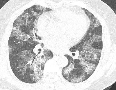
99 Case 9 : Answer 9.1 What is the main pattern of this HRCT? 1. Ground glass opacity 2. Reticulation 3. Septal thickening 4. All
100 Case 9 : Question 9.1 What is the main pattern of this HRCT? 1. Ground glass opacity 2. Reticulation 3. Septal thickening 4. All
101 Case 9 : Question 9.2 What is the diagnosis? 1. Pulmonary alveolar proteinosis 2. Hypersensitivity pneumonitis 3. NSIP


102 Case 9 : Answer 9.2 What is the diagnosis? 1. Pulmonary alveolar proteinosis 2. Hypersensitivity pneumonitis 3. NSIP

103 Histology Alveolar proteinosis
104 Confidence in diagnosis Group A: Confident CT diagnosis IPF Lymphangitic spreading: Bx Lymphagiomyomatosis Langerhans cell histocytosis Alveolar proteinosis: BAL, Bx Asbestosis Lung edema/ Left heart failure
105 Category1 No invasive procedure needed if clinical and CT feature are typical Category2 BAL Category3 Transbronchial Bx Category4 Surgical lung Bx UIP Lymphangiomyomatosis Langerhan's histiocytosis HP Pneumoconiosis Collagen vascular disease PAP Infection Sarcoidosis Lymphagitic carcinoma Lymphoperiferative disorders Malignancy Nonspecific CT appearance without a clinical explanation Typical CT appearance of condition with atypical features Typical clinical features of a condition with atypical CT
106 Case 9 Lung volume Normal Pattern Craniocaudal Lower Distribution Axial Diffuse Intralobular Associated finding - Diagnosis: PAP Histology : PAP GGO/ intralobular line Diffuse/ perilymphatic
107 Suspected diffused lung disease Chest radiograph Normal or equivocal Abnormal Suggestive of sarcoidosis HRCT with prone views HRCT Trans bronchial Bx Normal Abnormal See next slide Bx if clinical/physiologic evidence of disease See next slide
108 Suspected diffused lung disease Specific CT pattern(uip,eg,lam,h P, Lipoid Pneumonia) CT pattern suggestive of HP sarcoid, lymphagitic carcinoma,pap, alveolar carcinoma,eosinophillic pneumonia, BOOP Other CT pattern Accept CT diagnosis if clinical scenario is consistent Trans bronchial biopsy and/or brochoalveolar lavage (CT directed) Thoracosopic biopsy (CT directed)
109 Thank you
Case 1: Question. 1.1 What is the main pattern of this HRCT? 1. Intralobular line 2. Groundglass opacity 3. Perilymphatic nodule
 HRCT WORK SHOP Case 1 Case 1: Question 1.1 What is the main pattern of this HRCT? 1. Intralobular line 2. Groundglass opacity 3. Perilymphatic nodule Case 1: Question 1.2 What is the diagnosis? 1. Hypersensitivity
HRCT in Diffuse Interstitial Lung Disease Steps in High Resolution CT Diagnosis. Where are the lymphatics? Anatomic distribution
 Steps in High Resolution CT Diagnosis Pattern of abnormality Distribution of disease Associated findings Clinical history Tomás Franquet MD What is the diagnosis? Hospital de Sant Pau. Barcelona Secondary
Acute and Chronic Lung Disease
 KATHOLIEKE UNIVERSITEIT LEUVEN Faculty of Medicine Acute and Chronic Lung Disease W De Wever, JA Verschakelen Department of Radiology, University Hospitals Leuven, Belgium Clinical utility of HRCT To detect
Financial disclosure COMMON DIAGNOSES IN HRCT. High Res Chest HRCT. HRCT Pre test. I have no financial relationships to disclose. Anatomy Nomenclature
 Financial disclosure I have no financial relationships to disclose. Douglas Johnson D.O. Cardiothoracic Imaging Gaston Radiology COMMON DIAGNOSES IN HRCT High Res Chest Anatomy Nomenclature HRCT Sampling
Manish Powari Regional Training Day 10/12/2014
 Manish Powari Regional Training Day 10/12/2014 Large number of different types of Interstitial Lung Disease (ILD). Most are very rare Most patients present with one of a smaller number of commoner diseases
Daria Manos RSNA 2016 RC 401. https://medicine.dal.ca/departments/depar tment-sites/radiology/contact/faculty/dariamanos.html
 Daria Manos RSNA 2016 RC 401 https://medicine.dal.ca/departments/depar tment-sites/radiology/contact/faculty/dariamanos.html STEP1: Is this fibrotic lung disease? STEP 2: Is this a UIP pattern? If yes:
11/10/2014. Multi-disciplinary Approach to Diffuse Lung Disease: The Imager s Perspective. Radiology
 Multi-disciplinary Approach to Diffuse Lung Disease: The Imager s Perspective Radiology Pathology Clinical 1 Role of HRCT Diagnosis Fibrosis vs. inflammation Next step in management Response to treatment
Bronkhorst colloquium Interstitiële longziekten. Katrien Grünberg, klinisch patholoog
 Bronkhorst colloquium 2013-2014 Interstitiële longziekten De pathologie achter de CT Katrien Grünberg, klinisch patholoog K.grunberg@vumc.nl Preparing: introduction and 3 cases The introduction on microscopic
Outline Definition of Terms: Lexicon. Traction Bronchiectasis
 HRCT OF IDIOPATHIC INTERSTITIAL PNEUMONIAS Disclosures Genentech, Inc. Speakers Bureau Tadashi Allen, MD University of Minnesota Assistant Professor Diagnostic Radiology 10/29/2016 Outline Definition of
I have no relevant conflicts of interest to disclose
 I have no relevant conflicts of interest to disclose Diffuse parenchymal lung disease (DPLD) and its associations Secondary lobular anatomy DPLD History, clinical findings, temporal evolution, and exposures
A Review of Interstitial Lung Diseases. Paul J. Wolters, MD Associate Professor Department of Medicine University of California San Francisco
 A Review of Interstitial Lung Diseases Paul J. Wolters, MD Associate Professor Department of Medicine University of California San Francisco Outline Overview of diagnosis in ILD Why it is important Definition/Classification
A Review of Interstitial Lung Diseases
 Outline A Review of Interstitial Lung Diseases Paul J. Wolters, MD Associate Professor Department of Medicine University of California San Francisco Overview of diagnosis in ILD Why it is important Definition/Classification
Radiologic-pathologic correlation of pulmonary diseases
 The 1578 th Chest Conference/ 3 rd Biennial Clinical- Radiologic-Pathologic Correlation Radiologic-pathologic correlation of pulmonary diseases Harumi Itoh, M.D. University of Fukui, Japan Centriacinar
Usual Interstitial pneumonia and Nonspecific Interstitial Pneumonia. Nitra and the Gangs.
 Usual Interstitial pneumonia and Nonspecific Interstitial Pneumonia Nitra and the Gangs. บทน ำและบทท ๓, ๑๐, ๑๒, ๑๓, ๑๔, ๑๕, ๑๗ Usual Interstitial Pneumonia (UIP) Most common & basic pathologic pattern
Differential diagnosis
 Differential diagnosis Idiopathic pulmonary fibrosis (IPF) is part of a large family of idiopathic interstitial pneumonias (IIP), one of four subgroups of interstitial lung disease (ILD). Differential
5/9/2015. Multi-disciplinary Approach to Diffuse Lung Disease: The Imager s Perspective. No, I am not a pulmonologist! Radiology
 Multi-disciplinary Approach to Diffuse Lung Disease: The Imager s Perspective No, I am not a pulmonologist! Radiology Pathology Clinical 1 Everyone needs a CT Confidence in diagnosis Definitive HRCT +
Non-neoplastic Lung Disease II
 Pathobasic Non-neoplastic Lung Disease II Spasenija Savic Prince Pathology Program Systematic approach to surgical lung biopsies with ILD Examples (chronic ILD): Idiopathic interstitial pneumonias: UIP,
Mimics in chest disease: interstitial opacities
 Insights Imaging (2013) 4:9 27 DOI 10.1007/s13244-012-0207-7 PICTORIAL REVIEW Mimics in chest disease: interstitial opacities Anastasia Oikonomou & Panos Prassopoulos Received: 19 June 2012 / Revised:
Case Presentations in ILD. Harold R. Collard, MD Department of Medicine University of California San Francisco
 Case Presentations in ILD Harold R. Collard, MD Department of Medicine University of California San Francisco Outline Overview of diagnosis in ILD Definition/Classification High-resolution CT scan Multidisciplinary
Resident Case Review CHEST. Daria Manos CAR 2016
 Resident Case Review CHEST CAR 2016 Daria Manos Disclosure Speakers bureau, Roche CAR 2016 Daria Manos 1. Recognize common and critical chest radiograph and computed tomography signs and use these clues
The crazy-paving pattern: A radiological-pathological correlated and illustrated overview
 The crazy-paving pattern: A radiological-pathological correlated and illustrated overview Poster No.: C-0827 Congress: ECR 2010 Type: Educational Exhibit Topic: Chest Authors: W. F. M. De Wever, J. Coolen,
Epidemiology and classification of smoking related interstitial lung diseases
 Epidemiology and classification of smoking related interstitial lung diseases Šterclová M. Department of Respiratory Diseases, Thomayer Hospital, Prague, Czech Republic Supported by an IGA Grant No G 1207
Micronodular Lung Disease an algorithm
 Micronodular Lung Disease an algorithm H. Page McAdams, MD Department of Radiology Duke University Medical Center Durham, NC USA page.mcadams@duke.edu Question Which of the following lung diseases is MOST
Diffuse Interstitial Lung Diseases: Is There Really Anything New?
 : Is There Really Anything New? Sujal R. Desai, MBBS, MD ESTI SPEAKER SUNDAY Society of Thoracic Radiology San Antonio, Texas March 2014 Diffuse Interstitial Lung Disease The State of Play DILDs Is There
An Image Repository for Chest CT
 An Image Repository for Chest CT Francesco Frajoli for the Chest CT in Antibody Deficiency Group An Image Repository for Chest CT he Chest CT in Antibody Deficiency Group is an international and interdisciplinary
Liebow and Carrington's original classification of IIP
 Liebow and Carrington's original classification of IIP-- 1969 Eric J. Stern MD University of Washington UIP Usual interstitial pneumonia DIP Desquamative interstitial pneumonia BIP Bronchiolitis obliterans
Radiologic Approach to Smoking Related Interstitial Lung Disease
 Radiologic Approach to Smoking Related Interstitial Lung Disease Poster No.: C-1854 Congress: ECR 2013 Type: Educational Exhibit Authors: K.-N. Lee, J.-Y. Han, E.-J. Kang, J. Kang; Busan/KR Keywords: Toxicity,
INTERSTITIAL LUNG DISEASE. Radhika Reddy MD Pulmonary/Critical Care Long Beach VA Medical Center January 5, 2018
 INTERSTITIAL LUNG DISEASE Radhika Reddy MD Pulmonary/Critical Care Long Beach VA Medical Center January 5, 2018 Interstitial Lung Disease Interstitial Lung Disease Prevalence by Diagnosis: Idiopathic Interstitial
How to Analyse Difficult Chest CT
 How to Analyse Difficult Chest CT Complex diseases are:- - Large lesion - Unusual or atypical pattern - Multiple discordant findings Diffuse diseases are:- - Numerous findings in both sides 3 basic steps
Interstitial syndrome
 Interstitial syndrome Ground-glass attenuation Miliary and nodular images linear images Etienne Leroy Terquem Pierre L Her SPI / ISP Soutien Pneumologique International / International Support for Pulmonology
IPF: Epidemiologia e stato dell arte
 IPF: Epidemiologia e stato dell arte Clinical Classification Diffuse parenchimal lung diseases Exposure-related: - occupational - environmental - medication Desquamative interstitial pneumonia Idiopathic
Bronchoalveolar Lavage and Histopathologic Diagnosis Based on Biopsy
 Idiopathic Pulmonary Fibrosis Bronchoalveolar Lavage and Histopathologic Diagnosis Based on Biopsy JMAJ 46(11): 469 474, 2003 Yukihiko SUGIYAMA Professor, Division of Pulmonary Medicine, Department of
The radiological differential diagnosis of the UIP pattern
 5th International Conference on Idiopathic Pulmonary Fibrosis, Modena, 2015, June 12th The radiological differential diagnosis of the UIP pattern Simon Walsh King s College Hospital Foundation Trust London,
Radiologists toolbox to differentiate alveolar versus interstitial lung diseases
 Radiologists toolbox to differentiate alveolar versus interstitial lung diseases Dr Sumer Shikhare, Dr Trishna Shimpi, Dr Ashish Chawla Khoo Teck Puat Hospital Singapore. Relevant financial disclosures
American Thoracic Society European Respiratory Society Classification of the Idiopathic Interstitial Pneumonias: Advances in Knowledge since 20021
 This copy is for personal use only. To order printed copies, contact reprints@rsna.org American Thoracic Society European Respiratory Society Classification of the Idiopathic Interstitial Pneumonias: Advances
Chest imaging II. Interstitial lung diseases
 Chest imaging II. Interstitial lung diseases Dávid L. Tárnoki MD, PhD Ádám D. TárnokiMD, PhD Department of Radiology Semmelweis University Topics 1. Interstitial lung diseases 2. Occupational lung diseases
Diagnosis of TB: Radiology David Finlay, MD
 TB Intensive Tyler, Texas June 2-4, 2010 Diagnosis of TB: Radiology David Finlay, MD June 3, 2010 2stages stages- Tuberculosis 1. primary infection 2. reactivation, or post primary disease 2 1 Primary
Idiopathic interstitial pneumonias (IIPs) are a group of
 are a group of") SYMPOSIA C. Isabela S. Silva, MD, PhD and Nestor L. Müller, MD, PhD Abstract: The idiopathic interstitial pneumonias (IIPs) are a group of diffuse parenchymal lung diseases of unknown etiology characterized
SYMPOSIA C. Isabela S. Silva, MD, PhD and Nestor L. Müller, MD, PhD Abstract: The idiopathic interstitial pneumonias (IIPs) are a group of diffuse parenchymal lung diseases of unknown etiology characterized
Histopathologic Approach to Interstitial Lung Disease
 Histopathologic Approach to Interstitial Lung Disease Kirk D. Jones, MD UCSF Dept of Pathology kirk.jones@ucsf.edu Disclosures I have nothing to disclose 1 Why? Much of interstitial lung disease biopsies
Smoking-related Interstitial Lung Diseases: High-Resolution CT Findings
 Smoking-related Interstitial Lung Diseases: High-Resolution CT Findings Poster No.: C-2358 Congress: ECR 2013 Type: Educational Exhibit Authors: V. Cuartero Revilla, M. Nogueras Carrasco, P. Olmedilla
Typical and atypical findings of pulmonary sarcoidosis at high resolution CT
 Typical and atypical findings of pulmonary sarcoidosis at high resolution CT Poster No.: C-0169 Congress: ECR 2013 Type: Educational Exhibit Authors: L. Raposo Rodríguez, C. Mejía, B. Escobar Mallada,
Interstitial Syndrome Ground glass attenuation miliary and nodular images Linear images
 Interstitial Syndrome Ground glass attenuation miliary and nodular images Linear images Dr Etienne Leroy-Terquem Centre hospitalier de Meulan les Mureaux. France French-cambodian association for pneumology
Progress in Idiopathic Pulmonary Fibrosis
 Progress in Idiopathic Pulmonary Fibrosis David A. Lynch, MB Disclosures Progress in Idiopathic Pulmonary Fibrosis David A Lynch, MB Consultant: t Research support: Perceptive Imaging Boehringer Ingelheim
10/17/2016. Nuts and Bolts of Thoracic Radiology. Objectives. Techniques
 Nuts and Bolts of Thoracic Radiology October 20, 2016 Carleen Risaliti Objectives Understand the basics of chest radiograph Develop a system for interpreting chest radiographs Correctly identify thoracic
Interesting Cases. Pulmonary
 Interesting Cases Pulmonary 54M with prior history of COPD, hep B/C, and possible history of TB presented with acute on chronic dyspnea, and productive cough Hazy opacity overlying the left hemithorax
RADIOLOGICALL ANALYSIS OF INTERSTITIAL LUNG DISEASES
 Original Research Article RADIOLOGICALL ANALYSIS OF INTERSTITIAL LUNG DISEASES Meraj Rentia 1*, Himanshu Singla 1, Divya Malpani 2, Tushar Vaishnav 3, Pradeep Jhala 3 1 1 st year Resident, 2 3 rd year
Diagnostic Imaging of Diffuse Infiltrative Disease of the Lung
 Thematic Review Series Respiration 2004;71:4 19 DOI: 10.1159/000075642 Diagnostic Imaging of Diffuse Infiltrative Disease of the Lung Maurizio Zompatori a Claudio Bnà a Venerino Poletti c Enrica Spaggiari
IPF - Inquadramento clinico
 IPF - Inquadramento clinico Sergio Harari Unità Operativa di Pneumologia UTIR Servizio di Fisiopat. Resp. e Emodinamica Polmonare Ospedale S. Giuseppe, Milano Clinical Classification Diffuse parenchimal
Chest Radiology LYMPHANGITIC CARCINOMATOSIS CERTAIN CANCERS SPREAD BY PLUGGING THE LYMPHATICS
 2 Chest Radiology Includes plain film diagnosis, CT, MRI, and interventional techniques used in the diagnosis of diseases of the lungs, pleura, and mediastinum including the heart and great vessels. LYMPHANGITIC
Lines and crackles. Making sense of ILD
 Lines and crackles Making sense of ILD Case JM 65 year old male Gradual shortness of breath, going on over a year Some dry cough Ex-smoker, quit 10 years ago Crackles in the bases CXR presented Sent to
Workshop Cyst & Lucency. How to Approach
 Workshop Cyst & Lucency How to Approach To Approach Cystic Lung Disease True cysts? Cavitary disease Cystic bronchiectasis Mosaic attenuation Subpleural cysts Bullae Paraseptal emphysema Honeycombing Birt
Thoracic Sarcoidosis Imaging Updated: Jul 19, 2013
 Thoracic Sarcoidosis Imaging Updated: Jul 19, 2013 Overview Radiography Computed Tomography Magnetic Resonance Imaging Nuclear Imaging Show All Multimedia Library References Overview For patients with
T he diagnostic evaluation of a patient with
 546 REVIEW SERIES Challenges in pulmonary fibrosis? 1: Use of high resolution CT scanning of the lung for the evaluation of patients with idiopathic interstitial pneumonias Michael B Gotway, Michelle M
How to identify interstitial pneumonias.
 How to identify interstitial pneumonias. Poster No.: C-0804 Congress: ECR 2014 Type: Educational Exhibit Authors: S. claret loaiza, M. C. Cañete Moslero, R. Carreño Gonzalez, C. de la Torre; Malaga/ES
* * APPROACH TO NON- NEOPLASTIC LUNG DISEASE IN TRANSBRONCHIAL AND SURGICAL BIOPSIES. Financial Disclosures: NONE. BIOPSY TECHNIQUES Bronchoscopic
 APPROACH TO NON- NEOPLASTIC LUNG DISEASE IN TRANSBRONCHIAL AND SURGICAL BIOPSIES Thomas V. Colby, M.D. Mayo Clinic Arizona Geraldine C. Zeiler Professor of Cytopathology Mayo Clinic Arizona Financial Disclosures:
4/17/2010 C ini n ca c l a Ev E a v l a ua u t a ion o n of o ILD U dat a e t e i n I LDs
 Update in ILDs Diagnosis 101: Clinical Evaluation April 17, 2010 Jay H. Ryu, MD Mayo Clinic, Rochester MN Clinical Evaluation of ILD Outline General aspects of ILDs Classification of ILDs Clinical evaluation
An Introduction to Radiology for TB Nurses
 An Introduction to Radiology for TB Nurses Garold O. Minns, MD September 14, 2017 TB Nurse Case Management September 12 14, 2017 EXCELLENCE EXPERTISE INNOVATION Garold O. Minns, MD has the following disclosures
Role of Computed Tomography in Diagnosis of Diffuse Lung Diseases Chauhan Jayant 1*, Panchal Pankaj 2, Faruqui Tehzeeb 3
 ORIGINAL ARTICLE Role of Computed Tomography in Diagnosis of Diffuse Lung Diseases Chauhan Jayant 1*, Panchal Pankaj 2, Faruqui Tehzeeb 3 1 MD,DTCD,Additional Professor& HOD, 2,3 MBBS, 3 rd year resident
Disclosures. Fibrotic lung diseases: Basic Principles, Common Problems, and Reporting. Relevant financial relationships: None. Off-label usage: None
 Fibrotic lung diseases: Basic Principles, Common Problems, and Reporting Brandon T. Larsen, MD, PhD Senior Associate Consultant Department of Laboratory Medicine and Pathology Mayo Clinic Arizona Arizona
HYPERSENSITIVITY PNEUMONITIS
 HYPERSENSITIVITY PNEUMONITIS A preventable fibrosis MOSAVIR ANSARIE MB., FCCP INTERSTITIAL LUNG DISEASES A heterogeneous group of non infectious, non malignant diffuse parenchymal disorders of the lower
CT findings in multifocal or diffuse non-mucinous bronchioloalveolar carcinoma (BAC)
") CT findings in multifocal or diffuse non-mucinous bronchioloalveolar carcinoma (BAC) Poster No.: C-2192 Congress: ECR 2014 Type: Educational Exhibit Authors: I. Sandu, A. R. Popita, I.-A. Brumboiu; Cluj-Napoca/RO
CT findings in multifocal or diffuse non-mucinous bronchioloalveolar carcinoma (BAC) Poster No.: C-2192 Congress: ECR 2014 Type: Educational Exhibit Authors: I. Sandu, A. R. Popita, I.-A. Brumboiu; Cluj-Napoca/RO
Criteria for confident HRCT diagnosis of usual interstitial pneumonia (UIP)
") Criteria for confident HRCT diagnosis of usual interstitial pneumonia (UIP) Assem El Essawy (1) & Amr A. Nassef (٢) Abstract Identification of interstitial pneumonia (IP) was mainly based on histological
Criteria for confident HRCT diagnosis of usual interstitial pneumonia (UIP) Assem El Essawy (1) & Amr A. Nassef (٢) Abstract Identification of interstitial pneumonia (IP) was mainly based on histological
CT findings in multifocal or diffuse non-mucinous bronchioloalveolar carcinoma (BAC)
") CT findings in multifocal or diffuse non-mucinous bronchioloalveolar carcinoma (BAC) Poster No.: C-2192 Congress: ECR 2014 Type: Educational Exhibit Authors: I. Sandu, A. R. Popita, I.-A. Brumboiu; Cluj-Napoca/RO
CT findings in multifocal or diffuse non-mucinous bronchioloalveolar carcinoma (BAC) Poster No.: C-2192 Congress: ECR 2014 Type: Educational Exhibit Authors: I. Sandu, A. R. Popita, I.-A. Brumboiu; Cluj-Napoca/RO
Idiopathic Pulmonary Fibrosis: The Importance of Qualitative and Quantitative Phenotyping
 Idiopathic Pulmonary Fibrosis: The Importance of Qualitative and Quantitative Phenotyping K. R. Flaherty Division of Pulmonary and Critical Care Medicine, University of Michigan Health System, Ann Arbor,
Pulmonary Sarcoidosis - Radiological Evaluation
 Original Research Article Pulmonary Sarcoidosis - Radiological Evaluation Jayesh Shah 1, Darshan Shah 2*, C. Raychaudhuri 3 1 Associate Professor, 2 1 st Year Resident, 3 Professor and HOD Radiology Department,
Thoracic lung involvement in rheumatoid arthritis: Findings on HRCT
 Thoracic lung involvement in rheumatoid arthritis: Findings on HRCT Poster No.: C-2488 Congress: ECR 2015 Type: Educational Exhibit Authors: R. E. Correa Soto, M. J. Martín Sánchez, J. M. Fernandez 1 1
Pulmonary manifestations of Rheumatoid Arthritis: what is there waiting to be found?
 Pulmonary manifestations of Rheumatoid Arthritis: what is there waiting to be found? Poster No.: C-1795 Congress: ECR 2015 Type: Educational Exhibit Authors: M. S. C. Rodrigues, R. Correia, A. Carvalho,
INTERSTITIAL LUNG DISEASE Dr. Zulqarnain Ashraf
 Indep Rev Jul-Dec 2018;20(7-12) Dr. Zulqarnain Ashraf IR-653 Abstract: ILD is a group of diseases affect interstitium of the lung. Repeated insult to the lung cause the interstitium to be damaged. Similarly
Tuberculosis: The Essentials
 Tuberculosis: The Essentials Kendra L. Fisher, MD, PhD THORACIC TUBERCULOSIS: THE BARE ESSENTIALS Kendra Fisher MD, FRCP (C) Department of Radiology Loma Linda University Medical Center TUBERCULOSIS ()
International consensus statement on idiopathic pulmonary fibrosis
 Eur Respir J 2001; 17: 163 167 Printed in UK all rights reserved Copyright #ERS Journals Ltd 2001 European Respiratory Journal ISSN 0903-1936 PERSPECTIVE International consensus statement on idiopathic
UIP Possibile e Probabile
 UIP Possibile e Probabile Sergio Harari U.O. di Pneumologia e UTIR Servizio di Emodinamica e Fisiopatologia Respiratoria Ospedale San Giuseppe - Milano Current definition of IPF IPF is a distinct type
Pneumocystis jirovecci pneumonia: from mild disease to a real disaster. A pictorial review of the different radiologic patterns in acute settings
 Pneumocystis jirovecci pneumonia: from mild disease to a real disaster. A pictorial review of the different radiologic patterns in acute settings Poster No.: C-1425 Congress: ECR 2017 Type: Educational
Chronic Interstitial (Restrictive) Lung Disease
 Lung Disease") Chronic Interstitial (Restrictive) Lung Disease Fibrosing Usual interstitial pneumonia (idiopathic pulmonary fibrosis) IPF/UIP Nonspecific interstitial pneumonia(nsip) Cryptogenic organizing pneumonia(cop)
Chronic Interstitial (Restrictive) Lung Disease Fibrosing Usual interstitial pneumonia (idiopathic pulmonary fibrosis) IPF/UIP Nonspecific interstitial pneumonia(nsip) Cryptogenic organizing pneumonia(cop)
BRONCHO ALVEOLAR LAVAGE IN INTERSTITIAL LUNG DISEASES A USEFUL TOOL OR AN OUT DATED CONCEPT?
 1 BRONCHO ALVEOLAR LAVAGE IN INTERSTITIAL LUNG DISEASES A USEFUL TOOL OR AN OUT DATED CONCEPT? Dr. M. V. Nagarjuna AIMS 1. Technical Considerations in performing BAL 2. Role of BAL in ILD 1. Diagnosis
The Egyptian Journal of Hospital Medicine (July 2017) Vol.68 (2), Page
 Vol.68 (2), Page") The Egyptian Journal of Hospital Medicine (July 2017) Vol.68 (2), Page 1135-1140 Role of High Resolution Computed Tomography in Diagnosis of Interstitial Lung Diseases in Patients with Collagen Diseases
The Egyptian Journal of Hospital Medicine (July 2017) Vol.68 (2), Page 1135-1140 Role of High Resolution Computed Tomography in Diagnosis of Interstitial Lung Diseases in Patients with Collagen Diseases
The Imaging Analysis of Pulmonary Sarcodiosis
 www.cancercellresearch.org ISSN: 2161-2609 Article The Imaging Analysis of Pulmonary Sarcodiosis Xin He, Chuanyu Zhang* Department of Radiology, Affiliated Hospital of Qingdao University, Qingdao, China
Lung CT: Part 2, The Interstitial Pneumonias Clinical, Histologic, and CT Manifestations
 Integrative Imaging Review Ferguson and Berkowitz CT of Interstitial Pneumonia Integrative Imaging Review CME SAM Lung CT FOCUS ON: Emma C. Ferguson 1 Eugene A. Berkowitz 2 Ferguson EC, Berkowitz EA Keywords:
TB Radiology for Nurses Garold O. Minns, MD
 TB Nurse Case Management Salina, Kansas March 31-April 1, 2010 TB Radiology for Nurses Garold O. Minns, MD April 1, 2010 TB Radiology for Nurses Highway Patrol Training Center Salina, KS April 1, 2010
Comparison of High-resolution CT Findings between Miliary Metastases and Miliary Tuberculosis 1
 Comparison of High-resolution CT Findings between Miliary Metastases and Miliary Tuberculosis 1 Chan Sung Kim, M.D., Ki-Nam Lee, M.D., Jin Hwa Lee, M.D. Purpose: To compare the findings of high-resolution
Imaging findings in Hypersensitivity Pneumonitis - a pictorical review.
 Imaging findings in Hypersensitivity Pneumonitis - a pictorical review. Poster No.: C-1655 Congress: ECR 2014 Type: Educational Exhibit Authors: B. M. Araujo, A. F. S. Simões, M. S. C. Rodrigues, J. Pereira;
Idiopathic Pulmonary Fibrosis Treatable and Not Idiopathic
 Idiopathic Pulmonary Fibrosis Treatable and Not Idiopathic Brett Ley, MD University of California San Francisco CTS 1/26/18 Disclosures Speaker s bureau honorarium from Genentech (makers of pirfenidone)
Chest Radiology Interpretation: Findings of Tuberculosis
 Chest Radiology Interpretation: Findings of Tuberculosis Get out your laptops, smart phones or other devices pollev.com/chestradiology Case #1 1 Plombage Pneumonia Cancer 2 Reading the TB CXR Be systematic!
Micronodular lung pattern - Differential diagnosis
 Micronodular lung pattern - Differential diagnosis Poster No.: P-0074 Congress: ESTI 2015 Type: Educational Poster Authors: P. Ninitas, F. Marinho, P. Campos, I. Távora ; Lisbon/PT, 1 2 2 3 1 1 3 Funchal/PT,
Imaging Spectrum of Allergic Lung Disease: Hypersensitivity Reactions on the Lung Parenchyma
 Imaging Spectrum of Allergic Lung Disease: Hypersensitivity Reactions on the Lung Parenchyma Moon Sung Kim 1, Ki-Nam Lee 1, Won Jin Choi 1, Bo Ra Kim 1, Eun-Ju Kang 1 1 Department of Radiology, Dong-A
Spectrum of Findings on HRCT in Evaluation of Interstitial Lung Diseases: A Single Centre Prospective Observational Study
 IOSR Journal of Dental and Medical Sciences (IOSR-JDMS) e-issn: 2279-0853, p-issn: 2279-0861.Volume 17, Issue 11 Ver. 7 (November. 2018), PP 53-63 www.iosrjournals.org Spectrum of Findings on HRCT in Evaluation
Katerina M. Antoniou, MD, PhD As. Professor in Thoracic Medicine ERS ILD Group Secretary Medical School, University of Crete Prague, June 2014
 Hypersensitivity pneumonitis: Causes, clinical course, diagnosis and differential diagnosis, treatment Katerina M. Antoniou, MD, PhD As. Professor in Thoracic Medicine ERS ILD Group Secretary Medical School,
Thinking Through Pathology
 Thinking Through Pathology Pathology Radiology Alessandra Cancellieri Alberto Cavazza Giorgia Dalpiaz Introduction and anatomy Secondary lobule Page 28 Elementary lesions Defi ning lesions: neoplasm Defi
I don t need you. Disclosure Statement. Pathology Approach to ILD 11/5/2016. Kirk D. Jones, MD UCSF Dept of Pathology
 Pathology Approach to ILD Disclosure Statement Relevant financial relationships with a commercial interest: Boeringer Ingleheim, speaker Kirk D. Jones, MD UCSF Dept of Pathology kirk.jones@ucsf.edu I don
Pulmonary TB: HRCT findings
 Pulmonary TB: HRCT findings Tuberculosis: History BC 5000: Evidence of TB in neolithic man Jung-Gi Im, MD Department of Radiology Seoul National University Hospital BC 2900: Pyramid builders BC 1000 :
August 2018 Imaging Case of the Month: Dyspnea in a 55-Year-Old Smoker. Michael B. Gotway, MD
 August 2018 Imaging Case of the Month: Dyspnea in a 55-Year-Old Smoker Michael B. Gotway, MD Department of Radiology Mayo Clinic Arizona Scottsdale, AZ USA Clinical History: A 55 year old woman presented
Difficulties Diagnosing Idiopathic Pulmonary Fibrosis
 1. er Encuentro Entre Neumólogos y Radiólogos, Madrid, Spain, 2016, October 14th Difficulties Diagnosing Idiopathic Pulmonary Fibrosis Simon Walsh King s College Hospital Foundation Trust London, United
Systemic lupus erythematosus (SLE): Pleuropulmonary Manifestations
: Pleuropulmonary Manifestations") 08/30/10 09/26/10 Systemic lupus erythematosus (SLE): Pleuropulmonary Manifestations Camila Downey S. Universidad de Chile, School of Medicine, Year VII Harvard University, School of Medicine Sept 17,
08/30/10 09/26/10 Systemic lupus erythematosus (SLE): Pleuropulmonary Manifestations Camila Downey S. Universidad de Chile, School of Medicine, Year VII Harvard University, School of Medicine Sept 17,
Pictorial essay of unusual radiologic manifestations of pulmonary and airway metastasis at initial presentation of lung cancer
 Pictorial essay of unusual radiologic manifestations of pulmonary and airway metastasis at initial presentation of lung cancer Poster No.: C-2297 Congress: ECR 2012 Type: Educational Exhibit Authors: Y.
Overview of Idiopathic Pulmonary Fibrosis: Diagnosis and Therapy
 Overview of Idiopathic Pulmonary Fibrosis: Diagnosis and Therapy Jeff Swigris, DO, MS Director, ILD Program National Jewish Health Disclosures Speaker - Boehringer Ingelheim and Genentech Objectives Describe
September 2014 Imaging Case of the Month. Michael B. Gotway, MD. Department of Radiology Mayo Clinic Arizona Scottsdale, AZ
 September 2014 Imaging Case of the Month Michael B. Gotway, MD Department of Radiology Mayo Clinic Arizona Scottsdale, AZ Clinical History: A 57-year-old non-smoking woman presented to her physician as
Key words: CT scanners; interstitial lung diseases; polymyositis-dermatomyositis; x-ray
 Nonspecific Interstitial Pneumonia Associated With Polymyositis and Dermatomyositis* Serial High-Resolution CT Findings and Functional Correlation Hiroaki Arakawa, MD; Hidehiro Yamada, MD; Yasuyuki Kurihara,
The Dr. Jae Yang Lecture: An Overview of the Radiographic Picture of TB
 The Dr. Jae Yang Lecture: An Overview of the Radiographic Picture of TB Harvey H. Wong, MD FRCPC MScCH Assistant Professor Department of Medicine Division of Respirology University of Toronto Financial
COI: no conflicts of interest to declare
 Idiopathic versus secondary Usual Interstitial Pneumonia (UIP) pattern in a series of 96 consecutive surgical lung biopsies: The value of histologic ancillary findings in a multidisciplinary discussion