ADVANCES IN RADIATION TECHNOLOGIES IN THE TREATMENT OF CANCER
|
|
|
- Bruce Weaver
- 5 years ago
- Views:
Transcription
1 ADVANCES IN RADIATION TECHNOLOGIES IN THE TREATMENT OF CANCER Bro. Dr. Collie Miller
2 IARC/WHO Based on trends in the incidence of cancer, the International Agency for Research on Cancer (IARC) and WHO have predicted that the number of new cancer cases per year will increase to over 15 million in (10 million in 2008)
3 Incidence/Mortality
4 Incidence/Mortality
5 Status in Jamaica Breast and cervical cancers are the most common cause of death by cancers among females. For males, it is prostate cancer..
6 Status in Jamaica cont d Cancer is one of the leading causes of death in Jamaica. The most commonly occurring of these cancers respond positively to radiation treatment. Radiotherapy presents an effective and efficient treatment option.
7 Radiotherapy The use of radiation for the treatment of cancer and other radiosensitive conditions.
8 Radiation Radiation is energy that travels through space or matter in the form of waves or particles. 1. Electromagnetic Radiation (e.g. x-rays, gamma rays) 2. Particulate Radiation (e.g. electrons, protons, ß-particles, etc)

9 How is the radiation Produced? Atom - The smallest unit of a chemical substance that can exist by itself.

10 How is the radiation Produced? X-rays In an x-ray machine
11 Medical Applications Diagnostic Radiology Nuclear Medicine Radiotherapy - EBRT - Brachytherapy

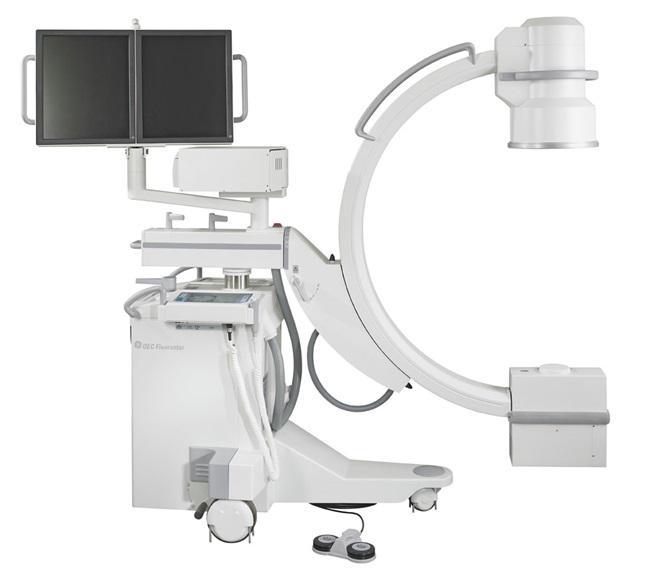
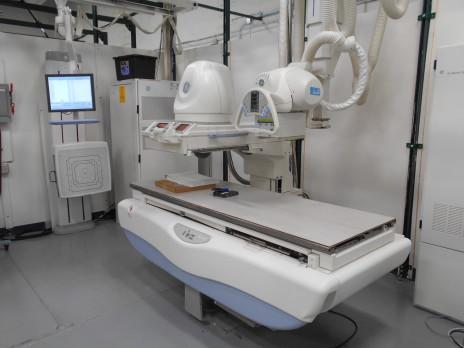
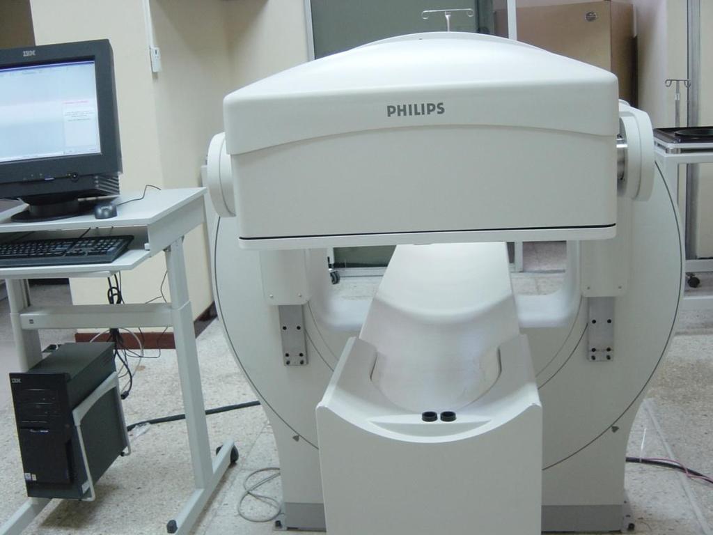
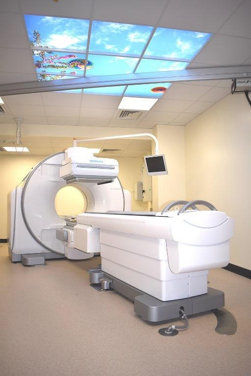
12 X-ray Tube & Machines
13 3 rd Generation Scanner 4 th Generation Scanner

14 CT Scanning (Sequential) A rotating beam of x- rays is passed through the patient. Measurements of the transmitted x-ray beam intensities are made by an array of detectors. CT images are obtained from these measurements.


15 Helical/Spiral Scanning

16 Multi-Row vs Single-Row Detectors Extremely fast light speed when compared with single slice scanner - completes scan in shorter time >>> cardiac Thin slices - high resolution - better image detail 64 slice scanner vs Single slice Protocols design to optimize detectability in the region of interest.

17 COMPUTED TOMOGRAPHY CT Scanner CT Imaging Process

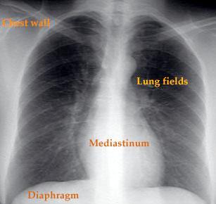
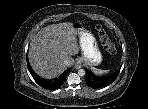
18 CT & Chest X-ray Images Cross-sectional image Chest X-ray
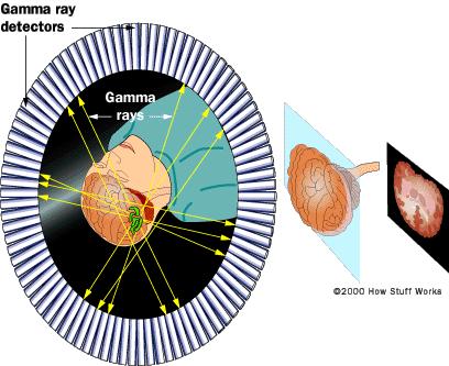
19 Nuclear Medicine

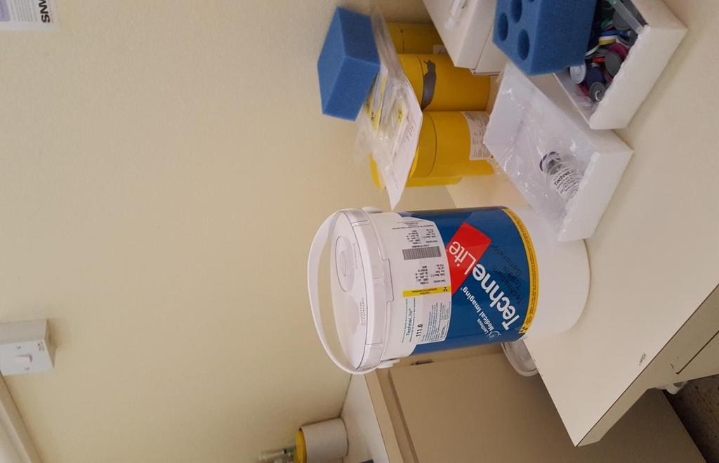
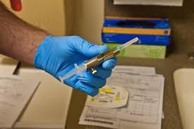
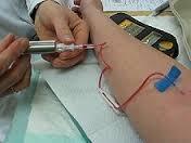
20 Nuclear Medicine Hot Lab Molybdenum- Technetium Generator
21
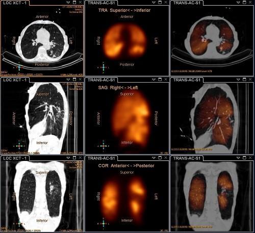
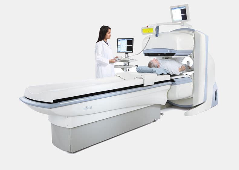
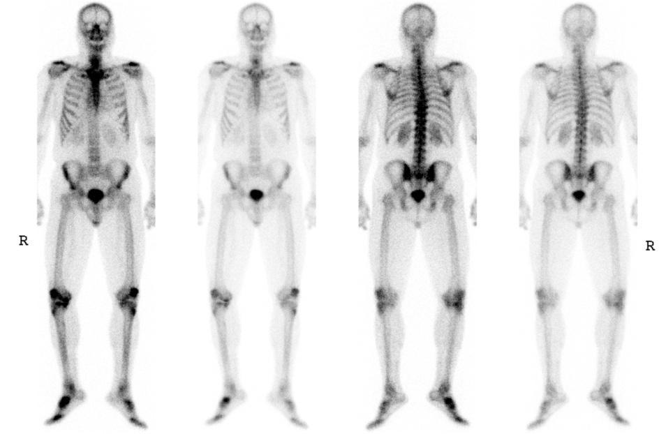
22 SPECT/CT
23 PET/CT Imaging
 and Computed Tomography University Lodge (CT) of the into West one")
24 PET/CT Scanner Advanced nuclear imaging technique that combines Positron Emission Tomography (PET) and Computed Tomography University Lodge (CT) of the into West one machine.
25 PET Imaging Principle Molecules significant for the condition studied, are labelled with a positron emitting isotope such as 18F. The labelled molecules are then injected into the patient and decay by positron emission. Radiopharmaceuticals that can be used for PET imaging include fluorodeoxyglucose (18F-FDG), used to study metabolism, and ammonia (13N), used to study perfusion, others.

26 Positron Emission Tomography - Principle PET is based on the simultaneous detection of two 511 kev annihilation photons produced when a positron loses its kinetic energy and combines with an electron

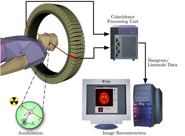
27 PET/CT

28 PET/CT Scanner A PET/CT scan reveals information about both the structure and function of cells and tissues in the body during a single imaging session..

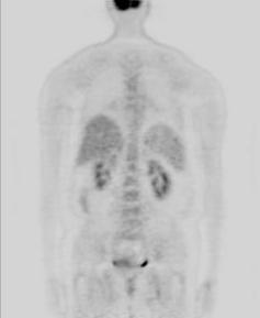
29 Normal PET - CT Body Scan

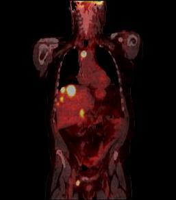
30 Abnormal PET - CT Body Scan


31 PET CT PET/CT H.S., FDG 15 mci Bed 1 min CT (1 min) KVs 130 kv mas 75 ma Slice 5 mm

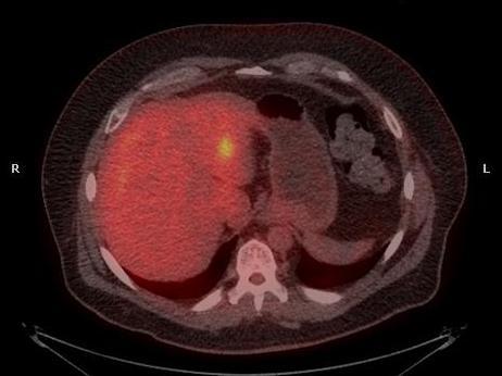
32 Enhanced Detection
33 PET/CT in Tumour Imaging Detect the presence of cancer in the body -molecular imaging Characterise radiographic abnormalities Determine the extent of the disease Evaluate response to treatment including -chemotherapy from biochemical changes
34 PET Imaging: Challenge Major limitation of PET is the cost to establish and operate the required imaging centre with an on-site cyclotron and radiopharmaceutical production facility.

35 Cyclotron - particle accelerator
36 Radiation Therapy EBRT Brachytherapy
37 Radiation Therapy Definition The application of ionizing radiation in the treatment of cancer and other radiation sensitive conditions Radiation Therapy Deliver a precisely measured dose of radiation to a defined cancer volume while minimizing damage to surrounding healthy tissue
38 External Beam Radiation Therapy in 21 st Century Several different types of energy used including photons, electrons, and heavy particles (carbon ions, neutrons, and protons) are used today. An evolution in the methods to deliver radiation treatment, progressing from 2-DRT to 3DCRT, and more recently, IMRT and others.
39 How does Radiation Therapy Work? A Moment of Radiobiology
40 Determinants of Biological Effects Radiation source related factors include: a) radiation quality b) quantity of radiation exposure c) dose rate
41 Conditional Factors These include: Linear Energy Transfer (LET) Dose Rate Fractionation Oxygen Enhancement Ratio (OER)
42 Determinants of Biological Effects System related factors include: a) radiosensitivity of tissue b) complexity of biological system - complex systems exhibit more sophisticated repair mechanisms
43 Interaction of Radiation with Tissue Direct DNA, RNA, protein or enzyme becomes ionized by an ionizing particle or photon passing through it. Indirect Cytoplasm
44
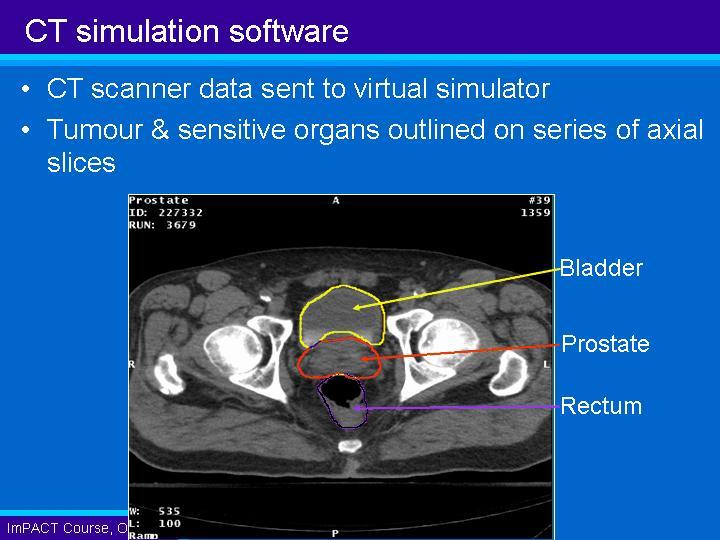
45 Interaction with Water Molecule The absorption of radiation by water molecule results in the formation of an ion pair (H 2 O +, H 2 O - ). H 2 O + h = H 2 O + + e H O + e = H 2 2O- (free electron capture)
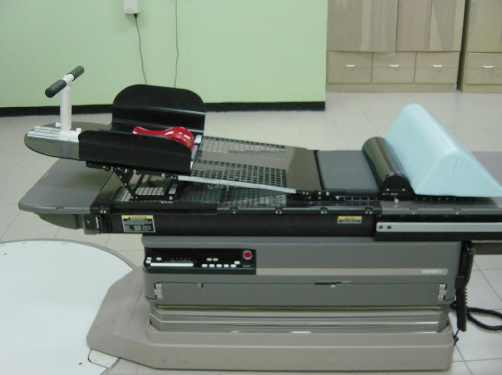
46 Interaction with Water Molecule The ions dissociate to form another ion and free radicals: H 2 O + H + + OH H 2 O H + OH Free Radicals are atomic or molecular species that have an unpaired orbital electron.
47 Reactions of Free Radicals Free radicals can combine with other free radicals to form water: H + OH H 2 O No biological damage
48 Reactions of Free Radicals Free radicals may combine with each other to form hydrogen peroxide which is highly toxic to the cell: OH + OH H 2 O 2
49 Reactions of Free Radicals Free radicals can act as strong oxidizing or reducing agent by combining directly with intact molecules: H + O 2 HO 2 Ø Oxygen combines with the hydrogen radical to form the highly reactive hydroperoxyl radical.
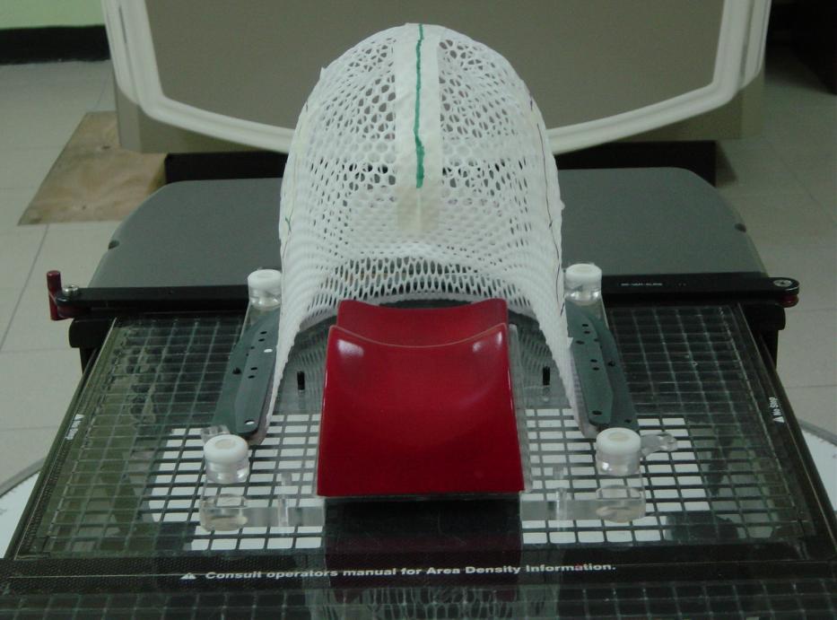
50
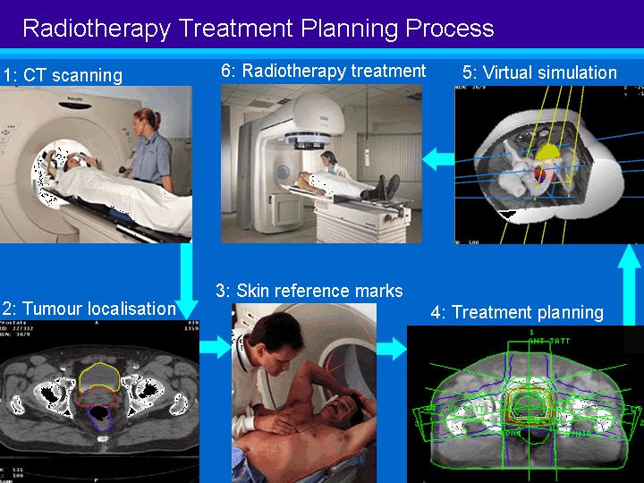
51 How does this Happen? To treat cancer we need to know exactly where the cancer is located in the body. We need to know if it is localised or spread to other parts of the body and where. Imaging is required
52 Treatment Planning (2-D) Visualize internal organs from any angle to locate and determine tumour volume. Pinpoints the precise area where the highenergy radiation will be aimed during treatment. Conventional Simulator
53 Treatment Planning (3-D) Provides cross-sectional data for 3-D Planning Virtual simulation for 3- D Conformal Treatment CT Simulator

54 Radiotherapy Treatment High Energy Treatment Machine used to deliver 3-D Conformal Radiation Therapy. Superior by far to Cobalt Units used in Public Hospitals Linear Accelerator (6 MV)


55 1. Simulation 2-D Treatment Process 5. Linac Treatment 2. AP Image 4. Verification 3. 2-D Plan
56 (3-D)

57 Treatment Position must be accurately reproduced Simulator CT Simulator Linear Accelerator 2-D 3-D
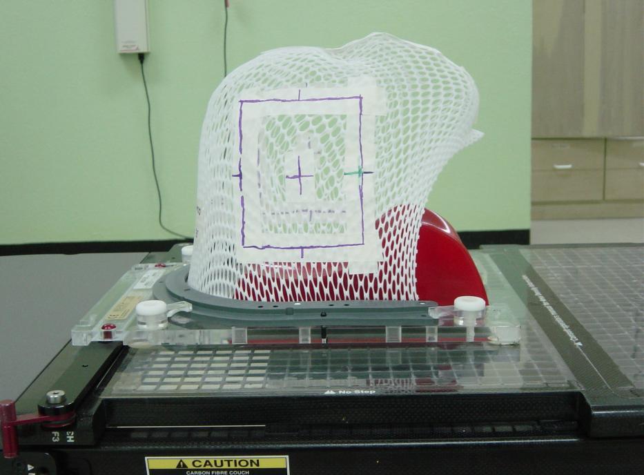
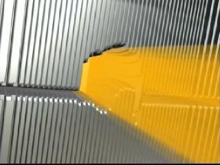
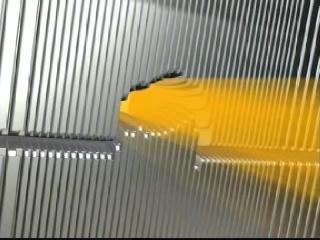
58 Immobilisation devices Indexed fixation points


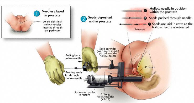
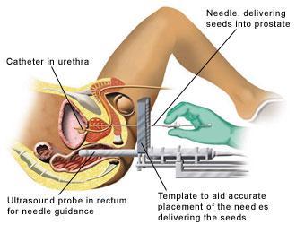
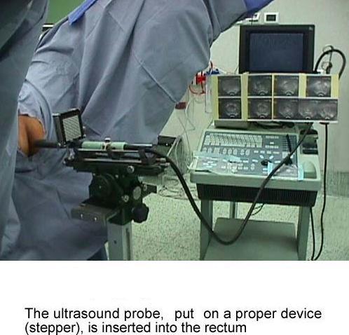
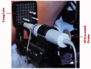
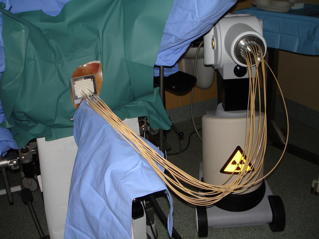
59 Treatment Planning (3-D) Lap Laser system Lap Computer

60 CT Simulation Whole Pelvis Slice thickness 3mm Patient aligned to the fiducials & laser marks IV contrast to enhance target & critical organs DICOM images transferred thro network Fiducial
61

62

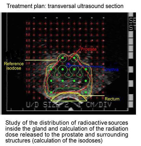
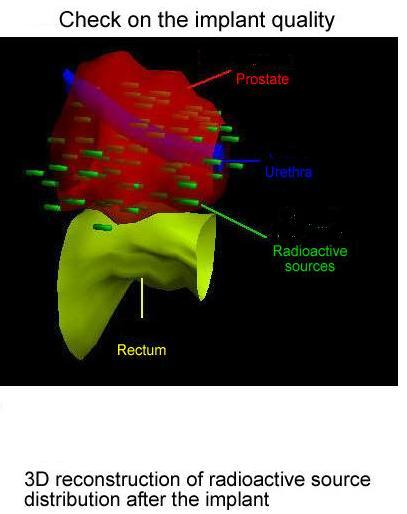
63 Medical Physics & Dosimetry Views with six beams

64 Medical Physics & Dosimetry 3-D Rendered with DRR

65 Dose Volume Histogram


66 Medical Physics & Dosimetry MLC Blades Beam s Eye View Computer Generated Shielding Block
67 Brachytherapy
68 Brachytherapy
69
70
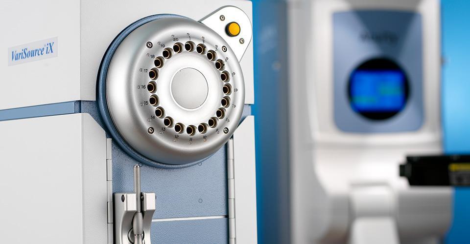
71

 and treatment plan.")

72 Transrectal ultrasound showing a series of prostate ultrasound images used to construct a 3-dimesnsional image of the prostate (volume study) and treatment plan. Key: Red line = prostate; University Blue Lodge line = of limit the West of radiation to be delivered
73

74 Volume Rendered

75
76
77
78 Manual Afterloading

79 High Dose Rate Brachytherapy Most modern brachytherapy is delivered using HDR Source - Iridium-192 Reasons? Outpatient procedure Optimization possible HDR Unit
80 HDR Brachytherapy Usually fractionated (e.g. 6 fractions of 6Gy) Either patient has new implant each time or stays in hospital for bi-daily treatments Time between treatments should be >6hours to allow normal tissue to repair all damage
81

82 Cervical Cancer Treatment
83 Other Advanced Technologies
84 Stereotactic Radiosurgery - Cyberknife A form of radiotherapy that focuses a high powered and precise radiation beam on a small area of the body. During treatment: A robotic arm controlled by a computer moves around the patient focusing radiation exactly on the area being treated. 1 to 5 Treatment sessions of about 30 min or more depending on dose required.


85 CyberKnife - Accuray


86 Treatment Applications Brain tumours Cancerous and non-cancerous Primary and metastatic spreading Parkinson s disease Epilepsy


87 Gamma Knife - Elekta - Uses 192 to 201 beams of highly focused gamma rays - Cobalt-60 sources


88 Xknife - Integra XKnife by Integra - can be adopted to linac - uses x-rays

89 Proton Therapy Machine
 Rapid distal dose fall-off Energy modulation (Spread-out Bragg peak) RBE close to")
90 Proton vs Photon Relatively low entrance dose (plateau) Maximum dose at depth (Bragg peak) Rapid distal dose fall-off Energy modulation (Spread-out Bragg peak) RBE close to unity
 Multi-leaf Collimator - typically")
91 Intensity Modulated Radiation Therapy (IMRT) Multi-leaf Collimator - typically 120

92 Image-Guide Radiotherapy (IGRT) system TRUE BEAM -Stereotactic mode - 6 and 18 MV x- ray beams, 6,9,12,15,18 Mev electrons -Portal imaging - Rapid Arc -- VMAT


93 IMRT with DMLC Also called as sliding window MLC leaves move when Beam is ON Dynamically moving leaf pair produces desired non-uniform intensity


94 IMRT for Prostate Critical structures sparing like Rectum & Bladder Improved target coverage 2D 3DCRT IMRT

95 True Dynamic Image Guided
96 VMAT Rapid ARC
97 THANK YOU
I. Equipments for external beam radiotherapy
 I. Equipments for external beam radiotherapy 5 linear accelerators (LINACs): Varian TrueBeam 6, 10 & 18 MV photons, 6-18 MeV electrons, image-guided (IGRT) and intensity modulated radiotherapy (IMRT),
MEDICAL MANAGEMENT POLICY
 PAGE: 1 of 8 This medical policy is not a guarantee of benefits or coverage, nor should it be deemed as medical advice. In the event of any conflict concerning benefit coverage, the employer/member summary
EORTC Member Facility Questionnaire
 Page 1 of 9 EORTC Member Facility Questionnaire I. Administrative Data Name of person submitting this questionnaire Email address Function Phone Institution Address City Post code Country EORTC No Enter
Medical Use of Radioisotopes
 Medical Use of Radioisotopes Therapy Radioisotopes prove to be useful in the application of brachytherapy, the procedure for using temporary irradiation close to the area of disease (i.e. cancer) 10% Medical
PHYS 383: Applications of physics in medicine (offered at the University of Waterloo from Jan 2015)
") PHYS 383: Applications of physics in medicine (offered at the University of Waterloo from Jan 2015) Course Description: This course is an introduction to physics in medicine and is intended to introduce
PHYS 383: Applications of physics in medicine (offered at the University of Waterloo from Jan 2015) Course Description: This course is an introduction to physics in medicine and is intended to introduce
PRINCIPLES and PRACTICE of RADIATION ONCOLOGY. Matthew B. Podgorsak, PhD, FAAPM Department of Radiation Oncology
 PRINCIPLES and PRACTICE of RADIATION ONCOLOGY Matthew B. Podgorsak, PhD, FAAPM Department of Radiation Oncology OUTLINE Physical basis Biological basis History of radiation therapy Treatment planning Technology
Nuclear Medicine and PET. D. J. McMahon rev cewood
 Nuclear Medicine and PET D. J. McMahon 150504 rev cewood 2018-02-15 Key Points Nuclear Medicine and PET: Imaging: Understand how Nuc Med & PET differ from Radiography & CT by the source of radiation. Be
An introduction to different types of radiotherapy
 An introduction to different types of radiotherapy Radiotherapy can cure cancer. It is delivered to around half of cancer patients and is a vital part of curative treatment in around 40% of patients 1.
First, how does radiation work?
 Hello, I am Prajnan Das, Faculty Member in the Department of Radiation Oncology at The University of Texas MD Anderson Cancer Center. We are going to talk today about some of the basic principles regarding
Proton and heavy ion radiotherapy: Effect of LET
 Proton and heavy ion radiotherapy: Effect of LET As a low LET particle traverses a DNA molecule, ionizations are far apart and double strand breaks are rare With high LET particles, ionizations are closer
A TREATMENT PLANNING STUDY COMPARING VMAT WITH 3D CONFORMAL RADIOTHERAPY FOR PROSTATE CANCER USING PINNACLE PLANNING SYSTEM *
 Romanian Reports in Physics, Vol. 66, No. 2, P. 394 400, 2014 A TREATMENT PLANNING STUDY COMPARING VMAT WITH 3D CONFORMAL RADIOTHERAPY FOR PROSTATE CANCER USING PINNACLE PLANNING SYSTEM * D. ADAM 1,2,
Therapeutic Medical Physics. Stephen J. Amadon Jr., Ph.D., DABR
 Therapeutic Medical Physics Stephen J. Amadon Jr., Ph.D., DABR Outline 1. Why physicists are needed in medicine 2. Branches of medical physics 3. Physics in Radiation Oncology 4. Treatment types and Treatment
Stereotactic Radiosurgery. Extracranial Stereotactic Radiosurgery. Linear accelerators. Basic technique. Indications of SRS
 Stereotactic Radiosurgery Extracranial Stereotactic Radiosurgery Annette Quinn, MSN, RN Program Manager, University of Pittsburgh Medical Center Using stereotactic techniques, give a lethal dose of ionizing
Option D: Medicinal Chemistry
 Option D: Medicinal Chemistry Basics - unstable radioactive nuclei emit radiation in the form of smaller particles alpha, beta, positron, proton, neutron, & gamma are all used in nuclear medicine unstable
Physical Bases : Which Isotopes?
 Physical Bases : Which Isotopes? S. Gnesin Institute of Radiation Physics, Lausanne University Hospital, Lausanne, Switzerland 1/53 Theranostic Bruxelles, 2 Octobrer 2017 Theranostic : use of diagnostic
FROM ICARO1 TO ICARO2: THE MEDICAL PHYSICS PERSPECTIVE. Geoffrey S. Ibbott, Ph.D. June 20, 2017
 FROM ICARO1 TO ICARO2: THE MEDICAL PHYSICS PERSPECTIVE Geoffrey S. Ibbott, Ph.D. June 20, 2017 1 DISCLOSURES My institution holds Strategic Partnership Research Agreements with Varian, Elekta, and Philips
The Physics of Oesophageal Cancer Radiotherapy
 The Physics of Oesophageal Cancer Radiotherapy Dr. Philip Wai Radiotherapy Physics Royal Marsden Hospital 1 Contents Brief clinical introduction Imaging and Target definition Dose prescription & patient
RADIOTHERAPY: TECHNOLOGIES AND GLOBAL MARKETS
 RADIOTHERAPY: TECHNOLOGIES AND GLOBAL MARKETS HLC176A February 2015 Neha Maliwal Project Analyst ISBN: 1-62296-043-2 BCC Research 49 Walnut Park, Building 2 Wellesley, MA 02481 USA 866-285-7215 (toll-free
Chapters from Clinical Oncology
 Chapters from Clinical Oncology Lecture notes University of Szeged Faculty of Medicine Department of Oncotherapy 2012. 1 RADIOTHERAPY Technical aspects Dr. Elemér Szil Introduction There are three possibilities
Advances in external beam radiotherapy
 International Conference on Modern Radiotherapy: Advances and Challenges in Radiation Protection of Patients Advances in external beam radiotherapy New techniques, new benefits and new risks Michael Brada
Radiotherapy. Marta Anguiano Millán. Departamento de Física Atómica, Molecular y Nuclear Facultad de Ciencias. Universidad de Granada
 Departamento de Física Atómica, Molecular y Nuclear Facultad de Ciencias. Universidad de Granada Overview Introduction Overview Introduction Brachytherapy Radioisotopes in contact with the tumor Overview
Linac or Non-Linac Demystifying And Decoding The Physics Of SBRT/SABR
 Linac or Non-Linac Demystifying And Decoding The Physics Of SBRT/SABR PhD, FAAPM, FACR, FASTRO Department of Radiation Oncology Indiana University School of Medicine Indianapolis, IN, USA Indra J. Das,
Sarcoma and Radiation Therapy. Gabrielle M Kane MB BCh EdD FRCPC Muir Professorship in Radiation Oncology University of Washington
 Sarcoma and Radiation Therapy Gabrielle M Kane MB BCh EdD FRCPC Muir Professorship in Radiation Oncology University of Washington Objective: Helping you make informed decisions Introduction Process Radiation
Present status and future of Proton beam therapy
 Present status and future of Proton beam therapy Description At present, the types of proven treatment for cancer are surgery, radiotherapy, and chemotherapy. Depending on the characteristics of cancer
Breast Cancer. What is breast cancer?
 Scan for mobile link. Breast Cancer Breast cancer is a malignant tumor in or around breast tissue. It usually begins as a lump or calcium deposit that develops from abnormal cell growth. Most breast lumps
NIA MAGELLAN HEALTH RADIATION ONCOLOGY CODING STANDARD. Dosimetry Planning
 NIA MAGELLAN HEALTH RADIATION ONCOLOGY CODING STANDARD Dosimetry Planning CPT Codes: 77295, 77300, 77301, 77306, 77307, 77321, 77316, 77317, 77318, 77331, 77399 Original Date: April, 2011 Last Reviewed
Prostate Cancer. What is prostate cancer?
 Scan for mobile link. Prostate Cancer Prostate cancer is a tumor of the prostate gland, which is located in front of the rectum and below the bladder. Your doctor may perform a physical exam, prostate-specific
Overview of Clinical and Research Activities at Georgetown University Hospital
 Overview of Clinical and Research Activities at Georgetown University Hospital Dalong Pang, Ph.D. Department of Radiation Medicine Georgetown University Hospital Clinical Operation Two Varian linear accelerators
Intensity Modulated RadioTherapy
 Intensity Modulated RadioTherapy A clinical application of a small accelerator University Medical Center Groningen A.A. van t Veld PhD I. Hoveijn PhD part 1 part2 CERN/KVI accelerator school, Zeegse, June
Radiation Therapy for Cancer: Questions and Answers
 Radiation Therapy for Cancer: Questions and Answers Key Points Radiation therapy uses ionizing radiation to kill cancer cells and shrink tumors (see Question 1). About half of all people with cancer are
Use of radiation to kill diseased cells. Cancer is the disease that is almost always treated when using radiation.
 Radiation Therapy Use of radiation to kill diseased cells. Cancer is the disease that is almost always treated when using radiation. One person in three will develop some form of cancer in their lifetime.
PHYSICS 2: HSC COURSE 2 nd edition (Andriessen et al) CHAPTER 20 Radioactivity as a diagnostic tool (pages 394-5)
 CHAPTER 20 Radioactivity as a diagnostic tool (pages 394-5)") PHYSICS 2: HSC COURSE 2 nd edition (Andriessen et al) CHAPTER 20 Radioactivity as a diagnostic tool (pages 394-5) 1. (a) A radioisotope is an isotope that is unstable and will emit particles from the nucleus
PHYSICS 2: HSC COURSE 2 nd edition (Andriessen et al) CHAPTER 20 Radioactivity as a diagnostic tool (pages 394-5) 1. (a) A radioisotope is an isotope that is unstable and will emit particles from the nucleus
Intensity-Modulated and Image- Guided Radiation Treatment. Outline. Conformal Radiation Treatment
 Intensity-Modulated and Image- Guided Radiation Treatment J. Daniel Bourland, PhD Professor Departments of Radiation Oncology, Physics, and Biomedical Engineering Wake Forest University School of Medicine
Breast Cancer. What is breast cancer?
 Scan for mobile link. Breast Cancer Breast cancer is a malignant tumor in or around breast tissue. It usually begins as a lump or calcium deposit that develops from abnormal cell growth. Most breast lumps
Understanding Radiation Therapy. For Patients and the Public
 Understanding Radiation Therapy For Patients and the Public Introduction to Radiation Oncology Radiation has been an effective tool for treating cancer for more than 100 years. Radiation oncologists are
X-ray Safety Discussion and Tour for Health Physicists
 X-ray Safety Discussion and Tour for Health Physicists April 1, 2016 Health Physics Society Spring Meeting Jerry Bai Environmental Administrator, Field Operations Bureau of Radiation Control Florida Department
HALF. Who gets radiotherapy? Who gets radiotherapy? Half of all cancer patients get radiotherapy. By 1899 X rays were being used for cancer therapy
 The Physical and Biological Basis of By 1899 X rays were being used for cancer therapy David J. Brenner, PhD, DSc Center for Radiological Research Department of Radiation Oncology Columbia University Medical
Chapter 7. What is Radiation Biology? Ionizing Radiation. Energy Transfer Determinants 09/21/2014
 Chapter 7 Molecular & Cellular Radiation Biology What is Radiation Biology? A branch of biology concerned with how ionizing radiation effects living systems. Biological damage that occurs from different
45 Hr PET Registry Review Course
 45 HR PET/CT REGISTRY REVIEW COURSE Course Control Document Timothy K. Marshel, MBA, R.T. (R), (N)(CT)(MR)(NCT)(PET)(CNMT) The PET/CT Training Institute, Inc. SNMMI-TS 028600-028632 45hr CEH s Voice Credits
Advances in Radiotherapy
 Advances in Radiotherapy Thomas Rockwell Mackie Professor Depts. Of Medical Physics, Human Oncology, and Engineering Physics University of Wisconsin Madison WI 53706 trmackie@wisc.edu Financial Disclosure
Medical Dosimetry Graduate Certificate Program IU Graduate School & The Department of Radiation Oncology IU Simon Cancer Center
 Medical Dosimetry Graduate Certificate Program IU Graduate School & The Department of Radiation Oncology IU Simon Cancer Center All students accepted into the Medical Dosimetry Graduate Certificate Program
APPLICATIONS OF RADIATION IN MEDICINE. Kirstin Murray BSc (Hons) Radiotherapy & Oncology
 Radiotherapy & Oncology") APPLICATIONS OF RADIATION IN MEDICINE Kirstin Murray BSc (Hons) Radiotherapy & Oncology CONTENTS Principles of Radiation Therapy Radiobiology Radiotherapy Treatment Modalities Special Techniques Nuclear
APPLICATIONS OF RADIATION IN MEDICINE Kirstin Murray BSc (Hons) Radiotherapy & Oncology CONTENTS Principles of Radiation Therapy Radiobiology Radiotherapy Treatment Modalities Special Techniques Nuclear
Prostate Cancer. What is prostate cancer?
 Scan for mobile link. Prostate Cancer Prostate cancer is a tumor of the prostate gland, which is located in front of the rectum, below the bladder and above the base of the penis. Your doctor may perform
Changing Paradigms in Radiotherapy
 Changing Paradigms in Radiotherapy Marco van Vulpen, MD, PhD Mouldroomdag-2015 Towards the elimination of invasion 1 NIH opinion on the future of oncology Twenty-five years from now,i hope that we won
Subject: Image-Guided Radiation Therapy
 04-77260-19 Original Effective Date: 02/15/10 Reviewed: 01/25/18 Revised: 01/01/19 Subject: Image-Guided Radiation Therapy THIS MEDICAL COVERAGE GUIDELINE IS NOT AN AUTHORIZATION, CERTIFICATION, EXPLANATION
Dosimetric Analysis of 3DCRT or IMRT with Vaginal-cuff Brachytherapy (VCB) for Gynaecological Cancer
 for Gynaecological Cancer") Dosimetric Analysis of 3DCRT or IMRT with Vaginal-cuff Brachytherapy (VCB) for Gynaecological Cancer Tan Chek Wee 15 06 2016 National University Cancer Institute, Singapore Clinical Care Education Research
Dosimetric Analysis of 3DCRT or IMRT with Vaginal-cuff Brachytherapy (VCB) for Gynaecological Cancer Tan Chek Wee 15 06 2016 National University Cancer Institute, Singapore Clinical Care Education Research
Prostate Cancer Treatment
 Scan for mobile link. Prostate Cancer Treatment Prostate cancer overview Prostate cancer is the most common form of cancer in American men, most prevalent in men over age 65 and fairly common in men 50-64
Expectations of Physics Knowledge for Certification
 Expectations of Physics Knowledge for Certification Bhudatt Paliwal, Ph.D. University of Wisconsin Medical School Department of Human Oncology Madison, Wisconsin 53792 USA Guiding Principles Test that
Additional Questions for Review 2D & 3D
 Additional Questions for Review 2D & 3D 1. For a 4-field box technique, which of the following will deliver the lowest dose to the femoral heads? a. 100 SSD, equal dmax dose to all fields b. 100 SSD, equal
Neutron Interactions Part 2. Neutron shielding. Neutron shielding. George Starkschall, Ph.D. Department of Radiation Physics
 Neutron Interactions Part 2 George Starkschall, Ph.D. Department of Radiation Physics Neutron shielding Fast neutrons Slow down rapidly by scatter in hydrogenous materials, e.g., polyethylene, paraffin,
COMPARISON OF RADIOBIOLOGICAL EFFECTS OF CARBON IONS TO PROTONS ON A RESISTANT HUMAN MELANOMA CELL LINE
 COMPARISON OF RADIOBIOLOGICAL EFFECTS OF CARBON IONS TO PROTONS ON A RESISTANT HUMAN MELANOMA CELL LINE I. Petrovi a, A. Risti -Fira a, L. Kori anac a, J. Požega a, F. Di Rosa b, P. Cirrone b and G. Cuttone
PGY-1. Resident Review Session Schedule
 1. August Simulation & Treatment 1.1. Sim Setup 1.2. Sim Techniques 1.3. 4DCT 1.4. Breath Hold / Gating 1.5. Treatment Setup 1.6. Treatment Delivery 1.7. Filming 1.7.1. Port film 1.7.2. kv 1.7.3. CBCT
Technological Advances in Radiotherapy for the Treatment of Localized Prostate Cancer - A Systematic Review
 Technological Advances in Radiotherapy for the Treatment of Localized Prostate Cancer - A Systematic Review Jayatissa R.M.G.C.S.B. (B.Sc.) Department of Radiography/Radiotherapy, Faculty of Allied Health
COMENIUS-Project: SM&CLIL Radiation & Medicine
 Medical imaging refers to the techniques and processes used to create images of the human body (or parts thereof) for clinical purposes. Thanks to modern mathematics and computer technology, medical imaging
Managing the imaging dose during Image-guided Radiotherapy. Martin J Murphy PhD Department of Radiation Oncology Virginia Commonwealth University
 Managing the imaging dose during Image-guided Radiotherapy Martin J Murphy PhD Department of Radiation Oncology Virginia Commonwealth University Radiographic image guidance has emerged as the new paradigm
Radiation Therapy 2013 The Role of Protons. Bob Gaston, D.O.
 Radiation Therapy 2013 The Role of Protons Bob Gaston, D.O. Disclosures Oklahoma ProCure Treatment Center Radiation Medicine Associates Goal of Radiation Therapy Increase the Therapeutic Ratio Therapeutic
Radiotherapy physics & Equipments
 Radiotherapy physics & Equipments RAD 481 Lecture s Title: An Overview of Radiation Therapy for Health Care Professionals Dr. Mohammed Emam Vision :IMC aspires to be a leader in applied medical sciences,
A Comparison of IMRT and VMAT Technique for the Treatment of Rectal Cancer
 A Comparison of IMRT and VMAT Technique for the Treatment of Rectal Cancer Tony Kin Ming Lam Radiation Planner Dr Patricia Lindsay, Radiation Physicist Dr John Kim, Radiation Oncologist Dr Kim Ann Ung,
Radiation physics and radiation protection. University of Szeged Department of Nuclear Medicine
 Radiation physics and radiation protection University of Szeged Department of Nuclear Medicine Radiation doses to the population 1 Radiation doses to the population 2 Sources of radiation 1 Radiation we
Questions may be submitted anytime during the presentation.
 Understanding Radiation Therapy and its Role in Treating Patients with Pancreatic Cancer Presented by Pancreatic Cancer Action Network www.pancan.org August 18, 2014 If you experience technical difficulty
A VMAT PLANNING SOLUTION FOR NECK CANCER PATIENTS USING THE PINNACLE 3 PLANNING SYSTEM *
 Romanian Reports in Physics, Vol. 66, No. 2, P. 401 410, 2014 A VMAT PLANNING SOLUTION FOR NECK CANCER PATIENTS USING THE PINNACLE 3 PLANNING SYSTEM * M. D. SUDITU 1,2, D. ADAM 1,2, R. POPA 1,2, V. CIOCALTEI
A Patient s Guide to SRS
 A Patient s Guide to SRS Stereotactic Radiosurgery 230 Nebraska St. Sioux City, IA 51101 NOTES 230 Nebraska St. Sioux City, IA 51101 Contents page Introduction 1 SRS and how it works 2 The technology involved
Guideline & Reports 医学物理学会教育委員会資料
 Guideline & Reports 医学物理学会教育委員会資料 2017/11 更新 report title year keyword 1 keyword 2 AAPM TG211 Classification and evaluation strategies of auto-segmentation approaches for PET 2017 PET autosegmentation
University of New Hampshire Scholars' Repository
 University of New Hampshire University of New Hampshire Scholars' Repository Honors Theses and Capstones Student Scholarship Spring 2017 Radiation Therapy Medical Physics Review Delivery, Interactions,
Managing the imaging dose during image-guided radiation therapy
 Managing the imaging dose during image-guided radiation therapy Martin J Murphy PhD Department of Radiation Oncology Virginia Commonwealth University Richmond VA Imaging during radiotherapy Radiographic
Brachytherapy. What is brachytherapy and how is it used?
 Scan for mobile link. Brachytherapy Brachytherapy places radioactive sources inside the patient on a temporary or permanent basis to damage cancer cells DNA and destroy their ability to divide and grow.
Comparison of high and low energy treatment plans by evaluating the dose on the surrounding normal structures in conventional radiotherapy
 Turkish Journal of Cancer Volume 37, No. 2, 2007 59 Comparison of high and low energy treatment plans by evaluating the dose on the surrounding normal structures in conventional radiotherapy MUHAMMAD BASIM
Radiotherapy and tumours in veterinary practice: part one
 Vet Times The website for the veterinary profession https://www.vettimes.co.uk Radiotherapy and tumours in veterinary practice: part one Author : Aleksandra Marcinowska, Jane Dobson Categories : Companion
Overview of Advanced Techniques in Radiation Therapy
 Overview of Advanced Techniques in Radiation Therapy Jacob (Jake) Van Dyk Manager, Physics & Engineering, LRCP Professor, UWO University of Western Ontario Acknowledgements Glenn Bauman Jerry Battista
Proton Treatment. Keith Brown, Ph.D., CHP. Associate Director, Radiation Safety University of Pennsylvania
 Proton Treatment Keith Brown, Ph.D., CHP Associate Director, Radiation Safety University of Pennsylvania Proton Dose vs. Depth Wilson,. R.R. Radiological use of fast protons. Radiology 47:487-491, 1946.
SHIELDING TECHNIQUES FOR CURRENT RADIATION THERAPY MODALITIES
 SHIELDING TECHNIQUES FOR CURRENT RADIATION THERAPY MODALITIES MELISSA C. MARTIN, M.S., FACR, FAAPM PRESIDENT AAPM - 2017 PRESIDENT - THERAPY PHYSICS INC., GARDENA, CA MELISSA@THERAPYPHYSICS.COM AAPM Spring
Has radiotherapy the potential being focal?
 Has radiotherapy the potential being focal? György Kovács & Alexander Schlaefer* Interdisciplinary Brachytherapy Unit and *Institute of Robotics and Cognitive Systems, University of Lübeck / 1 100% 90%
RADIATION THERAPY PROCEDURES REQUIRING PRECERTIFICATION FOR EVICORE HEALTHCARE ARRANGEMENT
 RADIATION THERAPY PROCEDURES REQUIRING PRECERTIFICATION FOR EVICORE HEALTHCARE ARRANGEMENT UnitedHealthcare Oxford Clinical Policy Policy Number: CANCER 014.14 T2 : December 1, 2017 Table of Contents Page
The future of radiation therapy. Safe and innovative options, including the CyberKnife System
 The future of radiation therapy Safe and innovative options, including the CyberKnife System Could a nonsurgical treatment be right for you? When you ve been diagnosed with a cancerous or noncancerous
Y FILMS DOSIMETR Nederland België / Belgique
 DOSIMETRY FILMS GAFCHROMIC Dosimetry Films The Self-developing Dosimetry Films that Allow You to Go Filmless Do away with the cost and headache of calibrating and maintaining a processor Do away with hazardous
Radiotherapy Advances
 Radiotherapy Advances Not Radiotherapy Principles IMRT IGRT Image Fusion Planning Introduction IMRT = Intensity Modulated RadioTherapy Restriction: IMRT with photon beams IMRT: Highly conformal technique
Learning Objectives. Clinically operating proton therapy facilities. Overview of Quality Assurance in Proton Therapy. Omar Zeidan
 Overview of Quality Assurance in Proton Therapy Omar Zeidan AAPM 2012 Charlotte, NC July 30 st, 2012 Learning Objectives Understand proton beam dosimetry characteristics and compare them to photon beams
Guidelines for the use of inversely planned treatment techniques in Clinical Trials: IMRT, VMAT, TomoTherapy
 Guidelines for the use of inversely planned treatment techniques in Clinical Trials: IMRT, VMAT, TomoTherapy VERSION 2.1 April 2015 Table of Contents Abbreviations & Glossary... 3 Executive Summary...
TomoTherapy. Michelle Roach CNC Radiation Oncology Liverpool Hospital CNSA. May 2016
 TomoTherapy Michelle Roach CNC Radiation Oncology Liverpool Hospital CNSA May 2016 TomoTherapy The Facts Greek Tomo = slice Advanced form of IMRT 3D computerised tomography (CT) imaging immediately prior
RADIOTHERAPY- CURRENT SITUATION AND FUTURE TRENDS
 HOSPITAL OF LITHUANIAN UNIVERSITY OF HEALTH SCIENCES KAUNO KLINIKOS RADIOTHERAPY- CURRENT SITUATION AND FUTURE TRENDS Prof. Elona Juozaitytė Perspectives of Czech- Lithuanian Research Partnerships About
Neutrons. ρ σ. where. Neutrons act like photons in the sense that they are attenuated as. Unlike photons, neutrons interact via the strong interaction
 Neutrons Neutrons act like photons in the sense that they are attenuated as I = I 0 e μx where Unlike photons, neutrons interact via the strong interaction μ = The cross sections are much smaller than
Positron Emission Tomography Computed Tomography (PET/CT)
") Positron Emission Tomography Computed Tomography (PET/CT) What is Positron Emission Tomography Computed Tomography (PET/CT) Scanning? What are some common uses of the procedure? How should I prepare for
Positron Emission Tomography Computed Tomography (PET/CT) What is Positron Emission Tomography Computed Tomography (PET/CT) Scanning? What are some common uses of the procedure? How should I prepare for
Molecular Imaging and Breast Cancer
 Molecular Imaging and Breast Cancer Breast cancer forms in tissues of the breast usually in the ducts, tubes that carry milk to the nipple, and lobules, the glands that make milk. It occurs in both men
Image Guided in Radiation Therapy (IGRT) Chumpot Kakanaporn Med Phys Radiation Oncology Siriraj Hospital
 Chumpot Kakanaporn Med Phys Radiation Oncology Siriraj Hospital") Image Guided in Radiation Therapy (IGRT) Chumpot Kakanaporn Med Phys Radiation Oncology Siriraj Hospital EBT Process Diagnosis Simulation Tx Planning Tx Verification Tx Delivery X-ray CT MRI NM Patho Process
Image Guided in Radiation Therapy (IGRT) Chumpot Kakanaporn Med Phys Radiation Oncology Siriraj Hospital EBT Process Diagnosis Simulation Tx Planning Tx Verification Tx Delivery X-ray CT MRI NM Patho Process
Implementation of advanced RT Techniques
 Implementation of advanced RT Techniques Tibor Major, PhD National Institute of Oncology Budapest, Hungary 2. Kongres radiološke tehnologije, Vukovar, 23-25. September 2016. Current RT equipments at NIO,
Outline. Chapter 12 Treatment Planning Combination of Beams. Opposing pairs of beams. Combination of beams. Opposing pairs of beams
 Chapter 12 Treatment Planning Combination of Beams Radiation Dosimetry I Text: H.E Johns and J.R. Cunningham, The physics of radiology, 4 th ed. http://www.utoledo.edu/med/depts/radther Outline Combination
PROGRESS IN RADIATION ONCOLOGY. Fox Chase Cancer Center. Jean Holland RN MSN AOCN Department of Radiation Oncology April 2017
 PROGRESS IN RADIATION ONCOLOGY Fox Chase Cancer Center Jean Holland RN MSN AOCN Department of Radiation Oncology April 2017 Radiation Therapy Therapeutic use of electromagnetic energy from a machine or
PROGRESS IN HADRONTHERAPY
 PROGRESS IN HADRONTHERAPY Saverio Braccini TERA Foundation for Oncological Hadrontherapy IPRD06 - Siena - 01.10.06 - SB 1 Outline Introduction Radiation therapy with X rays and hadrontherapy Hadrontherapy
Neutron dose evaluation in radiotherapy
 Neutron dose evaluation in radiotherapy Francesco d Errico University of Pisa, Italy Yale University, USA Radiation therapy with a linear accelerator (LINAC) Photoneutron production in accelerator head
Applications of Particle Accelerators
 Applications of Particle Accelerators Prof. Rob Edgecock STFC Rutherford Appleton Laboratory EMMA Accelerator at Daresbury Laboratory Applications >30000 accelerators in use world-wide: 44% for radiotherapy
Techniques and Technologies in Radiation Oncology 2018
 Techniques and Technologies in Radiation Oncology 2018 The Royal Australian and New Zealand College of Radiologists The Faculty of Radiation Oncology Name of document and version: Techniques and Technologies
Characterization and implementation of Pencil Beam Scanning proton therapy techniques: from spot scanning to continuous scanning
 Characterization and implementation of Pencil Beam Scanning proton therapy techniques: from spot scanning to continuous scanning Supervisors Prof. V. Patera PhD R. Van Roermund Candidate Annalisa Patriarca
Brain Tumor Treatment
 Scan for mobile link. Brain Tumor Treatment Brain Tumors Overview A brain tumor is a group of abnormal cells that grows in or around the brain. Tumors can directly destroy healthy brain cells. They can
TFY4315 STRÅLINGSBIOFYSIKK
 Norges teknisk-naturvitenskaplige universitet Institutt for fysikk EKSAMENSOPPGÅVER med løysingsforslag Examination papers with solution proposals TFY4315 STRÅLINGSBIOFYSIKK Biophysics of Ionizing Radiation
III. Proton-therapytherapy. Rome SB - 5/5 1
 Outline Introduction: an historical review I Applications in medical diagnostics Particle accelerators for medicine Applications in conventional radiation therapy II III IV Hadrontherapy, the frontier
Palliative RT in Ovarian cancer
 Outline Palliative RT in Ovarian cancer Case discussion Palliative treatment Radiation Techniques Asst Prof Pittaya Dankulchai, MD. Division of Radiation Oncology, Department of Radiology, Faculty of Medicine
Medical physics is beautiful
 Translational research in particle therapy Marco Durante Medical physics is beautiful Pisa, 31.10.2014 Relative dose 1. 2 1. 0 Tumor Durante & Loeffler, Nature Rev Clin Oncol 2010 0. 8 Normal tissue 0.
Out-of-field dosimetry in radiotherapy for input to epidemiological studies. Roger Harrison
 MELODI 7th Workshop, 9 11 November 2015 Helmholtz Zentrum München Next Generation Radiation Protection Research Out-of-field dosimetry in radiotherapy for input to epidemiological studies Roger Harrison
*Chien-Yi Yeh, Ji-Hong Hong * 葉健一洪志宏
 林口 Proton Therapy in Ti Taiwan *Chien-Yi Yeh, Ji-Hong Hong * 葉健一洪志宏 Chang Gung Memorial Hospital at Lin-Kou, Taiwan 林口長庚紀念醫院 OCPA2010 at Beijing, China Aug. 5, 2010 Outline 1. The principle of proton radiotherapy
Credentialing for the Use of IGRT in Clinical Trials
 Credentialing for the Use of IGRT in Clinical Trials James M. Galvin, DSc Thomas Jefferson University Hospital Jefferson Medical College Philadelphia, PA and The Radiation Therapy Oncology Group RADIATION