Introduction. Splenic pathologies. Polysplenia syndrome. Anatomical variants 21/08/2017. Ultrasound of the forgotten organ of the abdomen: the spleen
|
|
|
- Silvia Hutchinson
- 5 years ago
- Views:
Transcription


1 Ultrasound of the forgotten organ of the abdomen: the spleen Olivier Lucidarme Pitié Salpêtrière UPMC/Sorbonne University Paris France Malformations Introduction must look for other malformations frequently associated Indirect witness of more general diseases Specific spleen diseases Traumatism Organ the most involved in case of abdominal trauma Splenic pathologies Polysplenia syndrome Malformations must look for other malformations frequently associated Indirect witness of more general diseases Specific spleen diseases Traumatism Organ the most involved in case of abdominal trauma Polysplenia syndrome Multiple splenules + Heterotaxy syndrome or situs ambiguus or situs inversus Other characteristic features may include: congenital heart disease abnormal pulmonary venous return semi-annular pancreas / congenitally short pancreas midgut malrotation (80%) gallbladder agenesis (50%) renal agenesis intrahepatic IVC interruption with azygos/hemiazygos continuation preduodenal portal vein Accessory spleen : 40% Anatomical variants Same echogenicity of main spleen Moving with respiration = spleen differential diagnosis ADK of pancreatic tail (more hypo-echoic) Adrenal nodule (immobile) 1


 x 4.0 ± 0.")

 Agenesia situs ambigus, heterotaxia, isomerism, Ivemark synd")

2 Malformations Splenic pathologies must look for other malformations frequently associated Indirect witness of more general diseases Specific spleen diseases Traumatism Organ the most involved in case of abdominal trauma Homogeneous splenomegaly = general disease Mild Splenomegaly (< 700 g) Severe splenomegaly (> 700 g) Congestion Portal hypertension Venous thrombosis Heart failure Systemic infectious disease Infiltrative disease Acute leukemia Chronic lymphoid leukemia Thalasemia Lymphoma Sickle-cell anaemia Angiosarcoma Amyloïdosis Myeloproliferative disorders Sarcoïdosis Gaucher s disease Nymann-Pick disease Malaria Size of the spleen Normal volume of the spleen: between 100 and 265 ml (av :150 ml) Normal size 10.9 ± 1.4 cm (length) x 6.8 ± 0.71 cm (thickness) x 4.0 ± 0.45 cm (width) Splenic index : product of these 3 dimensions Must be lower than 480 cm 3 Longest oblique size of the spleen splenomegaly if > 12 cm Low interobserver reproducibility Do not allow comparisons with CT or MRI Size of the spleen The simplest: measure the true vertical height simple reproducible Comparable with CT and MRI Splenomegaly if > 10 cm Malformations Splenic pathologies must look for other malformations frequently associated Indirect witness of more general diseases Specific spleen diseases Traumatism Organ the most involved in case of abdominal trauma Invisible spleen 1) Agenesia situs ambigus, heterotaxia, isomerism, Ivemark synd 2) Ablation 3) Major atrophy Sickle-cell anaemia, thalassemia, Waquez repeated story of infarction = auto splenectomy 2
 abnormal location wandering spleen Focal lesions of the spleen Cystic Easy or affordable Solid Mess")

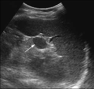
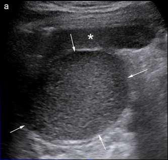
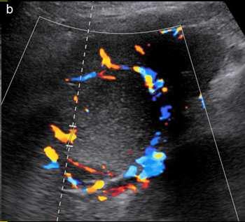
3 Invisible spleen 1) Agenesia situs ambigus, heterotaxia, isomerism, Ivemark synd 2) Ablation 3) Major atrophy Sickle-cell anaemia, thalassemia, Waquez repeated story of infarction = auto splenectomy 4) abnormal location wandering spleen Focal lesions of the spleen Cystic Easy or affordable Solid Mess Aswaniet al, JOP. J Pancreas (Online) 2015 Jan 31; 16(1): N DahiyaIndian Journal of Radiology and Imaging, Vol. 12, No. 3, July-September, 2002, Hypoechoic + wall Epidermoïde cyst Hematoma Pseudo Cysts Hypoechoic + wall Epidermoïde cyst Hematoma Pseudo Cysts Anechoic without wall Mesothelial simple Cyst Lymphangioma Peliosis 3


4 Be carreful: trap! Epidermoïde Cyst Epidermoïde Cyst 4
")


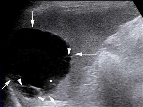
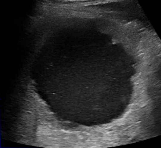
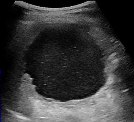
5 Hematoma (bike accident) Pseudo cyst Hypoechoic + wall Epidermoïde cyst Hematoma Pseudo Cysts Mesothelial simple cyst or cystic uniloculated lymphangioma ou hydatique cyst 5
:61 63.")

6 Multiloculated cystic lymphangioma Splenic peliosis Factors involved in peliosis are malignancy, tuberculosis, acquired immune deficiency, diabetes, drugs and parasites Above image from: Youssef Ghazzawi, MD, and ImadAbsah, MD. Visceral Leishmaniaas Unusual Cause of Splenic Peliosisin United States. Mayo Clinic College of Medicine, Rochester, MN. ACG Case Rep J 2013;1(1): Focal lesions of the spleen Hypoechoic + wall Epidermoïde cyst Hematoma Pseudo Cysts Anechoic without wall Mesothelial simple Cyst Lymphangioma Peliosis Cystic Easy or affordable Solid Mess Solid lesions Solid lesions hypoechoic hyperechoic Bénin hypoechoic hyperechoic Bénin Cysts Hemangiomas Cysts Hemangiomas littoral cell hemangioma littoral cell hemangioma Accute bleeding Complicated Cysts Accute bleeding Complicated Cysts Acute infarction Abcess Acute infarction Abcess Hemangiomas/ hamartomas Hematomas Hemangiomas/ hamartomas Hematomas Lymphangiomas Subacute or old infartion Lymphangiomas Subacute or old infartion SANT (sclerosis angiomatous transformation) Extramedullary Hematopoïesis Calcifications Gamna-Gandy bodies Peliosis Malin SANT (sclerosis angiomatous transformation) Extramedullary Hematopoïesis Calcifications Gamna-Gandy bodies Peliosis Malin Metastases Metastases Metastases Metastases Lymphomas Kaposi Sarcoma Lymphomas Kaposi Sarcoma 6


 but with anarchic")

7 Neoplasic context No neoplasic context: unifocal lesion Hemangioma Poumon Cancer du sein Mélanome No neoplasic context: unifocal lesion Hemangioma or Hamartoma Hemangioma or hamartoma Hemangioma Benign tumor the most frequent unique or multiple Cavernous hemangioma = hypoechoic, with cystic areas or completely cystic Capillary hemangioma = either isoechoic, or hyperechoic ronded or irregular whith sharp limits Hemangioma or hamartoma Hamartoma hypervascularized Composed of normal tissue of the spleen (mainly white and red pulp) but with anarchic organization Unifocal in 90% homogeneous, slightly hypoechoic No neoplasic context: unifocal lesion Hemangioma or Hamartoma 7


 Polylobulated")
(high frequency probe) macronodules")

8 Hemangioma or hamartoma Extramedulary hematopoiesis If lesions are multiple Malignant lesions lymphoma, metastases, Kaposi sarcoma Benign lesions Littoral cell hemangioma Sarcoïdosis, granulomatosis Infectious disease: Multiple micro abscesses... Lymphoma Most frequent malignant lesion Hodgkin or non-hodgkin lymphomas Spleen involved in 30 to 40% Splenomegaly = 50% (diffuse & infiltrative involvement, no focal lesion seen) Polylobulated multifocal lesions : 50% Micronodulary involvement (miliary like)(high frequency probe) macronodules hypoechoic, or quasi anechoic, mimicking cysts Borders of lesions are ill defined (in contrary to true cysts) Lymphoma (Hodgkin) Lymphoma (NHL) Lymphoma (NHL) 8


9 Littoral cell hemangioma Littoral cell hemangioma Rare benign disease of the spleen From the cells of the wall of the red pulp capillaries Specific to the spleen multiples nodules hyperechoic in an enlarged spleen If no oncologic context: Follow-up stable If oncologic context : Metastases must be considered until proof of contrary biopsie if spleen involvement is isolated Sarcoïdosis es Aboveimage from: Thomas Vancauwenberghe, AnnemiekSnoeckx, Dirk Vanbeckevoort, Steven Dymarkowski, Filip M Vanhoenacker: Imaging of the spleen: whatthe clinician needs to know Singapore Med J 2015; 56(3): Hyperechoic foci Gamna Gandy bodies Gamna Gandy bodies Portal hypertension +++ Sickle-cell anaemia ++ Hemolysis hémochromatosis Scar Old Micro-abcess Cysticercosis Calcified granuloma Pneumocystosis +++ Sarcoïdosis Tuberculosis histoplasmosis 9


10 Painful spleen = possible infarction due to acute venous obstruction Acute pancreatitis Pancreas adenocarcinoma (body and tail) hypercoagulability, drepanocytosis Traumatism Sickle-cell anaemia Spleen traumatism lacerations Intraparenchymal hematomas sub-capsular hematomas Sensitivity of US about 69% Echogenicity of fresh blood similar to spleen parenchyma Intra splenic vessel distribution in color Doppler Empty signal area FAST US must rather focus of free liquid in the abdomen CEUS improves ++ the detection of parenchymal injuries Spleen infaction due to Sickle-cell anaemia after Mountain hiking Gracias Olivier.lucidarme@aphp.fr 10
CT 101 :Pancreas and Spleen
 CT 101 :Pancreas and Spleen Shikha Khullar,, MD, MPH Division of Radiology University of South Alabama The Pancreas Normal Pancreas 3 Phase Pancreatic CT Non contrast Arterial phase : 30-35 35 second
Pediatric Hepatobiliary, Pancreatic & Splenic US
 Pediatric Hepatobiliary, Pancreatic & Splenic US Susan J. Back, MD Department of Radiology, The Children s Hospital of Philadelphia No Disclosures Objectives Normal Abnormal: cases and US advances Objectives
Appendix 5. EFSUMB Newsletter. Gastroenterological Ultrasound
 EFSUMB Newsletter 87 Examinations should encompass the full range of pathological conditions listed below A log book listing the types of examinations undertaken should be kept Training should usually
GENERAL ABDOMINAL IMAGING PERITONEAL SPACE, PANCREAS, & SPLEEN. VMB 960 March 25, 2013
 GENERAL ABDOMINAL IMAGING PERITONEAL SPACE, PANCREAS, & SPLEEN VMB 960 March 25, 2013 REFERENCE Chapters 35-36 Pages 650-678 Chapter 37 Pages 694-701 Chapter 3 Pages 38-49 OBJECTIVES Radiography and Ultrasound
Lách
 Lách Lách Lách Lách Splenogonadal fusion. Splenic tissue is attached to testicular tissue. Pseudocyst (false or secondary cyst). A, Outer aspect. Pseudocyst (false or secondary cyst). B, Inner surface.
Imaging of liver and pancreas
 Imaging of liver and pancreas.. Disease of the liver Focal liver disease Diffusion liver disease Focal liver disease Benign Cyst Abscess Hemangioma FNH Hepatic adenoma HCC Malignant Fibrolamellar carcinoma
Radiology of hepatobiliary diseases
 GI cycle - Lecture 14 436 Teams Radiology of hepatobiliary diseases Objectives 1. To Interpret plan x-ray radiograph of abdomen with common pathologies. 2. To know the common pathologies presentation.
Neckmasses in infancy and childhood: Clinical and radiological classification and imaging approaches M. Mearadji
 Neckmasses in infancy and childhood: Clinical and radiological classification and imaging approaches M. Mearadji International Foundation for Pediatric Imaging Aid Introduction Neck masses are a frequent
GASTROINTESTINAL IMAGING STUDY GUIDE
 GASTROINTESTINAL IMAGING STUDY GUIDE Pharynx Diverticula Foreign bodies Trauma o Motility Disorders Esophagus Diverticula Trauma Esophagitis Barrett esophagus Rings, webs, and strictures Varices Benign
Normal Sonographic Anatomy
 hapter 2:The Liver DUNSTAN ABRAHAM Normal Sonographic Anatomy Homogeneous, echogenic texture (Figure 2-1) Measures approximately 15 cm in length and 10 12.5 cm anterior to posterior; measurement taken
Imaging techniques to characterize spleen involvement in patients with Hodgkin lymphoma
 Imaging techniques to characterize spleen involvement in patients with Hodgkin lymphoma Marco Picardi, MD Ematologia, Azienda Ospedaliera Universitaria Federico II, Naples, Italy 5th International Workshop
Body MRI from the Liver to the Bladder
 Body MRI from the Liver to the Bladder I Want You! Audience Participation Methodist Hospital Continuing Education Seminar Jordan Swensson, MD November 7, 2015 Objectives Observe the uses of MRI for organs
Malignant Focal Liver Lesions
 Malignant Focal Liver Lesions Other Than HCC Pablo R. Ros, MD, MPH, PhD Departments of Radiology and Pathology University Hospitals Cleveland Medical Center Case Western Reserve University Pablo.Ros@UHhospitals.org
Imaging of common diseases of hepatobiliary and GI system
 Imaging of common diseases of hepatobiliary and GI system Natthaporn Tanpowpong, M.D. Diagnostic radiology Faculty of Medicine, Chulalongkorn University Normal plain radiograph A = Common bile duct
Evaluation of Liver Mass Lesions. American College of Gastroenterology 2013 Regional Postgraduate Course
 Evaluation of Liver Mass Lesions American College of Gastroenterology 2013 Regional Postgraduate Course Lewis R. Roberts, MB ChB, PhD Division of Gastroenterology and Hepatology Mayo Clinic College of
HEPATO-BILIARY IMAGING
 HEPATO-BILIARY IMAGING BY MAMDOUH MAHFOUZ MD PROF.OF RADIOLOGY CAIRO UNIVERSITY mamdouh.m5@gmail.com www.ssregypt.com CT ABDOMEN Indications Patient preparation Patient position Scanogram Fasting 4-6 hours
Contents. Basic Ultrasound Principles and Terminology. Ultrasound Nodule Characteristics
 Contents Basic Ultrasound Principles and Terminology Basic Ultrasound Principles... 1 Ultrasound System... 2 Linear Transducer for Superficial Images and Ultrasound-Guided FNA... 3 Scanning Planes... 4
Case-based discussion:
 Case-based discussion: Pailin Kongmebhol, M.D. Department of Radiology Faculty of Medicine Chiang Mai University There are many guidelines for managing thyroid nodules Two important guidelines: 2015 American
The Spleen. Dr Fahad Ullah
 The Spleen BY Dr Fahad Ullah Spleen The spleen is an largest lymphoid organ shaped like a shoe that lies relative to the 9th and 11th ribs and is located in the left hypochondrium. Thus, the spleen is
Case Discussion Splenic Abscess
 Case Discussion Splenic Abscess Personal Data Gender: male Birth Date: 1928/Mar/06th Allergy: Mefenamic Smoking: 0.5 PPD for 55 years Alcohol: negative (?) 4 Months Ago Abdominal pain: epigastric area
Without and With Dynamic Gadolinium Enhancement
 MRI of Focal Splenic Lesions bdominal Imaging Pictorial Essay Downloaded from www.ajronline.org by 46.3.204.37 on 01/28/18 from IP address 46.3.204.37. Copyright RRS. For personal use only; all rights
Chief Complaint. Retroperitoneal cystic mass incidentally found at health examination center.
 Personal Information Age: 34 y/o Sex: female Past history: major systemic medical history(-) surgical history(-), family history(-) Denied food or drug allergy Chief Complaint Retroperitoneal cystic mass
IT 의료융합 1 차임상세미나 복부질환초음파 이재영
 IT 의료융합 1 차임상세미나 2013-4-3 복부질환초음파 이재영 나는오늘누구를위하여 종을울리나? 전통적의료 의사 공학설계자 의사 최첨단진단장비들 USG, CT, MRI 환자 환자 현대의료 사용자중심의사고 US in the Abdomen Detection DDx Look Behavior Response by external stimuli Guiding Tool
Appendix 9: Endoscopic Ultrasound in Gastroenterology
 Appendix 9: Endoscopic Ultrasound in Gastroenterology This curriculum is intended for clinicians who perform endoscopic ultrasonography (EUS) in gastroenterology. It includes standards for theoretical
CTA/MRA of Pediatric Hepatic Masses Radiology-Pathology Correlation
 Acta Radiológica Portuguesa, Vol.XVIII, nº70, pág. 41-50, Abr.-Jun., 2006 CTA/MRA of Pediatric Hepatic Masses Radiology-Pathology Correlation Marilyn J. Siegel Mallinckrodt Institute of Radiology, Washington
Extraosseous myeloma: imaging features
 Extraosseous myeloma: imaging features C. Santos Montón, R. Corrales, J. M. Bastida Bermejo, M. Villanueva Delgado, R. E. Correa Soto, J. M. Alonso Sánchez; Salamanca/ES Learning objectives -To review
Interesting Cases from Liver Tumor Board. Jeffrey C. Weinreb, M.D.,FACR Yale University School of Medicine
 Interesting Cases from Liver Tumor Board Jeffrey C. Weinreb, M.D.,FACR Yale University School of Medicine jeffrey.weinreb@yale.edu Common Liver Diseases Hemangioma Cyst FNH Focal Fat/Sparing THID Non-Cirrhotic
Approach to the Patient with Liver Disease
 Approach to the Patient with Liver Disease Diagnosis of liver disease Careful history taking Physical examination Laboratory tests Radiologic examination and imaging studies Liver biopsy Liver diseases
Thyroid in a Nutshell Dublin Catherine Kirkpatrick Consultant Sonographer ULHT
 Thyroid in a Nutshell Dublin 2017 Catherine Kirkpatrick Consultant Sonographer ULHT Acknowledgements Dr. Steve Colley Dr. Rhodri Evans Dr. Rhian Rhys Dr. Andrew McQueen Aims Anatomy & Physiology Incidence
A Classic Case Of Polysplenia Syndrome With A Pancreatic Mass And SOLs In Liver
 ISPUB.COM The Internet Journal of Radiology Volume 13 Number 2 A Classic Case Of Polysplenia Syndrome With A Pancreatic Mass And SOLs In Liver V Gupta, N Agarwal Citation V Gupta, N Agarwal. A Classic
Job Task Analysis for ARDMS Abdomen Data Collected: June 30, 2011
 Job Task Analysis for ARDMS Abdomen Data Collected: June 30, 2011 Reported: Analysis Summary for: Abdomen Examination Survey Dates 06/13/2011-06/26/2011 Invited Respondents 6,000 Surveys with Demographics
1. Department of Radiology, Policlinico Universitario Paolo Giaccone, Palermo, Italy
 Hamartoma of the spleen (splenoma) with calcifications in a child with beta-thalassemia: Dario Giambelluca 1, Dario Picone 1, Giuseppe Lo Re 1*, Salvatore Pappalardo 2, Placido Romeo 2 1. Department of
FOR YOUR EYES ONLY: A Guide to Accurate Detection of Diffuse Infiltrators in the Liver Eric C. Ehman, MD 1
 FOR YOUR EYES ONLY: A Guide to Accurate Detection of Diffuse Infiltrators in the Liver Eric C. Ehman, MD 1 INFILTRATOR Brian T. Welch, MD 1 Naoki Takahashi, MD 1 Christine O. Menias, MD 2 Ajit H. Goenka,
CASE OF HETEROTAXY SYNDROME WITH POLYSPLENIA AND INTESTINAL MALROTATION
 CASE OF HETEROTAXY SYNDROME WITH POLYSPLENIA AND INTESTINAL MALROTATION *Sagar H S, Basanta Manjari Swain, Jayashree Mohanty and Sasmita Parida Department of Radio diagnosis, S.C.B. Medical College, Cuttack
TIN COLLOID LIVER/SPLEEN SCINTIGRAPHY THIRD YEAR MEDICAL STUDENTS DIAGNOSTIC BLOCK
 TIN COLLOID LIVER/SPLEEN SCINTIGRAPHY THIRD YEAR MEDICAL STUDENTS DIAGNOSTIC BLOCK Porta hepatis = division part between R and L lobes. Contains hepatic artery, portal vein and the common bile duct ANATOMY(liver)
GENERAL ABDOMINAL IMAGING PERITONEAL SPACE, PANCREAS, & SPLEEN
 GENERAL ABDOMINAL IMAGING PERITONEAL SPACE, PANCREAS, & SPLEEN VMB 960 March 25, 2013 REFERENCE Chapters 35-36 Pages 650-678 Chapter 37 Pages 694-701 Chapter 3 Pages 38-49 OBJECTIVES Radiography and Ultrasound
Role of imaging in RCC. Ultrasonography. Solid lesion. Cystic RCC. Solid RCC 31/08/60. From Diagnosis to Treatment: the Radiologist Perspective
 Role of imaging in RCC From Diagnosis to Treatment: the Radiologist Perspective Diagnosis Staging Follow up Imaging modalities Limitations and pitfalls Duangkamon Prapruttam, MD Department of Therapeutic
Zoltan Harkanyi M.D., Ph.D. Department of Radiology, Heim Pal Children s Hospital, Budapest, Hungary
 Zoltan Harkanyi M.D., Ph.D. Department of Radiology, Heim Pal Children s Hospital, Budapest, Hungary CEUS expereince 10 years Department of Radiology, Heim Pal Children s Hospital, Budapest US N o 1 study
Lesion Imaging Characteristics Mass, Favoring Benign Circumscribed Margins Intramammary Lymph Node
 Lesion Imaging Characteristics Mass, Favoring Benign Circumscribed Margins Intramammary Lymph Node Oil Cyst Mass, Intermediate Concern Microlobulated Margins Obscured Margins Mass, Favoring Malignant Indistinct
Renal masses - the role of diagnostic imaging
 Renal masses - the role of diagnostic imaging Poster No.: C-2471 Congress: ECR 2015 Type: Educational Exhibit Authors: V. Rai#; Bjelovar/HR Keywords: Cysts, Cancer, Structured reporting, Ultrasound, MR,
US in non-traumatic acute abdomen. Lalita, M.D. Radiologist Department of radiology Faculty of Medicine ChiangMai university
 US in non-traumatic acute abdomen Lalita, M.D. Radiologist Department of radiology Faculty of Medicine ChiangMai university Sagittal Orientation Transverse (Axial) Orientation Coronal Orientation Intercostal
Sonography of soft-tissue vascular lesions
 Sonography of soft-tissue vascular lesions Oscar M. Navarro Associate Professor, University of Toronto Dept. of Diagnostic Imaging, The Hospital for Sick Children Toronto, Canada Declaration of Disclosure
Guidelines, Policies and Statements D5 Statement on Abdominal Scanning
 Guidelines, Policies and Statements D5 Statement on Abdominal Scanning Disclaimer and Copyright The ASUM Standards of Practice Board have made every effort to ensure that this Guideline/Policy/Statement
of Thyroid Lesions Comet Tail Crystals
 2 Ultrasound Features of Thyroid Lesions There are many different features indicating a certain benign or malignant tumor type, but many of these are overlapping signs. Combining several features is considered
Vascular Imaging in the Pediatric Abdomen. Jonathan Swanson, MD
 Vascular Imaging in the Pediatric Abdomen Jonathan Swanson, MD Goals and Objectives To understand the imaging approach, appearance, and clinical manifestations of the common pediatric abdominal vascular
Contrast Enhanced Ultrasound of Parenchymal Masses in Children
 Contrast Enhanced Ultrasound of Parenchymal Masses in Children Sue C Kaste, DO On behalf of Beth McCarville, MD St. Jude Children s Research Hospital Memphis, TN Overview Share St. Jude experience with
Abdomen Sonography Examination Content Outline
 Abdomen Sonography Examination Content Outline (Outline Summary) # Domain Subdomain Percentage 1 2 3 Anatomy, Perfusion, and Function Pathology, Vascular Abnormalities, Trauma, and Postoperative Anatomy
From the Archives of the AFIP
 AFIP ARCHIVES 1137 CME FEATURE See the questionnaire on pp 1211 1217. LEARNING OBJECTIVES FOR TEST 6 After reading this article and taking the test, the reader will be able to: Identify the pathologic
The UGent Institutional Repository is the electronic archiving and dissemination platform for
 biblio.ugent.be The UGent Institutional Repository is the electronic archiving and dissemination platform for all UGent research publications. Ghent University has implemented a mandate stipulating that
Pathology of the Hematopoietic System. Case studies
 Pathology of the Hematopoietic System Case studies Shannon Martinson, September 2015 Signalment: 9 yr-old MC cat Case Study 1 History: Cat had been anorexic and developed bleeding in the eyes Physical
Common and unusual CT and MRI manifestations of pancreatic adenocarcinoma: a pictorial review
 Review Article Common and unusual CT and MRI manifestations of pancreatic adenocarcinoma: a pictorial review Min-Jie Yang, Su Li, Yong-Guang Liu, Na Jiao, Jing-Shan Gong Department of Radiology, Shenzhen
Radiology Pathology Conference
 Radiology Pathology Conference Nadia F. Yusaf, M.D. PGY-3 1/29/2010 Presentation material is for education purposes only. All rights reserved. 2010 URMC Radiology Page 1 of 90 Case 1 60 year- old man presents
Characterization of adrenal lesions on CT and MRI: all that a radiologist must know
 Characterization of adrenal lesions on CT and MRI: all that a radiologist must know Poster No.: C-2476 Congress: ECR 2013 Type: Educational Exhibit Authors: N. Benzina, S. MAJDOUB, C. H. ZARRAD, H. Zaghouani,
Report of a case of pancreatic hemangioma: A difficult preoperative diagnosis
 www.edoriumjournals.com CASE REPORT PEER REVIEWED OPEN ACCESS Report of a case of pancreatic hemangioma: A difficult preoperative diagnosis AL Hashmi Al Warith, Lagrange Xavier, Fara Régis, Camerlo Antoine
Thyroid Nodules: US Risk Stratification. Alex Tessnow, MD, FACE, ECNU University of Texas Southwestern Associate Professor of Medicine Dallas, Texas
 Thyroid Nodules: US Risk Stratification Alex Tessnow, MD, FACE, ECNU University of Texas Southwestern Associate Professor of Medicine Dallas, Texas Which of the following is true? A. All echogenic foci
Interpreting the Thyroid Ultrasound Report
 Interpreting the Thyroid Ultrasound Report Michael Neuman, MD Radiology Specialists of the Northwest February 2, 2018 Goals Review indications for thyroid ultrasound Review the role of ultrasound in evaluation
Radiological Investigations of Abdominal Trauma
 76 77 Investigations of Abdominal Trauma Introduction: Trauma to abdominal organs is a common cause of patient morbidity and mortality among trauma patients. Causes of abdominal trauma include blunt injuries,
Pediatric Ocular Sonography
 Pediatric Ocular Sonography Cicero J Torres A Silva, MD Associate Professor of Radiology 2016 SPR Pediatric Ultrasound Course Yale University School of Medicine None Disclosures Objectives of Presentation
Splenic Cystic Lesions - Differential Diagnosis
 Splenic Cystic Lesions - Differential Diagnosis Poster No.: C-1343 Congress: ECR 2013 Type: Educational Exhibit Authors: N. Neto, P. G. M. G. Ferreira, A. Vasconcelos ; Lisboa/PT, 1 2 2 3 1 3 Amadora/PT,
How to Analyse Difficult Chest CT
 How to Analyse Difficult Chest CT Complex diseases are:- - Large lesion - Unusual or atypical pattern - Multiple discordant findings Diffuse diseases are:- - Numerous findings in both sides 3 basic steps
Central role: - Regulating the immune system - Influencing metabolic and endocrine functions
 Spleen Central role: - Regulating the immune system - Influencing metabolic and endocrine functions Anatomy: An encapsulated mass of vascular and lymphatic tissue The largest RES organ 9-11 th ribs 4 impressions
CT abdomen and pelvis
 CT abdomen and pelvis General indications: Assessment of vague abdominal symptoms (pain, colics,distenstion,...) Varifecation of a lesion discovered by other diagnostic modalities as US, barium,ivp, Staging
Sex: 女 Age: 51 Occupation: 無 Admission date:92/07/22
 Sex: 女 Age: 51 Occupation: 無 Admission date:92/07/22 Chief complaint Unknown fever for one month Hand tremor and left huge renal tumor was noted Present illness Suffered from fever for one month, hand
Pediatric Abdominal Masses. Andrew Phelps MD Assistant Professor of Pediatric Radiology UCSF Benioff Children's Hospital
 Pediatric Abdominal Masses Andrew Phelps MD Assistant Professor of Pediatric Radiology UCSF Benioff Children's Hospital No Disclosures Take Home Message All you need to remember are the 5 common masses
Ultrasound of malignant testicular lesions. Arne Hørlyck Department of Radiology Aarhus University Hospital, Skejby
 Ultrasound of malignant testicular lesions Arne Hørlyck Department of Radiology Aarhus University Hospital, Skejby Testis Ultrasound is fantastic!! Scrotum Extratesticular mass: Benign Intratesticular
Policies, Standards, and Guidelines. Guidelines for Abdominal Ultrasound Examination
 Policies, Standards, and Guidelines Guidelines for Abdominal Ultrasound Examination Approved by Council Feb 2018 Disclaimer and Copyright The ASUM Standards of Practice Board have made every effort to
Hepatic Imaging: What Every Practitioner Should Know
 Hepatic Imaging: What Every Practitioner Should Know Shuchi K. Rodgers, MD Section Chief, Abdominal Imaging Director of Ultrasound Department of Radiology Einstein Medical Center rodgerss@einstein.edu
Hematologic Malignancies of the Liver : Spectrum of Disease. Zhou Jian
 Hematologic Malignancies of the Liver : Spectrum of Disease Zhou Jian 2015-7-8 Hematologic malignancies include a wide spectrum of lymphoproliferative and myeloproliferative disorders with nodal and extranodal
Ultrasound of soft-tissue vascular anomalies
 Ultrasound of soft-tissue vascular anomalies Oscar M. Navarro Associate Professor, University of Toronto Dept. of Diagnostic Imaging, The Hospital for Sick Children Toronto, Canada Declaration of Disclosure
CT EVALUATION OF GASTRIC LESIONS:
 CT EVALUATION OF GASTRIC LESIONS: Pictural essay Hasni Bouraoui I, Kahloun A, Jemni H, Elouni F, Moulahi H, Daadoucha A, Ben Ali A, Sriha B, Tlili Graies K Departments of Radiology, Gastro enterology,
Liver Cancer (Hepatocellular Carcinoma or HCC) Overview
 Overview") Liver Cancer (Hepatocellular Carcinoma or HCC) Overview Recent advances in liver cancer care seek to address the rising incidence of liver cancer, which has steadily increased over the past three decades.
Liver Cancer (Hepatocellular Carcinoma or HCC) Overview Recent advances in liver cancer care seek to address the rising incidence of liver cancer, which has steadily increased over the past three decades.
EFSUMB EUROPEAN FEDERATION OF SOCIETIES FOR ULTRASOUND IN MEDICINE AND BIOLOGY Building a European Ultrasound Community
 MINIMUM TRAINING REQUIREMENTS FOR THE PRACTICE OF MEDICAL ULTRASOUND IN EUROPE Appendix 9: Endoscopic Ultrasound in Gastroenterology This curriculum is intended for clinicians who perform endoscopic ultrasonography
Abdominal ultrasound:
 Abdominal ultrasound: Non-traumatic acute abdomen Wittanee Na-ChiangMai, MD Department of Radiology ChiangMai University 26/04/2017 Contents Technique of examination Normal anatomy Emergency conditions
Autoimmune Pancreatitis: A Great Imitator
 Massachusetts General Hospital Harvard Medical School Autoimmune Pancreatitis: A Great Imitator Dushyant V Sahani MD dsahani@partners.org Autoimmune Pancreatitis: Learning Objectives Clinical manifestations
Adina Alazraki, MD, FAAP Assistant Professor Radiology and Pediatrics Emory University and Children s Healthcare of Atlanta
 Adina Alazraki, MD, FAAP Assistant Professor Radiology and Pediatrics Emory University and Children s Healthcare of Atlanta Review recently published pediatric guidelines for management of thyroid nodules
Newcastle HPB MDM updated radiology imaging protocol recommendations. Author Dr John Scott. Consultant Radiologist Freeman Hospital
 Newcastle HPB MDM updated radiology imaging protocol recommendations Author Dr John Scott. Consultant Radiologist Freeman Hospital This document is intended as a guide to aid radiologists and clinicians
CT & MRI of Benign Liver Neoplasms Srinivasa R Prasad
 CT & MRI of Benign Liver Neoplasms Srinivasa R Prasad No financial disclosures Acknowledgements Many thanks to Drs. Heiken, Narra & Menias (MIR) Dr. Sahani (MGH) for sharing images Benign Liver Tumors:
HEALTH SERVICES POLICY & PROCEDURE MANUAL
 PAGE 1 of 6 PURPOSE To establish basic understanding of indications and contraindications for transplantation of various organs. POLICY The N.C. Department of Correction, Division of Prisons, Health Services
Traumatic and Non Traumatic Adrenal Emergencies
 Traumatic and Non Traumatic Adrenal Emergencies Michael N. Patlas, MD, FRCPC (1), Christine O. Menias, MD (2), Douglas S. Katz, MD, FACR (3), Ania Z. Kielar, MD, FRCPC (4), Alla M. Rozenblit, MD (5), Jorge
Dr Claire Smith, Consultant Radiologist St James University Hospital Leeds
 Dr Claire Smith, Consultant Radiologist St James University Hospital Leeds Imaging in jaundice and 2ww pathway Image protocol Staging Limitations Pancreatic cancer 1.2.4 Refer people using a suspected
Adrenal glands transabdominal ultrasonography pictorial essay
 Pictorial essay Med Ultrason 2017, Vol. 19, no. 3, 318-323 DOI: 10.11152/mu-978 Adrenal glands transabdominal ultrasonography pictorial essay Romeo Ioan Chira 1, Alexandra Chira 2, Roberta-Maria Manzat-Saplacan
Thyroid and Parathyroid Ultrasound Protocol
 Thyroid and Parathyroid Ultrasound Protocol Reviewed By: Anna Ellermeier, MD Last Reviewed: December 2017 Contact: (866) 761-4200, Option 1 **NOTE for all examinations: 1. If documenting possible flow
Chief Complain. Liver lesion found in routine health check 41 days ago
 Chief Complain Liver lesion found in routine health check 41 days ago Present Illness On 2005-7-26 at 台北署立醫院 he underwent a health check for the first time. Abdominal US showed suspicious of a 6*5 cm hepatoma,
Ultrasound Evaluation of Masses
 Ultrasound Evaluation of Masses Jon A. Jacobson, M.D. Professor of Radiology Director, Division of Musculoskeletal Radiology University of Michigan Disclosures: Consultant: Bioclinica Advisory Panel: GE,
Pediatric Retroperitoneal Masses Radiologic-Pathologic Correlation
 Acta Radiológica Portuguesa, Vol.XVIII, nº 70, pág. 61-70, Abr.-Jun., 2006 Pediatric Retroperitoneal Masses Radiologic-Pathologic Correlation Marilyn J. Siegel Mallinckrodt Institute of Radiology, Washington
Anatomy Jessica Ferguson Ashley Dobos May 31, 2006 LIVER
 Anatomy Jessica Ferguson Ashley Dobos May 31, 2006 LIVER 1) Other Names: Reidel s Lobe normal anatomic variant; projection of the right lobe that can extend as far as the iliac crest (Tempkin, p.54, Anatomy).
The clinical sign of pleurisy is characterized by a
 Contrast-Enhanced Sonography for Differential Diagnosis of Pleurisy and Focal Pleural Lesions of Unknown Cause* Christian Görg, MD; Tillmann Bert, MD; and Konrad Görg, MD Background: Ultrasound enables
Abdomen and Retroperitoneum Ultrasound Protocols
 Abdomen and Retroperitoneum Ultrasound Protocols Reviewed By: Anna Ellermeier, MD Last Reviewed: March 2018 Contact: (866) 761-4200, Option 1 **NOTE for all examinations: 1. If documenting possible flow
Financial Disclosure
 Benign Liver Masses Adil Abdalla, MBBS Creighton University-CHI Health August 25, 2018 Financial Disclosure Nothing to disclose Financial Disclosure 1 Objectives To assess patients with benign liver tumors
INTERDISCIPLINARY DISCUSSIONS IN LOCALISED RCC DIAGNOSIS AND SURGICAL STRATEGIES FOR ATYPICAL RENAL CYSTIC LESIONS. Maria Cova
 INTERDISCIPLINARY DISCUSSIONS IN LOCALISED RCC DIAGNOSIS AND SURGICAL STRATEGIES FOR ATYPICAL RENAL CYSTIC LESIONS Maria Cova Radiology Department University of Trieste (IT) Eleventh European International
Simplifying liver assessment in internal medicine
 Ultrasound Customer story Simplifying liver assessment in internal medicine Philips Affiniti ultrasound for elastography and contrast-enhanced ultrasound (CEUS) Where Sonography Institute, Uster, Switzerland
Principal Site Investigator ENHANCE (Evaluation of Thyroid FNA Genomic Signature) study: An IRB approved study with funding to Rochester Regional
 study: An IRB approved study with funding to Rochester Regional") October 20 th 2018 Principal Site Investigator ENHANCE (Evaluation of Thyroid FNA Genomic Signature) study: An IRB approved study with funding to Rochester Regional Health from Veracyte Review ultrasound
October 20 th 2018 Principal Site Investigator ENHANCE (Evaluation of Thyroid FNA Genomic Signature) study: An IRB approved study with funding to Rochester Regional Health from Veracyte Review ultrasound
MALIGNANT HEPATIC NEOPLASMS: USING ULTRASONOGRAPHY AS A MEANS OF DEFINING HEPATIC LESIONS. 1.5 Contact Hours. Presented by: CEU Professor 7
 MALIGNANT HEPATIC NEOPLASMS: USING ULTRASONOGRAPHY AS A MEANS OF DEFINING HEPATIC LESIONS 1.5 Contact Hours Presented by: CEU Professor 7 www.ceuprofessoronline.com Copyright 8 2007 The Magellan Group,
Scrotum Kacey Morrison Amanda Baxter Sabrina Tucker July 18, 2006 SCROTUM
 Scrotum Kacey Morrison Amanda Baxter Sabrina Tucker July 18, 2006 SCROTUM 1) Other Names: Scrotum None Testicles Testes (Curry Tempkin, p. 236, 2/3/2) Ductus deferens spermatic cord (Tempkin, p. 279, Anatomy
Use of Ultrasound in NAFLD
 Institute for Liver and Digestive Health Use of Ultrasound in NAFLD Dr. Davide Roccarina Specialist in General Medicine Specialist Doctor in Clinical Ultrasound and non-invasive liver assessment Hepatology
BODY IMAGING CORE LECTURE CURRICULUM
 BODY IMAGING CORE LECTURE CURRICULUM LECTURE TITLE Presenter INTRODUCTORY (5 lectures) INTRODUCTION TO FLUOROSCOPY INTRODUCTIONS TO ABDOMINAL PLAIN FILMS INTRODUCTION TO CT Scanners MDCT Technique & dose
Chest Radiology Interpretation: Findings of Tuberculosis
 Chest Radiology Interpretation: Findings of Tuberculosis Get out your laptops, smart phones or other devices pollev.com/chestradiology Case #1 1 Plombage Pneumonia Cancer 2 Reading the TB CXR Be systematic!
Alice Fung, MD Oregon Health and Science University
 Alice Fung, MD Oregon Health and Science University Disclosure Comments The speaker Alice Fung, MD Has relevant financial relationships to disclose. Received honorarium from (Guerbet). This individual
Abdominal Solid Organ Injury
 Abdominal Solid Organ Injury 9th Nordic Trauma Radiology Course Aarhus, Denmark May 23-26, 2016 K.SHANMUGANATHAN M.D. ABDOMINAL TRAUMA OBJECTIVES Splenic injury Late arterial / early p-v phase imaging