The Ear. Dr. Heba Kalbouneh Assistant Professor of Anatomy and Histology
|
|
|
- Aubrey Reed
- 5 years ago
- Views:
Transcription


1 The Ear Dr. Heba Kalbouneh Assistant Professor of Anatomy and Histology

2 The Ear The ear consists of the external ear; the middle ear (tympanic cavity); and the internal ear (labyrinth), which contains the organs of hearing and balance. Sound is captured by the external ear Mechanical signals in the middle ear The internal ear converts these signals into electrical signals to transfer information to the brain The internal ear also contain receptors that detect motion and position
 Internal ear")
3 External ear Middle ear (tympanic cavity) Internal ear (labyrinth)
4 Auricle (pinna) External Ear External auditory meatus The auricle has a characteristic shape It collects sound waves (air vibrations) It consists of a thin plate of elastic cartilage covered by skin. The external auditory meatus is a curved tube that leads from the auricle to the tympanic membrane It conducts sound waves from the auricle to the tympanic membrane


5 1. Helix. 2. Crus of helix 3. Auricular tubercle. 4. Antihelix. 5. Crura of antihelix. 6. Triangular fossa. 7. Scaphoid fossa. 8. Concha of auricle. 9. External acoustic meatus. 10. Tragus. 11. Antitragus. 12. Intertragic notch. 13. Lobule of auricle. The cartilage of the auricle is arranged in a pattern of elevations and depressions Prominent ears (also known as bat ears) are caused by the absence or inadequacy of an antihelical fold. Anotia is complete absence of the external ear, and is most likely caused by a developmental disturbance between the seventh and eighth gestational week.

6 Nerve supply of the auricle Auriculotemporal nerve: upper part of the outer surface Lesser occipital nerve: the upper part of the inner surface Great auricular nerve: the lower part of both inner and outer surfaces Note that the Auricular branches of vagus and facial supply deeper parts of the auricle

7 The external auditory meatus (canal) The outer third of the meatus is elastic cartilage The inner two thirds is bone The meatus is lined by skin The outer third is provided with hairs and sebaceous and ceruminous glands Ceruminous glands are modified sweat glands that secrete a yellowish brown wax. (cerumen=earwax)) The hairs and the wax provide a sticky barrier that prevents the entrance of foreign bodies. Outer third Cartilagenous Inner two thirds Bony

8 External acoustic meatus

9 External acoustic meatus Outer third is directed upward and backward Opposite Inner two thirds are directed downward and forward Move the mobile part to make meatus straight Otoscopic examination of the tympanic membrane is facilitated by first straightening the external auditory meatus by gently pulling the auricle upward and backward in the adult

10 The Tympanic membrane (ear drum) Is a thin, fibrous membrane The inner surface of tympanic membrane is fixed to handle of Malleus Is formed of: 1-Outer layer: Skin 2- Middle layer: Fibrous tissue 3-Inner layer: Mucous membrane It is concave laterally Umbo is small depression produced by the tip of the handle of the malleus. The membrane is obliquely placed, facing downward, forward, and laterally
11 Remember that the middle fibrous layer is present in the major parts of the ear drum which called pars tensa Posterior malleolar fold Pars flaccida (thin and slack) Anterior malleolar folds However, this layer is absent in the upper part of the ear drum which is called pars flaccida Handle of malleus The pars tensa and flaccida are separated from each other by two folds called the anterior and posterior malleolar folds Pars tensa (Thick and taut) Umbo The tympanic membrane is extremely sensitive to pain
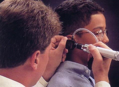
12 Otoscopic Examination

13 When the tympanic membrane is illuminated through an otoscope, the concavity produces a cone of light, which radiates anteriorly and inferiorly from the umbo Cone of light

14 Lateral surface of tympanic membrane Handle of malleus Cone of light Note it is translucent, concave laterally The antero-inferior quadrant of the ear drum is called The cone of light (because it reflects the light coming from the otoscope)

15 Otitis media
16 Middle Ear (Tympanic Cavity) Is an air-containing cavity in the petrous part of the temporal bone Is lined with mucous membrane It contains the auditory ossicles, whose function is to transmit the vibrations of the tympanic membrane (eardrum) to the inner ear. The middle ear has ROOF FLOOR ANTERIOR WALL POSTERIOR WALL LATERAL WALL MEDIAL WALL
17 Tympanic cavity (middle ear) is a narrow, oblique, slitlike cavity whose long axis lies approximately parallel to the plane of the tympanic membrane
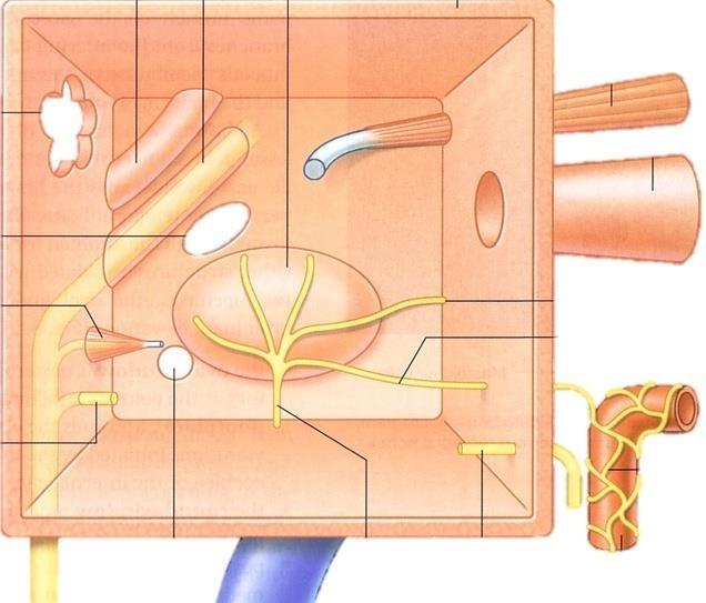
18 Walls of middle ear Roof Medial wall Posterior wall Anterior wall Floor
 It separates the tympanic cavity from the meninges and")

19 Roof TEGMENTAL WALL Is formed by tegmen tympani (part of the petrous temporal bone) It separates the tympanic cavity from the meninges and the temporal lobe of the brain in the middle cranial fossa. Floor JUGULAR WALL It separates the tympanic cavity from the internal jugular vein


20 Tegmen tympani

21 Anterior wall Eustachian tube Tensor tympani Internal carotid artery At the upper part of the anterior wall are the openings into two canals. The lower and larger of these leads into the auditory tube The upper and smaller is the entrance into the canal for the tensor tympani muscle

,")
22 Round window: Below and behind the promontory lies the round window (fenestra cochleae), which is round and closed by the secondary tympanic membrane. Medial wall Promontory is a rounded projection, which results from the underlying first turn of the cochlea The horizontal part of the facial nerve arching above the promontory Oval window: Above and behind the promontory lies the oval window (fenestra vestibuli), which is oval shaped and closed by the base of the stapes The medial wall is formed by the lateral wall of the inner ear.
23 Stapes The base of stapes closes the oval window of the internal ear

24 Internal acoustic meatus 7 th Brain stem 8 th


25 The horizontal part of the facial nerve Stylomastoid foramen Note facial nerve is related to medial and posterior walls of the middle ear

26 Posterior wall 1-Has in its upper part a large, irregular opening, the Aditus to the mastoid 2-Below this is a small, hollow, conical projection, the pyramid, from whose apex emerges the tendon of the stapedius muscle.

27 Mastoid Antrum communicates with the middle ear by the aditus
28 Lateral wall The lateral wall is largely formed by the tympanic membrane (ear drum) tympanic membrane
 Its posterior inner third is bony Its anterior two thirds is cartilaginous Bony")
29 EUSTACHIAN TUBE: Pharyngo-tympanic tube Auditory tube Eustachian tube It connects the anterior wall of the tympanic cavity to the nasopharynx It serves to equalize air pressures in the tympanic cavity and the nasopharynx (on both sides of Tympanic membrane) Its posterior inner third is bony Its anterior two thirds is cartilaginous Bony Cartilaginous
 are shorter and more")

30 Note: Eustachian tubes (auditory tubes) are shorter and more horizontal in children than they are in adults Otitis media is more common in children

31 Opening of Eustachian tube into nasopharynx (nasopharynx is the back of throat)
32 Roof Roof Tegmen tympani
33 Floor Floor Int. jugular vein
34 Anterior wall Tensor tympani m. Auditory tube Internal carotid art
35 Canal for facial N. Promontory Medial wall Oval window Round window Tympanic plexus

36 Aditus Posterior wall Pyramid Vertical part of Facial canal Chorda tympani
37 Chorda tympani Chorda tympani
38

39 Chorda tympani
 into the mastoid antrum The posterior wall of the mastoid antrum is related to the sigmoid venous sinus.")
40 Infections and Otitis Media The meninges and the temporal lobe of the brain lie superiorly meningitis and a cerebral abscess in the temporal lobe. (acute mastoiditis) into the mastoid antrum The posterior wall of the mastoid antrum is related to the sigmoid venous sinus. If the infection spreads in this direction, a thrombosis in the sigmoid sinus may take place through the auditory tube from the nasal part of the pharynx. Medial wall: A spread of the infection in this direction can cause a facial nerve palsy and labyrinthitis with vertigo

41 CONTENTS OF THE MIDDLE EAR Malleus Incus. Stapes. A-3 Auditory Ossicles B-2 muscles C-2 nerves (tympanic plexus and chorda tympani) D-air
 Incus Malleus Limbs Head Stapes Neck")
42 Malleus articulation Ossicles Head Anterior process Incus articulation Short process Lateral process Neck Body Handle Long process Foot plate (base) Incus Malleus Limbs Head Stapes Neck
43 1-The malleus is the largest ossicle and possesses head, a neck, a long process or handle, an anterior process, and a lateral process. its head is rounded and articulates posteriorly with the incus The incus possesses: a large body and two processes: The body articulates with the head of the malleus. The long process articulates with the head of the stapes The stapes has a head, a neck, two limbs, and a base The head articulates with the long process of the incus. The neck is narrow and receives the insertion of the stapedius muscle. The two limbs diverge from the neck and are attached to the oval base which closes the oval window of the internal ear

44 Malleus The handle is firmly attached to the medial surface of the tympanic membrane
45 Incus
46 Stapes The base of stapes closes the oval window of the internal ear
47 Muscles of middle ear Muscle Nerve supply Action Tensor tympani Mandibular division of trigeminal nerve Dampens down vibrations of tympanic membrane Stapedius Facial nerve Dampens down vibrations of stapes

48 Tensor tympani muscle is inserted into malleus Stapedius muscle is inserted into stapes Stapes Tympanic membrane
49 Inner Ear (labyrinth)
50 The inner ear is divided into: 1- Bony labyrinth 2- Membranous labyrinth Semicircular canals Vestibule Cochlea The vestibule is the central part of the bony labyrinth
51 Semicircular canals Vestibule Cochlea Bony labyrinith

52 Membranous labyrinith Semicircular ducts Saccule and utricle Cochlear duct The membranous labyrinth is lodged within the bony labyrinth It is filled with endolymph and surrounded by perilymph
53 The cochlea resembles a snail shell Cochlea
54 The duct of the cochlea lies within the bony cochlea Cochlear duct
55 Posterior semicircular canal
56 Posterior semicircular duct Note: Lodged within the canals are the semicircular ducts
57 Superior semicircular canal
58 Superior semicircular duct
59 Lateral semicircular canal

60 Lateral semicircular duct
61 Bony ampulla Each canal has a swelling at one end called the ampulla
62 Membranous ampulla
63 Utricle Utricle and Saccule are lodged in the bony vestibule
64 Saccule Utricle and Saccule are lodged in the bony vestibule
65 Vestibulo-cochlear nerve Vestibular nerve Cochlear nerve

66 Vestibular nerve carries impulses from the utricle, the saccule, and the ampullae of the semicircular ducts

67 Cochlear nerve carries impulses from organ of Corti in cochlea (contains the sensory receptors for hearing)

68 Transmission of sound Incus Oval window Malleus Stapes Tympanic membrane Round window closed with 2dry tympanic membrane
The Ear The ear consists of : 1-THE EXTERNAL EAR 2-THE MIDDLE EAR, OR TYMPANIC CAVITY 3-THE INTERNAL EAR, OR LABYRINTH 1-THE EXTERNAL EAR.
 The Ear The ear consists of : 1-THE EXTERNAL EAR 2-THE MIDDLE EAR, OR TYMPANIC CAVITY 3-THE INTERNAL EAR, OR LABYRINTH 1-THE EXTERNAL EAR Made of A-AURICLE B-EXTERNAL AUDITORY MEATUS A-AURICLE It consists
AUDITORY APPARATUS. Mr. P Mazengenya. Tel 72204
 AUDITORY APPARATUS Mr. P Mazengenya Tel 72204 Describe the anatomical features of the external ear Describe the tympanic membrane (ear drum) Describe the walls of the middle ear Outline the structures
Dr. Sami Zaqout Faculty of Medicine IUG
 Auricle External Ear External auditory meatus The Ear Middle Ear (Tympanic Cavity) Auditory ossicles Internal Ear (Labyrinth) Bony labyrinth Membranous labyrinth External Ear Auricle External auditory
Gross Anatomy of the. TEMPORAL BONE, EXTERNAL EAR, and MIDDLE EAR
 Gross Anatomy of the TEMPORAL BONE, EXTERNAL EAR, and MIDDLE EAR M1 Gross and Developmental Anatomy 9:00 AM, December 11, 2008 Dr. Milton M. Sholley Professor of Anatomy and Neurobiology Assignment: Head
Gross Anatomy of the. TEMPORAL BONE, EXTERNAL EAR, and MIDDLE EAR. Assignment: Head to Toe Temporomandibular Joint (TMJ)
") Gross Anatomy the TEMPORAL BONE, EXTERNAL EAR, and MIDDLE EAR M1 Gross and Developmental Anatomy 9:00 AM, December 11, 2008 Dr. Milton M. Sholley Pressor Anatomy and Neurobiology Assignment: Head to Toe
Gross Anatomy the TEMPORAL BONE, EXTERNAL EAR, and MIDDLE EAR M1 Gross and Developmental Anatomy 9:00 AM, December 11, 2008 Dr. Milton M. Sholley Pressor Anatomy and Neurobiology Assignment: Head to Toe
Anatomy of the Ear Region. External ear Middle ear Internal ear
 Ear Lecture Objectives Make a list of structures making the external, middle, and internal ear. Discuss the features of the external auditory meatus and tympanic membrane. Describe the shape, position,
Unit VIII Problem 9 Anatomy of The Ear
 Unit VIII Problem 9 Anatomy of The Ear - The ear is an organ with 2 functions: Hearing. Maintenance of equilibrium/balance. - The ear is divided into 3 parts: External ear. Middle ear (which is also known
Anatomy of the ear: Lymphatics
 Anatomy of the ear: 1. External ear which consist of auricle and external auditory canal. The auricle has a framework of cartilage except the lobule, the skin is closely adherent to perichonderium at the
The ear: some applied basic science
 Chapter 1 The ear: some applied basic science The pinna The external ear or pinna is composed of cartilage with closely adherent perichondrium and skin. It is developed from six tubercles of the first
Anatomy and Physiology of Hearing
 Anatomy and Physiology of Hearing The Human Ear Temporal Bone Found on each side of the skull and contains the organs for hearing and balance Divided into four major portions: - squamous - mastoid - tympanic
Kingdom of Bahrain Arabian Gulf University College of Medicine and Medical Sciences Year 6 ENT SMC Otitis Media (Dr.
 Kingdom of Bahrain Arabian Gulf University College of Medicine and Medical Sciences Year 6 ENT SMC Otitis Media (Dr. Jalal Almarzooq) - Anatomy of the ear: The ear is divided into 3 parts: External ear.
THE EAR AND HEARING Be sure you have read and understand Chapter 16 before beginning this lab. INTRODUCTION: hair cells outer ear tympanic membrane
 BIOLOGY 211: HUMAN ANATOMY & PHYSIOLOGY ****************************************************************************************************** THE EAR AND HEARING ******************************************************************************************************
REVIEW/PREVIEW OF HEAD AND NECK ANATOMY FOR ENT EXAM
 REVIEW/PREVIEW OF HEAD AND NECK ANATOMY FOR ENT EXAM - 2017 PALPATE CAROTID ARTERY: AT LEVEL OF CAROTID BIFURCATION VERTEBRAL LEVEL C4 Sternocleidomastoid Muscle INTERNAL CAROTID EXTERNAL CAROTID COMMON
Anatomy of External and Middle ear. Dr Sai Manohar
 Anatomy of External and Middle ear. Dr Sai Manohar 1 Human Ear For Anatomical description, Ear is divided into Auricle (or pinna) The external auditory canal The Middle Ear and its derivatives The Inner
Activity 1: Anatomy of the Eye and Ear Lab
 Activity 1: Anatomy of the Eye and Ear Lab 1. Launch the view! Launch Human Anatomy Atlas. Navigate to Quizzes/Lab Activities, find the Eye and Ear Lab section. Launch Augmented Reality mode and scan the
Chapter 17, Part 2! The Special Senses! Hearing and Equilibrium!
 Chapter 17, Part 2! The Special Senses! Hearing and Equilibrium! SECTION 17-5! Equilibrium sensations originate within the inner ear, while hearing involves the detection and interpretation of sound waves!
Chapter 17, Part 2! Chapter 17 Part 2 Special Senses! The Special Senses! Hearing and Equilibrium!
 Chapter 17, Part 2! The Special Senses! Hearing and Equilibrium! SECTION 17-5! Equilibrium sensations originate within the inner ear, while hearing involves the detection and interpretation of sound waves!
SENSORY SYSTEM VII THE EAR PART 1
 SENSORY SYSTEM VII THE EAR PART 1 Waves Sound is a compression wave The Ear Ear Outer Ear Pinna Outer ear: - Made up of the pinna and the auditory canal Auditory Canal Outer Ear Pinna (also called the
ENT 318 Artificial Organs Physiology of Ear
 ENT 318 Artificial Organs Physiology of Ear Lecturer: Ahmad Nasrul Norali The Ear The Ear Components of hearing mechanism - Outer Ear - Middle Ear - Inner Ear - Central Auditory Nervous System Major Divisions
For the following questions, indicate the letter that corresponds to the SINGLE MOST APPROPRIATE ANSWER
 GROSS ANATOMY EXAMINATION May 15, 2000 For the following questions, indicate the letter that corresponds to the SINGLE MOST APPROPRIATE ANSWER 1. Pain associated with an infection limited to the middle
The Organs of Special Senses
 8 The Organs of Special Senses Special senses are those other than touch, pain, temperature, and proprioception. Vision, hearing, and equilibrium are the special senses discussed in this chapter. The Eye
MECHANISM OF HEARING
 MECHANISM OF HEARING Sound: Sound is a vibration that propagates as an audible wave of pressure, through a transmission medium such as gas, liquid or solid. Sound is produced from alternate compression
1. Axial view, left temporal bone. Plane through the upper antrum (A), superior semicircular canal (SSC) and IAC.
, superior semicircular canal (SSC) and IAC.") PA IAC SSC A 1. Axial view, left temporal bone. Plane through the upper antrum (A), superior semicircular canal (SSC) and IAC. IAC VII M I LSC Plane through the IAC, malleus head and incus and the lateral
PA IAC SSC A 1. Axial view, left temporal bone. Plane through the upper antrum (A), superior semicircular canal (SSC) and IAC. IAC VII M I LSC Plane through the IAC, malleus head and incus and the lateral
Sensory system. Dr. Carmen E. Rexach Anatomy 35 Mt San Antonio College
 Sensory system Dr. Carmen E. Rexach Anatomy 35 Mt San Antonio College Sensory receptors Detect stimuli Classified by structure Origin Distribution Modality Structural Classification naked nerve endings
The white of the eye and the part that maintains its shape is know n as the:
 Scrub In The white of the eye and the part that maintains its shape is know n as the: a. Cornea b. Pupil c. Retina d. Sclera The structure that is found in the ear and contains the organ of hearing is
Dr. Sami Zaqout, IUG Medical School
 The skull The skull is composed of several separate bones united at immobile joints called sutures. Exceptions? Frontal bone Occipital bone Vault Cranium Sphenoid bone Zygomatic bones Base Ethmoid bone
Chapter 15 Hearing & Equilibrium
 Chapter 15 Hearing & Equilibrium ANATOMY OF THE OUTER EAR EAR PINNA is the outer ear it is thin skin covering elastic cartilage. It directs incoming sound waves to the EXTERNAL AUDITORY CANAL, which is
Skull-2. Norma Basalis Interna Norma Basalis Externa. Dr. Heba Kalbouneh Associate Professor of Anatomy and Histology
 Skull-2 Norma Basalis Interna Norma Basalis Externa Dr. Heba Kalbouneh Associate Professor of Anatomy and Histology Norma basalis interna Base of the skull- superior view The interior of the base of the
Chapter 1: Applied Anatomy, Physiology and Embryology of the Ear. Anatomy and Physiology. The Outer Ear. The Pinna. The External Ear Canal
 Chapter 1: Applied Anatomy, Physiology and Embryology of the Ear The ear contains two specialized sensory organs, the cochlea and the vestibular apparatus, enclosed in the extremely hard protective casing
LESSON ASSIGNMENT , Irrigate an Obstructed Ear. After completing this lesson, you should be able to:
 LESSON ASSIGNMENT LESSON 6 Irrigate an Obstructed Ear. TEXT ASSIGNMENT Paragraphs 6-1 through 6-9. TASK TAUGHT LESSON OBJECTIVES 081-833-0059, Irrigate an Obstructed Ear. After completing this lesson,
The Senses. Chapter 10 7/8/11. Introduction
 Chapter 10 The Senses Introduction A. Sensory receptors detect changes in the environment and stimulate neurons to send nerve impulses to the brain. B. A sensation is formed based on the sensory input.
Structure, Energy Transmission and Function. Gross Anatomy. Structure, Function & Process. External Auditory Meatus or Canal (EAM, EAC) Outer Ear
 Outer Ear") Gross Anatomy Structure, Energy Transmission and Function IE N O ME 1 Structure, Function & Process 4 External Auditory Meatus or Canal (EAM, EAC) Outer third is cartilaginous Inner 2/3 is osseous Junction
Gross Anatomy Structure, Energy Transmission and Function IE N O ME 1 Structure, Function & Process 4 External Auditory Meatus or Canal (EAM, EAC) Outer third is cartilaginous Inner 2/3 is osseous Junction
Cone Beam CT Atlas of the Normal Suspensory Apparatus of the Middle Ear Ossicles
 Cone Beam CT Atlas of the Normal Suspensory Apparatus of the Middle Ear Ossicles Poster No.: C-2036 Congress: ECR 2013 Type: Authors: Educational Exhibit B. Smet 1, I. De Kock 2, P. Gillardin 2, M. Lemmerling
Assisting in Otolaryngology
 Assisting in Otolaryngology Learning Objectives Identify the structures and explain the functions of the external, middle, and internal ear. Describe the conditions that can lead to hearing loss, including
Dr.Noor Hashem Mohammad Lecture (5)
") Dr.Noor Hashem Mohammad Lecture (5) 2016-2017 If the mandible is discarded, the anterior part of this aspect of the skull is seen to be formed by the hard palate. The palatal processes of the maxillae
Dr.Noor Hashem Mohammad Lecture (5) 2016-2017 If the mandible is discarded, the anterior part of this aspect of the skull is seen to be formed by the hard palate. The palatal processes of the maxillae
Taste buds Gustatory cells extend taste hairs through a narrow taste pore
 The Special Senses Objectives Describe the sensory organs of smell, and olfaction. Identify the accessory and internal structures of the eye, and explain their function. Explain how light stimulates the
Hearing. By: Jimmy, Dana, and Karissa
 Hearing By: Jimmy, Dana, and Karissa Anatomy - The ear is divided up into three parts - Sound enters in through the outer ear and passes into the middle where the vibrations are received and sent to the
Skull-2. Norma Basalis Interna. Dr. Heba Kalbouneh Assistant Professor of Anatomy and Histology
 Skull-2 Norma Basalis Interna Dr. Heba Kalbouneh Assistant Professor of Anatomy and Histology Norma basalis interna Base of the skull- superior view The interior of the base of the skull is divided into
College of Medicine Dept. of Medical physics Physics of ear and hearing /CH
 College of Medicine Dept. of Medical physics Physics of ear and hearing /CH 13 2017-2018 ***************************************************************** o Introduction : The ear is the organ that detects
SPECIAL SENSES PART I: OLFACTION & GUSTATION
 SPECIAL SENSES PART I: OLFACTION & GUSTATION 5 Special Senses Olfaction Gustation Vision Equilibrium Hearing Olfactory Nerves Extend through cribriform plate into nasal cavity on both sides of nasal septum
o A cushion of fat surrounds most of the eye
 Name Period SPECIAL SENSES The Senses of touch o Temperature o Pressure o Pain o Smell o Taste o Sight o Hearing o Equilibrium The Eye and Vision are in the eyes has over a o Most of the eye is enclosed
Bony and membranous labyrinth. Vestibular system. János Hanics M.D.
 Bony and membranous labyrinth. Vestibular system. János Hanics M.D. The position of the inner ear The labyrinthes of the inner ear - Continuous cavity system in the petrous part of temporal bone - Cavity
For this lab you will use parts of Exercise #18 in your Wise lab manual. Please be sure to read those sections before coming to lab
 Bio 322 Human Anatomy Objectives for the laboratory exercise The Eye and Ear Required reading before beginning this lab: Saladin, KS: Human Anatomy 5 th ed (2017) Chapter 17 For this lab you will use parts
THE COCHLEA AND AUDITORY PATHWAY
 Dental Neuroanatomy Suzanne S. Stensaas, PhD February 23, 2012 Reading: Waxman, Chapter 16, Review pictures in a Histology book Computer Resources: http://www.cochlea.org/ - Promenade around the Cochlea
Infratemporal fossa: Tikrit University college of Dentistry Dr.Ban I.S. head & neck Anatomy 2 nd y.
 Infratemporal fossa: This is a space lying beneath the base of the skull between the lateral wall of the pharynx and the ramus of the mandible. It is also referred to as the parapharyngeal or lateral pharyngeal
Essential questions. What are the structures of the sensory system? 3.03 Remember the structures of the sensory system 2
 Essential questions What are the structures of the sensory system? 3.03 Remember the structures of the sensory system 2 The Senses Eyes Sight Ears Hearing Nose Smell Tongue Taste Skin Touch 3.03 Remember
Tikrit University collage of dentistry Dr.Ban I.S. head & neck anatomy 2 nd y. Lec [5] / Temporal fossa :
![Tikrit University collage of dentistry Dr.Ban I.S. head & neck anatomy 2 nd y. Lec [5] / Temporal fossa :](/thumbs/88/115294566.jpg "Tikrit University collage of dentistry Dr.Ban I.S. head & neck anatomy 2 nd y. Lec [5] / Temporal fossa :") Lec [5] / Temporal fossa : Borders of the Temporal Fossa: Superior: Superior temporal line. Inferior: gap between zygomatic arch and infratemporal crest of sphenoid bone. Anterior: Frontal process of the
Lec [5] / Temporal fossa : Borders of the Temporal Fossa: Superior: Superior temporal line. Inferior: gap between zygomatic arch and infratemporal crest of sphenoid bone. Anterior: Frontal process of the
PCM1 Physical Exam Skills Session: Head and Neck FACILITATOR & STUDENT COPY
 PATIENT CENTERED MEDICINE - 1 GOALS & OUTCOMES: PCM1 Physical Exam Skills Session: Head and Neck FACILITATOR & STUDENT COPY 1. To introduce the applied anatomy relevant for the examination of the head
Hearing. By Jack & Tori
 Hearing By Jack & Tori 3 Main Components of the Human Ear. Outer Ear. Middle Ear. Inner Ear Outer Ear Pinna: >Visible part of ear and ear canal -Acts as a funnel to direct sound Eardrum: >Airtight membrane
Printable version - Hearing - OpenLearn - The Open University
 Skip to content Accessibility Sign in Contact Search the OU The Open University Study at the OU Research at the OU OU Community About the OU Hearing Printable page generated Saturday, 12 November 2011,
Unit VIII Problem 9 Physiology: Hearing
 Unit VIII Problem 9 Physiology: Hearing - We can hear a limited range of frequency between 20 Hz 20,000 Hz (human hearing acuity is between 1000 Hz 4000 Hz). - The ear is divided into 3 parts. Those are:
Ear. Utricle & saccule in the vestibule Connected to each other and to the endolymphatic sac by a utriculosaccular duct
 Rahaf Jreisat *You don t have to go back to the slides. Ear Inner Ear Membranous Labyrinth It is a reflection of bony labyrinth but inside. Membranous labyrinth = set of membranous tubes containing sensory
Major Anatomic Components of the Orbit
 Major Anatomic Components of the Orbit 1. Osseous Framework 2. Globe 3. Optic nerve and sheath 4. Extraocular muscles Bony Orbit Seven Bones Frontal bone Zygomatic bone Maxillary bone Ethmoid bone Sphenoid
4. Which letter in figure 9.1 points to the fovea centralis? Ans: b
 Chapter 9: The Sensory System 1. Proprioceptors are involved in the sense of A) pain. B) temperature. C) pressure. D) movement of limbs. 2. Which are chemoreceptors? A) taste B) olfactory C) proprioceptors
Dr.Ban I.S. head & neck anatomy 2 nd y. جامعة تكريت كلية طب االسنان املرحلة الثانية أ.م.د. بان امساعيل صديق 6102/6102
 جامعة تكريت كلية طب االسنان التشريح مادة املرحلة الثانية أ.م.د. بان امساعيل صديق 6102/6102 Parotid region The part of the face in front of the ear and below the zygomatic arch is the parotid region. The
Introduction. Senses our perception of what is out there 2 groups. General senses Special senses
 Introduction Senses our perception of what is out there 2 groups General senses Special senses Central Processing and Adaptation Adaptation the loss of sensitivity after continuous stimulation Tonic receptors
TASTE: Taste buds are the sense organs that respond to gustatory stimuli. Chemoreceptors that respond to chemicals broken down from food in the saliva
 UNIT 5: Nervous System- Senses Somatic Senses Somatic senses are associated with receptors in the skin, muscles, joints, and viscera (organs of the body) Include senses of touch, pressure, temperature,
Lec [8]: Mandibular nerve:
![Lec [8]: Mandibular nerve:](/thumbs/94/121295776.jpg "Lec [8]: Mandibular nerve:") Lec [8]: Mandibular nerve: The mandibular branch from the trigeminal ganglion lies in the middle cranial fossa lateral to the cavernous sinus. With the motor root of the trigeminal nerve [motor roots lies
Lec [8]: Mandibular nerve: The mandibular branch from the trigeminal ganglion lies in the middle cranial fossa lateral to the cavernous sinus. With the motor root of the trigeminal nerve [motor roots lies
Sound. Audition. Physics of Sound. Properties of sound. Perception of sound works the same way as light.
 Sound Audition Perception of sound works the same way as light. Have receptors to convert a physical stimulus to action potentials Action potentials are organized in brain structures You apply some meaning
Audition. Sound. Physics of Sound. Perception of sound works the same way as light.
 Audition Sound Perception of sound works the same way as light. Have receptors to convert a physical stimulus to action potentials Action potentials are organized in brain structures You apply some meaning
Presentation On SENSATION. Prof- Mrs.Kuldeep Kaur
 Presentation On SENSATION Prof- Mrs.Kuldeep Kaur INTRODUCTION:- Sensation is a specialty area within Psychology that works at understanding how are senses work and how we perceive stimuli in the environment.
Chapter 13 Physics of the Ear and Hearing
 Hearing 100 times greater dynamic range than vision Wide frequency range (20 ~ 20,000 Hz) Sense of hearing Mechanical system that stimulates the hair cells in the cochlea Sensors that produce action potentials
Dr.Ban I.S. head & neck anatomy 2 nd y. جامعة تكريت كلية طب االسنان املرحلة الثانية
 جامعة تكريت كلية طب االسنان التشريح مادة املرحلة الثانية أ.م.د. بان امساعيل صديق 6102-6102 1 The Palate The palate forms the roof of the mouth and the floor of the nasal cavity. It is divided into two
SKULL / CRANIUM BONES OF THE NEUROCRANIUM (7) Occipital bone (1) Sphenoid bone (1) Temporal bone (2) Frontal bone (1) Parietal bone (2)
 Occipital bone (1) Sphenoid bone (1) Temporal bone (2) Frontal bone (1) Parietal bone (2)") Important! 1. Memorizing these pages only does not guarantee the succesfull passing of the midterm test or the semifinal exam. 2. The handout has not been supervised, and I can not guarantee, that these
Important! 1. Memorizing these pages only does not guarantee the succesfull passing of the midterm test or the semifinal exam. 2. The handout has not been supervised, and I can not guarantee, that these
THE COCHLEA AND AUDITORY PATHWAY
 Dental Neuroanatomy Suzanne S. Stensaas, PhD April 14, 2010 Reading: Waxman, Chapter 16, Review pictures in a Histology book Computer Resources: http://www.cochlea.org/ - Promenade around the Cochlea HyperBrain
20-20,000 Hertz range of human hearing
 20-20,000 Hertz range of human hearing accommodation automatic adjustment in focal length of the lens of the eye; changing the shape of the lens aqueous humor Watery fluid in the anterior chambers of the
Cranial Nerve VII - Facial Nerve. The facial nerve has 3 main components with distinct functions
 Cranial Nerve VII - Facial Nerve The facial nerve has 3 main components with distinct functions Somatic motor efferent Supplies the muscles of facial expression; posterior belly of digastric muscle; stylohyoid,
Structure Location Function
 Frontal Bone Cranium forms the forehead and roof of the orbits Occipital Bone Cranium forms posterior and inferior portions of the cranium Temporal Bone Cranium inferior to the parietal bone forms the
Unit # 10 B Assessment of Ears
 In The Name of God (A PROJECT OF NEW LIFE HEALTH CARE SOCIETY KARACHI) Unit # 10 B Assessment of Ears Shahzad Bashir RN, BScN, DCHN, MScN (Std. DUHS) Instructor New Life College of Nursing Updated, January
Skull Base Course. Dissection with fresh temporal bones and half heads
 Skull Base Course Dissection with fresh temporal bones and half heads 711 November 2016 Gruppo Otologico Via Emmanueli 42 Piacenza 29122 t +39 0523 754 362 fax +39 0523 453 708 www.gruppootologico.com
Otoconia: Calcium carbonate crystals Gelatinous mass. Cilia. Hair cells. Vestibular nerve. Vestibular ganglion
 VESTIBULAR SYSTEM (Balance/Equilibrium) The vestibular stimulus is provided by Earth s, and. Located in the of the inner ear, in two components: 1. Vestibular sacs - gravity & head direction 2. Semicircular
A&P 1. Ear, Hearing & Equilibrium Lab. Basic Concepts. These notes follow Carl s Talk at the beginning of lab
 A&P 1 Ear, Hearing & Equilibrium Lab Basic Concepts These notes follow Carl s Talk at the beginning of lab In this "Lab Exercise Guide", we will be looking at the basics of hearing and equilibrium. NOTE:
3-Deep fascia: is absent (except over the parotid gland & buccopharngeal fascia covering the buccinator muscle)
") The Face 1-Skin of the Face The skin of the face is: Elastic Vascular (bleed profusely however heal rapidly) Rich in sweat and sebaceous glands (can cause acne in adults) It is connected to the underlying
The Face 1-Skin of the Face The skin of the face is: Elastic Vascular (bleed profusely however heal rapidly) Rich in sweat and sebaceous glands (can cause acne in adults) It is connected to the underlying
The cochlea: auditory sense. The cochlea: auditory sense
 Inner ear apparatus 1- Vestibule macula and sacculus sensing acceleration of the head and direction of gravity 2- Semicircular canals mainly for sensing direction of rotation of the head 1 3- cochlea in
Chapter Fourteen. The Hearing Mechanism. 1. Introduction.
 Chapter Fourteen The Hearing Mechanism 1. Introduction. 2. Hearing. 3. The Ear. 4. The External Ear. 5. The Inner Ear. 6. Frequency Discrimination. 7. The Organ of Corti. 8. Tests and Exrecises. 9. References.
Scrub In. What is the function of cerumen? Which part of the ear collects sound waves and directs them into the auditory canal?
 Scrub In What is the function of cerumen? a. Keeps the ear canal from collapsing b. Helps transmit sound waves c. Protection d. Lubrication Which part of the ear collects sound waves and directs them into
Temporal fossa Infratemporal fossa Pterygopalatine fossa Terminal branches of external carotid artery Pterygoid venous plexus
 Outline of content Temporal fossa Infratemporal fossa Pterygopalatine fossa Terminal branches of external carotid artery Pterygoid venous plexus Boundary Content Communication Mandibular division of trigeminal
A&P 1. Ear, Hearing & Equilibrium Lab. Basic Concepts. Pre-lab Exercises
 A&P 1 Ear, Hearing & Equilibrium Lab Basic Concepts Pre-lab Exercises In this "Lab Exercise Guide", we will be looking at the basics of hearing and equilibrium. NOTE: these notes do not follow the order
Parotid Gland, Temporomandibular Joint and Infratemporal Fossa
 M1 - Anatomy Parotid Gland, Temporomandibular Joint and Infratemporal Fossa Jeff Dupree Sanger 9-057 jldupree@vcu.edu Parotid gland: wraps around the mandible positioned between the mandible and the sphenoid
to vibrate the fluid. The ossicles amplify the pressure. The surface area of the oval window is
 Page 1 of 6 Question 1: How is the conduction of sound to the cochlea facilitated by the ossicles of the middle ear? Answer: Sound waves traveling through air move the tympanic membrane, which, in turn,
Acquired Deafness Loss of hearing that occurs or develops sometime in the course of a lifetime, but is not present at birth.
 Page 1 of 5 URMC» Audiology Glossary of Terms A Acoustic Neuroma A tumor, usually benign, which develops on the hearing and balance nerves and can cause gradual hearing loss, tinnitus, and dizziness. Acquired
Anatomy and Pathophysiology
 Anatomy and Pathophysiology for ICD-10 2014 Module 11 Disclaimer This course was current at the time it was published. This course was prepared as a tool to assist the participant in understanding how
The Nervous System: General and Special Senses Pearson Education, Inc.
 18 The Nervous System: General and Special Senses Introduction Sensory information arrives at the CNS Information is picked up by sensory receptors Sensory receptors are the interface between the nervous
Receptors / physiology
 Hearing: physiology Receptors / physiology Energy transduction First goal of a sensory/perceptual system? Transduce environmental energy into neural energy (or energy that can be interpreted by perceptual
The orbit-1. Dr. Heba Kalbouneh Assistant Professor of Anatomy and Histology
 The orbit-1 Dr. Heba Kalbouneh Assistant Professor of Anatomy and Histology Orbital plate of frontal bone Orbital plate of ethmoid bone Lesser wing of sphenoid Greater wing of sphenoid Lacrimal bone Orbital
is the clear, transparent part at the front of the eye. It allows light to enter the eye and it also refracts (focuses) the light onto the retina.
 the light onto the retina.") Senses- Vision Light is a small part (1/70th) of the total electromagnetic (EM) spectrum. The EM band extends from radio waves at one extreme to x-rays at the other. The eye detects light and converts
Senses- Vision Light is a small part (1/70th) of the total electromagnetic (EM) spectrum. The EM band extends from radio waves at one extreme to x-rays at the other. The eye detects light and converts
Anatomy and Physiology. Bones, Sutures, Teeth, Processes and Foramina of the Human Skull
 Anatomy and Physiology Chapter 6 DRO Bones, Sutures, Teeth, Processes and Foramina of the Human Skull Name: Period: Bones of the Human Skull Bones of the Cranium: Frontal bone: forms the forehead and the
o A cushion of fat surrounds most of the eye
 Name Period SPECIAL SENSES The Senses General senses of touch o Temperature o Pressure o Pain Special senses o Smell o Taste o Sight o Hearing o Equilibrium The Eye and Vision 70 percent of all sensory
a) Central sulcus- shallow groove that runs across brain sagitally
 Central sulcus- shallow groove that runs across brain sagitally") KEY BRAIN Brain Gross Anatomy Terms 1) Explain each of the following in terms of structure of the brain a) Central sulcus- shallow groove that runs across brain sagitally b) Lateral fissure- deep groove
KEY BRAIN Brain Gross Anatomy Terms 1) Explain each of the following in terms of structure of the brain a) Central sulcus- shallow groove that runs across brain sagitally b) Lateral fissure- deep groove
Before we talk about the auditory system we will talk about the sound and waves
 The Auditory System PHYSIO: #3 DR.LOAI ZAGOUL 24/3/2014 Refer to the slides for some photos. Before we talk about the auditory system we will talk about the sound and waves All waves have basic characteristics:
AXIAL SKELETON SKULL
 AXIAL SKELETON SKULL CRANIAL BONES (8 total flat bones w/ 2 paired) 1. Frontal forms forehead & upper portion of eyesocket (orbital) 2. Parietal paired bones; form superior & lateral walls of cranium 3.
Chapter 15 Lecture Outline
 Chapter 15 Lecture Outline See separate PowerPoint slides for all figures and tables preinserted into PowerPoint without notes. Copyright 2016 McGraw-Hill Education. Permission required for reproduction
The sebaceous glands (glands of Zeis) open directly into the eyelash follicles, ciliary glands (glands of Moll) are modified sweat glands that open
 open directly into the eyelash follicles, ciliary glands (glands of Moll) are modified sweat glands that open") The Orbital Region The orbits are a pair of bony cavities that contain the eyeballs; their associated muscles, nerves, vessels, and fat; and most of the lacrimal apparatus upper eyelid is larger and more
The Orbital Region The orbits are a pair of bony cavities that contain the eyeballs; their associated muscles, nerves, vessels, and fat; and most of the lacrimal apparatus upper eyelid is larger and more
Special Senses. Unit 6.7 (6 th Edition) Chapter 7.7 (7 th Edition)
 Chapter 7.7 (7 th Edition)") Special Senses Unit 6.7 (6 th Edition) Chapter 7.7 (7 th Edition) 1 Learning Objectives Identify the five special senses. Identify the four general senses. Trace the pathway of light rays as they pass
Special Senses Unit 6.7 (6 th Edition) Chapter 7.7 (7 th Edition) 1 Learning Objectives Identify the five special senses. Identify the four general senses. Trace the pathway of light rays as they pass
Functional components
 Facial Nerve VII cranial nerve Emerges from Pons Two roots Functional components: 1. GSA (general somatic afferent) 2. SA (Somatic afferent) 3. GVE (general visceral efferent) 4. BE (Special visceral/branchial
Chapter 7. Audition, the Body Senses, and the Chemical Senses. Copyright Allyn & Bacon 2004
 Chapter 7 Audition, the Body Senses, and the Chemical Senses This multimedia product and its contents are protected under copyright law. The following are prohibited by law: any public performance or display,
The Visible Ear Simulator Dissection Manual.
 The Visible Ear Simulator Dissection Manual. Stereoscopic Tutorialized Version 3.1, August 2017 Peter Trier Mikkelsen, the Alexandra Institute A/S, Aarhus, Denmark Mads Sølvsten Sørensen & Steven Andersen,