THE COCHLEA AND AUDITORY PATHWAY
|
|
|
- Maurice Page
- 5 years ago
- Views:
Transcription
1 Dental Neuroanatomy Suzanne S. Stensaas, PhD April 14, 2010 Reading: Waxman, Chapter 16, Review pictures in a Histology book Computer Resources: - Promenade around the Cochlea HyperBrain 9, Objectives: THE COCHLEA AND AUDITORY PATHWAY 1. Trace the trajectory of a sound wave of a high note from the tympanic membrane to the production of a nerve impulse. 2. Contrast conduction deafness and neural deafness. 3. Describe where in the nervous system a lesion would have to be for a person to have deafness in the ipsilateral ear. 4. Explain why lesions of the auditory pathway are harder to localize and of less clinical value than those of the DCML system. 5. Two people have strokes in part of the distribution of the middle cerebral artery. One does not understand what family members are saying and the other understands but cannot answer. How can a lesion of the same artery produce such different results? 6. Describe the structures that could be affected and the clinical findings associated with an acoustic neuroma (benign Schwann cell tumor) on the VIII nerve in the cerebellopontine angle. Would it be different if it were in the internal auditory meatus? How? I. General Anatomy of the Ear A. Position in the skull. B. Middle ear. 1. Tympanic membrane. 2. Ossicles=mechanical multipliers of tympanic membrane motion. a. Malleus b. Incus c. Stapes--attachment to oval window of inner ear. 3. Middle ear muscles dampen sound, improve discrimination. a. Tensor tympani--attachment on malleus--acts to decrease sensitivity of the ear to loud sounds. (CNV) b. Stapedius--attachment to stapes--acts to decrease sensitivity. (CN VII) C. Inner Ear 1. The bony labyrinth. a. Contains the watery perilymph high in sodium, which is in contact with the subarachnoid space. The membranous labyrinth is suspended in the perilymph inside the bony labyrinth. b. Oval window (which is closed by the stapes) leads into the vestibule, the middle part of the bony labyrinth. 1

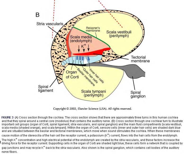
2 c. Anteriorly, the vestibule merges with the bony cochlea, which has the form of a 2-1/2-turn spiral cone. d. Posteriorly, the vestibule merges with the bony semicircular canals. 2. The membranous labyrinth. Location of the sensory receptors a. Contains the viscous fluid endolymph, which is high in potassium. b. The bony vestibule itself contains two membranous vestibular sensory organs. (1) Saccule (2) Utricle c. Three bony semicircular canals emanate from the vestibule at right angles to each other; these contain the membranous semicircular ducts, which communicate with the utriculus and are filled with endolymph. c. The bony cochlea contains the cochlear canal in which sits the membranous cochlear duct filled with endolymph. 2

3 Cochlear Fluid Web site: 1. The perilymphatic space above the cochlear duct is the scala vestibuli, which opens onto the vestibule. Frank Netter 3
4 4

5 3. The cochlear duct is separated from the scala vestibuli by the vestibular membrane. The scala vestibuli merges with the scala tympani at the helicotrema, which is at the apical tip of the cochlea. Thus the pressure wave is continuous and goes "up" the scala vestibuli from the oval window and down the scala tympani to the round window 4. The Organ of Corti. 1. Position in cochlear duct. 2. Sits on basilar membrane. a. Its width varies along the cochlear duct. Influences discrimination. Membrane shorter at base (high tones) and wider at the apex (low tones) b. The spiral ligament=lateral attachment of basilar membrane. c. The stria vascularis forms the lateral surface of the cochlear duct and produces the endolymph high in K+. From The Digital Anatomist Interactive Brain Syllabus. John Sundsten and Kate Mulligan, Univ.Washington School of Medicine From The Digital Anatomist Interactive Brain Syllabus. John Sundsten and Kate Mulligan, Univ.Washington School of Medicine
6 3. Histology of Organ of Corti A. Supporting cells hold the sensory hair cells in position below the tectorial membrane. B. Inner and outer hair cells. Inner are more important and necessary for hearing. 1. Stereocilia of sensory hairs touch the tectorial membrane. 2. Afferent (95%) and efferent (5%) synaptic endings on outer hair cells form on base of cells. 95% of axons in auditory nerve innervate inner hair cells. 3. Outer hair cells appear to be contractile and change the stiffness of the basilar membrane in response to electrical currents. 6


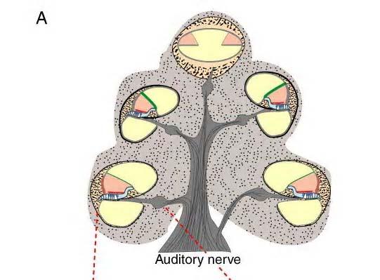
7 THE AUDITORY PATHWAY THE AUDITORY PATHWAY II. Auditory Pathway Frank Netter A. Organizing principles. 1, Tonotopic organization: there is a topographic map of the cochlea at each level of the ascending auditory pathway. This results in an orderly representation of sound frequencies (tones) at each level. 2. Because of the many crossing pathways, there is bilateral representation of the cochlea at most levels of the auditory system. 7
8 Medial Geniculate Nuclei Superior Temporal Gyrus Commissure of the Inferior Colliculi Inferior Colliculus Lateral Lemniscus Sublentiform Part of Internal Capsule Nucleus of the Lateral Lemniscus Dorsal Cochlear Nucleus Superior Olivary Nucleus Cochlear Nerve Trapezoid Body Inferior Cerebellar Peduncle Spiral Ganglion Ventral Cochlear Nucleus Source? B Sequence of relays and processing of information 1. Spiral ganglion neurons innervate the inner and outer cochlear hair cells. 8


9 Suzanne S. Stensaas Suzanne S. Stensaas 2. Medullopontine Junction: Cochlear Nuclei A. All cochlear nerve fibers (axons of spiral ganglion neurons) make synapses on neurons in the ipsilateral cochlear nucleus. B. Each entering axon forms an ascending and a descending branch as it enters the brain. Thus, two separate representations of the cochlea are formed in the cochlear nucleus. 9

10 1. The ascending branch forms synapses on neurons in the dorsal cochlear nucleus. 2. The descending branch forms synapses in the ventral cochlear nucleus. C. Different types of neuron in the two-cochlear nuclei send their axons through two major pathways to higher auditory centers. 1. The dorsal cochlear nucleus next to the inferior cerebellar peduncle, crosses the midline, and ascends in a tract called the lateral lemniscus. The axons carry information about the frequency spectrum of sound stimuli. 2. The ventral cochlear nucleus, also next to the inferior cerebellar peduncle and then join the lateral lemniscus From The Digital Anatomist Interactive Brain Syllabus. John Sundsten and Kate Mulligan, Univ.Washington School of Medicine


11 Suzanne S. Stensaas 3. Mesencephalon: Inferior Colliculus A. Lateral lemniscal axons end in the ipsilateral inferior colliculus. Map of auditory space. B. The inferior colliculus is involved in auditory reflexes and sound localization. C. Neurons in the colliculus project to the medial geniculate body of the thalamus through the brachium of the inferior colliculus. 11
12 Suzanne S. Stensaas 12


13 Suzanne S. Stensaas 4. Thalamus: Medial Geniculate Body A. Medial geniculate neurons receive ascending auditory input from the brachium of the inferior colliculus. B. Project to the ipsilateral transverse temporal gyrus of the cerebral cortex via the auditory radiations in the internal capsule. This is primary auditory cortex. 13


14 From The Digital Anatomist Interactive Brain Syllabus. John Sundsten and Kate Mulligan, Univ.Washington School of Medicine
15 15
 receive the thalamocortical projections from the medial geniculate body. B.")

16 Source? Suzanne S. Stensaas III. Auditory Cortex A. The primary auditory cortical region is located on the dorsal surface of the temporal lobe deep within the lateral fissure. Two prominent transverse temporal gyri (Heschl's gyri) receive the thalamocortical projections from the medial geniculate body. B. Surrounding this primary auditory cortex are secondary auditory association areas in the temporal lobe C. As elsewhere, there are extensive projections from primary and secondary auditory cortical areas to various association areas of neocortex. IV. Association Areas - The Posterior Superior Temporal Lobe and Language Function A. The posterior superior temporal lobe (area 22) Unlike the primary auditory cortex, it is not activated by simple auditory stimuli (e.g., tones or clicks) but during language comprehension tasks that involve auditory or phonetic processing and short-term memory of words. We call this Wernicke s area B. Aphasia: an impairment in language or communication 1. Comprehension 2. Production, verbalization, fluency 3. Writing 4. Reading 5. Signing 16


17 Scientific American, Freeman Press. C. Two major types of Aphasia result from strokes in dominant hemisphere. 1. Receptive or sensory aphasia typically involves damage of the posterior part of the left superior temporal gyrus. This is also called (Wernicke's aphasia or Wernicke's area. Other terms are fluent aphasia since they produce words but they often use the wrong words or unrelated words. Sometimes described as "word salad". Grammar is intact. Don t understand what they are saying, what you say or what they hear or read. 1. Expressive aphasia is non-fluent. The patient understands but has difficulty speaking. Typical of frontal motor cortex lesions particularly in the inferior frontal gyrus or Broca's area 44. Very frustrating and often only expletives come out. 2. (Conduction Aphasia involves the arcuate connections between the two areas. Can usually comprehend) 3. (Global aphasia affects all areas such as a massive middle cerebral artery occlusion.) 17
? 2. Unlike the primary auditory cortex, is not activated by simple auditory stimuli (e.g., tones or clicks) but is activated during language comprehension tasks that involve auditory or phonetic processing of words 18")
18 Scientific American, Freeman Press D. The planum temporale, which is the portion of the dorsal surface of the temporal lobe just posterior to the transverse gyrus (primary auditory cortex), 1. Is normally about one-third larger in the language-dominant hemisphere (the left hemisphere in 97% of people)? 2. Unlike the primary auditory cortex, is not activated by simple auditory stimuli (e.g., tones or clicks) but is activated during language comprehension tasks that involve auditory or phonetic processing of words 18
THE COCHLEA AND AUDITORY PATHWAY
 Dental Neuroanatomy Suzanne S. Stensaas, PhD February 23, 2012 Reading: Waxman, Chapter 16, Review pictures in a Histology book Computer Resources: http://www.cochlea.org/ - Promenade around the Cochlea
Auditory and Vestibular Systems
 Auditory and Vestibular Systems Objective To learn the functional organization of the auditory and vestibular systems To understand how one can use changes in auditory function following injury to localize
SPECIAL SENSES: THE AUDITORY SYSTEM
 SPECIAL SENSES: THE AUDITORY SYSTEM REVISION OF PHYSICS: WAVES A wave is an oscillation of power, sound waves have two main characteristics: amplitude, which is the maximum displacement or the power of
Cranial Nerve VIII (The Vestibulo-Cochlear Nerve)
") Cranial Nerve VIII (The Vestibulo-Cochlear Nerve) Please view our Editing File before studying this lecture to check for any changes. Color Code Important Doctors Notes Notes/Extra explanation Objectives
Cranial Nerve VIII (The Vestibulo-Cochlear Nerve) Please view our Editing File before studying this lecture to check for any changes. Color Code Important Doctors Notes Notes/Extra explanation Objectives
AUDITORY APPARATUS. Mr. P Mazengenya. Tel 72204
 AUDITORY APPARATUS Mr. P Mazengenya Tel 72204 Describe the anatomical features of the external ear Describe the tympanic membrane (ear drum) Describe the walls of the middle ear Outline the structures
Auditory System. Barb Rohrer (SEI )
") Auditory System Barb Rohrer (SEI614 2-5086) Sounds arise from mechanical vibration (creating zones of compression and rarefaction; which ripple outwards) Transmitted through gaseous, aqueous or solid medium
Auditory System Barb Rohrer (SEI614 2-5086) Sounds arise from mechanical vibration (creating zones of compression and rarefaction; which ripple outwards) Transmitted through gaseous, aqueous or solid medium
Chapter 17, Part 2! The Special Senses! Hearing and Equilibrium!
 Chapter 17, Part 2! The Special Senses! Hearing and Equilibrium! SECTION 17-5! Equilibrium sensations originate within the inner ear, while hearing involves the detection and interpretation of sound waves!
Chapter 17, Part 2! Chapter 17 Part 2 Special Senses! The Special Senses! Hearing and Equilibrium!
 Chapter 17, Part 2! The Special Senses! Hearing and Equilibrium! SECTION 17-5! Equilibrium sensations originate within the inner ear, while hearing involves the detection and interpretation of sound waves!
Auditory System Feedback
 Feedback Auditory System Feedback Using all or a portion of the information from the output of a system to regulate or control the processes or inputs in order to modify the output. Central control of
Otoconia: Calcium carbonate crystals Gelatinous mass. Cilia. Hair cells. Vestibular nerve. Vestibular ganglion
 VESTIBULAR SYSTEM (Balance/Equilibrium) The vestibular stimulus is provided by Earth s, and. Located in the of the inner ear, in two components: 1. Vestibular sacs - gravity & head direction 2. Semicircular
Ear. Utricle & saccule in the vestibule Connected to each other and to the endolymphatic sac by a utriculosaccular duct
 Rahaf Jreisat *You don t have to go back to the slides. Ear Inner Ear Membranous Labyrinth It is a reflection of bony labyrinth but inside. Membranous labyrinth = set of membranous tubes containing sensory
THE VESTIBULAR APPRATUS AND PATHWAY
 Dental Neuroanatomy February 23, 2012 Suzanne Stensaas, Ph.D. Reading: Waxman Chapter 17 Also pp 105-108 on control of eye movments Computer Resources: HyperBrain Ch. 8 Vestibulospinal Pathway Quiz http://library.med.utah.edu/kw/animations/hyperbrain/pathways/
Auditory Physiology Richard M. Costanzo, Ph.D.
 Auditory Physiology Richard M. Costanzo, Ph.D. OBJECTIVES After studying the material of this lecture, the student should be able to: 1. Describe the morphology and function of the following structures:
Unit VIII Problem 9 Physiology: Hearing
 Unit VIII Problem 9 Physiology: Hearing - We can hear a limited range of frequency between 20 Hz 20,000 Hz (human hearing acuity is between 1000 Hz 4000 Hz). - The ear is divided into 3 parts. Those are:
to vibrate the fluid. The ossicles amplify the pressure. The surface area of the oval window is
 Page 1 of 6 Question 1: How is the conduction of sound to the cochlea facilitated by the ossicles of the middle ear? Answer: Sound waves traveling through air move the tympanic membrane, which, in turn,
Unit VIII Problem 9 Anatomy of The Ear
 Unit VIII Problem 9 Anatomy of The Ear - The ear is an organ with 2 functions: Hearing. Maintenance of equilibrium/balance. - The ear is divided into 3 parts: External ear. Middle ear (which is also known
Structure, Energy Transmission and Function. Gross Anatomy. Structure, Function & Process. External Auditory Meatus or Canal (EAM, EAC) Outer Ear
 Outer Ear") Gross Anatomy Structure, Energy Transmission and Function IE N O ME 1 Structure, Function & Process 4 External Auditory Meatus or Canal (EAM, EAC) Outer third is cartilaginous Inner 2/3 is osseous Junction
Gross Anatomy Structure, Energy Transmission and Function IE N O ME 1 Structure, Function & Process 4 External Auditory Meatus or Canal (EAM, EAC) Outer third is cartilaginous Inner 2/3 is osseous Junction
Chapter 15 Hearing & Equilibrium
 Chapter 15 Hearing & Equilibrium ANATOMY OF THE OUTER EAR EAR PINNA is the outer ear it is thin skin covering elastic cartilage. It directs incoming sound waves to the EXTERNAL AUDITORY CANAL, which is
9.01 Introduction to Neuroscience Fall 2007
 MIT OpenCourseWare http://ocw.mit.edu 9.01 Introduction to Neuroscience Fall 2007 For information about citing these materials or our Terms of Use, visit: http://ocw.mit.edu/terms. 9.01 Recitation (R02)
Deafness and hearing impairment
 Auditory Physiology Deafness and hearing impairment About one in every 10 Americans has some degree of hearing loss. The great majority develop hearing loss as they age. Hearing impairment in very early
Required Slide. Session Objectives
 Auditory Physiology Required Slide Session Objectives Auditory System: At the end of this session, students will be able to: 1. Characterize the range of normal human hearing. 2. Understand the components
Before we talk about the auditory system we will talk about the sound and waves
 The Auditory System PHYSIO: #3 DR.LOAI ZAGOUL 24/3/2014 Refer to the slides for some photos. Before we talk about the auditory system we will talk about the sound and waves All waves have basic characteristics:
Taste buds Gustatory cells extend taste hairs through a narrow taste pore
 The Special Senses Objectives Describe the sensory organs of smell, and olfaction. Identify the accessory and internal structures of the eye, and explain their function. Explain how light stimulates the
The cochlea: auditory sense. The cochlea: auditory sense
 Inner ear apparatus 1- Vestibule macula and sacculus sensing acceleration of the head and direction of gravity 2- Semicircular canals mainly for sensing direction of rotation of the head 1 3- cochlea in
Anatomy of the Ear Region. External ear Middle ear Internal ear
 Ear Lecture Objectives Make a list of structures making the external, middle, and internal ear. Discuss the features of the external auditory meatus and tympanic membrane. Describe the shape, position,
Laith Sorour. Facial nerve (vii):
:") Laith Sorour Cranial nerves 7 & 8 Hello, there are edited slides please go back to them to see pictures, they are not that much important in this lecture but still, and yes slides are included :p Let s
Laith Sorour Cranial nerves 7 & 8 Hello, there are edited slides please go back to them to see pictures, they are not that much important in this lecture but still, and yes slides are included :p Let s
Cranial Nerve VII & VIII
 Cranial Nerve VII & VIII Lecture Objectives Follow up the course of facial nerve from its point of central connections, exit and down to its target areas. Follow up the central connections of the facial
What is the effect on the hair cell if the stereocilia are bent away from the kinocilium?
 CASE 44 A 53-year-old man presents to his primary care physician with complaints of feeling like the room is spinning, dizziness, decreased hearing, ringing in the ears, and fullness in both ears. He states
Sensory Systems Vision, Audition, Somatosensation, Gustation, & Olfaction
 Sensory Systems Vision, Audition, Somatosensation, Gustation, & Olfaction Sarah L. Chollar University of California, Riverside sarah.chollar@gmail.com Sensory Systems How the brain allows us to see, hear,
SOMATIC SENSATION PART I: ALS ANTEROLATERAL SYSTEM (or SPINOTHALAMIC SYSTEM) FOR PAIN AND TEMPERATURE
 FOR PAIN AND TEMPERATURE") Dental Neuroanatomy Thursday, February 3, 2011 Suzanne S. Stensaas, PhD SOMATIC SENSATION PART I: ALS ANTEROLATERAL SYSTEM (or SPINOTHALAMIC SYSTEM) FOR PAIN AND TEMPERATURE Reading: Waxman 26 th ed, :
Dental Neuroanatomy Thursday, February 3, 2011 Suzanne S. Stensaas, PhD SOMATIC SENSATION PART I: ALS ANTEROLATERAL SYSTEM (or SPINOTHALAMIC SYSTEM) FOR PAIN AND TEMPERATURE Reading: Waxman 26 th ed, :
For this lab you will use parts of Exercise #18 in your Wise lab manual. Please be sure to read those sections before coming to lab
 Bio 322 Human Anatomy Objectives for the laboratory exercise The Eye and Ear Required reading before beginning this lab: Saladin, KS: Human Anatomy 5 th ed (2017) Chapter 17 For this lab you will use parts
Hearing. By Jack & Tori
 Hearing By Jack & Tori 3 Main Components of the Human Ear. Outer Ear. Middle Ear. Inner Ear Outer Ear Pinna: >Visible part of ear and ear canal -Acts as a funnel to direct sound Eardrum: >Airtight membrane
Chapter 3: Anatomy and physiology of the sensory auditory mechanism
 Chapter 3: Anatomy and physiology of the sensory auditory mechanism Objectives (1) Anatomy of the inner ear Functions of the cochlear and vestibular systems Three compartments within the cochlea and membranes
Chapter 11: Sound, The Auditory System, and Pitch Perception
 Chapter 11: Sound, The Auditory System, and Pitch Perception Overview of Questions What is it that makes sounds high pitched or low pitched? How do sound vibrations inside the ear lead to the perception
ENT 318 Artificial Organs Physiology of Ear
 ENT 318 Artificial Organs Physiology of Ear Lecturer: Ahmad Nasrul Norali The Ear The Ear Components of hearing mechanism - Outer Ear - Middle Ear - Inner Ear - Central Auditory Nervous System Major Divisions
Gathering information the sensory systems; Vision
 Visual System Gathering information the sensory systems; Vision The retina is the light-sensitive receptor layer at the back of the eye. - Light passes through the cornea, the aqueous chamber, the lens,
MECHANISM OF HEARING
 MECHANISM OF HEARING Sound: Sound is a vibration that propagates as an audible wave of pressure, through a transmission medium such as gas, liquid or solid. Sound is produced from alternate compression
Chapter 7. Audition, the Body Senses, and the Chemical Senses. Copyright Allyn & Bacon 2004
 Chapter 7 Audition, the Body Senses, and the Chemical Senses This multimedia product and its contents are protected under copyright law. The following are prohibited by law: any public performance or display,
General Sensory Pathways of the Face Area, Taste Pathways and Hearing Pathways
 General Sensory Pathways of the Face Area, Taste Pathways and Hearing Pathways Lecture Objectives Describe pathways for general sensations (pain, temperature, touch and proprioception) from the face area.
Vestibular/Auditory Systems
 Vestibular/Auditory Systems Jay Zenner on February 3, 2012 Dental Neuroanatomy Scott Rogers Office: SOM 2C132 Boney Labyrinth Vestibular Apparatus Two Major Divisions Cochlea (anterior) VIII VII Semicircular
Auditory and vestibular system
 Auditory and vestibular system Sensory organs on the inner ear inner ear: audition (exteroceptor) and vestibular apparatus (proprioceptor) bony and membranous labyrinths within the temporal bone (os temporale)
THE EAR AND HEARING Be sure you have read and understand Chapter 16 before beginning this lab. INTRODUCTION: hair cells outer ear tympanic membrane
 BIOLOGY 211: HUMAN ANATOMY & PHYSIOLOGY ****************************************************************************************************** THE EAR AND HEARING ******************************************************************************************************
Activity 1: Anatomy of the Eye and Ear Lab
 Activity 1: Anatomy of the Eye and Ear Lab 1. Launch the view! Launch Human Anatomy Atlas. Navigate to Quizzes/Lab Activities, find the Eye and Ear Lab section. Launch Augmented Reality mode and scan the
Carlson (7e) PowerPoint Lecture Outline Chapter 7: Audition, the Body Senses, and the Chemical Senses
 PowerPoint Lecture Outline Chapter 7: Audition, the Body Senses, and the Chemical Senses") Carlson (7e) PowerPoint Lecture Outline Chapter 7: Audition, the Body Senses, and the Chemical Senses This multimedia product and its contents are protected under copyright law. The following are prohibited
Carlson (7e) PowerPoint Lecture Outline Chapter 7: Audition, the Body Senses, and the Chemical Senses This multimedia product and its contents are protected under copyright law. The following are prohibited
Cranial Nerves VII to XII
 Cranial Nerves VII to XII MSTN121 - Neurophysiology Session 13 Department of Myotherapy Cranial Nerve VIII: Vestibulocochlear Sensory nerve with two distinct branches. Vestibular branch transmits information
Presentation On SENSATION. Prof- Mrs.Kuldeep Kaur
 Presentation On SENSATION Prof- Mrs.Kuldeep Kaur INTRODUCTION:- Sensation is a specialty area within Psychology that works at understanding how are senses work and how we perceive stimuli in the environment.
Receptors / physiology
 Hearing: physiology Receptors / physiology Energy transduction First goal of a sensory/perceptual system? Transduce environmental energy into neural energy (or energy that can be interpreted by perceptual
Anatomy and Physiology of Hearing
 Anatomy and Physiology of Hearing The Human Ear Temporal Bone Found on each side of the skull and contains the organs for hearing and balance Divided into four major portions: - squamous - mastoid - tympanic
Anatomy of the ear: Lymphatics
 Anatomy of the ear: 1. External ear which consist of auricle and external auditory canal. The auricle has a framework of cartilage except the lobule, the skin is closely adherent to perichonderium at the
BCS 221: Auditory Perception BCS 521 & PSY 221
 BCS 221: Auditory Perception BCS 521 & PSY 221 Time: MW 10:25 11:40 AM Recitation: F 10:25 11:25 AM Room: Hutchinson 473 Lecturer: Dr. Kevin Davis Office: 303E Meliora Hall Office hours: M 1 3 PM kevin_davis@urmc.rochester.edu
The Nervous System: General and Special Senses Pearson Education, Inc.
 18 The Nervous System: General and Special Senses Introduction Sensory information arrives at the CNS Information is picked up by sensory receptors Sensory receptors are the interface between the nervous
Hearing: Physiology and Psychoacoustics
 9 Hearing: Physiology and Psychoacoustics Click Chapter to edit 9 Hearing: Master title Physiology style and Psychoacoustics The Function of Hearing What Is Sound? Basic Structure of the Mammalian Auditory
Intro to Audition & Hearing
 Intro to Audition & Hearing Lecture 16 Chapter 9, part II Jonathan Pillow Sensation & Perception (PSY 345 / NEU 325) Fall 2017 1 Sine wave: one of the simplest kinds of sounds: sound for which pressure
The speed at which it travels is a function of the density of the conducting medium.
 Sound is a compression wave which (unlike light) must have a medium to conduct it. If the medium is uniform in density, the sound will spread at as a uniform ring of circles (actually spheres). The speed
PSY 215 Lecture 10 Topic: Hearing Chapter 7, pages
 PSY 215 Lecture 10 Topic: Hearing Chapter 7, pages 189-197 Corrections: NTC 09-1, page 3, the Superior Colliculus is in the midbrain (Mesencephalon). Announcements: Movie next Monday: Case of the frozen
A&P 1. Ear, Hearing & Equilibrium Lab. Basic Concepts. These notes follow Carl s Talk at the beginning of lab
 A&P 1 Ear, Hearing & Equilibrium Lab Basic Concepts These notes follow Carl s Talk at the beginning of lab In this "Lab Exercise Guide", we will be looking at the basics of hearing and equilibrium. NOTE:
Hearing. By: Jimmy, Dana, and Karissa
 Hearing By: Jimmy, Dana, and Karissa Anatomy - The ear is divided up into three parts - Sound enters in through the outer ear and passes into the middle where the vibrations are received and sent to the
C:\Documents and Settings\sstensaas\Desktop\dental visual 2010\VisualPath dental 2010.docVisualPath dental 2010.doc
 Neuroanatomy Suzanne Stensaas April 8, 2010, 10:00-12:00 p.m. Reading: Waxman Ch. 15, Computer Resources: HyperBrain Ch 7 THE VISUAL PATHWAY Objectives: 1. Describe the pathway of visual information from
Printable version - Hearing - OpenLearn - The Open University
 Skip to content Accessibility Sign in Contact Search the OU The Open University Study at the OU Research at the OU OU Community About the OU Hearing Printable page generated Saturday, 12 November 2011,
Central Auditory System Basics and the Effects of Abnormal Auditory Input to the Brain. Amanda M. Lauer, Ph.D. July 3,
 Central Auditory System Basics and the Effects of Abnormal Auditory Input to the Brain Amanda M. Lauer, Ph.D. July 3, 2012 1 Overview Auditory system tasks Peripheral auditory system Central pathways -Ascending
The Senses. Chapter 10 7/8/11. Introduction
 Chapter 10 The Senses Introduction A. Sensory receptors detect changes in the environment and stimulate neurons to send nerve impulses to the brain. B. A sensation is formed based on the sensory input.
Sensory system. Dr. Carmen E. Rexach Anatomy 35 Mt San Antonio College
 Sensory system Dr. Carmen E. Rexach Anatomy 35 Mt San Antonio College Sensory receptors Detect stimuli Classified by structure Origin Distribution Modality Structural Classification naked nerve endings
Νευροφυσιολογία και Αισθήσεις
 Biomedical Imaging & Applied Optics University of Cyprus Νευροφυσιολογία και Αισθήσεις Διάλεξη 11 Ακουστικό και Αιθουσιαίο Σύστημα (Auditory and Vestibular Systems) Introduction Sensory Systems Sense of
4. Which letter in figure 9.1 points to the fovea centralis? Ans: b
 Chapter 9: The Sensory System 1. Proprioceptors are involved in the sense of A) pain. B) temperature. C) pressure. D) movement of limbs. 2. Which are chemoreceptors? A) taste B) olfactory C) proprioceptors
The Ear. Dr. Heba Kalbouneh Assistant Professor of Anatomy and Histology
 The Ear Dr. Heba Kalbouneh Assistant Professor of Anatomy and Histology The Ear The ear consists of the external ear; the middle ear (tympanic cavity); and the internal ear (labyrinth), which contains
Dr. Sami Zaqout Faculty of Medicine IUG
 Auricle External Ear External auditory meatus The Ear Middle Ear (Tympanic Cavity) Auditory ossicles Internal Ear (Labyrinth) Bony labyrinth Membranous labyrinth External Ear Auricle External auditory
HEARING AND COCHLEAR IMPLANTS
 HEARING AND COCHLEAR IMPLANTS FRANCIS CREIGHTON, MD NEUROTOLOGY & SKULL BASE SURGERY FELLOW JOHNS HOPKINS SCHOOL OF MEDICINE NOV 9 TH, 2017 THANKS TO: CHARLIE DELLA SANTINA, HEIDI NAKAJIMA AND DOUG MATTOX
Physiology of human perception
 Physiology of human perception Vision Hearing Thermal and tactile sensations Basic introduction and the list and description of the tasks to be carried out Visible light: 400-700 nm. Vision or sight Anatomy
THE VISUAL PATHWAY FOR DENTAL STUDENTS
 Neuroanatomy Suzanne S. Stensaas, Ph.D. February 16, 2012 Objectives: THE VISUAL PATHWAY FOR DENTAL STUDENTS A. Draw the expected visual fields seen in classic lesions of the nerve, chiasm, thalamus, optic
a) Central sulcus- shallow groove that runs across brain sagitally
 Central sulcus- shallow groove that runs across brain sagitally") KEY BRAIN Brain Gross Anatomy Terms 1) Explain each of the following in terms of structure of the brain a) Central sulcus- shallow groove that runs across brain sagitally b) Lateral fissure- deep groove
KEY BRAIN Brain Gross Anatomy Terms 1) Explain each of the following in terms of structure of the brain a) Central sulcus- shallow groove that runs across brain sagitally b) Lateral fissure- deep groove
The Nervous System: Sensory and Motor Tracts of the Spinal Cord
 15 The Nervous System: Sensory and Motor Tracts of the Spinal Cord PowerPoint Lecture Presentations prepared by Steven Bassett Southeast Community College Lincoln, Nebraska Introduction Millions of sensory
Systems Neuroscience Oct. 16, Auditory system. http:
 Systems Neuroscience Oct. 16, 2018 Auditory system http: www.ini.unizh.ch/~kiper/system_neurosci.html The physics of sound Measuring sound intensity We are sensitive to an enormous range of intensities,
Chapter Fourteen. The Hearing Mechanism. 1. Introduction.
 Chapter Fourteen The Hearing Mechanism 1. Introduction. 2. Hearing. 3. The Ear. 4. The External Ear. 5. The Inner Ear. 6. Frequency Discrimination. 7. The Organ of Corti. 8. Tests and Exrecises. 9. References.
Major Anatomic Components of the Orbit
 Major Anatomic Components of the Orbit 1. Osseous Framework 2. Globe 3. Optic nerve and sheath 4. Extraocular muscles Bony Orbit Seven Bones Frontal bone Zygomatic bone Maxillary bone Ethmoid bone Sphenoid
ID# Exam 2 PS 325, Fall 2003
 ID# Exam 2 PS 325, Fall 2003 As always, the Honor Code is in effect and you ll need to write the code and sign it at the end of the exam. Read each question carefully and answer it completely. Although
COM3502/4502/6502 SPEECH PROCESSING
 COM3502/4502/6502 SPEECH PROCESSING Lecture 4 Hearing COM3502/4502/6502 Speech Processing: Lecture 4, slide 1 The Speech Chain SPEAKER Ear LISTENER Feedback Link Vocal Muscles Ear Sound Waves Taken from:
HEARING. Structure and Function
 HEARING Structure and Function Rory Attwood MBChB,FRCS Division of Otorhinolaryngology Faculty of Health Sciences Tygerberg Campus, University of Stellenbosch Analyse Function of auditory system Discriminate
b. The groove between the two crests is called 2. The neural folds move toward each other & the fuse to create a
 Chapter 13: Brain and Cranial Nerves I. Development of the CNS A. The CNS begins as a flat plate called the B. The process proceeds as: 1. The lateral sides of the become elevated as waves called a. The
Bony and membranous labyrinth. Vestibular system. János Hanics M.D.
 Bony and membranous labyrinth. Vestibular system. János Hanics M.D. The position of the inner ear The labyrinthes of the inner ear - Continuous cavity system in the petrous part of temporal bone - Cavity
Copyright 2009 Pearson Education, Inc.
 Outline Nervous System Sensory Systems I. II. III. IV. V. VI. Biol 105 Lecture 11 Chapter 9 Senses Sensory receptors Touch Vision Hearing and balance Smell Senses Sensory receptor cells Sensory receptors
Note: Waxman is very sketchy on today s pathways and nonexistent on the Trigeminal.
 Dental Neuroanatomy Thursday, February 3, 2011 Suzanne Stensaas, PhD Note: Waxman is very sketchy on today s pathways and nonexistent on the Trigeminal. Resources: Pathway Quiz for HyperBrain Ch. 5 and
Auditory Physiology PSY 310 Greg Francis. Lecture 29. Hearing
 Auditory Physiology PSY 310 Greg Francis Lecture 29 A dangerous device. Hearing The sound stimulus is changes in pressure The simplest sounds vary in: Frequency: Hertz, cycles per second. How fast the
PSY 214 Lecture 16 (11/09/2011) (Sound, auditory system & pitch perception) Dr. Achtman PSY 214
 (Sound, auditory system & pitch perception) Dr. Achtman PSY 214") PSY 214 Lecture 16 Topic: Sound, auditory system, & pitch perception Chapter 11, pages 268-288 Corrections: None needed Announcements: At the beginning of class, we went over some demos from the virtual
PSY 214 Lecture 16 Topic: Sound, auditory system, & pitch perception Chapter 11, pages 268-288 Corrections: None needed Announcements: At the beginning of class, we went over some demos from the virtual
PSY 310: Sensory and Perceptual Processes 1
 Auditory Physiology PSY 310 Greg Francis Lecture 29 A dangerous device. Hearing The sound stimulus is changes in pressure The simplest sounds vary in: Frequency: Hertz, cycles per second. How fast the
Sensory Physiology. Sensory Range Varies. Introduction to the Special Senses. How do we sense the world around us?
 Sensory Physiology How do we sense the world around us? We do not see things as they are; we see things as we are. --Anais Nin Anais Nin, French author 1903-1977 Sensory Range Varies Introduction to the
SENSORY SYSTEM VII THE EAR PART 1
 SENSORY SYSTEM VII THE EAR PART 1 Waves Sound is a compression wave The Ear Ear Outer Ear Pinna Outer ear: - Made up of the pinna and the auditory canal Auditory Canal Outer Ear Pinna (also called the
Innervation of the Cochlea. Reading: Yost Ch. 8
 Innervation of the Cochlea Reading: Yost Ch. 8 Fine Structure of the Organ of Corti Auditory Nerve Auditory nerve (AN) is a branch of the VIII th cranial nerve (other branch is vestibular). AN is composed
Introduction. Senses our perception of what is out there 2 groups. General senses Special senses
 Introduction Senses our perception of what is out there 2 groups General senses Special senses Central Processing and Adaptation Adaptation the loss of sensitivity after continuous stimulation Tonic receptors
Special Senses. Mechanoreception Electroreception Chemoreception Others
 Special Senses Mechanoreception Electroreception Chemoreception Others Recall our receptor types Chemically regulated: Respond to particular chemicals Voltage regulated: respond to changing membrane potential
P. Hitchcock, Ph.D. Department of Cell and Developmental Biology Kellogg Eye Center. Wednesday, 16 March 2009, 1:00p.m. 2:00p.m.
 Normal CNS, Special Senses, Head and Neck TOPIC: CEREBRAL HEMISPHERES FACULTY: LECTURE: READING: P. Hitchcock, Ph.D. Department of Cell and Developmental Biology Kellogg Eye Center Wednesday, 16 March
 Question 1: Briefly describe the structure of the following: (a) Brain (b) Eye (c) Ear (A) Brain: Brain is the main coordinating centre of the body. It is a part of nervous system that controls and monitors
Homework Week 2. PreLab 2 HW #2 Synapses (Page 1 in the HW Section)
") Homework Week 2 Due in Lab PreLab 2 HW #2 Synapses (Page 1 in the HW Section) Reminders No class next Monday Quiz 1 is @ 5:30pm on Tuesday, 1/22/13 Study guide posted under Study Aids section of website
Homework Week 2 Due in Lab PreLab 2 HW #2 Synapses (Page 1 in the HW Section) Reminders No class next Monday Quiz 1 is @ 5:30pm on Tuesday, 1/22/13 Study guide posted under Study Aids section of website
SPECIAL SENSES PART I: OLFACTION & GUSTATION
 SPECIAL SENSES PART I: OLFACTION & GUSTATION 5 Special Senses Olfaction Gustation Vision Equilibrium Hearing Olfactory Nerves Extend through cribriform plate into nasal cavity on both sides of nasal septum
Unit VIII Problem 3 Neuroanatomy: Brain Stem, Cranial Nerves and Scalp
 Unit VIII Problem 3 Neuroanatomy: Brain Stem, Cranial Nerves and Scalp - Brain stem: It is connected to the cerebellum and cerebral hemispheres. Rostral end of brain stem: diencephalon is the area which
CNS pathways. topics. The auditory nerve, and the cochlear nuclei of the hindbrain
 CNS pathways topics The auditory nerve, and the cochlear nuclei of the hindbrain Sensory channels of information flow in CNS Pathways to medial geniculate body of thalamus Functional categorization of
Cranial Nerve VII - Facial Nerve. The facial nerve has 3 main components with distinct functions
 Cranial Nerve VII - Facial Nerve The facial nerve has 3 main components with distinct functions Somatic motor efferent Supplies the muscles of facial expression; posterior belly of digastric muscle; stylohyoid,
BIOLOGICAL PSYCHOLOGY I (2012) MIDTERM EXAM 2
 MIDTERM EXAM 2") BIOLOGICAL PSYCHOLOGY I (2012) MIDTERM EXAM 2 Mark the ONE BEST letter choice (either A, B, C, D, or E) on the computer-graded sheet in NUMBER TWO PENCIL. If you need to erase, do so completely! You MUST
BIOLOGICAL PSYCHOLOGY I (2012) MIDTERM EXAM 2 Mark the ONE BEST letter choice (either A, B, C, D, or E) on the computer-graded sheet in NUMBER TWO PENCIL. If you need to erase, do so completely! You MUST
The Ear The ear consists of : 1-THE EXTERNAL EAR 2-THE MIDDLE EAR, OR TYMPANIC CAVITY 3-THE INTERNAL EAR, OR LABYRINTH 1-THE EXTERNAL EAR.
 The Ear The ear consists of : 1-THE EXTERNAL EAR 2-THE MIDDLE EAR, OR TYMPANIC CAVITY 3-THE INTERNAL EAR, OR LABYRINTH 1-THE EXTERNAL EAR Made of A-AURICLE B-EXTERNAL AUDITORY MEATUS A-AURICLE It consists
For more information about how to cite these materials visit
 Author(s): Matthew Velkey, 2009 License: Unless otherwise noted, this material is made available under the terms of the Creative Commons Attribution Non-commercial Share Alike 3.0 License: http://creativecommons.org/licenses/by-nc-sa/3.0/
The Central Nervous System I. Chapter 12
 The Central Nervous System I Chapter 12 The Central Nervous System The Brain and Spinal Cord Contained within the Axial Skeleton Brain Regions and Organization Medical Scheme (4 regions) 1. Cerebral Hemispheres