Fundamental & Preventive Curvatures of Teeth and Tooth Development. Lecture Three Chapter 15 Continued; Chapter 6 (parts) Dr. Margaret L.
|
|
|
- Scot Campbell
- 5 years ago
- Views:
Transcription
1 Fundamental & Preventive Curvatures of Teeth and Tooth Development Lecture Three Chapter 15 Continued; Chapter 6 (parts) Dr. Margaret L. Dennis

2 Proximal contact areas Contact areas are on the mesial and distal surfaces of teeth where one tooth touches it s neighbor in the same arch. It prevents food from being packed between the teeth. Also offers support to neighboring tooth.
3 Contact areas Anterior teeth - contact area located closer to the incisal surface. Posterior teeth - contact area located closer to the middle third of the crown. ***The more posterior the tooth is located in the mouth, the more cervical the contact area. Exception: the maxillary canine

 of the mesial and distal contours of")
4 Location of contact areas: proximal contact areas are flattened surfaces, not pointed. located at the widest part (height of contour) of the mesial and distal contours of the teeth.
5 Contact area vs. contact point Contact area Where one tooth touches its neighbor in the same arch. It is a flattened surface. Located on mesial and distal surfaces. Contact point Where occlusal cusp touches opposing tooth in the opposite arch. It is a point of contact. Located on occlusal surfaces.

6 Embrasures This feature of the dental arches is found occlusal to the contact area and is designed to direct the flow of food away from the interproximal space. Facial, lingual, incisal, and occlusal embrasures are named for their location.

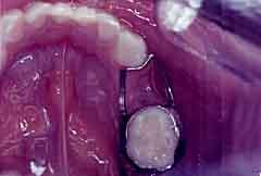
7 Interproximal spaces Triangular shaped spaces between the teeth. In health, these spaces are filled with gingiva called interdental papillae In disease, the tissue no longer fills the space and a void exists between the teeth.
8 Healthy interdental tissue completely fills the interdental space which is also called the cervical embrasure
9 Interdental voids

10 Embrasures function to: Prevent food impaction Reduce occlusal trauma Self- cleansing Permit stimulation to gingiva by frictional massage and protect gingiva.

11 Embrasures Embrasures widen as you move in a posterior direction in the oral cavity.
12 Embrasure Review Anterior Contact area near incisal of teeth Narrow embrasure Posterior Contact area near middle third of teeth Wide embrasure Tall interproximal spaces Short interproximal spaces

13 Adult with contact areas between the teeth. Exception: It is normal for children to have spaces between the teeth, no contact areas on anterior teeth, until the first molars erupt (around 6 years old).

14 Teeth Shapes 14


15 Teeth Shapes Anterior teeth Canines 15


16 Teeth Shapes Premolars Molars 16
17 Curvatures and Alignment of Teeth. Function to: Maintain the teeth within dental arch Protect gingiva and periodontium Disperse occlusal trauma and biting forces. Aid in preventing disease, calculus build-up, bacterial invasion. Increase the life expectancy of the tooth.
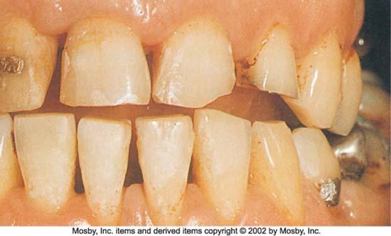
18 Facial & Lingual Contours The FACIAL height of contour (HOC) is in the gingival third of the crown.
19 Lingual Surfaces Anterior teeth HOC is in the cervical third Posterior teeth HOC is located in the middle third closer to the occlusal. 19

20 Height of Contour buccal and lingual 20
21 Terms/Definitions Crest of Curvature - refers to the widest part of the crown of the tooth. Height of Contour(HOC) - same as the crest of curvature, it is the widest part of the crown of the tooth. Curvature of CEJ (cementoenamel junction) the CEJ (cervical line) is a continuous landmark around the circumference of the tooth. The curvature of the CEJ is greater on anterior teeth than it is on posterior teeth.
22 Curvature of the CEJ The more anterior a tooth is located the greater the curvature of the CEJ. This provides more cementum for bony attachment and, thus, more stability.
23 Curvature of the CEJ The mesial curvature is always greater than the distal curvature on the same tooth. Distal curvatures on posterior teeth are very slight. Mesial Distal

24 Line Angles To further specify location on a tooth, the concept of line angles and point angles is utilized. A line angle is the part of a crown where two surfaces intersect forming an imaginary line. Fig

25 Point Angles The intersection of three surfaces of the tooth crown marks a point angle. Fig

26 What is an overhanging restoration? A condition where the margin of a restoration extends far beyond the tooth surface. Usually a rough surface or ledge where food and plaque collect. Can cause : recurrent caries(decay) hard deposit buildup calculus formation periodontal disease


27 May also be detected with an explorer during the clinical exam. Overhangs, if present, may be detected on dental radiographs on the proximal surface, depending on the quality of the radiograph.

28 Recession of the gingiva due to disease opens this space to bacteria, food and debris. Beneath the contact, a space results that was formerly filled with gingiva; that space is called the cervical embrasure. As additional bone is lost, the cervical embrasure widens and enlarges.
29 Are teeth self-cleaning? Consider the following: Shapes of teeth Chewing & eating habits Health of periodontium Missing teeth Teeth, by virtue of the morphology and positioning in the dental arches, are largely selfcleansing. Their shape directs food toward the awaiting digestive system and their smooth enamel surface reduces adherence of food.
30 Self-cleaning properties Enamel is smooth Shape of teeth aid cleaning by stimulating and deflecting food from gingiva Tongue and cheek muscles aid by forcing food onto chewing surface and by helping to remove food particles after eating.
31 Development and Rules of Eruption: Mandibular teeth precede maxillary teeth of the same type (ex: mandibular central incisors erupt before maxillary central incisors). Teeth in both jaws erupt in pairs (one on the right and one on the left erupt at the same time).
32 Rules of Eruption Teeth erupt at an earlier age in girls than in boys. If eruption of primary teeth is at an early age, then eruption of the permanent teeth will be at an early age.
33 Development, Form & Eruption Teeth first develop within the alveolar bone before they erupt in the mouth. Teeth first erupt in the infant around 6 months of age. When teeth erupt in the mouth the roots are not yet fully developed. The roots are the last thing to calcify, after the crown.

34 0 months 2 years 6 months 3 years 9 months 4 years 12 months 5 years 34

35 Tooth Germ tooth bud The tissue that will develop into a tooth. It develops within the alveolar bone and contains the cells that will form the tissues of the teeth (enamel, pulp, dentin, cementum).

36 Tooth Bud 36
37 Calcification when organic tissue becomes hardened by deposits of calcium salts. Occurs over a period of time.
38 Resorption physiological removal of the roots of the primary teeth.
39 Exfoliation shedding or natural loss of primary teeth.
40 Exfoliation 40
41 Stages of Eruption: Preeruptive - begins with development of the crown, calcification. Eruptive - begins with the development of the root. Post eruptive - begins when teeth come into occlusion and continues throughout life.
42 Primary Teeth Table 18-1 P. 263 Approximate Eruption and Shedding Ages for Primary Teeth By age 3 the primary dentition is fully erupted.

43 Order of Eruption of Primary and Permanent Teeth Table 6-22 Pg. 70 and 71
44 Permanent Teeth Appendix D Page 319 Approximate Eruption Schedule & Root Completion for Permanent Teeth First Permanent tooth to erupt!

45 Impacted a tooth that is not completely erupted and is partially or completely covered by bone.

46 Impacted wisdom tooth

47 Impacted 3 rd Molar 47

48 Congenitally Missing condition in which the tooth never developed. Hereditary trait involving missing tooth buds.

49 Missing tooth buds

50 Missing Permanent premolar 50

51 Mesial drift is a phenomenon of the movement of teeth toward the midline. Teeth continue to move mesially after eruption.
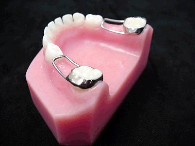
52 Space Maintainers

53 Occlusal Plane- as the teeth erupt they meet the opposing tooth in the opposite arch.

54 Curve of Spee the anatomical line beginning at the tip of canines and following cusps of the premolars and molars.
 that extends onto the occlusal surface Buccal Pit - a depression formed")
55 Landmarks of the Tooth Apex tip of the tooth root Apical Foramen the hole in the apex through which nerves and blood vessels enter and leave the tooth Buccal Groove a groove on the buccal surface of molars (particularly mandibular) that extends onto the occlusal surface Buccal Pit - a depression formed by

56 Cingulum a bulge adjacent to the gingiva on the lingual surface of anterior teeth Cingulum Cusp a pointed or rounded projection on the chewing surface of a tooth Cusp of Carabelli the fifth cusp located on the mesiolingual surface of maxillary first molars

57 Developmental Groove linear depression formed by the joining of tooth lobes during development Fissure a developmental groove where decay often occurs

58 Fossa a shallow depression Furcation space where roots divide in a multirooted tooth
59 Lobes tooth crowns begin in pieces called lobes that fuse together to form the whole (not pictured)

60 Mamelons three bumps on the incisal edge of newly erupted incisors; they usually wear away rapidly Marginal Ridges elevated enamel ridges that form the mesial and distal borders of occlusal surfaces of posterior teeth
61 Oblique Ridge an enamel elevation that crosses the occlusal surface of molars obliquely Pit a depression formed by intersecting grooves

62 Ridge - an elevated portion of the crown of the tooth Supplemental Groove smaller grooves that radiate from the developmental grooves
63 Transverse Ridge the union of two triangular ridges across the occlusal surface of a posterior tooth Triangular Ridge enamel ridge that starts at the cusp tip and widens into a triangle as it descends to the middle of the occlusal surface The End
Morphology of an Anatomic Crown. By: Assistant Professor Dr. Baydaa Ali Al - Rawi
 Morphology of an Anatomic Crown By: Assistant Professor Dr. Baydaa Ali Al - Rawi October 4, 2009 Elevated landmarks Depressed landmarks A) Elevated landmarks : 1. Dental lobe : is one of the primary centers
1. What is the highest and sharpest cusp on the lower first deciduous molar? 2. Which of the following is NOT the correct location of an embrasure?
 1 1. What is the highest and sharpest cusp on the lower first deciduous molar? a. mesiobuccal b. distobuccal c. distolingual d.mesiolingual 2. Which of the following is NOT the correct location of an embrasure?
6610 NE 181st Street, Suite #1, Kenmore, WA
 660 NE 8st Street, Suite #, Kenmore, WA 9808 www.northshoredentalacademy.com.08.900 READ CHAPTER The Professional Dental Assistant (p.-9) No Key Terms Recall Questions:,,,, and 6 CLASS SYLLABUS DAY READ
Lecture 2 Maxillary central incisor
 Lecture 2 Maxillary central incisor Generally The deciduous tooth appears in the mouth at 3 18 months of age, with 6 months being the average and is replaced by the permanent tooth around 7 8 years of
Dental Anatomy High Yield Notes. **Atleast 35 questions comes from these areas of old lectures**
 Dental Anatomy High Yield Notes **Atleast 35 questions comes from these areas of old lectures** This review notes compiled and prepared by my sister for her own study, as a last day review session for
Dental Morphology and Vocabulary
 Dental Morphology and Vocabulary Palate Palate Palate 1 2 Hard Palate Rugae Hard Palate Palate Palate Soft Palate Palate Palate Soft Palate 4 Palate Hard Palate Soft Palate Maxillary Arch (Maxilla) (Uppers)
Primary Teeth Chapter 18. Dental Anatomy 2016
 Primary Teeth Chapter 18 Dental Anatomy 2016 Primary Teeth - Introduction Synonyms deciduous teeth, baby teeth, temporary teeth, milk teeth. There are 20 primary teeth, designated as A thru T in the Universal
Central Incisor DR.Ahmed Al-Jobory B.D.S.,M.Sc. Conservative Department
 Dental Anatomy Lecture 3 Central Incisor DR.Ahmed Al-Jobory B.D.S.,M.Sc. Conservative Department The permanent maxillary Incisors Maxillary incisor are four in number. The maxillary central incisor is
Medical NBDE-II. Dental Board Exams Part I.
 Medical NBDE-II Dental Board Exams Part I http://killexams.com/exam-detail/nbde-ii Question: 149 Anatomically, the term "clinical root" can be defined as which of the following: A. The space in the tooth
ORAL ANATOMY AND PHYSIOLOGY
 CHAPTER 7 ORAL ANATOMY AND PHYSIOLOGY INTRODUCTION This chapter covers the oral anatomy and physiology of the teeth, the histology of the tissues and supporting structures, and concentrates on the external
Arrangement of the artificial teeth:
 Lecture Prosthodontic Dr. Osama Arrangement of the artificial teeth: It s the placement of the teeth on a denture with definite objective in mind or it s the setting of teeth on temporary bases. Rules
CHAPTER 4 ORAL ANATOMY
 CHAPTER 4 ORAL ANATOMY This chapter covers the oral anatomy and physiology of the teeth, the histology of their tissues and supporting structures, and concentrates on the external features of the teeth.
SPRINGFIELD TECHNICAL COMMUNITY COLLEGE ACADEMIC AFFAIRS
 SPRINGFIELD TECHNICAL COMMUNITY COLLEGE ACADEMIC AFFAIRS Course Number: DHYG 103 Department: Dental Hygiene Course Title: Oral Anatomy 1 Semester: Spring Year: 1997 Objectives/ 1. Utilize appropriate clinical
Periodontal Disease. Radiology of Periodontal Disease. Periodontal Disease. The Role of Radiology in Assessment of Periodontal Disease
 Radiology of Periodontal Disease Steven R. Singer, DDS srs2@columbia.edu 212.305.5674 Periodontal Disease! Includes several disorders of the periodontium! Gingivitis! Marginal Periodontitis! Localized
Dental Anatomy and Occlusion
 CHAPTER 53 Dental Anatomy and Occlusion Ma Lou C. Sabino DDS, and Emily G. Smythe, DDS What numerical system is used most commonly in the United States for designating the adult dentition? Pediatric dentition?
Advanced Probing Techniques
 Module 21 Advanced Probing Techniques MODULE OVERVIEW The clinical periodontal assessment is one of the most important functions performed by dental hygienists. This module begins with a review of the
Oral Embryology and Histology
 Oral Embryology and Histology Chapter 8 Copyright 2018, Elsevier Inc. All Rights Reserved. 1 Learning Objectives Lesson 8.1: Oral Embryology 1. Pronounce, define, and spell the key terms. 2. Define embryology
1B Getting Ready for Instrumentation: Mathematical Principles and Anatomic Descriptors
 MODULE 1B Getting Ready for Instrumentation: Mathematical Principles and Anatomic Descriptors Module Overview This module contains a review of the mathematical principles and anatomic descriptors used
The Premolars. Chapter 17 Permanent Posterior Teeth (p )
") The Premolars Chapter 17 Permanent Posterior Teeth (p. 230-244) General Information Function: u Hold and grind food u Work with molars in mastication. u Even without molars one may be able to chew well
The Premolars Chapter 17 Permanent Posterior Teeth (p. 230-244) General Information Function: u Hold and grind food u Work with molars in mastication. u Even without molars one may be able to chew well
Lecture. Permanent maxillary premolars
 Lecture Permanent maxillary premolars Permanent premolars The maxillary premolars are four in number: two in the right and two in the left. They are posterior to the canines and anterior to the molars.
Tooth Variations. Suruedee Chinthakanan
 Tooth Variations Suruedee Chinthakanan Tooth variations Dental anomalies Cause : hereditary factor Developmental disturbances of teeth www.ectodermaldysplsia.org Tooth variations Enamel is formed from
Eruption and Shedding of Teeth
 Eruption and Shedding of Teeth Mixed Dentition: Presence of both dentitions Figure from Ten Cate s Oral Histology, Ed., Antonio Nanci, 6 th edition Tooth eruption is the process by which developing teeth
Arrangement of posterior artificial teeth Standardized parameters Curve of Wilson Curve of Spee
 . Arrangement of posterior artificial teeth Posterior teeth are set up in tight centric occlusion. The mandibular teeth are set in the wax occlusion rim over the residual ridge in their ideal buccolingual
Key points for starting off
 Key points for starting off First off, the five questions to ask yourself about a loose tooth before identifying it are: 1. 2. 3. 4. 5. Category (incisor, canine, premolar or molar)? Permanent or deciduous?
Permanent 2 nd Maxillary Molars
 Permanent 2 nd Maxillary Molars In comparison to the first max molar First molars appears in the oral cavity at the age of 6 years old.. While 2 nd molar 3 rd molar Max. 2 nd molar have long roots (sometimes
Lec. 3-4 Dr. Saif Alarab Clinical Technique for Class I Amalgam Restorations The outline form
 Lec. 3-4 Dr. Saif Alarab Clinical Technique for Class I Amalgam Restorations Class I refers to -Restorations on the occlusal surfaces of posterior teeth, - The occlusal two thirds of facial and lingual
NEW YORK CITY COLLEGE OF TECHNOLOGY DEPARTMENT OF THE CITY UNIVERSITY OF NEW YORK RESTORATIVE DENTISTRY
 NEW YORK CITY COLLEGE OF TECHNOLOGY DEPARTMENT OF THE CITY UNIVERSITY OF NEW YORK RESTORATIVE DENTISTRY DEPARTMENT: COURSE CODE: COURSE TITLE: COURSE DESCRIPTION: CLASS HOURS & CREDITS: NUMBER OF WEEKS:
Indications The selection of amalgam as a restorative material for class V cavity should involve the following considerations:
 1 Lec.7 د.عبد املنعم اخلفاجي CLASS V CAVITY PREPARATION FOR AMAGLAM Indications The selection of amalgam as a restorative material for class V cavity should involve the following considerations: 1- Caries:
Tooth eruption and movement
 Tooth eruption and movement Dr. Krisztián Nagy Diphydont dentition Deciduous dentition primary dentition Diphydont dentition Permanent dentition secondary dentition Mixed Dentition: Presence of both dentitions
Attachment G. Orthodontic Criteria Index Form Comprehensive D8080. ABBREVIATIONS CRITERIA for Permanent Dentition YES NO
 First Review IL HFS Dental Program Models Second Review Ortho cad Attachment G Orthodontic Criteria Index Form Comprehensive D8080 Ceph Film X-Rays Photos Narrative Patient Name: DOB: ABBREVIATIONS CRITERIA
PERMANENT MANDIBULAR INCISORS
 PERMANENT MANDIBULAR INCISORS (Central and Lateral) DR.AHMED AL-JOBORY LEC. 5 PERMANENT MANDIBULAR INCISORS ARE 4 IN NUMBER : 2 CENTRAL (RIGHT &LEFT) AND 2 LATERAL INCISORS (RIGHT &LEFT). CHARACTERISTIC
Texas A&M College of Dentistry Caruth School of Dental Hygiene
 Texas A&M College of Dentistry Caruth School of Dental Hygiene Course Number and Name: 3120 Dental Anatomy Course Type: Lecture Laboratory Seminar Academic Year/Semester Offered: 2016/Fall Semester Course
Prosthetic Options in Implant Dentistry. Hakimeh Siadat, DDS, MSc Associate Professor
 Prosthetic Options in Dentistry Hakimeh Siadat, DDS, MSc Associate Professor Dental Research Center, Department of Prosthodontics & Dental s Faculty of Dentistry, Tehran University of Medical Sciences
Development of occlusion
 Development of occlusion The development of dentition is an important part of craniofacial growth as the formation, eruption, exfoliation and exchange of teeth take place during this period. Term occlusion
1. Anterior-posterior movement of the mandible (APM):
:") Dental Anatomy A. Terminology: Over the past few years there have been several acronyms introduced into the equine dentistry industry. These terms should help you describe and understand equine dental
Dental Anatomy A. Terminology: Over the past few years there have been several acronyms introduced into the equine dentistry industry. These terms should help you describe and understand equine dental
Radiology. & supporting structures. Lec. 14 Common diseases of teeth Dr. Areej
 Radiology Lec. 14 Common diseases of teeth Dr. Areej & supporting structures A radiograph is only one part of the diagnostic process. Usually one does NOT make a diagnosis solely from a radiograph. A diagnosis
Concepts of occlusion Balanced occlusion. Monoplane occlusion. Lingualized occlusion. Figure (10-1)
") Any contact between teeth of opposing dental arches; usually, referring to contact between the occlusal surface. The static relationship between the incising or masticatory surfaces of the maxillary or
Any contact between teeth of opposing dental arches; usually, referring to contact between the occlusal surface. The static relationship between the incising or masticatory surfaces of the maxillary or
Dental Anatomy and Physiology for Clinical Dental Technicians. with Marnie Hayward
 Dental Anatomy and Physiology for Clinical Dental Technicians with Marnie Hayward Salivary glands Parotid Submandibular Sublingual Salivary glands position Parotid glands Lie below ear and behind angle
Upper arch. 1Prosthodontics. Dr.Bassam Ali Al-Turaihi. Basic anatomy & & landmark of denture & mouth
 1Prosthodontics Lecture 2 Dr.Bassam Ali Al-Turaihi Basic anatomy & & landmark of denture & mouth Upper arch Palatine process of maxilla: it form the anterior three quarter of the hard palate. Horizontal
Tooth and Surface Identification (TID and SID)
") Tooth and Surface Identification (TID and SID) Dental treatment documentation and billing require to properly identify teeth and tooth surfaces. Incorrect TID and SID are frequent reasons for claim denial
Tooth and Surface Identification (TID and SID) Dental treatment documentation and billing require to properly identify teeth and tooth surfaces. Incorrect TID and SID are frequent reasons for claim denial
أ.م. هدى عباس عبد اهلل CROWN AND BRIDGE جامعة تكريت كلية. Lec. (2) طب االسنان
 طب االسنان") Lec. (2) CROWN AND BRIDGE أ.م. هدى عباس عبد اهلل Patient selection and examination A thorough diagnosis must first be made of the patient's dental condition, considering both hard and soft tissues. this
Lec. (2) CROWN AND BRIDGE أ.م. هدى عباس عبد اهلل Patient selection and examination A thorough diagnosis must first be made of the patient's dental condition, considering both hard and soft tissues. this
An Overview of Dental Anatomy
 Continuing Education Brought to you by An Overview of Dental Anatomy Course Author(s): Vickie Parrish Foster, RDH, MEd CE Credits: 1 hour Intended Audience: Dental Hygienists, Dental Assistants, Dental
FIRST YEAR RDS 111 DENTAL ANATOMY, MORPHOLOGY AND INTRODUCTION TO OPERATIVE DENTISTRY. Course Director (DUC) PROF. ALI M. EL-SAHN
 PROF. ALI M. EL-SAHN") FIRST YEAR RDS 111 DENTAL ANATOMY, MORPHOLOGY AND INTRODUCTION TO OPERATIVE DENTISTRY Course Director (DUC) PROF. ALI M. EL-SAHN Course Contributors: Dr. M. Benhameurlaine, Dr. T. Al Qunaian, Dr. Abdullah
FIRST YEAR RDS 111 DENTAL ANATOMY, MORPHOLOGY AND INTRODUCTION TO OPERATIVE DENTISTRY Course Director (DUC) PROF. ALI M. EL-SAHN Course Contributors: Dr. M. Benhameurlaine, Dr. T. Al Qunaian, Dr. Abdullah
part TWO Communication
 part TWO Communication chapter FOUR Dental Terminology OBJECTIVES After completing this chapter, you should be able to do the following: Spell and define key terms Discuss the purposes of teeth Identify
NATIONAL EXAMINING BOARD FOR DENTAL NURSES
 NATIONAL EXAMINING BOARD FOR DENTAL NURSES NATIONAL DIPLOMA EXAMINATION DENTAL CHARTING NEBDN is a limited company registered in England & Wales No. 5580200 Registered with the Charity Commisioners No.
ANATOMY OF THE PERIODONTIUM. Dr. Fatin Awartani
 ANATOMY OF THE PERIODONTIUM Part II Cementum and Alveolar bone Associate Professor Periodontal division King Saud university Cementum Calcified mesenchymal tissue that forms the outer covering of the anatomic
Intrusion of Incisors to Facilitate Restoration: The Impact on the Periodontium
 Note: This is a sample Eoster. Your EPoster does not need to use the same format style. For example your title slide does not need to have the title of your EPoster in a box surrounded with a pink border.
ORTHODONTICS Treatment of malocclusion Assist.Lec.Kasem A.Abeas University of Babylon Faculty of Dentistry 5 th stage
 Lec: Treatment of class I malocclusion Class I occlusion can be defined by Angles, classification as the mesiobuccal cusp of the upper 1 st permanent molar occlude with the developmental groove of the
Educational Training Document
 Educational Training Document Table of Contents Part 1: Resource Document Disclaimer Page: 2 Part 2: Line Item Grade Sheets Page: 3 Release: 11/2016 Page 1 of 6 Part 1: Resource Document Disclaimer The
Smile Design. Daniel H Ward DDS 1080 Polaris Pkwy Ste 130 Columbus OH
 Smile Design Daniel H Ward DDS 1080 Polaris Pkwy Ste 130 Columbus OH 43240 614-430-8990 dward@columbus.rr.com Incisal Placement Incisal plane parallel to the interpupillary line Vertical midline in the
An Overview of Dental Anatomy
 An Overview of Dental Anatomy Vickie P. Overman, RDH, MEd Continuing Education Units: 1 hour Online Course: www.dentalcare.com/en-us/professional-education/ce-courses/ce500 Disclaimer: Participants must
TASKS. 2. Apply a disclosing agent to make the plaque visible.
 TASKS EQUIPMENT AND MATERIALS Personal protective equipment Assortment of nylon, soft-bristled toothbrushes Assortment of dental Floss, waxed and nonwaxed Disclosing solution Face mirror 3. Demonstrate
IMPACTED CANINES. Unfortunately, this important tooth is the second most common tooth to be impacted after third molars
 IMPACTED CANINES After we talked about impacted third molars, today we ll discuss about maxillary impacted canines in upper dental arch, how to manage these cases as a dental surgeon. You will study about
Development of teeth. 5.DM - Pedo
 Development of teeth 5.DM - Pedo Tooth development process of continuous changes in predetermined order starts from dental lamina A band of ectodermal cells growing from the epithelium of the embryonic
CLASS II CAVITY PREPARATION CHARACTERISTICS OF AN IDEAL CLASS II
 CLASS II CAVITY PREPARATION CHARACTERISTICS OF AN IDEAL CLASS II Contact area carious lesion Proximal view Vertical section - Buccal view Class II carious lesions are diagnosed using bitewing radiographs.
Appendix 4. Case Summary Template. Social history. Patient (Name and Date of birth): Presenting complaints. History Medical history
: Presenting complaints. History Medical history") Appendix 4 Case Summary Template Patient (Name and Date of birth): Presenting complaints Chief complaint Subsidiary complaints History of complaints Patient s expectations History Medical history Medical
Appendix 4 Case Summary Template Patient (Name and Date of birth): Presenting complaints Chief complaint Subsidiary complaints History of complaints Patient s expectations History Medical history Medical
PLATE 1. Outline Form
 PLATE 1 Outline Form A. A standard radiograph (left) in buccolingual projection provides only a two-dimensional view of what is actually a three-dimensional problem. If a mesiodistal x-ray projection could
Plaque and Occlusion in Periodontal Disease Wednesday, February 25, :54 AM
 Plaque and Occlusion in Periodontal Disease Wednesday, February 25, 2015 9:54 AM 1. The definition of Trauma From Occlusion: Primary TFO, Secondary TFO, and Combined TFO 2. Clinical and Radiographic signs
Treatment planning of nonskeletal problems. in preadolescent children
 In the name of GOD Treatment planning of nonskeletal problems in preadolescent children Presented by: Dr Somayeh Heidari Orthodontist Reference: Contemporary Orthodontics Chapter 7 William R. Proffit,
Structure of an Incisor
 MAMMALIAN TEETH Mammals have different types and shapes of teeth and they are thus termed Heterodonts. Those which have teeth of the same size and shapes are termed as Homodonts. In mammals teeth consist
Dr.Adel F.Ibraheem Partial Veneer Crown(Three quarter crown) Three quarter (¾ )crown: Uses: Indications ---- For posterior teeth ;
 Three quarter (¾ )crown: Uses: Indications ---- For posterior teeth ;") Lecture.9 Dr.Adel F.Ibraheem Partial Veneer Crown(Three quarter crown) *It is a cast metal crown restoration that cover only a part of the clinical crown, most commonly used type of partial veneer crown
Lecture.9 Dr.Adel F.Ibraheem Partial Veneer Crown(Three quarter crown) *It is a cast metal crown restoration that cover only a part of the clinical crown, most commonly used type of partial veneer crown
ALABAMA DENTAL HYGIENE PROGRAM 50 QUESTIONS PRE ENTRANCE EXAM
 ALABAMA DENTAL HYGIENE PROGRAM 50 QUESTIONS PRE ENTRANCE EXAM NAME DATE RETURN COMPLETED EXAM WITH APPLICATION You may copy for future reference 1. One cause of decay is the Streptococcus mutans bacteria
14/09/15. Assessment of Periodontal Disease. Outline. Why is Periodontal assessment needed? The Basics of Periodontal assessment
 Assessment of Periodontal Disease Dr Wendy Turner Outline Why is Periodontal assessment needed? The Basics of Periodontal assessment Probing: Basic Periodontal Examination for adults and children. Detailed
Position and Structure of Human Teeth *
 OpenStax-CNX module: m66077 1 Position and Structure of Human Teeth * Marcos Gridi-Papp This work is produced by OpenStax-CNX and licensed under the Creative Commons Attribution License 4.0 Abstract Humans
Common Equine Dental Malocclusions Molars
 Common Equine Dental Malocclusions Molars CAUDAL HOOKS Definition - Dominant lower or upper last molar overhanging opposing molar. Etiology - Hereditary - May result from horse born with over or under
Periapical Radiography
 Periapical Radiography BARBARA E. DIXON B.D.S., M.Sc., D.P.D.S. Main Indications Detection of Apical infection/inflammation Assessment of the periodontal status After trauma Assessment of Unerupted teeth
European Veterinary Dental College
 European Veterinary Dental College EVDC Training Support Document Preparation of Radiograph Sets (Cat and Dog) Document version : evdc-tsd-radiograph_positioning_(dog_and_cat)-20120121.docx page 1 of 13
Preclinical Dentistry. I. Dental Caries Non carious lesions: trauma, erosion. abrasion, wedge shaped defects. Lenka Roubalíková
 Preclinical Dentistry I. Dental Caries Non carious lesions: trauma, erosion. abrasion, wedge shaped defects Lenka Roubalíková Understanding dental caries crown pulp chamber neck root canal root Dental
BASCD Trainers Pack for Caries Prevalence Studies. Updated: June 2014 for UK Training & Calibration exercise for the Deciduous Dentition
 BASCD Trainers Pack for Caries Prevalence Studies Updated: June 2014 for UK Training & Calibration exercise for the Deciduous Dentition Prepared by Helen Paisley, Cynthia Pine and Girvan Burnside Administrative
Principles of. By: Dr. Ahmad Rabah
 Principles of By: Dr. Ahmad Rabah 1. Utilize what's present: Whenever possible, select a design that fits the teeth and soft tissues, rather than choosing one that requires tissue alteration. When minimal
OCCLUSION. Principles & Treatment. José dos Santos, Jr, DDS, PhD. São Paulo, Brazil
 OCCLUSION Principles & Treatment José dos Santos, Jr, DDS, PhD São Paulo, Brazil Former Professor Division of Occlusion Department of Restorative Dentistry University of Texas Health Science Center at
Morphological Scoring of Dental Casts Using the Arizona State University Dental Anthropology System
 University of Tennessee, Knoxville Trace: Tennessee Research and Creative Exchange University of Tennessee Honors Thesis Projects University of Tennessee Honors Program 5-2004 Morphological Scoring of
T O O T H A T L A S C O U R S E G U I D E A S S I S T A N T E D I T I O N
 T O O T H A T L A S C O U R S E G U I D E A S S I S T A N T E D I T I O N The information in this guide was prepared by ehuman with contributions from: Cara Miyasaki, RDHEF, MS, Foothill College Kay Murphy,
MAXILLARY INJECTION TECHNIQUE. Chinthamani Laser Dental Clinic
 MAXILLARY INJECTION TECHNIQUE Chinthamani Laser Dental Clinic Introduction A number of injection techniques are available to aid in providing clinically adequate anesthesia of the teeth and soft and hard
#45 Ortho-Tain, Inc PREVENTIVE ERUPTION GUIDANCE -- PREVENTIVE OCCLUSAL DEVELOPMENT
 #45 Ortho-Tain, Inc. 1-800-541-6612 PREVENTIVE ERUPTION GUIDANCE -- PREVENTIVE OCCLUSAL DEVELOPMENT Analysis and Diagnosis of Occlusion: The ideal child of 5 y ears of age that probably has the best chance
Development of occlusion:
 : Dr.Issam Aljorani (BDS, MSc. Ortho.) Postnatal development of the dentition When a child is born, mineralization of all the primary tooth crowns is well underway, with this process also beginning in
Proceedings of the 12th International Congress of the World Equine Veterinary Association WEVA
 www.ivis.org Proceedings of the 12th International Congress of the World Equine Veterinary Association WEVA November 2-5, 2011 Hyderabad, India Reprinted in IVIS with the Permission of WEVA Organizers
Dr.Sepideh Falah-kooshki
 Dr.Sepideh Falah-kooshki MAXILLA Premaxillary/median palatal suture (radiolucent). Incisive fossa and foramen (radiolucent). Nasal passages (radiolucent). Nasal septum (radiopaque). Anterior nasal spine
Elevators. elevators:- There are three major components of the elevator are:-
 Elevators Elevators:- Are exo-levers, instrument designed to elevate or luxate the teeth or roots from their bony socket in close or surgical method of extraction to force a tooth or root along the line
Techniques of local anesthesia in the mandible
 Techniques of local anesthesia in the mandible The technique of choice for anesthesia of the mandible is the block injection and this is attributed to the absence of the advantages which are present in
You know you would like to stop swearing at the computer after each shot. Troubleshooting oral radiography
 You know you would like to stop swearing at the computer after each shot Troubleshooting oral radiography Goals of oral radiology Achieve diagnostic images of the teeth and surrounding bone. Images should
Amalgam restoration of posterior proximal cavities with deep and concave gingival outlines
 Amalgam restoration of posterior proximal cavities with deep and concave gingival outlines M. Darveniza, MDSc, FRACDS* Abstract A technique for making a matrix and wedges for amalgam restoration of deep
Our Teeth. History Of Equine Dentistry EQUINE DENTISTRY. Who Should Do Equine Dentistry? Some Facts To Know About Teeth
 EQUINE DENTISTRY Mike Black, DVM Nebraska Equine Veterinary Clinic Omaha NE History Of Equine Dentistry Some evidence of equine dentistry dates back to 2,000 B.C. Bit-Seats: Information on bit-seats dates
Aggressive Periodontitis Monday, August 31, 2015
 Aggressive Periodontitis Monday, August 31, 2015 8:01 AM Biofilm levels of dicernable plaque would match the disease state of the patient Supra and sub gingival calculus Anticipate gram - anaerobes but
DENT Advanced Topics in Removable Prosthodontics, Winter 2008
 University of Michigan Deep Blue deepblue.lib.umich.edu 2008-01 DENT 718 - Advanced Topics in Removable Prosthodontics, Winter 2008 Shotwell, Jeffrey Shotwell, J. (2008, April 23) Advanced Topics in Removable
Everything You Wanted to Know About Extractions but Were Afraid to Ask
 Everything You Wanted to Know About Extractions but Were Afraid to Ask Tooth extraction is a surgical procedure with serious potential complications and should only be performed by a trained veterinarian.
Chapter 20: Pathology
 : Pathology There are many pathologic conditions, which occur in and around the mouth. Some of them have already been discussed in Chapters 7, 13, 15, 16, 17, 18 and 19. This chapter will cover a few more
AAO / AAPD Scottsdale 2018
 AAO / AAPD Scottsdale 2018 Missing Premolars : What are the Options? David Kennedy Clinical Professor UBC Vancouver Canada drdavidkennedy@yahoo.ca At what age can you know second premolars are absent?
Clinical UM Guideline
 Clinical UM Guideline Subject: Clinical Crown Lengthening Guideline #: 04-206 Current Effective Date: 03/24/2017 Status: New Last Review Date: 02/08/2017 Description This document addresses the procedure
CLINICAL APPLICATION GUIDE DIAGNOSTIC INSTRUMENTS & PERIODONTAL SCALERS/ CURETTES
 CLINICAL APPLICATION GUIDE DIAGNOSTIC INSTRUMENTS & PERIODONTAL SCALERS/ CURETTES DIAGNOSTIC INSTRUMENTATION EXPLORERS Explorers are used to examine tooth surfaces for calculus, decalcified and carious
Alveolar Growth in Japanese Infants: A Comparison between Now and 40 Years ago
 Bull Tokyo Dent Coll (2017) 58(1): 9 18 Original Article doi:10.2209/tdcpublication.2016-0500 Alveolar Growth in Japanese Infants: A Comparison between Now and 40 Years ago Hiroki Imai 1), Tetsuhide Makiguchi
Figure (2-6): Labial frenum and labial notch.
: Labial frenum and labial notch.") The anatomy of the edentulous ridge in the maxilla and mandible is very important for the design of a complete denture. The consistency of the mucosa and architecture of the underlying bone is different
The anatomy of the edentulous ridge in the maxilla and mandible is very important for the design of a complete denture. The consistency of the mucosa and architecture of the underlying bone is different
2. Gap closure and replacement of the missing tooth 35 with directly modelled bridge region 34-36
 GrandTEC Test Kit Dear User, This Test Kit has been put together to enable you to test GrandTEC on the model before using it in a clinical situation. GrandTEC is a resin-impregnated glass fibre strip.
PREMATURE PRIMARY TOOTH LOSS
 Disclaimer This movie is an educational resource only and should not be used to manage your dental health. All decisions about the management of premature primary tooth loss must be made in conjunction
Clinical UM Guideline
 Clinical UM Guideline Subject: Non-Medically Necessary Orthodontia Care Guideline #: #08-002 Current Publish Date: 10/16/2017 Status: Reviewed Last Review Date: 10/11/2017 Description This document addresses
6/8/12 THE LEGEND OF WORMS
 1 INTRODUCTION DEFINITION EARLY THEORIES Legend of worms Humoral theory Vital theory Chemical theory Parasitic theory Chemicoparasitic theory Proteolytic theory Proteolysis- chelation theory Sucrose chelation
Digestion. Final Products of Digestion Carbohydrates Disaccharides (sucrose, maltose) Proteins Short polypeptides Individual amino acids
 Proteins Short polypeptides Individual amino acids") Digestion Process has three stages: 1. Physical Digestion: mechanical process of breaking down the food. Knives and forks, teeth grinding and tearing 2. Chemical Digestion: enzymes and stomach acid breaks
Digestion Process has three stages: 1. Physical Digestion: mechanical process of breaking down the food. Knives and forks, teeth grinding and tearing 2. Chemical Digestion: enzymes and stomach acid breaks
The periodontium attempts to accommodate to the forces exerted to the crown. This adaptive capacity varies in different persons and in the same person
 The periodontium attempts to accommodate to the forces exerted to the crown. This adaptive capacity varies in different persons and in the same person at different times. The effect of occlusal forces
RETENTION AND RELAPSE
 RETENTION AND RELAPSE DEFINITION Maintaining newly moved teeth long enough to aid in stabilizing their correction MOYERS loss of any correction achieved by any orthodontic treatment RELAPSE CAUSES OF RELAPSE
Principles of Periodontal flap surgery. Dr.maryam khosravi
 Principles of Periodontal flap surgery Dr.maryam khosravi Goals of periodontal SURGICAL phase 1 - Controlling or eliminating periodontal disease. 2 Correcting anatomic conditions that may a. favor periodontal