ANATOMY OF THE PERIODONTIUM. Dr. Fatin Awartani
|
|
|
- Marjorie Baker
- 6 years ago
- Views:
Transcription
1 ANATOMY OF THE PERIODONTIUM
2 Part II Cementum and Alveolar bone Associate Professor Periodontal division King Saud university
3 Cementum
4 Calcified mesenchymal tissue that forms the outer covering of the anatomic root
5 Cementum: is calcified tissue that covers the root of the tooth and provides a means of attachments for the periodontal ligament fibers to the tooth. It consists of calcified collagen fibers and interfibriller ground substance. It is made up of 45% to 50% inorganic material and 50% to 55% organic matter and water.
6 Width varies from 16 to 60 microns in the coronal half of the root and 150 to 200 microns in the apical third of the root. Width increases with age, 95 microns at age 20 and 215 microns at age 60.
7 Cementum What are the sources of collagen fibers in cementum? Extrinsic sharpeys fibers formed by Fibroblasts. Intrinsic Fibers of the cementum matrix formed by cement oblasts.
8 Two types of cementum acellular and cellular acellular cementum is found on the coronal areas of the root. cellular cementum is found in the apical areas of the roots and in the furcation areas of multirooted teeth.

9 TWO MAIN FORMS OF CEMENTUM ACELLULAR - first to be formed covers approximately the cervical third of half of the root does not contain cells formed before the tooth reaches occlusal plane sharpey s fibers make up most of the structure of acellular cmentum

10 CELLULAR formed after tooth reaches occlusal plane contains cells in lacunae less calcified than acellular more common on apical half of tooth greatest increase with age is cellular type in apical half of root
11 Types of Cementum Schroeder's Classification Acellular Afibrillar Cementum Acellular Extrinsic Fiber Cellular intrinsic Fiber cementum. Cellular mixed stratified cementum Intermediate Cementum
12 Cementoenamel junction: the area where enamel and cementum meet at the cervical region of the tooth. Three different relationships among the enamel and cementum: 60% to 65% of the cases the cementum overlaps the enamel 30% of the cases edge to edge 5% to 10% cementum fails to meet enamel resulting in exposed dentin
13 CEMENTOENAMEL JUNCTION

14 Alveolar Bone
15 Alveolar bone: are the parts of the maxilla and mandible providing the housing for the roots of the teeth. Bone that forms and support tooth sockets
 2- trabecular bone 3- compact")
16 Alveolar bone: 1- alveolar bone proper (lamina dura in radiographs) 2- trabecular bone 3- compact bone
17 1)Alveoli: The space in the alveolar bone that accommodate the roots of the teeth (tooth socket).
18 Alveoli: covered lined with a layer of bone know as alveolar bone proper or the cribriform plate. This layer of bone shows as a white line on radiographs and called lamina dura. This layer also covers the crest of interproximal bone and called crestal lamina dura.
19 2)Supporting alveolar bone: cancellous and cortical bone that surrounds the alveolar bone proper
20 3)Interproximal bone (interdental septum): bone located between the roots of adjacent teeth. 4)Interradicular bone: bone located between the roots of multirooted teeth

21 5)Radicular bone: alveolar process located on the facial or lingual surfaces of the roots of teeth
22 CELLS OF ALVEOLAR BONE Calcified matrix with osteocytes enclosed in lacunae Constantly changing Osteoblasts deposit Osteoclasts resorb Matrix deposited by osteoblasts is not mineralized and is termed osteoid. As new osteoid is deposited the old osteoid mineralizes. Osteoclasts are large multinucleated cells that are often on surface or in Howship s lacunae. Main function is resorption of bone.

23 FENESTRATION - isolated areas which the root is denuded of bone and root surface is covered only by periosteum and overlying gingiva DEHISCENCE - denuded areas extend through the marginal bone

24 Fenestration: some bone present in the most coronal portion Dehiscence: the bone coverage is missing at the coronal portion of the roots
25 Summary: Periodontium consists of 4 different tissues: Gingiva Cementum PDL Alveolar bone They are anatomically separate, but functionally, they all depends on another in maintaining a viable, healthy supporting structure for the tooth.
Periodontal ligament
 Periodontal ligament The periodontium The periodontium includes: The gingiva Cementum Periodontal ligament Alveolar bone Def: The periodontal ligament is the dense fibrous connective tissue that occupies
PERIODONTAL REVIEW DENTALELLE TUTORING ANDREA TWAROWSKI
 PERIODONTAL REVIEW DENTALELLE TUTORING ANDREA TWAROWSKI Periodontics Questions 1. What is the periodontium composed of? 2. What type of mucosa is the gingiva composed of? 3. Is it normal to have melanin
Oral Histology. Alveolar bone or process: Functions of alveolar bone: Chemical composition: Development of the alveolar process: Dr.
 Oral Histology Lec.12 Alveolar bone or process: Dr. Nada Al-Ghaban Alveolar bone is a specialized part of the mandibular and maxillary bones that forms the primary support structure for teeth. Although
Oral Embryology and Histology
 Oral Embryology and Histology Chapter 8 Copyright 2018, Elsevier Inc. All Rights Reserved. 1 Learning Objectives Lesson 8.1: Oral Embryology 1. Pronounce, define, and spell the key terms. 2. Define embryology
Periodontal Disease. Radiology of Periodontal Disease. Periodontal Disease. The Role of Radiology in Assessment of Periodontal Disease
 Radiology of Periodontal Disease Steven R. Singer, DDS srs2@columbia.edu 212.305.5674 Periodontal Disease! Includes several disorders of the periodontium! Gingivitis! Marginal Periodontitis! Localized
Pre op Failed endodontic treatment with sinus involvement.
 Case #1 of 10 consecutive extraction sockets grafted with Socket Graft Putty, covered with Socket Seal and sealed with Periacryl. I D # HEU This patient is a 66 year old female. Pre op Failed endodontic
Eruption and Shedding of Teeth
 Eruption and Shedding of Teeth Mixed Dentition: Presence of both dentitions Figure from Ten Cate s Oral Histology, Ed., Antonio Nanci, 6 th edition Tooth eruption is the process by which developing teeth
06 Tooth Development and Eruption
 + 06 Tooth Development and Eruption Tooth development Root development PDL and alveolar bone development Primary tooth eruption and shedding Permanent tooth eruption Q. Where and how tooth starts to form?
Fundamental & Preventive Curvatures of Teeth and Tooth Development. Lecture Three Chapter 15 Continued; Chapter 6 (parts) Dr. Margaret L.
 Dr. Margaret L.") Fundamental & Preventive Curvatures of Teeth and Tooth Development Lecture Three Chapter 15 Continued; Chapter 6 (parts) Dr. Margaret L. Dennis Proximal contact areas Contact areas are on the mesial and
Fundamental & Preventive Curvatures of Teeth and Tooth Development Lecture Three Chapter 15 Continued; Chapter 6 (parts) Dr. Margaret L. Dennis Proximal contact areas Contact areas are on the mesial and
Dental Assisting COURSE ONE
 Dental Assisting COURSE ONE 1 History of Dentistry False teeth used to be made of wood or ivory Often painful dental treatments were done by "guzzling some vodka" Wilhelmina Conrad Roentgen discovered
BONE TISSUE. Dr. Heba Kalbouneh Associate Professor of Anatomy and Histology
 BONE TISSUE Dr. Heba Kalbouneh Associate Professor of Anatomy and Histology BONE FUNCTION Support Protection (protect internal organs) Movement (provide leverage system for skeletal muscles, tendons, ligaments
Morphology of periodontal defects, indications of periodontal surgery
 Morphology of periodontal defects, indications of periodontal surgery Dr. Ferenc Dőri PhD Semmelweis University Dept. of Periodontology Periodontium Gingiva + Cementum PDL (periodontal ligament) Alveolar
Advanced Probing Techniques
 Module 21 Advanced Probing Techniques MODULE OVERVIEW The clinical periodontal assessment is one of the most important functions performed by dental hygienists. This module begins with a review of the
Medical NBDE-II. Dental Board Exams Part I.
 Medical NBDE-II Dental Board Exams Part I http://killexams.com/exam-detail/nbde-ii Question: 149 Anatomically, the term "clinical root" can be defined as which of the following: A. The space in the tooth
Biology. Dr. Khalida Ibrahim
 Biology Dr. Khalida Ibrahim BONE TISSUE Bone tissue is a specialized form of connective tissue and is the main element of the skeletal tissues. It is composed of cells and an extracellular matrix in which
Radiology. & supporting structures. Lec. 14 Common diseases of teeth Dr. Areej
 Radiology Lec. 14 Common diseases of teeth Dr. Areej & supporting structures A radiograph is only one part of the diagnostic process. Usually one does NOT make a diagnosis solely from a radiograph. A diagnosis
ORAL ANATOMY AND PHYSIOLOGY
 CHAPTER 7 ORAL ANATOMY AND PHYSIOLOGY INTRODUCTION This chapter covers the oral anatomy and physiology of the teeth, the histology of the tissues and supporting structures, and concentrates on the external
Dental Morphology and Vocabulary
 Dental Morphology and Vocabulary Palate Palate Palate 1 2 Hard Palate Rugae Hard Palate Palate Palate Soft Palate Palate Palate Soft Palate 4 Palate Hard Palate Soft Palate Maxillary Arch (Maxilla) (Uppers)
Quiz 6. Cartilage and Bone
 Quiz 6 Cartilage and Bone MCQs X type (true or false): 1. Cartilage tissue: a. Has a rich blood supply. b. Develops from mesenchyme. c. Has ability for a quick regeneration. d. Has chondrocytes as precursor
Tooth eruption and movement
 Tooth eruption and movement Dr. Krisztián Nagy Diphydont dentition Deciduous dentition primary dentition Diphydont dentition Permanent dentition secondary dentition Mixed Dentition: Presence of both dentitions
The treatment of destructive periodontal disease, due to specific periodontopathic
 1 1 INTRODUCTION The treatment of destructive periodontal disease, due to specific periodontopathic bacteria, aims at the regeneration of a periodontal attachment composed of new cementum, alveolar bone
14/09/15. Assessment of Periodontal Disease. Outline. Why is Periodontal assessment needed? The Basics of Periodontal assessment
 Assessment of Periodontal Disease Dr Wendy Turner Outline Why is Periodontal assessment needed? The Basics of Periodontal assessment Probing: Basic Periodontal Examination for adults and children. Detailed
CHAPTER 4 ORAL ANATOMY
 CHAPTER 4 ORAL ANATOMY This chapter covers the oral anatomy and physiology of the teeth, the histology of their tissues and supporting structures, and concentrates on the external features of the teeth.
Dental Anatomy and Physiology for Clinical Dental Technicians. with Marnie Hayward
 Dental Anatomy and Physiology for Clinical Dental Technicians with Marnie Hayward Salivary glands Parotid Submandibular Sublingual Salivary glands position Parotid glands Lie below ear and behind angle
PREVALENCE AND CHARACTERIZATION OF ALVEOLAR DEHISCENCES AND FENESTRATIONS SEEN IN A SAMPLE OF SOUTH AFRICAN HUMAN
 PREVALENCE AND CHARACTERIZATION OF ALVEOLAR DEHISCENCES AND FENESTRATIONS SEEN IN A SAMPLE OF SOUTH AFRICAN HUMAN SKULLS. SANDRA KOUTRAS A research report submitted to the Faculty of Health Sciences, University
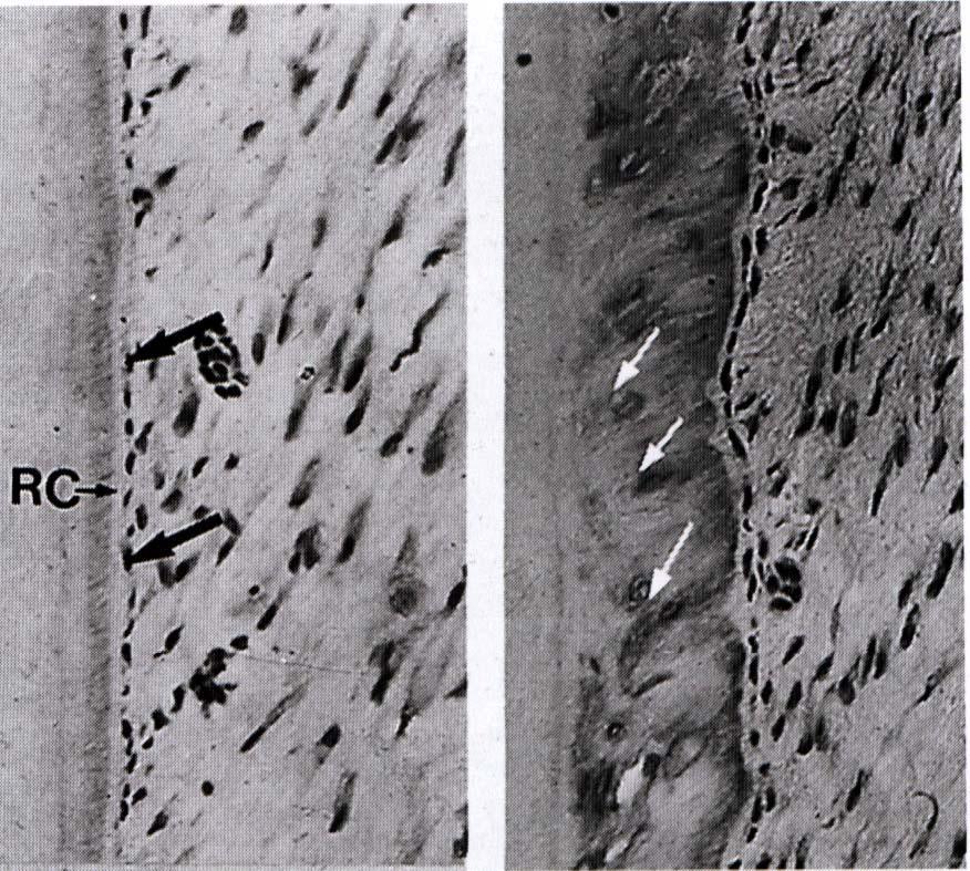
Figure 1. Morphology of a normal furcation area (hematoxylin and eosin [H&E] staining), a Low magnification view of a furcation area.
![Figure 1. Morphology of a normal furcation area (hematoxylin and eosin [H&E] staining), a Low magnification view of a furcation area.](/thumbs/94/120315208.jpg "Figure 1. Morphology of a normal furcation area (hematoxylin and eosin [H&E] staining), a Low magnification view of a furcation area.") 905 Errata that were The following figures are four-color representations of figures incorrectly published in black and white in the July and August issues. Immunohistochemical Expression of Extracellular
905 Errata that were The following figures are four-color representations of figures incorrectly published in black and white in the July and August issues. Immunohistochemical Expression of Extracellular
Lec. 11 & 12 Dr. Ali H. Murad Dental pulp 1- Coronal pulp
 Lec. 11 & 12 Dr. Ali H. Murad Dental pulp Is the soft connective tissue located in the central portion of each tooth. All pulps have similar morphologic characteristic, such as a soft, gelatinous consistency
Sheets 16&17. Dr. Heba Kalbouneh. Dr. Heba Kalbouneh. Dr. Heba Kalbouneh
 Sheets 16&17 Dr. Heba Kalbouneh Dr. Heba Kalbouneh Dr. Heba Kalbouneh Ossification (formation of bone) - Osteoblasts are responsible for producing the extracellular matrix of the bone and these osteoblasts
Osteology. Dr. Carmen E. Rexach Anatomy 35 Mt San Antonio College
 Osteology Dr. Carmen E. Rexach Anatomy 35 Mt San Antonio College Functions of the Skeletal System: Support Movement Protection Hemopoiesis Electrolyte balance (Ca ++ /PO -3 4 ) Acid-base balance Storage
Bones. The division of bones anatomically is : long, short, irregular, flat and sesamoid.
 Bones Osteocytes : Are responsible for maintenance of bones Present in lacunae, and send processes. Unable to divide. The division of bones anatomically is : long, short, irregular, flat and sesamoid.
ANATOMY & PHYSIOLOGY - CLUTCH CH. 8 - BONE AND CARTILAGE.
 !! www.clutchprep.com CONCEPT: BONE CLASSIFICATIONS There are four classifications of bones based on their 1. Long bones are greater in length than in width - Found in the upper and lower limbs (ex: arm,
The periodontium attempts to accommodate to the forces exerted to the crown. This adaptive capacity varies in different persons and in the same person
 The periodontium attempts to accommodate to the forces exerted to the crown. This adaptive capacity varies in different persons and in the same person at different times. The effect of occlusal forces
Root Proximity Characteristics and Type of Alveolar Bone Loss: A Case-control Study
 Journal of the International Academy of Periodontology 011 13/3: 73-79 Root Proximity Characteristics and Type of Alveolar Bone Loss: A Case-control Study 1 1 1 Maria-Anna Loukideli, Alexandra Tsami, Eudoxie
Bone Formation, Growth, and Remodeling
 Bone Formation, Growth, and Remodeling Pre-natal Ossification Embryonic skeleton: fashioned from fibrous membranes or cartilage to accommodate mitosis. 2 types of pre-natal ossification (bone formation)
BONE LABORATORY DEMONSTRATIONS. These demonstrations are found on the bulletin boards outside the MCO Bookstore.
 BONE LABORATORY DEMONSTRATIONS These demonstrations are found on the bulletin boards outside the MCO Bookstore. COMPACT & TRABECULAR BONE - LM When viewed under the polarizing light microscope, the layering
Chapter 6: Skeletal System: Bones and Bone Tissue
 Chapter 6: Skeletal System: Bones and Bone Tissue I. Functions A. List and describe the five major functions of the skeletal system: 1. 2. 3.. 4. 5.. II. Cartilage A. What do chondroblasts do? B. When
CAP STAGE. Ans 1 The following are the stages of tooth development :
 Ans 1 The following are the stages of tooth development : 1. Bud stage 2. Cap stage 3. Bell stage 4. Advanced bell stage 5. Formation of Hertwig s epithelial root sheath BUD STAGE 1. Around the eighth
Chapter 4. Cartilage and Bone. Li Shu-Lei instructor. Dept. Histology and Embryology, School of Basic Medical Sciences, Jilin University
 Chapter 4 Cartilage and Bone Li Shu-Lei instructor Dept. Histology and Embryology, School of Basic Medical Sciences, Jilin University I Cartilage a specialized connective tissue Characterizers: Cartilage
Development of teeth. 5.DM - Pedo
 Development of teeth 5.DM - Pedo Tooth development process of continuous changes in predetermined order starts from dental lamina A band of ectodermal cells growing from the epithelium of the embryonic
Alveolar bone development after decoronation of ankylosed teeth
 Endodontic Topics 2006, 14, 35 40 All rights reserved Copyright r Blackwell Munksgaard ENDODONTIC TOPICS 2008 1601-1538 Alveolar bone development after decoronation of ankylosed teeth BARBRO MALMGREN,
An Overview of Dental Anatomy
 Continuing Education Brought to you by An Overview of Dental Anatomy Course Author(s): Vickie Parrish Foster, RDH, MEd CE Credits: 1 hour Intended Audience: Dental Hygienists, Dental Assistants, Dental
Evaluation of osteoclastic activity in the canine supraalveolar periodontal defect model : an exploratory study
 Oregon Health & Science University OHSU Digital Commons Scholar Archive 5-2015 Evaluation of osteoclastic activity in the canine supraalveolar periodontal defect model : an exploratory study James Yoon
DENTAL RADIOGRAPH INTERPRETATION
 DENTAL RADIOGRAPH INTERPRETATION Brook A. Niemiec, DVM Diplomate, American Veterinary Dental College Fellow, Academy of Veterinary Dentistry www.vetdentaltraning.com www.vetdentalrad.com Interpreting dental
Dimensional ridge alterations following tooth extraction. An experimental study in the dog
 J Clin Periodontol 2005; 32: 212 218 doi: 10.1111/j.1600-051X.2005.00642.x Copyright r Blackwell Munksgaard 2005 Dimensional ridge alterations following tooth extraction. An experimental study in the dog
Dental Anatomy and Occlusion
 CHAPTER 53 Dental Anatomy and Occlusion Ma Lou C. Sabino DDS, and Emily G. Smythe, DDS What numerical system is used most commonly in the United States for designating the adult dentition? Pediatric dentition?
FRACTURES AND LUXATIONS OF PERMANENT TEETH
 FRACTURES AND LUXATIONS OF PERMANENT TEETH 1. Treatment guidelines and alveolar bone Followup Procedures INFRACTION Clinical findings Radiographic findings Treatment Follow-Up Favorable Outcome Unfavorable
A Clinical Evaluation of Anatomic Features of Gingiva in Dental Students in Tabriz, Iran
 Received 26 October 2007; Accepted 11 February 2008 A Clinical Evaluation of Anatomic Features of Gingiva in Dental Students in Tabriz, Iran Adileh Shirmohammadi 1 Masoumeh Faramarzie 1 * Ardeshir Lafzi
CHAPTER 6 LECTURE OUTLINE
 CHAPTER 6 LECTURE OUTLINE I. INTRODUCTION A. Bone is made up of several different tissues working together: bone, cartilage, dense connective tissue, epithelium, various blood forming tissues, adipose
Cartilage & bone. Red: important. Black: in male female slides. Gray: notes extra. Editing File
 Cartilage & bone Red: important. Black: in male female slides. Gray: notes extra. Editing File OBJECTIVES describe the microscopic structure, distribution and growth of the different types of Cartilage
高雄醫學大學 口腔醫學院 口腔病理影像科 牙科 X 光影像判讀 教學範例
 高雄醫學大學 口腔醫學院 口腔病理影像科 牙科 X 光影像判讀 教學範例 Content Image No. 001 Dentigerous cyst over left upper embedded canine--------------------- 頁 1 Image No. 002---------------------------------------------------------------
Anatomy Sheet: Oral cavity Done by: rasha Rakan edited by: khansaa Mahmoud
 Anatomy Sheet: Oral cavity Done by: rasha Rakan edited by: khansaa Mahmoud The oral cavity has 2 parts: 1. Oral vestibule: outer part that consists of outside the teeth, between the teeth, the cheeks and
Lecture 2: Skeletogenesis
 Jilin University School of Stomatology Skeletogenesis Lecture 2: Skeletogenesis Aug. 18, 2015 Yuji Mishina, Ph.D. mishina@umich.edu Student will describe Development of Bone - the general anatomy of bone
Periodontal disease: Aetiopathogenesis
 Periodontal disease: Aetiopathogenesis RACHEL PERRY BSc (VetSc), BVM&S, MANZCVS (Small Animal Dentistry and Oral Surgery), MRCVS e: info@perrydentalvet.co.uk twitter:@perrydentalvet Periodontal disease
COMBINED PERIODONTAL-ENDODONTIC LESION. By Dr. P.K. Agrawal Sr. Prof and Head Dept. Of Periodontia Govt. Dental College, Jaipur
 COMBINED PERIODONTAL-ENDODONTIC LESION By Dr. P.K. Agrawal Sr. Prof and Head Dept. Of Periodontia Govt. Dental College, Jaipur Differential diagnosis For differential diagnostic purposed the endo-perio
An Overview of Dental Anatomy
 An Overview of Dental Anatomy Vickie P. Overman, RDH, MEd Continuing Education Units: 1 hour Online Course: www.dentalcare.com/en-us/professional-education/ce-courses/ce500 Disclaimer: Participants must
6610 NE 181st Street, Suite #1, Kenmore, WA
 660 NE 8st Street, Suite #, Kenmore, WA 9808 www.northshoredentalacademy.com.08.900 READ CHAPTER The Professional Dental Assistant (p.-9) No Key Terms Recall Questions:,,,, and 6 CLASS SYLLABUS DAY READ
For more information about how to cite these materials visit
 Author(s): University of Michigan Medical School, Department of Cell and Developmental Biology License: Unless otherwise noted, the content of this course material is licensed under a Creative Commons
Surgical Procedure in Guided Tissue Regeneration with the. Inion GTR Biodegradable Membrane System
 Surgical Procedure in Guided Tissue Regeneration with the Inion GTR Biodegradable Membrane System 1 Introduction This presentation familiarizes you with the basic steps how to use the Inion GTR membrane
FORMATION OF BONE. Intramembranous Ossification. Bone-Lec-10-Prof.Dr.Adnan Albideri
 FORMATION OF BONE All bones are of mesodermal origin. The process of bone formation is called ossification. We have seen that formation of most bones is preceded by the formation of a cartilaginous model,
Topographie Classification of Deformities of the Alveolar
 Topographie Classification of Deformities of the Alveolar Process* Kenneth W. Karn.f Howard P. Shockett^ William C. Moffitt and Jonathan L. Gray Accepted for publication 1 August 1983 A system of nomenclature
Principles of Periodontal flap surgery. Dr.maryam khosravi
 Principles of Periodontal flap surgery Dr.maryam khosravi Goals of periodontal SURGICAL phase 1 - Controlling or eliminating periodontal disease. 2 Correcting anatomic conditions that may a. favor periodontal
Healing of external inflammatory root resorption - a case report
 Healing of external inflammatory root resorption - a case report Mithra N. Hegde * Deepak Pardal ** ABSTRACT Case report describes a radiographic follow-up of healing of external inflammatory root resorption
KEY CONCEPTS Unit 6 THE SKELETAL SYSTEM
 ANATOMY & PHYSIOLOGY 1 (101-805 - AB) PAUL ANDERSON 2011 KEY CONCEPTS Unit 6 THE SKELETAL SYSTEM A Overview of The Skeletal System 1. Definition: Anatomically the SKELETAL SYSTEM consists of bones, cartilages,
What are the parts of the skeletal system? Chapter 6- Part I Bones and Skeletal Tissues. Growth of Cartilage. Bones come in many shapes
 Chapter 6- Part I Bones and Skeletal Tissues Components of the skeletal system Classification of Bone (bone shapes) Functions of bone Bone structure Microscopic structure of bone and bone cells What are
Dr. Heba Kalbouneh. Saba Alfayoumi. Heba Kalbouneh
 11 Dr. Heba Kalbouneh Saba Alfayoumi Heba Kalbouneh 2- Bone Bone tissue is also classified into primary bone and secondary bone. In the beginning, the first bone that is deposited by the osteoblasts is
Human Anatomy and Physiology I Laboratory
 Human Anatomy and Physiology I Laboratory Skeletal Tissue: Cartilage and Bone This lab involves study of the laboratory exercise Overview of the Skeleton, Classification and Structure of Bones and Cartilages,
SKELETAL TISSUES CHAPTER 7 INTRODUCTION TO THE SKELETAL SYSTEM TYPES OF BONES
 SKELETAL TISSUES CHAPTER 7 By John McGill Supplement Outlines: Beth Wyatt Original PowerPoint: Jack Bagwell INTRODUCTION TO THE SKELETAL SYSTEM STRUCTURE Organs: Bones Related Tissues: Cartilage and Ligaments
Everything You Wanted to Know About Extractions but Were Afraid to Ask
 Everything You Wanted to Know About Extractions but Were Afraid to Ask Tooth extraction is a surgical procedure with serious potential complications and should only be performed by a trained veterinarian.
Masking Buccal Plate Remodeling in the Esthetic Zone with Connective Tissue Grafts: Concepts and Techniques with Immediate Implants
 Peer-Reviewed and Indexed Annual Implant Issue Masking Buccal Plate Remodeling in the Esthetic Zone with Connective Tissue Grafts: Concepts and Techniques with Immediate Implants of Continuing Education
Prospective, comparative assessment of alveolar ridge preservation using Guidor Easy-Graft Classic in atrumatic extraction socket
 University of Iowa Iowa Research Online Theses and Dissertations Summer 2017 Prospective, comparative assessment of alveolar ridge preservation using Guidor Easy-Graft Classic in atrumatic extraction socket
Educational Training Document
 Educational Training Document Table of Contents Part 1: Resource Document Disclaimer Page: 2 Part 2: Line Item Grade Sheets Page: 3 Release: 11/2016 Page 1 of 6 Part 1: Resource Document Disclaimer The
NOTES: Skeletal System (Ch 5, part 1)
") NOTES: Skeletal System (Ch 5, part 1) Individual bones are the organs of the skeletal system. A bone contains very active tissues. BONE STRUCTURE: *Bone structure reflects its function. Parts of a long
NOTES: Skeletal System (Ch 5, part 1) Individual bones are the organs of the skeletal system. A bone contains very active tissues. BONE STRUCTURE: *Bone structure reflects its function. Parts of a long
Extraction with Immediate Implant Placement and Ridge Preservation in the Posterior
 Extraction with Immediate Implant Placement and Ridge Preservation in the Posterior by Timothy F. Kosinski, DDS, MAGD The following case presentation illustrates the diagnosis, planning and treatment for
Clinical UM Guideline
 Clinical UM Guideline Subject: Clinical Crown Lengthening Guideline #: 04-206 Current Effective Date: 03/24/2017 Status: New Last Review Date: 02/08/2017 Description This document addresses the procedure
Socket Graft Plus. Case Presentation. Ideal Bone Graft for All Socket Grafting Situations. Case #1
 Socket Graft Plus Ideal Bone Graft for All Socket Grafting Situations Case Presentation Case #1 Tooth #3 presents with a buccal parulis, ensuring a buccal alveolar fenestration. The tooth will be remove
5.3. The Nature of Cartilage Matrix The components of cartilage matrix include a high component of fibers, and proteoglycans. Proteoglycans are a
 Chapter 5 Supportive Tissues Support in Animals is carried out by Cartilage and Bone. A. Cartilage 5.1. Nature of Cartilage Cartilage is a highly resilient c..t that provides strength and support in areas
The Effect of X Ray Vertical Angulation on Radiographic Assessment of Alveolar Bone Loss
 1 The Effect of X Ray Vertical Angulation on Radiographic Assessment of Alveolar Bone Loss ABSTRACT M. Mehdizadeh MD*, M. Amintavakoli MD**, M. Allahverdi MD*** Introduction: Radiographs provide unique
Occlusion periodontal health
 9 Occlusion and periodontal health J. De Boever, A. De Boever Synopsis Periodontal structures depend on functional occlusal forces to activate the periodontal mechanoreceptors in the neuromuscular physiology
Car$lage and Bone. Kris$ne Kra0s, M.D.
 Car$lage and Bone Kris$ne Kra0s, M.D. Car$lage and Bone Lecture Objec$ves Describe the general func$ons of car$lage and bone. Compare the func$on and composi$on of the three types of car$lage. Describe
DENTIN It a hard vital tissue, surrounds the pulp & underlies the enamel on the crown & the cementum on the roots of the teeth.
 Lec. 7 Dr. Ali H.Murad DENTIN It a hard vital tissue, surrounds the pulp & underlies the enamel on the crown & the cementum on the roots of the teeth. Physical properties: 1-Dentin is pale yellow in color,
SURGICAL TREATMENT OF GINGIVAL RECESSION WITH SOFT TISSUE GRAFT PROCEDURE
 Journal of IMAB ISSN: 1312-773X https://www.journal-imab-bg.org https://doi.org/10.5272/jimab.2018243.2149 Journal of IMAB - Annual Proceeding (Scientific Papers). 2018 Jul-Sep;24(3) Literature review
Chapter 6 Skeletal System
 Chapter 6 Skeletal System Functions of the skeletal system/bone 1. Support skeletal system is the internal framework of the body 2. Protection protects internal organs 3. Movement muscles & bones work
Oral Surgery. Basic Techniques of Dental Local Anesthesia. A variety of techniques used in administration and deposition of local anesthesia:
 Oral Surgery Lecture: 9 Dr. Saif Saadedeen Basic Techniques of Dental Local Anesthesia A variety of techniques used in administration and deposition of local anesthesia: 1. Topical anesthesia 2. Infiltration
DHYG 121 Winter, 2009 COURSE OUTLINE
 CAMOSUN COLLEGE School of Health & Human Services Dental Hygiene Department DHYG 121 Winter, 2009 COURSE OUTLINE The Approved Course Description is available on the web @ http://www.camosun.bc.ca/calendar/current/web/dhyg.html#dhyg121
Due in Lab. Due next week in lab - Scientific America Article Select one article to read and complete article summary
 Due in Lab 1. Skeletal System 33-34 2. Skeletal System 26 3. PreLab 6 Due next week in lab - Scientific America Article Select one article to read and complete article summary Cell Defenses and the Sunshine
EVALUATION OF TRANSVERSE, BODILY TOOTH MOVEMENT AND ITS EFFECTS ON THE SURROUNDING HARD TISSUE
 EVALUATION OF TRANSVERSE, BODILY TOOTH MOVEMENT AND ITS EFFECTS ON THE SURROUNDING HARD TISSUE A Thesis by CHAD JASON CAPPS Submitted to the Office of Graduate and Professional Studies of Texas A&M University
RAJ M. SAINI, DDS, MSD
 Restoring and Maintaining Periodontal Health with Orthodontic Treatment RAJ M. SAINI, DDS, MSD rajmsaini@yahoo.com Diplomate Of The American Board Of Orthodontics Clinical Professor Of Orthodontics New
Biological Considerations Related To Osseointegration
 IOSR Journal of Dental and Medical Sciences (IOSR-JDMS) e-issn: 2279-0853, p-issn: 2279-0861.Volume 14, Issue 9 Ver. II (Sep. 2015), PP 53-59 www.iosrjournals.org Biological Considerations Related To Osseointegration
The Skeletal System PART A. PowerPoint Lecture Slide Presentation by Patty Bostwick-Taylor, Florence-Darlington Technical College
 PowerPoint Lecture Slide Presentation by Patty Bostwick-Taylor, Florence-Darlington Technical College The Skeletal System 5 PART A The Skeletal System Parts of the skeletal system Bones (skeleton) Joints
Semester Credits: 3 Lecture Hours: 3. Prerequisites:
 Revised: Fall 2015 Semester Credits: 3 Lecture Hours: 3 21THistology DNH 115 Admission into dental hygiene program. Prerequisites: Course Description: Presents a study of the microscopic and macroscopic
Comparative analysis of the stress distribution in five anatomical types of maxillary central incisor
 Technology and Health Care 25 (2017) S53 S62 DOI 10.3233/THC-171306 IOS Press S53 Comparative analysis of the stress distribution in five anatomical types of maxillary central incisor Lei Sun a, Xifeng
Chapter 7. Skeletal System
 Chapter 7 Skeletal System 1 Introduction: A. Bones are very active, living tissues B. Each bone is made up of several types of tissues and so is an organ. C. Bone functions include: muscle attachment,
INTERPRETATION RADIOGRAPHIC INTERPRETATION. Law of Symmetry. We will be reviewing: 7/30/16
 RADIOGRAPHIC INTERPRETATION Pam Wood, CDA, RDH, M.Ed, CAGS Community College of Rhode Island pwood@ccri.edu INTERPRETATION the ability to read what is revealed on a dental radiograph any dental professional
DENTIN-PULP COMPLEX. Erlina Sih Mahanani. School of Dental sciences Universiti Sains Malaysia. Erlina Sih Mahanani
 DENTIN-PULP COMPLEX School of Dental sciences Universiti Sains Malaysia Introduction Overview anatomy & histology of dentin and pulp. Development of dentin and pulp Structure of dentin and pulp Dentin
and Non-Human MODULE No.17: Structural Variation in Teeth- Human and Non-Human
 SUBJECT Paper No. and Title Module No. and Title Module Tag MODULE No.17: Structural Variation in Teeth- Human and FSC_P11_M17 TABLE OF CONTENTS 1. Learning Outcomes 2. Introduction 3. Structure of Human
Root resorptions are not multifactorial, complex, controversial or polemical!
 O r t h o d o n t i c I n s i g h t The concept of root resorptions or Root resorptions are not multifactorial, complex, controversial or polemical! Alberto Consolaro* Abstract The mechanisms of root resorptions
THE JAWS AND TEETH IN PAGET'S DISEASE OF BONE
 J. clin. Path. (1955), 8, 195. THE JAWS AND TEETH IN PAGET'S DISEASE OF BONE BY R. B. LUCAS From the Department of Pathology, Royal Dental Hospital of the London School of Dental Surgery (RECEIVED FOR
Controlling Tissue Contours with a Prosthetically Driven Approach to Implant Dentistry
 Controlling Tissue Contours with a Prosthetically Driven Approach to Implant Dentistry Go online for in-depth content by Timothy F. Kosinski, DDS, MAGD With continual improvements in the design and production
Double Papillary Flap - A Treatment for Gingival Recession
 World Journal of Medical Sciences 10 (2): 117-121, 2014 ISSN 1817-3055 IDOSI Publications, 2014 DOI: 10.5829/idosi.wjms.2014.10.2.82175 Double Papillary Flap - A Treatment for Gingival Recession 1 1 1